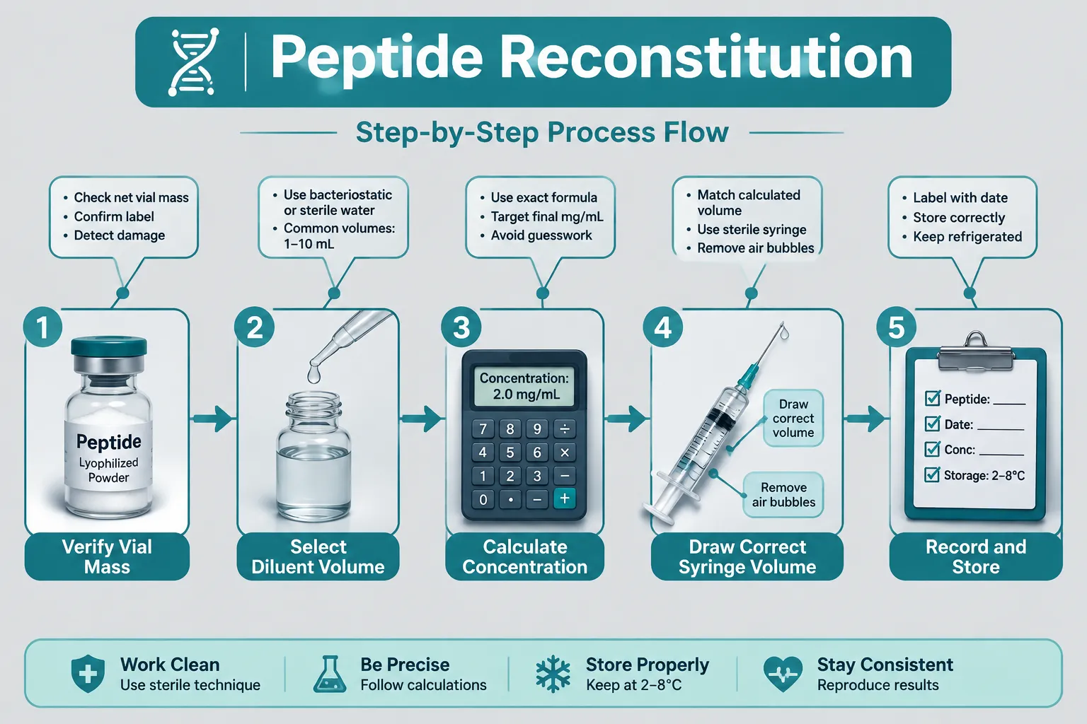
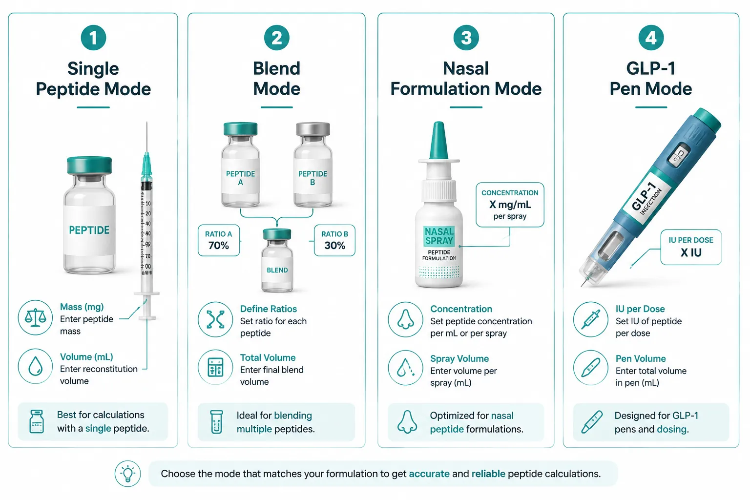
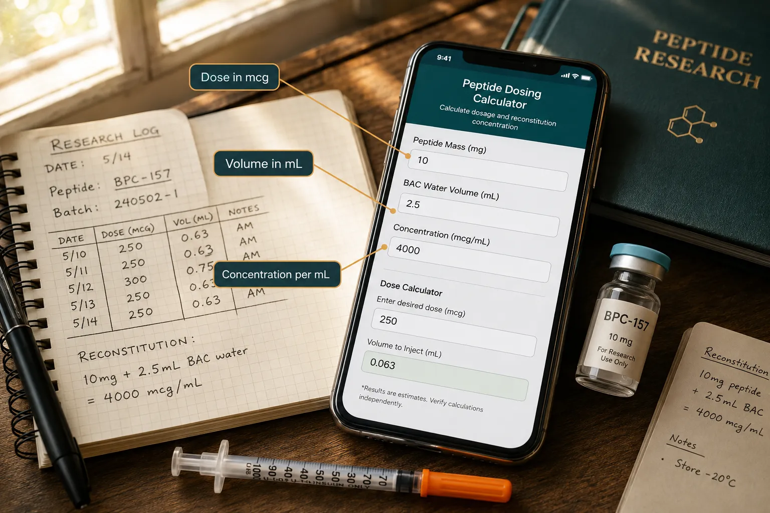
Biologically Effective Dose in Peptide Experiments: Translating Nominal Concentration Into Exposure and Response
Fewer than one in five in vitro peptide studies formally account for the gap between the concentration written on a vial label and the concentration that actually reaches a cellular receptor. That gap is not a minor rounding error. It can span an order of magnitude, and it is the central problem that the concept of biologically effective dose in peptide experiments: translating nominal concentration into exposure and response is designed to solve.
Nominal concentration, administered amount, molar concentration, exposure duration, and observed biological response are five distinct measurements. Treating them as interchangeable is one of the most common sources of irreproducible results in peptide research.
Key Takeaways
- Nominal concentration is a calculated convenience metric, not a direct measure of what reaches the target site.
- Protein binding, plastic adsorption, and cell-surface interactions can dramatically reduce the bioavailable fraction of a peptide in an assay.
- The freely dissolved concentration (C_free) in the medium is a far better proxy for biologically effective dose than the nominal value.
- Mass-balance and PBPK models can translate nominal doses into target-site exposure estimates when direct measurement is unavailable.
- Exposure duration and time-dependent concentration profiles matter as much as peak concentration for predicting biological response.
Why Nominal Concentration Is Not the Same as Biologically Effective Dose
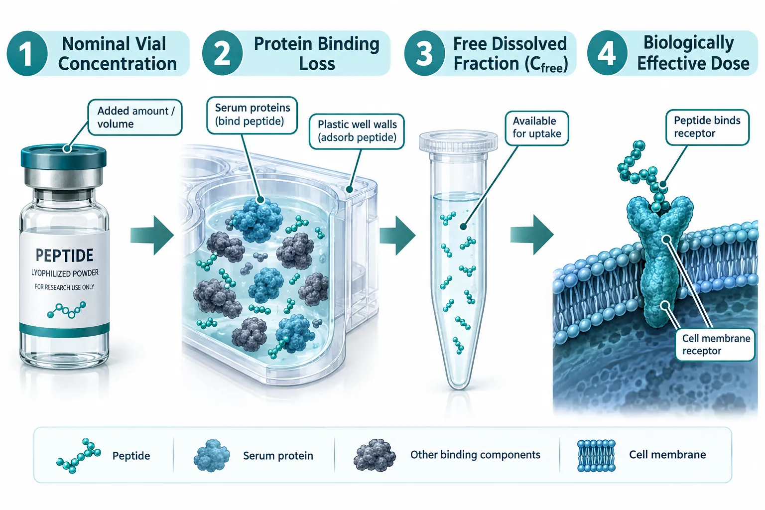
When a researcher dissolves a peptide in cell culture medium and records "100 nM," that number describes what was added to the well, not what is available to the cell. Regulatory and scientific guidance is explicit: biologically effective dose (BED) is the amount of a compound at the organ, cell, or macromolecular target that is available to interact and cause a biological effect.
Several physical and chemical processes erode the nominal concentration before any peptide molecule encounters a receptor:
- Serum protein binding – A significant fraction of added peptide binds to albumin and other serum proteins, removing it from the freely dissolved pool.
- Plastic adsorption – Polystyrene plates and tubing can adsorb hydrophobic or amphipathic peptides, reducing the medium concentration over time.
- Cell-surface sequestration – Cationic peptides, including mitochondria-targeted sequences like those found in SS31 peptide research, can associate with negatively charged membrane phospholipids before reaching their intended intracellular target.
- Evaporation and degradation – Volatile solvents and enzymatic cleavage further reduce the effective concentration during extended incubations.
The result is that nominal concentration systematically overestimates in vitro potency relative to free concentration. Studies comparing dose-response curves built on nominal versus free concentrations consistently show that free concentration yields more consistent results across assay conditions and better predicts in vivo response.
"The freely dissolved, unbound concentration in the medium is generally a better proxy for biologically effective dose than the nominal concentration added to the well."
For peptides like those explored in peptide dosing research, this distinction has direct consequences for how minimum effective doses and therapeutic windows are interpreted.
A Framework for Translating Nominal Concentration Into Exposure and Response
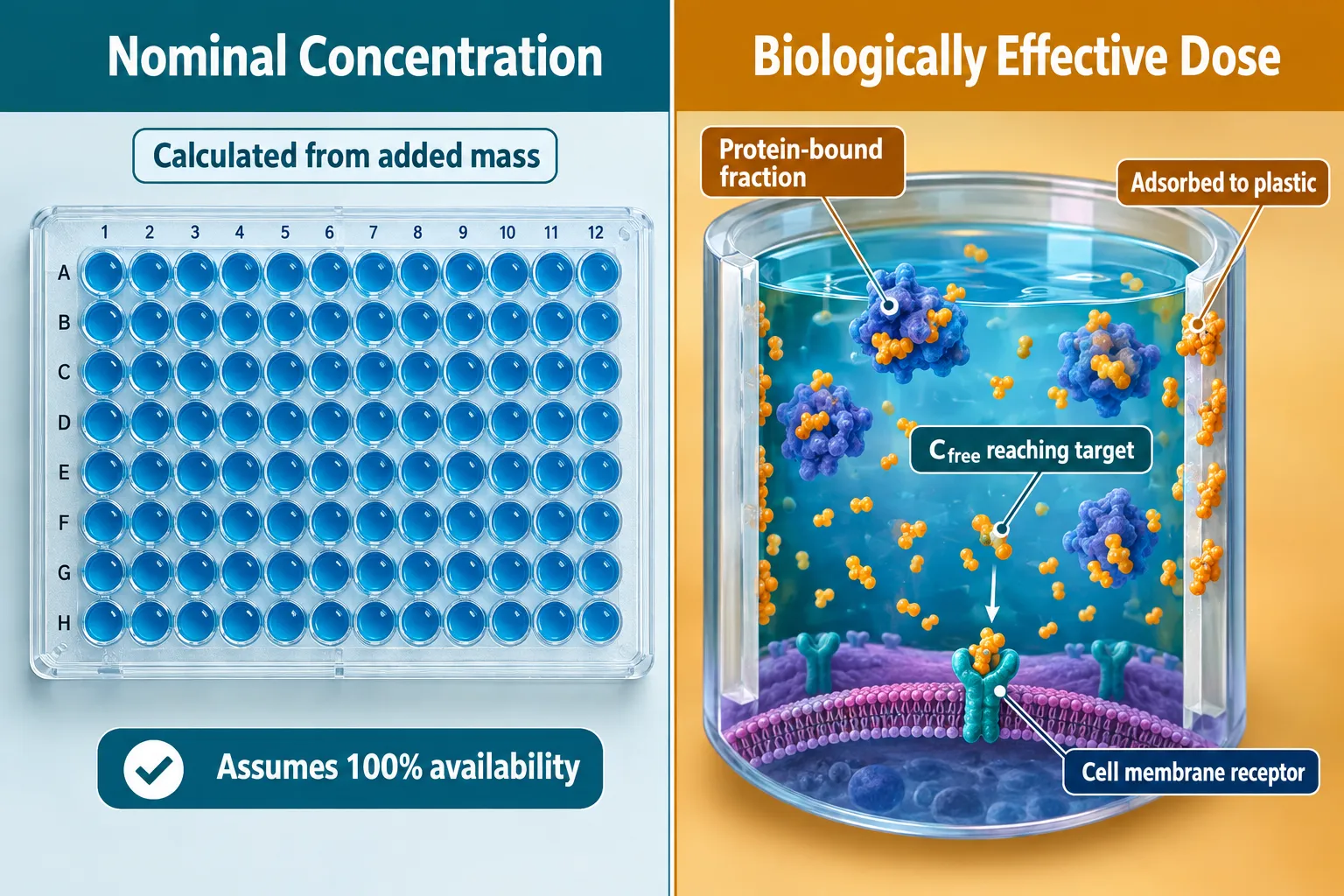
Understanding the problem is only the first step. A practical framework for biologically effective dose in peptide experiments: translating nominal concentration into exposure and response involves a hierarchy of dose metrics, each closer to the actual site of action.
The Dose Metric Hierarchy
| Dose Metric | Definition | Proximity to BED |
|---|---|---|
| Nominal concentration | Added mass divided by medium volume | Lowest |
| Loaded/released dose | Amount released from formulation or carrier | Moderate |
| C_free (freely dissolved) | Unbound concentration in aqueous medium | High |
| Cell-associated concentration | Peptide measured in or on cells | Very high |
| Target-site concentration | Concentration at receptor or organelle | Highest (true BED) |
When direct measurement of C_free or cell-associated concentrations is unavailable, current scientific guidance requires that researchers either use dynamic mass-balance models or explicitly characterize the uncertainty introduced by relying on nominal values.
Applying Mass-Balance Models
Dynamic dosimetry models estimate medium and intracellular BED from a peptide's physicochemical properties: molecular weight, log P, protein binding fraction, and assay geometry. These in silico approaches allow researchers to predict how much active peptide remains in solution over time, accounting for partitioning into plastics and proteins.
For peptides such as those studied in GLP-3 peptide research or RETA peptide experiments, where receptor binding kinetics are central to the dose-response relationship, these corrections can shift the apparent EC50 substantially.
Time-Dependent Exposure Matters
A single nominal concentration value collapses a dynamic process into a static number. Quantitative in vitro-to-in vivo extrapolation (QIVIVE) frameworks emphasize that time-dependent concentration-response relationships are more informative than simple endpoint measurements. A peptide that degrades rapidly in serum may produce a high C_free spike followed by near-zero exposure, a profile that a nominal 100 nM label cannot describe.
Practical Steps for Implementing Biologically Effective Dose in Peptide Experiments
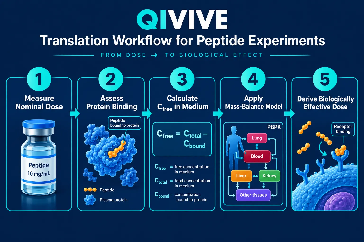
Translating biologically effective dose in peptide experiments: translating nominal concentration into exposure and response from a theoretical concept into laboratory practice involves concrete, sequential actions.
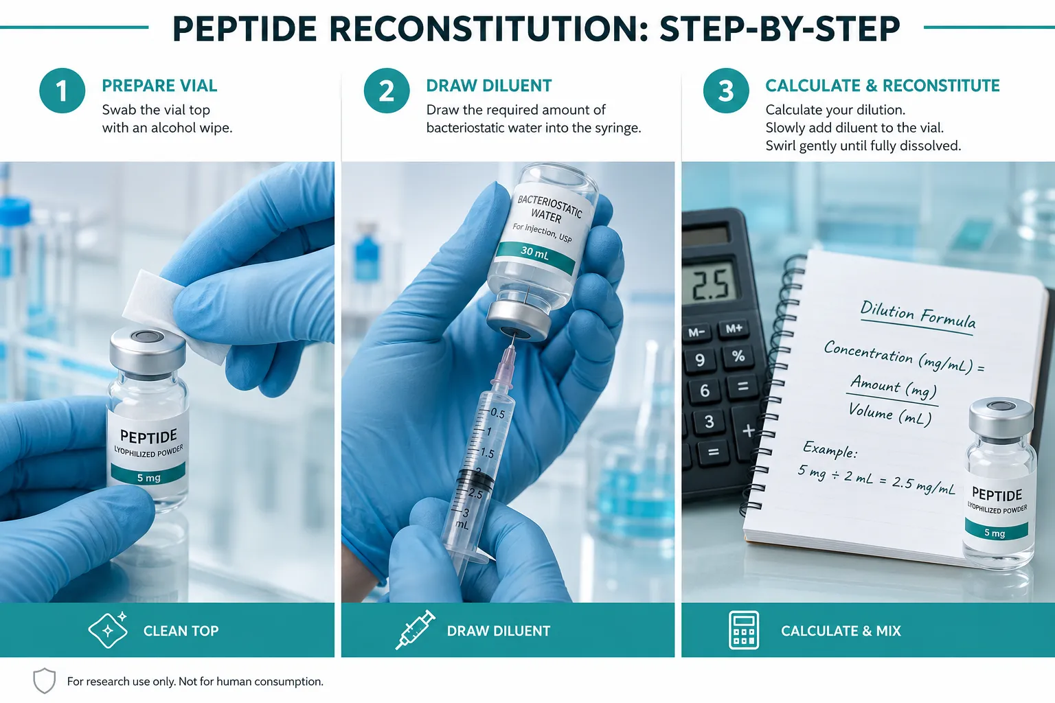
Step 1 – Characterize protein binding. Use equilibrium dialysis or rapid equilibrium dialysis (RED) assay to determine the unbound fraction in the specific medium formulation used. Serum percentage matters: a 10% FBS medium and a 1% FBS medium will yield very different C_free values for the same nominal dose.
Step 2 – Assess plastic adsorption. Measure peptide recovery from wells at multiple time points. Significant losses over 24-48 hours signal adsorption, particularly relevant for amphipathic or cationic sequences.
Step 3 – Apply a mass-balance correction. Use the measured binding and adsorption data to calculate C_free at each time point. For complex peptides, consider PBPK-informed models.
Step 4 – Build time-resolved concentration-response curves. Plot response against C_free at the relevant time point, not against nominal concentration at time zero. This approach aligns with QIVIVE best practices and improves cross-study comparability.
Step 5 – Report both metrics. Publish nominal concentration alongside the calculated or measured C_free and any BED estimate. This supports reproducibility and allows other researchers to apply corrections for their own assay conditions.
For researchers working with mitochondria-targeted peptides, the synergy of LL-37 and SS31 provides a relevant case study in how membrane partitioning can dramatically alter the effective intracellular dose relative to the nominal medium concentration. Similarly, resources on tesa peptide benefits illustrate how administered dose and tissue-level exposure diverge in in vivo settings, reinforcing the same principle at a different scale.
When nominal doses are the only available metric, current guidance treats them as a starting point that requires explicit uncertainty characterization, not as a reliable proxy for BED.
Conclusion
The vial concentration is where a peptide experiment begins, not where it ends. Nominal concentration, administered amount, molar concentration, exposure duration, and biological response each occupy a distinct position in the chain of events between a researcher's bench and a cell's receptor. Conflating them produces dose-response curves that cannot be reproduced, translated to in vivo settings, or compared across laboratories.
Actionable next steps for researchers in 2026:
- Measure protein binding and plastic adsorption for every new peptide-assay combination before interpreting dose-response data.
- Report C_free alongside nominal concentration in all publications and internal records.
- Apply mass-balance or PBPK corrections when direct measurement is impractical.
- Build time-resolved exposure profiles rather than relying on single-timepoint nominal values.
- Treat nominal concentration as a convenience metric with documented uncertainty, not as a synonym for biologically effective dose.
Adopting these practices moves peptide research closer to the standard that regulators and leading toxicology groups now expect: dose-response relationships grounded in actual exposure at the target site.