Call or Text 727-513-9780
  • Shopping Cart Shopping Cart
    0Shopping Cart
Pure Tested Peptides | America's most trusted Peptides for sale online
  • Peptides for sale
    • Oral Peptides for sale
      • Peptide Capsules for sale
      • BPC 157 Capsules 1000mcg
      • SLU-PP-332 Capsules | 1000 mcg
      • 5-Amino-1MQ 50mg Capsules
      • Tesofensine 500mcg
    • All Peptides for sale
    • Peptide Sprays
      • BPC 157 Nasal Spray Kit
      • BPC-157 TB500 Nasal Spray Kit
      • Semax Nasal Spray 10mg
      • Selank – Nasal Spray Kit – 10mg
      • Epithalon 50MG Nasal Spray Kit
      • Ipamorelin 10mg Nasal Spray
      • Klow Nasal Spray (BPC-157 + TB-500 + GHK-Cu + KPV) | 80mg
      • Hulk Nasal Spray Tesa / Ipa Blend 6/3 MG
      • Klow Nasal Spray
      • NAD + 500 mg Nasal Spray
      • PT-141 Nasal Spray Kit
    • GHRH Peptides
      • Ipa Peptides
      • CJC-1295 Peptides
        • CJC-1295 with DAC 5 mg
        • CJC-1295 without DAC 5 mg
        • CJC-1295 Ipa 10mg
      • Tesa Peptides
        • Tesa Peptide
        • Tesa 20 mg
    • GHK-Cu Peptides
      • All GHK-Cu Peptides
      • GHK-Cu 100mg
      • KLOW Peptide Blend – Buy KLOW blend online
    • BPC Peptides
      • All BPC Peptides
      • BPC-157
      • BPC-157 TB-500
      • BPC 157 capsules 1000mcg
    • SLU-PP-332 Peptides
      • All SLU-PP-332 Peptides
      • SLU-PP-332 5mg
    • GLP3 Peptides
      • GLP3-R
      • GLP3-R CAG 10mg
      • GLP3-R 20mg
    • PT-141 Peptides
      • PT-141 Peptides for sale
      • PT-141 10mg
      • PT-141 Nasal Spray
    • CAG Peptides
      • Lipo-C Peptide Blend
      • CAG 5mg
      • CAG 10mg
    • MOTS-C Peptides
      • MOTS-C Peptides for sale
      • MOTS-c peptide
      • MOTS-c 10mg *6 pack*
    • 5 Amino 1MQ Peptides
      • 5 Amino 1MQ Peptides for sale
      • 5-Amino-1MQ 50mg Capsules
      • 5-Amino-1MQ 5mg
    • Epithalon Peptides
      • Epithalon Peptides for sale
      • Epithalon 10mg
      • Epithalon 50mg
  • Shop
    • GLPs
      • 5-Amino-1MQ 50mg Capsules
      • 5-Amino-1MQ 5mg
      • GLP3-Reta
      • L-Carnitine 500mg/ml
      • Tesofensine 500mcg
      • SLU-PP-332 5mg
      • MOTS-c 10mg *6 pack*
    • Epithalon & BPC Peptides
      • Epithalon 10mg
      • Epithalon 50mg
      • BPC-157
      • BPC 157 capsules 1000mcg
      • BPC-157 TB-500
      • BPC-157 TB500 Nasal Spray Kit
      • BPC 157 Nasal Spray Kit
    • BPC TB-500 & NAD+ Peptides
      • NAD+ 500 mg
      • KLOW Peptide Blend – Buy KLOW blend online
      • GLOW Peptide Blend
      • TB 500 5mg
      • BPC 157 capsules 1000mcg – Supplement
      • BPC 157 Nasal Spray Kit
      • BPC-157
      • BPC-157 TB500 Nasal Spray Kit
      • BPC-157 TB-500
      • BPC 157 capsules 1000mcg
    • LL-37 Peptide
      • LL-37 10 mg
    • MOTS-C & Selank
      • MOTS-c peptide
      • Selank 10mg
    • GHK Peptides
      • GHK-Cu 100mg
      • GLOW Peptide Blend
      • KLOW Peptide Blend – Buy KLOW blend online
  • COAs
  • Wholesale
    • Wholesale Peptides for sale
  • PTP FAQ
  • Affiliates
    • Selank Peptide: Advanced Pharmacological Mechanisms Underlying Its Anxiolytic and Nootropic Effects in Research
      • 5-Amino-1MQ Peptide: Detailed Mechanisms of NNMT Inhibition and Its Impact on Cellular Metabolism Research
        • MOTS-C Peptide: Unraveling Its Role in Mitochondrial Dynamics and Energy Metabolism Research
          • Epithalon Peptide and Telomerase Regulation: Investigating Its Impact on Cellular Senescence and Lifespan Research Models
            • GLP2-T vs GLP2 Tirz Peptide: Understanding the Naming, Mechanistic Nuances, and Research Implications for Gut Health
      • Peptides and Polypeptides in Modern Pharmacology: What Research on Metoprolol, Prednisone, and Amlodipine Reveals
        • Polypeptide Peptides vs NSAIDs: What Naproxen and Diclofenac Teach Tissue-Repair Researchers About BPC‑157 and TB‑500
          • Peptides and Polypeptides in Endocrine Pharmacology: How GLP-1, GLP-2, and GLP-3 Retatrutide Differ From Classic Drugs Like Prednisone and Amlodipine
          • 5‑Amino‑1MQ Peptide and NNMT Inhibition: How It Compares With Statins Like Atorvastatin in Adipose and Lipid Metabolism Research
      • Enclomiphene, Estrogen Receptor Signaling, and Luteinizing Phase Biology: What Hormone Researchers Should Measure
        • Mesenchymal Stem Cells, BPC‑157, and GHK‑Cu: How Tissue Repair Peptides Compare With Classic NSAIDs Like Naproxen in Injury Models
          • Peptide Calculator Use Cases Beyond Growth Hormone: Working Through GLP‑3 Retatrutide, MOTS‑c, and BPC‑157 Research Dosing
        • Polypeptide Peptides in Endocrine and Metabolic Pharmacology: Lessons From Amlodipine, Prednisone, and Metoprolol
        • Peptides Calculator for Advanced Blends: Worked Examples for Tesamorelin, CJC‑1295, and Ipamorelin Stacks
          • Peptides and Polypeptides in Human Physiology: How Molecular Size Shapes Research Applications
            • Selank Peptide: Advanced Pharmacological Mechanisms Underlying Its Anxiolytic and Nootropic Effects in Research
          • 5-Amino-1MQ Peptide: Detailed Mechanisms of NNMT Inhibition and Its Impact on Cellular Metabolism Research
            • MOTS-C Peptide: Unraveling Its Role in Mitochondrial Dynamics and Energy Metabolism Research
              • Epithalon Peptide and Telomerase Regulation: Investigating Its Impact on Cellular Senescence and Lifespan Research Models
                • GLP2-T vs GLP2 Tirz Peptide: Understanding the Naming, Mechanistic Nuances, and Research Implications for Gut Health
                  • Peptides and Polypeptides in Endocrine Pharmacology: How GLP-1, GLP-2, and GLP-3 Retatrutide Differ From Classic Drugs Like Prednisone and Amlodipine
                    • Polypeptide Peptides vs NSAIDs: What Naproxen and Diclofenac Teach Tissue-Repair Researchers About BPC‑157 and TB‑500
                      • Peptides and Polypeptides in Modern Pharmacology: What Research on Metoprolol, Prednisone, and Amlodipine Reveals
                        • 5‑Amino‑1MQ Peptide and NNMT Inhibition: How It Compares With Statins Like Atorvastatin in Adipose and Lipid Metabolism Research
                        • Enclomiphene, Estrogen Receptor Signaling, and Luteinizing Phase Biology: What Hormone Researchers Should Measure
                        • Mesenchymal Stem Cells, BPC‑157, and GHK‑Cu: How Tissue Repair Peptides Compare With Classic NSAIDs Like Naproxen in Injury Models
                        • Peptide Calculator Use Cases Beyond Growth Hormone: Working Through GLP‑3 Retatrutide, MOTS‑c, and BPC‑157 Research Dosing
                        • Polypeptide Peptides in Endocrine and Metabolic Pharmacology: Lessons From Amlodipine, Prednisone, and Metoprolol
                        • Peptides Calculator for Advanced Blends: Worked Examples for Tesamorelin, CJC‑1295, and Ipamorelin Stacks
                        • Peptides and Polypeptides in Human Physiology: How Molecular Size Shapes Research Applications
                        • Selank Peptide: Advanced Pharmacological Mechanisms Underlying Its Anxiolytic and Nootropic Effects in Research
                        • MOTS-C Peptide: Unraveling Its Role in Mitochondrial Dynamics and Energy Metabolism Research
                        • Peptides and Polypeptides in Modern Pharmacology: What Research on Metoprolol, Prednisone, and Amlodipine Reveals
                        • Peptides and Polypeptides in Endocrine Pharmacology: How GLP-1, GLP-2, and GLP-3 Retatrutide Differ From Classic Drugs Like Prednisone and Amlodipine
                        • 5-Amino-1MQ Peptide: Detailed Mechanisms of NNMT Inhibition and Its Impact on Cellular Metabolism Research
                        • Mesenchymal Stem Cells, BPC‑157, and GHK‑Cu: How Tissue Repair Peptides Compare With Classic NSAIDs Like Naproxen in Injury Models
                        • Peptide Calculator Use Cases Beyond Growth Hormone: Working Through GLP‑3 Retatrutide, MOTS‑c, and BPC‑157 Research Dosing
                        • GLP2-T vs GLP2 Tirz Peptide: Understanding the Naming, Mechanistic Nuances, and Research Implications for Gut Health
                        • Polypeptide Peptides in Endocrine and Metabolic Pharmacology: Lessons From Amlodipine, Prednisone, and Metoprolol
                        • Polypeptide Peptides vs NSAIDs: What Naproxen and Diclofenac Teach Tissue-Repair Researchers About BPC‑157 and TB‑500
                        • Peptides and Polypeptides in Human Physiology: How Molecular Size Shapes Research Applications
                        • Peptides Calculator for Advanced Blends: Worked Examples for Tesamorelin, CJC‑1295, and Ipamorelin Stacks
                        • 5‑Amino‑1MQ Peptide and NNMT Inhibition: How It Compares With Statins Like Atorvastatin in Adipose and Lipid Metabolism Research
                        • Enclomiphene, Estrogen Receptor Signaling, and Luteinizing Phase Biology: What Hormone Researchers Should Measure
                        • Epithalon Peptide and Telomerase Regulation: Investigating Its Impact on Cellular Senescence and Lifespan Research Models
                        • Best research protocol Klow blend
                        • best time to take BPC-157
                        • best time to take DSIP (Delta Sleep Inducing Peptide)
                        • best time to take CJC-1295
                        • best time to take AOD-9604
                        • best time to take Follistatin 344
                        • best time to take Ipamorelin
                        • best time to take MK-677 (Ibutamoren)
                        • best time to take Ligandrol (LGD-4033) — research compound
                        • best time to take Ostarine (MK-2866) — research compound
                        • best time to take GHK-CU
                        • best time to take TB-500
                        • best time to take MOTS-c
                        • best time to take Semax
                        • best time to take RAD-140 (Testolone) — research compound
                        • best time to take Thymosin Alpha-1
                        • best time to take PEG-MGF
                        • Biolife Plasma, Octapharma Plasma, and Research Peptides: How Plasma Donation Labs Differ From Peptide Suppliers
                        • best time to take YK-11 — research compound
                        • best time to take PT-141 (Bremelanotide)
                        • Best research protocol Klow blend
                        • 5-Amino-1MQ and MOTS-C Synergy: Metabolic Signaling, Mitochondria, and Research Design
                        • BPC-157 and TB-500: Investigating Their Combined Effects on Angiogenesis and Cellular Migration in Tissue Repair Models
                        • BPC-157 Peptide: Gut Barrier Function, Inflammation, and Tissue-Recovery Research
                        • 5‑Amino‑1MQ and MOTS‑c Synergy in Metabolic Research: Designing NNMT and Mitochondrial Biogenesis Stacks
                        • CJC-1295 with DAC vs. Without DAC: Half-Life, Release Kinetics, and Research Implications
                        • CJC‑1295 with DAC vs. Without DAC: Expanding on Half‑Life Differences Using Tesamorelin and Ipamorelin Blend Case Studies
                        • Collagen Biology and Copper‑Binding Peptides: How GHK‑Cu, Glow Blend, and Klow Blend Interact with Skin and Connective Tissue
                        • Collagen Biology and Regenerative Peptides: How GHK‑Cu, Glow Blend, and Klow Blend Affect Extracellular Matrix Research
                    • DNA, Telomeres, and Longevity Peptides: Positioning Epithalon and MOTS‑c in Genetic Aging Research
                      • Enclomiphene Citrate: serm Mechanism, Testosterone Research, and Stack Compatibility
                        • Enclomiphene vs Enclomiphene Citrate: Formulation, Bioavailability, and Research Distinctions
                        • Epithalon Peptide Research: Telomerase Activation, Aging, and Pineal Gland Function
                        • Estrogen Receptor Signaling and Enclomiphene: How Selective Modulators Compare with Classic Polypeptide Hormones
                        • GHK-Cu Peptide: Advanced Mechanisms in Extracellular Matrix Remodeling and Wound Healing Research
                        • GHK-Cu Peptide: Collagen Synthesis, Wound Repair, and Skin-Barrier Research Models
                        • GLP-1 vs GLP-3 vs GLP-2: Peptide Classification and Research Applications
                        • GLP-2 Peptide Research Guide: Gut Barrier Function, Nutrient Absorption, and Intestinal Recovery Models
                        • GLP-3 Retatrutide vs. GLP-1 Drugs: What Triple-Agonist Biology Changes in Research Models
                        • Ipamorelin and Tesamorelin Combination: Synergistic GH Secretagogue Research and Dosing Protocols
                        • GLP2 Tirz Peptide: What It Is, Why the Name Exists, and How Researchers Should Interpret It
                        • Klow Blend Peptide Nasal Spray: What the Formulation Is Trying to Do in Cognitive Research
                        • Mitochondria, NNMT Inhibition, and Peptide Modulators: Where MOTS‑c and 5‑Amino‑1MQ Fit in Cellular Energy Research
                        • MOTS-c Peptide: Mitochondrial Function, Energy Metabolism, and What Researchers Measure
                        • MOTS-c vs. 5-Amino-1MQ: Which Metabolic Research Questions Each Compound Actually Answers
                        • Nasal Spray Peptides: Bioavailability, Administration, and Semax/Selank Research Applications
                        • PT-141 Peptide Research: Mechanism of Action and Melanocortin Receptor Signaling
                        • Retatrutide for Research: Mechanism, Structure, and GLP-1/GLP-3 Dual Action
                        • Retatrutide for Obesity and Type 2 Diabetes: What the Latest Trial Data Suggest
                        • Tesofensine Peptide Research: Mechanism, Appetite Suppression, and Neuropeptide Y Pathways
  • Contact
    • Contact Customer Service
    • Text Customer Support
  • About US
  • Shop all peptides
  • Affiliate Program
    • Affiliate Signup
  • Login / Register Login / Register Page Link Login / Register Page Link
  • Click to open the search input field Click to open the search input field Search
  • Menu Menu
Peptides: How Researchers Classify Chains, Polypeptides, and Hormone Analogues in Lab Use

Peptides: How Researchers Classify Chains, Polypeptides, and Hormone Analogues in Lab Use

August 18, 2026/0 Comments/in Uncategorized/by

Less than two percent of naturally occurring peptides have been fully characterized at the structural level, yet these short amino acid chains govern everything from appetite regulation to cellular repair. Understanding how researchers classify chains, polypeptides, and hormone analogues in lab use is not just academic housekeeping. Terminology directly shapes synthesis protocols, analytical workflows, and how results are interpreted across studies. When a lab team disagrees on whether a 25-residue chain is a "peptide" or a "polypeptide," it can affect purification strategy, storage conditions, and even regulatory framing. This article clarifies the nomenclature, explains where the boundaries lie, and explains why precise classification matters in practice.

Key Takeaways

  • Peptides are chains of two or more amino acids; the sub-categories, dipeptide, oligopeptide, polypeptide, are defined primarily by chain length.
  • Oligopeptides are generally defined as 2-20 residues; polypeptides as 20 or more residues; proteins as folded polypeptides typically exceeding 50 residues or 10 kDa.
  • These length-based cut-offs are conventions, not strict rules, thresholds vary across textbooks and institutions.
  • A single molecule can carry multiple simultaneous labels: structural (oligopeptide), biosynthetic (polypeptide), and functional (hormone analogue).
  • In lab practice, classification guides synthesis methods, analytical choices, and how hormone analogues are sourced and described in literature.

Defining the Building Blocks: Chain Length and Nomenclature

The most fundamental way researchers classify peptides is by counting residues, the individual amino acid units linked by peptide bonds.

Defining the Building Blocks: Chain Length and Nomenclature

The hierarchy looks straightforward on paper, but the boundaries are deliberately flexible:

Term Residue Range Common Lab Context
Dipeptide 2 Smallest possible peptide unit
Tripeptide 3 Common in enzyme substrate studies
Oligopeptide 2-20 (varies) Solid-phase synthesis, signaling research
Polypeptide 20+ residues Longer chains, may fold partially
Protein ~50+ residues / 10 kDa+ Stable 3D fold, distinct function

Why the variation? Some biochemistry texts define oligopeptides as fewer than 10 residues; others extend the range to 15 or even 20. The key point is that these are descriptive conventions, not codified regulatory categories. A research team working on a 12-residue signaling chain may call it an oligopeptide, a short peptide, or simply a peptide, all three are technically defensible.

The transition from polypeptide to protein is equally nuanced. A chain of 40 residues is typically still called a polypeptide. Once it exceeds roughly 50 residues or a molecular mass of about 10,000 Daltons and adopts a stable three-dimensional fold with a defined biological function, the scientific community generally calls it a protein. Length alone does not make a protein, structure and function must follow.

For researchers exploring longer signaling chains, resources on growth hormone research illustrate how polypeptide length and receptor specificity intersect in practice.

How Researchers Classify Chains, Polypeptides, and Hormone Analogues in Lab Use

Understanding structural classification is only half the picture. In modern research, the same molecule often carries overlapping labels depending on the context of discussion.

How Researchers Classify Chains, Polypeptides, and Hormone Analogues in Lab Use

Structural vs. Functional Labels

A synthetic peptide used in metabolic research might be:

  • Structurally: an oligopeptide (18 residues, below the 20-residue threshold)
  • Biosynthetically: derived from a longer polypeptide precursor
  • Functionally: a hormone analogue that mimics glucagon-like signaling

None of these labels contradicts the others. Researchers in biochemistry and pharmacology routinely layer structural and functional terminology. The GLP peptide family is a strong example, these molecules are structurally short enough to qualify as oligopeptides or small polypeptides, yet they are primarily discussed as hormone analogues in the literature. The GLP-1, GLP-2, and GLP-3 peptide family guide breaks down how this family is categorized across structural and functional dimensions.

Functional Classification Categories

Beyond chain length, lab-focused resources increasingly organize peptides by role:

  • Signaling peptides: Include hormone analogues, neuropeptides, and receptor agonists. Examples include GLP-1 analogues and growth hormone secretagogues.
  • Structural peptides: Contribute to tissue architecture; collagen fragments fall here.
  • Therapeutic peptides: Synthetic or semi-synthetic chains designed for targeted biological activity in research models.

"A synthetic peptide hormone analogue may be structurally classified as an oligopeptide while simultaneously regulated and discussed in the literature as a peptide therapeutic, the same molecule, described through two different lenses."

This overlap is particularly visible in hormone research protocols, where the same compound is referenced by its structural class in synthesis documents and by its functional class in bioassay reports.

Neuropeptide research follows a similar pattern. Chains like those studied in Semax and Selank comparative neurogenesis research are short enough to be oligopeptides structurally, yet they are classified functionally as neuroprotective or nootropic agents.

Applying Classification in the Lab: Synthesis, Analysis, and Sourcing

Classification is not purely theoretical. It has direct consequences for how researchers design experiments, choose analytical tools, and source materials.

Applying Classification in the Lab: Synthesis, Analysis, and Sourcing

Synthesis and Handling

Chains shorter than roughly 20-30 residues are typically produced using solid-phase peptide synthesis (SPPS), a well-established method suited to oligopeptides. Longer chains approaching or exceeding 50 residues introduce folding complexity and often require recombinant expression systems or specialized ligation strategies. This practical divide reinforces why the oligopeptide/polypeptide distinction matters even when the exact residue cut-off is debated.

Storage and formulation also vary by length. Shorter peptides are generally more stable as lyophilized powders and more straightforward to reconstitute. Longer polypeptides may require controlled temperature conditions and careful buffer selection to prevent aggregation.

Analytical Methods

The choice of analytical technique often follows chain length:

  • Mass spectrometry (MS): Effective across all chain lengths; essential for confirming molecular weight and sequence integrity.
  • HPLC: Standard for purity assessment; gradient conditions differ between short oligopeptides and longer polypeptides.
  • NMR spectroscopy: More practical for shorter chains; longer polypeptides may require advanced techniques.

For researchers working with mitochondria-targeted peptides, resources like the MOTS-C peptide and mitochondrial biogenesis research guide demonstrate how structural classification informs both analytical selection and biological interpretation.

Sourcing Considerations

When sourcing peptides for research, classification terminology directly affects catalog navigation and specification review. A compound listed as a "polypeptide" in one supplier's catalog may appear as a "peptide" in another's, both descriptions can be accurate. Researchers should verify residue count, molecular weight, and purity data independently of the label used.

Reference standard benchmarking, as discussed in resources on Bachem and reference standards for peptide benchmarks, provides a structured approach to confirming that sourced materials meet the structural specifications a study requires. For practical sourcing guidance, the where to buy peptides resource outlines key quality and traceability considerations.

Conclusion

Peptide classification is a layered system, not a single scale. Researchers classify chains by residue count, dipeptide, oligopeptide, polypeptide, protein, while simultaneously applying functional labels such as hormone analogue, signaling peptide, or therapeutic peptide. These categories overlap by design, because the same molecule can be described structurally, biosynthetically, and pharmacologically at the same time.

Actionable next steps for researchers in 2026:

  1. Always confirm residue count and molecular weight from supplier documentation, do not rely on catalog labels alone.
  2. Use structural classification (oligopeptide vs. polypeptide) to guide synthesis method and analytical protocol selection.
  3. Apply functional classification (hormone analogue, signaling peptide) when framing biological assay design and literature comparisons.
  4. When reviewing published studies, note which classification system the authors use, structural or functional, to avoid misinterpreting results.
  5. Cross-reference sourcing decisions against reference standards to ensure experimental reproducibility.

Precise terminology is not bureaucratic formality. It is the foundation on which reproducible, credible peptide research is built.

https://www.puretestedpeptides.com/wp-content/uploads/2026/08/peptides-how-researchers-classify-chains-polypeptides-and-hormone-analogues-in-l.webp 1024 1536 https://www.puretestedpeptides.com/wp-content/uploads/2026/01/buy-peptides-online.jpg 2026-08-18 13:08:462026-08-18 13:08:46Peptides: How Researchers Classify Chains, Polypeptides, and Hormone Analogues in Lab Use
Adenosine Triphosphate, Mitochondrial Function, and Why Peptide Researchers Care About Cellular Energy

Adenosine Triphosphate, Mitochondrial Function, and Why Peptide Researchers Care About Cellular Energy

August 18, 2026/0 Comments/in Uncategorized/by

Every cell in the human body runs on a single molecular currency. Without a steady supply of adenosine triphosphate, neurons stop firing, muscles stop contracting, and repair processes stall within seconds. Understanding adenosine triphosphate, mitochondrial function, and why peptide researchers care about cellular energy is not an abstract exercise in biochemistry, it is the foundation for interpreting nearly every efficacy claim and endpoint choice in modern mitochondria-targeted peptide research.

Key Takeaways

  • Adenosine triphosphate (ATP) is produced primarily through oxidative phosphorylation inside mitochondria, making mitochondrial health the central determinant of cellular energy output.
  • Peptides such as SS-31 (elamipretide) target the inner mitochondrial membrane directly, stabilizing cardiolipin-dependent respiratory complexes and improving ATP synthesis efficiency.
  • Mitochondria-derived peptides (MDPs), including MOTS-c and humanin, are encoded by mitochondrial DNA and act as systemic regulators of energy metabolism and stress resistance.
  • In September 2025, elamipretide became the first FDA-approved mitochondria-targeted peptide drug, validating ATP modulation as a clinically recognized therapeutic endpoint.
  • Peptide researchers measure ATP turnover, reactive oxygen species, and mitochondrial membrane potential as primary outcomes because these metrics directly reflect whether an intervention is working at the cellular energy level.

How Mitochondria Produce ATP and Why the Process Fails

The mitochondrion is often called the powerhouse of the cell, but that shorthand understates its complexity. Inside the inner mitochondrial membrane, five large protein complexes, collectively known as the electron transport chain and ATP synthase, work in sequence to convert nutrients into usable energy. Electrons stripped from glucose and fatty acids travel through Complexes I through IV, driving protons across the membrane and creating an electrochemical gradient. Complex V, the F1Fo ATP synthase, then uses that gradient to phosphorylate ADP into ATP.

How Mitochondria Produce ATP and Why the Process Fails

What makes this system fragile is its dependence on a specialized phospholipid called cardiolipin. Cardiolipin anchors the respiratory complexes into functional supercomplexes on the inner membrane. When cardiolipin is oxidized or depleted, as happens with aging, metabolic disease, or genetic disorders, the supercomplexes destabilize, electron flow becomes inefficient, and ATP output drops. Simultaneously, electron leakage increases reactive oxygen species (ROS), which further damage the membrane in a self-reinforcing cycle.

This is precisely why peptide researchers focus on ATP and mitochondrial function as primary endpoints rather than downstream symptoms. Measuring ATP turnover, mitochondrial membrane potential, and ROS levels gives researchers a direct, quantifiable window into whether an intervention is actually working at the cellular level.

Adenosine Triphosphate, Mitochondrial Function, and the Rise of Targeted Peptides

The connection between adenosine triphosphate, mitochondrial function, and why peptide researchers care about cellular energy became clinically concrete in September 2025, when the FDA granted accelerated approval to elamipretide, sold under the brand name Forzinity, for Barth syndrome. This made elamipretide the first drug to directly target mitochondrial dysfunction and the first mitochondria-targeted peptide to reach regulatory approval.

Elamipretide is also known as SS-31, a synthetic tetrapeptide with the sequence D-Arg-Dmt-Lys-Phe-NH2. It crosses mitochondrial membranes and binds directly to cardiolipin on the inner membrane, stabilizing respiratory chain supercomplexes and improving ATP production efficiency. Mechanistic reviews confirm that its benefits extend well beyond simple antioxidant activity, it modulates membrane electrostatic potentials and supports the assembly of cardiolipin-dependent protein complexes. Researchers interested in this area can explore detailed SS-31 mitochondrial research themes and the broader topic of SS31 mitochondrial dynamics for mechanistic context.

Earlier in vivo work demonstrated that a single injection of SS-31 could restore mitochondrial energetics to "young" levels in aged mouse skeletal muscle within one hour, normalizing both ATP synthesis and the cellular redox environment. That finding gave the field a mechanistic foundation: peptides could rapidly recalibrate cellular energy output rather than simply slowing its decline.

Adenosine Triphosphate, Mitochondrial Function, and the Rise of Targeted Peptides

Beyond SS-31, plant-derived peptides such as roseltide rT1 have demonstrated the ability to increase cellular ATP production by hyperpolarizing the mitochondrial membrane and directly interacting with ATP synthase subunit O, the intramitochondrial component of the F1Fo complex. This finding is significant because it shows that peptide researchers can tune cellular energy at the level of ATP synthase itself, not only at upstream electron transport steps.

Mitochondria-Derived Peptides and the Broader Cellular Energy Architecture

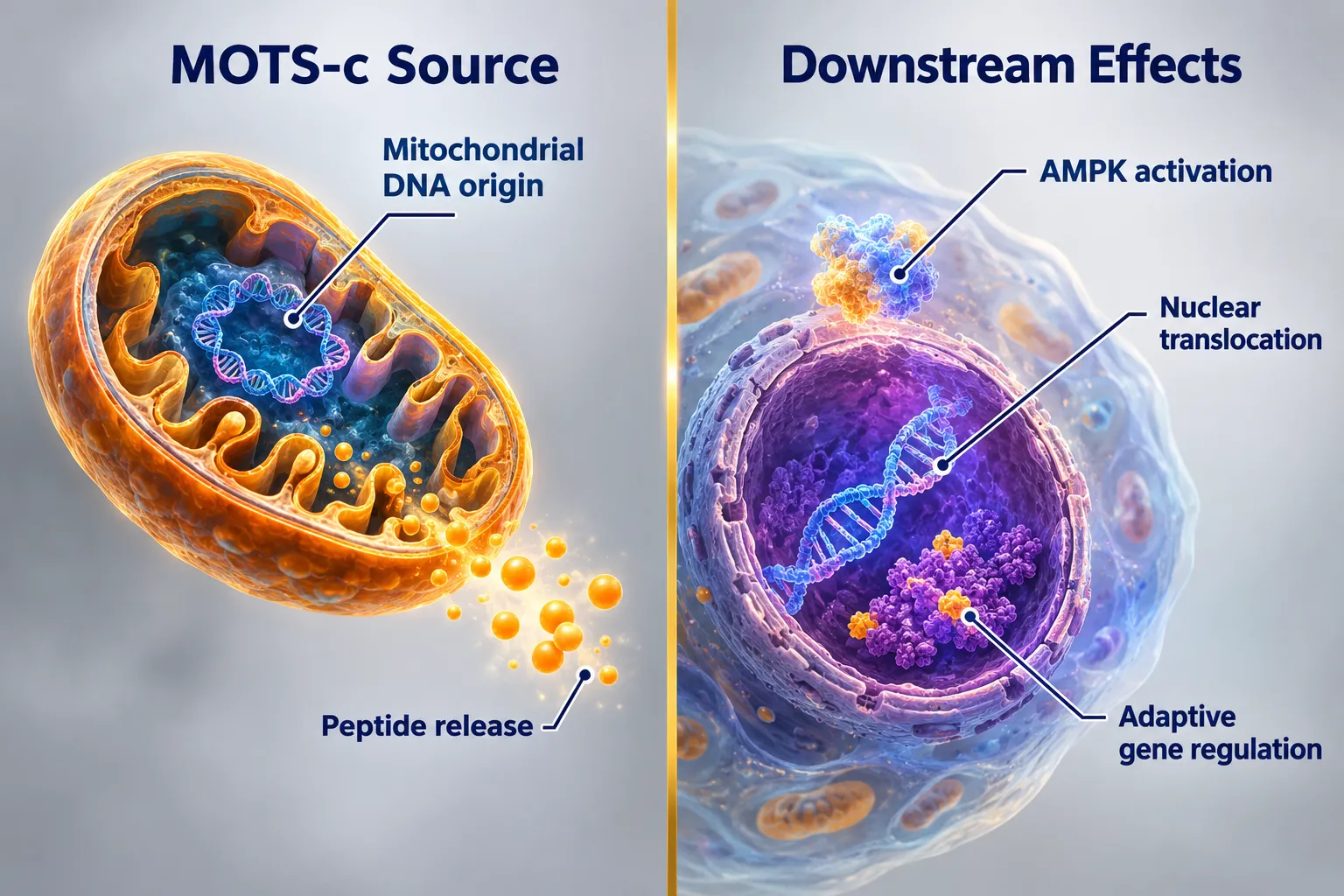
A parallel and rapidly expanding area of research concerns mitochondria-derived peptides (MDPs), bioactive microproteins encoded by short open reading frames within mitochondrial DNA itself. The best-characterized MDPs include MOTS-c, humanin, and the small humanin-like peptides (SHLPs). These molecules influence glucose and lipid metabolism, stress resistance, and longevity pathways, making them central to any peptide strategy aimed at optimizing ATP production and metabolic resilience.

MOTS-c, a 16-amino-acid MDP, has produced some of the most compelling human data to date. A 2026 trial reported in the Journal of Cellular Biochemistry found that twice-weekly subcutaneous dosing of 5-10 mg MOTS-c increased skeletal muscle ATP turnover by approximately 18-22% over 12 weeks in 84 adults aged 35-55. Separate work in diabetic models shows that MOTS-c can restore mitochondrial function and improve metabolic parameters under insulin-resistant conditions, extending its relevance beyond rare diseases.

Humanin and the SHLPs are also under active investigation for neurodegenerative diseases, Alzheimer's, Parkinson's, and Huntington's, where maintaining neuronal ATP supply and limiting mitochondrial stress are critical survival factors for neurons.

Research Insight: Peptide researchers now categorize mitochondria-targeting compounds into three mechanistic classes: those that support mitochondrial biogenesis (MOTS-c, humanin), those that directly enhance ATP synthesis (SS-31/elamipretide), and those optimized for NAD+ synergy, often combined with NMN (~500 mg/day) or NR (~300 mg/day) to simultaneously support electron transport chain substrate availability.

This three-class framework helps explain endpoint selection. A researcher studying an SS-31 analog will measure cardiolipin integrity and ATP synthase flux. A researcher studying MOTS-c will track glucose uptake, mitochondrial biogenesis markers, and ATP turnover rates. The choice of endpoint is not arbitrary, it follows directly from the peptide's mechanism of action.

Next-generation delivery platforms are also entering the picture. A 2026 study introduced a cationic liposomal system that co-delivers SS-31 with metabolic agents directly to adipose-tissue mitochondria, aiming to modulate fat-cell energy metabolism in obesity models. While this platform remains preclinical, it illustrates how cellular energy and ATP output have become central design constraints for advanced drug delivery research.

For researchers sourcing compounds for controlled laboratory investigations, quality documentation is a critical prerequisite. Resources covering peptide Certificate of Analysis standards and sourcing guides such as the GHK-Cu copper peptide research sourcing guide provide useful frameworks for evaluating purity and traceability before beginning any cellular energy study.

Mitochondria-Derived Peptides and the Broader Cellular Energy Architecture

The synergy between different peptide classes is also drawing attention. Combining compounds that target different nodes of the mitochondrial energy network, membrane stabilization, biogenesis signaling, and substrate availability, reflects how researchers now think in terms of integrated cellular energy architectures rather than single-target interventions. The documented synergy of LL-37 and SS-31 offers one example of how multi-peptide approaches are being explored in preclinical settings.

Conclusion

Adenosine triphosphate, mitochondrial function, and why peptide researchers care about cellular energy ultimately comes down to measurement and mechanism. ATP is not just a biological detail, it is the most direct indicator of whether a mitochondria-targeted intervention is producing a real cellular effect. The FDA approval of elamipretide, the human data on MOTS-c, and the expanding library of MDPs all point toward the same conclusion: peptides offer unusually precise tools for modulating cellular energy, and researchers who understand the underlying ATP biology are better equipped to design studies, select endpoints, and interpret results.

Actionable next steps for researchers:

  • Identify which mechanistic class a candidate peptide belongs to (biogenesis support, direct ATP enhancement, or NAD+ synergy) before selecting outcome measures.
  • Use ATP turnover rate, mitochondrial membrane potential, and ROS levels as primary endpoints rather than relying solely on downstream functional markers.
  • Review available mechanistic literature on cardiolipin-targeted peptides, particularly SS-31 research, to establish a baseline for comparing novel compound data.
  • Prioritize sourcing compounds with verified Certificates of Analysis to ensure that purity variables do not confound cellular energy measurements.
  • Consider multi-class peptide combinations in study design, informed by the growing body of work on integrated mitochondrial energy architectures.
https://www.puretestedpeptides.com/wp-content/uploads/2026/08/adenosine-triphosphate-mitochondrial-function-and-why-peptide-researchers-care-a.webp 1024 1536 https://www.puretestedpeptides.com/wp-content/uploads/2026/01/buy-peptides-online.jpg 2026-08-18 13:08:442026-08-18 13:08:44Adenosine Triphosphate, Mitochondrial Function, and Why Peptide Researchers Care About Cellular Energy
Collagen Research Peptides: Where GHK-Cu, Glow Blend, and Skin-Focused Formulas Fit in Laboratory Models

Collagen Research Peptides: Where GHK-Cu, Glow Blend, and Skin-Focused Formulas Fit in Laboratory Models

August 18, 2026/0 Comments/in Uncategorized/by

Copper-binding tripeptide GHK-Cu has appeared in peer-reviewed literature for more than five decades, yet its role inside modern laboratory frameworks is still evolving rapidly. As of 2026, the compound sits at the center of a broader conversation about collagen research peptides: where GHK-Cu, Glow Blend, and skin-focused formulas fit in laboratory models, a question that matters to researchers who want to understand what the data actually supports versus what is extrapolated from single-peptide studies.

This article compares the major research frameworks, clarifies how multi-peptide "glow" stacks are positioned relative to direct clinical evidence, and outlines what distinguishes rigorous laboratory models from commercially motivated formulations.

Key Takeaways

  • GHK-Cu has the strongest direct preclinical and emerging clinical evidence among collagen-focused peptides in 2026.
  • Glow blends are multi-peptide stacks that extrapolate from single-peptide data rather than carrying independent clinical trial support.
  • Epigenetic and gene-expression profiling is reshaping how researchers understand GHK-Cu's mechanism in aging skin.
  • Laboratory models distinguish between peptides with direct ECM evidence and those relying on mechanistic synergy arguments.
  • All glow blend and GHK-Cu formulations discussed here are research-grade compounds, not approved therapeutics.

GHK-Cu: The Anchor of Collagen Peptide Research

GHK-Cu: The Anchor of Collagen Peptide Research

GHK-Cu (glycyl-L-histidyl-L-lysine copper complex) is a naturally occurring tripeptide found in human plasma, saliva, and urine. Its concentration declines with age, and that decline correlates with measurable reductions in skin collagen density. This biological context makes it the logical anchor for any discussion of collagen research peptides in laboratory settings.

In preclinical models, GHK-Cu consistently demonstrates several well-documented actions:

  • Upregulation of collagen I and III synthesis in dermal fibroblasts
  • Inhibition of matrix metalloproteinases (MMPs), the enzymes that degrade the extracellular matrix (ECM)
  • Stimulation of elastin and glycosaminoglycan production
  • Antioxidant and anti-inflammatory signaling through copper-dependent pathways

What makes 2026 research particularly compelling is the shift toward epigenetic profiling. Recent studies have moved beyond simple protein expression assays to examine how GHK-Cu modulates gene networks associated with aging skin. This mechanistic depth gives researchers a more complete picture of why the peptide affects collagen turnover rather than just confirming that it does.

A Phase 2 clinical trial currently underway is testing a topical GHK-Cu gel specifically for acute wound re-epithelialization, marking a significant step from preclinical evidence toward controlled human data. Additionally, modified GHK constructs embedded in hydrogel scaffolds have shown enhanced wound repair in diabetic animal models, broadening the peptide's research scope beyond cosmetic applications.

For researchers sourcing this compound, the GHK-Cu peptide purchase and copper peptide research sourcing guide provides practical guidance on purity standards and documentation requirements.

Glow Blends: How Multi-Peptide Stacks Are Positioned in Lab Discussions

Glow Blends: How Multi-Peptide Stacks Are Positioned in Lab Discussions

The term "glow blend" refers to standardized multi-peptide stacks built around GHK-Cu, typically combined with complementary compounds such as matrikines, antioxidant peptides, or growth-factor analogs. These formulations are designed to address multiple pathways in collagen synthesis and ECM maintenance simultaneously.

How glow blends differ from single-peptide models:

Feature Single-Peptide GHK-Cu Glow Blend Stack
Clinical evidence base Direct RCT and preclinical data Extrapolated from component studies
Mechanism clarity Well-characterized Synergy assumed, not always tested
Research utility Mechanistic endpoint studies Exploratory multi-pathway screening
Regulatory status Research-grade Research-grade

The critical distinction is that glow blends extrapolate from existing single-peptide data rather than carrying independent clinical trial support. Commercially available "Glow Mix" formulations in 2026 emphasize mechanistic synergy, the idea that combining peptides with complementary targets produces additive or synergistic ECM effects. This is a scientifically reasonable hypothesis, but it is not the same as demonstrated clinical efficacy.

In laboratory discussions, this matters because researchers need to know whether they are working with a validated model or a plausible construct. Glow blends are best understood as exploratory frameworks for multi-pathway screening rather than as replacements for single-peptide mechanistic studies.

This is consistent with how other multi-peptide research blends are evaluated. Researchers familiar with stacks like the Tesamorelin CJC-1295 Ipamorelin 12mg blend will recognize the same principle: combining peptides with complementary mechanisms requires careful interpretation of which component drives which endpoint.

Skin-Focused Formulas and the Evolving Laboratory Framework

Skin-Focused Formulas and the Evolving Laboratory Framework

Skin-focused peptide research in 2024 through 2026 has been consolidating smaller randomized controlled trials into more comprehensive summaries. Human imaging studies, including high-frequency ultrasound and reflectance confocal microscopy, have linked GHK-Cu formulations to measurable gains in collagen and elastin density in vivo, providing a bridge between cell culture data and real-world skin biology.

This consolidation is reshaping how laboratory models are structured. Key developments include:

  1. Gene-expression profiling as a standard endpoint alongside protein assays
  2. Epigenetic markers of skin aging used to assess peptide efficacy over time
  3. Hydrogel and scaffold delivery systems that improve peptide stability and localized concentration
  4. Diabetic wound models as a secondary research context for GHK-Cu constructs

The industry sentiment in 2026 reflects a "growth surge" in GHK-Cu interest, driven partly by its anti-aging positioning and partly by the accumulating mechanistic data. However, researchers are advised to maintain clear boundaries between compounds with direct evidence and those whose benefits are inferred.

For broader context on how purity and sourcing affect research validity, the lab tested peptides resource and the high purity peptide sourcing tag page offer relevant quality benchmarks. Researchers exploring adjacent metabolic peptide frameworks may also find value in the top 5 research peptides for metabolic health buyer's guide.

A note on regulatory context: All glow blend and GHK-Cu formulations discussed in this article are sold as research-grade compounds. They are not approved therapeutics, and findings from laboratory models should not be extrapolated to human clinical use without appropriate trial design and regulatory oversight.

Conclusion

The landscape of collagen research peptides in 2026 is more nuanced than a simple ranking of compounds. GHK-Cu holds the strongest direct evidence base, supported by decades of preclinical work, emerging Phase 2 clinical data, and increasingly sophisticated epigenetic profiling. Glow blends occupy a legitimate but distinct space, useful for exploratory multi-pathway research, but dependent on extrapolated rather than independent clinical evidence.

Actionable next steps for researchers:

  • Prioritize single-peptide GHK-Cu models when mechanistic clarity is the goal
  • Use glow blends for hypothesis-generating, multi-pathway screening protocols
  • Verify purity documentation and third-party testing before incorporating any compound into a study
  • Follow ongoing Phase 2 trial data on topical GHK-Cu to understand how preclinical findings translate to human endpoints
  • Distinguish between ECM-direct evidence and synergy-based arguments when evaluating formulation claims

Understanding where each formula sits within the evidence hierarchy is not a minor detail, it determines the validity of every endpoint a laboratory model is designed to measure.

https://www.puretestedpeptides.com/wp-content/uploads/2026/08/collagen-research-peptides-where-ghk-cu-glow-blend-and-skin-focused-formulas-fit.webp 1024 1536 https://www.puretestedpeptides.com/wp-content/uploads/2026/01/buy-peptides-online.jpg 2026-08-18 13:08:362026-08-18 13:08:36Collagen Research Peptides: Where GHK-Cu, Glow Blend, and Skin-Focused Formulas Fit in Laboratory Models
Peptides and Polypeptides: Complete Research Guide for GLP-1, GLP-2, GLP-3, and Growth Hormone Peptides

Peptides and Polypeptides: Complete Research Guide for GLP-1, GLP-2, GLP-3, and Growth Hormone Peptides

August 18, 2026/0 Comments/in Uncategorized/by

Over 100 distinct peptide-based drugs are currently in active clinical development worldwide, yet most researchers encounter these molecules without a clear structural map of how they relate to one another. This guide on Peptides and Polypeptides: Complete Research Guide for GLP-1, GLP-2, GLP-3, and Growth Hormone Peptides addresses that gap directly, building a scientific foundation before diving into specific compound families.

Key Takeaways

  • Peptides are short amino acid chains; polypeptides are longer chains that fold into functional proteins, size determines receptor specificity and research use.
  • GLP-1, GLP-2, and GLP-3 all originate from the same proglucagon gene but act on entirely different receptor systems with distinct biological roles.
  • GLP-1 agonists represent the most clinically active peptide class in 2026, with oral, injectable, and ultra-long-acting formats now available or in late-stage trials.
  • Growth hormone-releasing peptides and analogs operate through the hypothalamic-pituitary axis, making them mechanistically distinct from GLP-class compounds.
  • Purity and structural integrity are non-negotiable in peptide research, third-party testing is the baseline standard.

Understanding Peptide and Polypeptide Structure

Understanding Peptide and Polypeptide Structure

A peptide is any chain of two or more amino acids linked by peptide bonds. The classification system is straightforward:

Term Chain Length Example
Dipeptide 2 amino acids Carnosine
Oligopeptide 3-20 amino acids GLP-1 (30 aa)
Polypeptide 20-50+ amino acids Growth hormone fragments
Protein 50+ amino acids Full-length GH (191 aa)

The distinction matters in research because chain length directly influences receptor selectivity, half-life, and delivery route. Shorter peptides often cross biological barriers more easily but degrade faster. Longer polypeptides may require injectable delivery to preserve their three-dimensional structure.

Receptor binding is the next critical concept. Most research peptides act on G-protein coupled receptors (GPCRs), triggering intracellular signaling cascades rather than directly altering gene expression. This mechanism produces rapid, dose-dependent responses that researchers can measure with precision, a key advantage in preclinical models.

"Peptide size, charge, and secondary structure are not incidental features, they are the mechanism."

For researchers building a broader framework, the top 5 research peptides for metabolic health buyer's guide offers a practical starting point for compound selection within this structural context.

GLP-1, GLP-2, and GLP-3: The Proglucagon Peptide Family

GLP-1, GLP-2, and GLP-3: The Proglucagon Peptide Family

All three glucagon-like peptides derive from a single precursor protein called proglucagon, encoded by the GCG gene. Post-translational processing in different tissues produces distinct peptide fragments with entirely separate biological roles.

GLP-1: The Dominant Research Target

GLP-1 (glucagon-like peptide-1) is a 30-amino-acid incretin hormone secreted by intestinal L-cells. It stimulates insulin secretion, suppresses glucagon, slows gastric emptying, and signals satiety through the central nervous system. These combined actions make it the most studied metabolic peptide in modern pharmacology.

In 2026, the GLP-1 landscape has expanded dramatically:

  • Oral non-peptide GLP-1 agonists such as orforglipron (Foundayo, Eli Lilly) have received approval for chronic weight management, making oral GLP-1 a mainstream modality for the first time.
  • High-dose injectable semaglutide (Wegovy HD, 7.2 mg weekly) extends efficacy for patients requiring greater weight reduction.
  • Ultra-long-acting monthly injectables, including Pfizer's PF-3944/MET-097i, have shown robust Phase 2b results, potentially reducing injection frequency to once per month.
  • Multi-agonist peptides combining GLP-1 with GIP and glucagon receptor activity show the highest weight-loss efficacy seen in late-stage trials to date.

Emerging research also points to non-metabolic applications: addiction neuroscience, mood regulation, and neuroinflammation are active areas of investigation, though these remain speculative outside controlled settings.

Researchers sourcing compounds in this class should review GLP-1 peptide buying: generational research concepts and sourcing notes for structured guidance on acquisition standards. Those evaluating specific product options can also browse GLP-1 peptides available for research.

GLP-2: Intestinal Repair and Nutrient Absorption

GLP-2 is a 33-amino-acid peptide co-secreted with GLP-1 from L-cells. Its receptor is expressed almost exclusively in the gastrointestinal tract. GLP-2 promotes intestinal epithelial growth, reduces gut permeability, and enhances nutrient absorption. Research applications center on short bowel syndrome, inflammatory bowel conditions, and intestinal barrier function.

Researchers working with this compound can find relevant sourcing information under GLP-2 peptide research products.

GLP-3: The Least Characterized Fragment

GLP-3 is a proglucagon-derived fragment whose receptor biology remains incompletely mapped. Public research output on GLP-3 is limited compared to GLP-1 and GLP-2, and no approved therapeutic agents target this peptide as of 2026. It represents an early-stage area where foundational receptor characterization work is still ongoing. Researchers interested in this compound can explore GLP-3 peptide sourcing options as a starting reference.

Growth Hormone Peptides: Axis, Mechanism, and Research Context

Growth Hormone Peptides: Axis, Mechanism, and Research Context

Growth hormone (GH) peptides operate through a fundamentally different axis than GLP-class compounds. The hypothalamic-pituitary-somatotropic axis governs GH release, and research peptides in this category generally work by modulating one or more points along that pathway.

Key categories include:

  • GHRH analogs, mimic growth hormone-releasing hormone to stimulate pulsatile GH secretion from the anterior pituitary. Tesamorelin is the most studied example; researchers can review tesa peptide benefits and research context for a detailed breakdown.
  • GHRPs (growth hormone-releasing peptides), act on ghrelin receptors (GHSR-1a) to amplify GH pulses, often synergistically with GHRH analogs.
  • GH fragments, truncated polypeptide sequences derived from full-length growth hormone, studied for specific downstream effects on fat metabolism and tissue repair.

Downstream from GH release, IGF-1 production in the liver drives many of the tissue-level effects researchers are interested in: protein synthesis, cellular repair, and metabolic substrate utilization. Understanding this cascade is essential for interpreting research data correctly.

Research Standards: Purity, Benchmarking, and Sourcing

The structural complexity of peptides makes quality control non-negotiable. A single incorrect amino acid, oxidized residue, or truncated sequence can produce misleading results or no activity at all.

Minimum standards for research-grade peptides:

  • HPLC purity of 98% or greater
  • Mass spectrometry confirmation of molecular weight
  • Third-party certificate of analysis (CoA) from an independent laboratory
  • Sterility and endotoxin testing for injectable preparations

Reference standards from established manufacturers provide the benchmark against which research samples should be validated. The article on Bachem reference standards and building robust peptide benchmarks outlines how to use certified reference materials effectively.

Researchers should also confirm that suppliers offer lab-tested peptides with verifiable documentation before committing to a source.

Conclusion

The Peptides and Polypeptides: Complete Research Guide for GLP-1, GLP-2, GLP-3, and Growth Hormone Peptides framework presented here gives researchers a reliable map before engaging with any specific compound. The actionable next steps are clear:

  1. Establish structural literacy first, know whether a target peptide is an oligopeptide or polypeptide, and how that affects delivery and receptor interaction.
  2. Match the compound to the correct receptor family, GLP-1, GLP-2, and GLP-3 are not interchangeable despite sharing a common precursor.
  3. Understand the signaling axis, GH peptides require knowledge of the hypothalamic-pituitary cascade to interpret results meaningfully.
  4. Demand verified purity, third-party CoA documentation is the baseline, not a bonus.
  5. Stay current, the GLP-1 field in particular is evolving rapidly, with oral formats, multi-agonists, and monthly injectables reshaping the research landscape throughout 2026 and beyond.

A strong structural foundation makes every downstream research decision more defensible and more productive.

https://www.puretestedpeptides.com/wp-content/uploads/2026/08/peptides-and-polypeptides-complete-research-guide-for-glp-1-glp-2-glp-3-and-grow.webp 1024 1536 https://www.puretestedpeptides.com/wp-content/uploads/2026/01/buy-peptides-online.jpg 2026-08-18 13:08:362026-08-18 13:08:36Peptides and Polypeptides: Complete Research Guide for GLP-1, GLP-2, GLP-3, and Growth Hormone Peptides
Mitochondria and Research Peptides: What Labs Measure When Studying Cellular Energy Signaling

Mitochondria and Research Peptides: What Labs Measure When Studying Cellular Energy Signaling

August 18, 2026/0 Comments/in Uncategorized/by

Fewer than 1% of the roughly 1,500 proteins inside a human mitochondrion have been fully characterized for their role in energy output, yet that small fraction already underpins some of the most active areas in peptide research today. Understanding Mitochondria and Research Peptides: What Labs Measure When Studying Cellular Energy Signaling is no longer a niche concern for biochemists alone. In 2026, it sits at the center of metabolic disease research, rare disease trials, and next-generation therapeutic design.

Key Takeaways

  • Mitochondrial function is measured through oxygen consumption rate (OCR) and ATP production assays, most commonly using Seahorse XF technology.
  • Research peptides such as SS-31 and MOTS-c target distinct nodes in the mitochondrial energy network and produce measurable, quantifiable effects.
  • Labs convert raw OCR data into ATP production rates using established bioenergetic equations, enabling direct comparison across studies.
  • Mitochondrial targeting sequences (MTS) are engineered design features that labs validate through membrane potential and proteomics assays.
  • Peptide purity and certificate of analysis standards directly affect the reliability of cellular energy signaling data.

Why Mitochondrial Energy Signaling Demands Precise Measurement

Why Mitochondrial Energy Signaling Demands Precise Measurement

Mitochondria are not static power generators. They are dynamic organelles that constantly shift their output in response to nutrient availability, stress signals, and intercellular communication. When a research peptide enters this environment, it can alter membrane potential, modulate electron transport chain activity, or change the rate at which ATP synthase produces adenosine triphosphate.

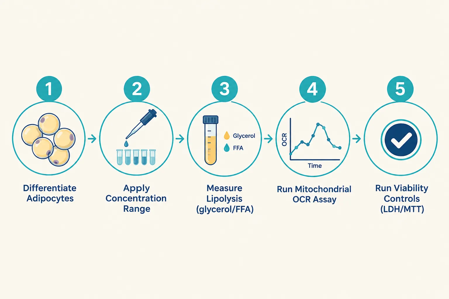
Labs studying cellular energy signaling need quantitative endpoints, not qualitative impressions. The most widely adopted platform for this work is the Seahorse XF Analyzer, which measures oxygen consumption rate (OCR) and extracellular acidification rate (ECAR) in live cells in real time. These two metrics together reveal how a cell balances oxidative phosphorylation against glycolysis, a balance that research peptides can shift in measurable ways.

"The OCR value alone tells you how hard the mitochondria are working. The ATP production rate tells you how efficiently that work translates into usable energy."
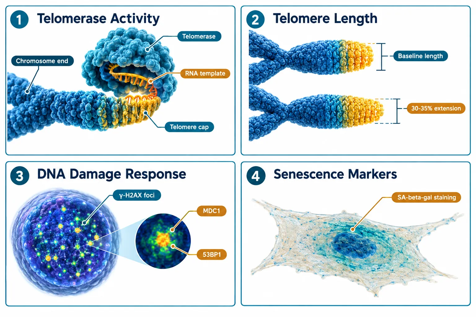
Beyond OCR, labs also track:

  • Mitochondrial membrane potential (using dyes such as JC-1 or TMRE)
  • Reactive oxygen species (ROS) output via fluorescent probes
  • NAD+/NADH ratios as indicators of redox balance
  • Mitochondrial fusion and fission dynamics through live-cell imaging

A 2025 study examining LRRK2 gene mutations used this exact framework to quantify ATP deficits in patient-derived neurons, demonstrating how OCR-to-ATP conversion math can anchor a disease mechanism to a specific molecular target.

Core Assays in Mitochondria and Research Peptides Studies

Core Assays in Mitochondria and Research Peptides Studies

The Seahorse XF Mito Stress Test

The standard Seahorse mito stress test injects three compounds in sequence, oligomycin, FCCP, and rotenone/antimycin A, each blocking a different step in the respiratory chain. The resulting OCR curve reveals:

Parameter What It Reflects
Basal respiration Baseline mitochondrial workload
ATP-linked respiration OCR directly coupled to ATP synthesis
Maximal respiration Full electron transport chain capacity
Spare respiratory capacity Metabolic reserve under stress
Proton leak Membrane integrity and uncoupling

Labs convert ATP-linked OCR to an ATP production rate by applying a stoichiometric factor derived from the P/O ratio, roughly 2.73 ATP per oxygen atom consumed during oxidative phosphorylation. This conversion is critical when comparing peptide-treated cells against controls.

Proteomics and Interaction Networks

SS-31 (elamipretide) has become a model compound for understanding how a peptide reshapes the mitochondrial protein interaction landscape. Proteomics studies have mapped SS-31's binding to cardiolipin on the inner mitochondrial membrane, revealing downstream stabilization of cristae architecture and suppression of cytochrome c release. Labs use co-immunoprecipitation and proximity labeling (BioID) to build these interaction networks.

For researchers sourcing this compound, the SS-31 10mg research peptide considerations page outlines purity and handling requirements that directly affect assay reproducibility. Additional context on SS-31 mitochondrial dynamics research is also available for deeper background.

Comparing SS-31 and MOTS-c: Two Distinct Signaling Profiles

Comparing SS-31 and MOTS-c: Two Distinct Signaling Profiles

In 2026 analyses, researchers have drawn a sharper line between SS-31 and MOTS-c, two peptides that both influence mitochondrial energy output but through fundamentally different mechanisms.

SS-31 acts at the inner mitochondrial membrane. It binds cardiolipin, reduces ROS production, and stabilizes the electron transport chain supercomplexes. Its primary measurable effect is an increase in ATP-linked OCR and a reduction in proton leak, outcomes directly visible in Seahorse assay data. Clinical trials in Barth syndrome and primary mitochondrial myopathy (MMPOWER-3) used functional endpoints such as the six-minute walk test and fatigue scores alongside these bioenergetic markers, though MMPOWER-3 showed limited efficacy improvements over placebo.

MOTS-c, by contrast, is a mitochondria-derived peptide that translocates to the nucleus under metabolic stress. It activates AMPK signaling, upregulates antioxidant gene expression, and shifts cellular metabolism toward glucose utilization. Labs measure its effects through AMPK phosphorylation assays, gene expression panels, and glucose uptake assays rather than pure OCR data.

This distinction matters for experimental design. Researchers exploring metabolic health applications can review the top 5 research peptides for metabolic health to understand how these compounds compare in applied research contexts.

Engineering Mitochondrial Targeting Sequences

Beyond naturally occurring peptides, labs now engineer mitochondrial targeting sequences (MTS), short amphipathic helical peptides that guide attached cargo into the mitochondrial matrix. Key metrics labs validate include:

  • Import efficiency (measured by protease protection assays)
  • Membrane potential dependence (collapsed by CCCP treatment)
  • Submitochondrial localization (outer membrane vs. matrix)

Plant-derived peptides such as roseltide rT1 have served as structural models for MTS design, demonstrating that even non-mammalian sequences can modulate ATP production in cell-free and cell-based systems. Reference standards for these comparisons are discussed in detail in the Bachem and reference standards guide for building robust peptide benchmarks.

Data Quality and Peptide Sourcing in Energy Signaling Research

The reliability of any cellular energy signaling dataset depends on the quality of the peptide used. Impurities in a research-grade compound can independently alter OCR, membrane potential, or ROS output, confounding results in ways that are difficult to detect post hoc.

Labs should require:

  • Certificate of Analysis (CoA) with HPLC purity above 98%
  • Mass spectrometry confirmation of molecular weight
  • Endotoxin testing results for cell-based assays
  • Lot-specific documentation for reproducibility across experiments

Researchers can find guidance on evaluating supplier documentation through peptide CoA standards and documentation and peptide supplier comparison resources. For those studying kidney-specific mitochondrial applications, SS-31 kidney health research provides organ-specific context for interpreting bioenergetic data.

Conclusion

The intersection of Mitochondria and Research Peptides: What Labs Measure When Studying Cellular Energy Signaling is defined by rigorous quantitative methods, Seahorse OCR profiling, ATP rate calculations, proteomics interaction mapping, and MTS validation assays. SS-31 and MOTS-c exemplify how two peptides targeting the same organelle can produce entirely different measurable signatures, requiring distinct experimental frameworks.

Actionable next steps for research teams:

  1. Standardize OCR-to-ATP conversion using published P/O ratios before comparing datasets across labs.
  2. Pair Seahorse assay data with at least one orthogonal endpoint (membrane potential or ROS) to validate findings.
  3. Confirm peptide purity via CoA and mass spectrometry before any cell-based energy assay.
  4. Select peptide compounds based on the specific node of the energy signaling pathway under investigation, membrane-targeted versus nuclear-translocating mechanisms require different readouts.
  5. Document lot numbers and storage conditions for every experiment to support reproducibility.

As mitochondrial peptide research matures in 2026, the labs that invest in measurement precision will produce the datasets that hold up to scrutiny, and drive the field forward.

https://www.puretestedpeptides.com/wp-content/uploads/2026/08/mitochondria-and-research-peptides-what-labs-measure-when-studying-cellular-ener.webp 1024 1536 https://www.puretestedpeptides.com/wp-content/uploads/2026/01/buy-peptides-online.jpg 2026-08-18 13:08:342026-08-18 13:08:34Mitochondria and Research Peptides: What Labs Measure When Studying Cellular Energy Signaling
Epithalon Peptide Formulations: How Labs Compare Lyophilized vs Solution Stability in Telomere Research

Epithalon Peptide Formulations: How Labs Compare Lyophilized vs Solution Stability in Telomere Research

August 17, 2026/0 Comments/in Uncategorized/by

A reconstituted Epithalon solution left at room temperature can lose meaningful biological activity within a matter of days, a detail that can quietly invalidate weeks of telomere-length data if it goes unnoticed. For labs running telomerase activation assays or tracking telomere elongation across multiple time points, the choice between lyophilized and solution formulations is not a minor logistical preference. It is a core experimental variable.

This article focuses specifically on degradation kinetics, storage conditions, and formulation selection for Epithalon peptide formulations, practical intelligence for researchers already familiar with the peptide's mechanism and looking to optimize their experimental design.

Key Takeaways

  • Lyophilized Epithalon stored at minus 20 C retains greater than 95% purity for up to 24 months; reconstituted solutions in bacteriostatic water are limited to approximately 28 days at 2 to 8 C.
  • Solutions prepared in plain sterile water (no preservative) should be discarded within 24 hours.
  • Moisture and light are the primary degradation drivers for dry powder; hydrolysis, oxidation, and temperature stress govern solution stability.
  • Multi-site telomere studies increasingly ship only lyophilized vials and reconstitute locally just before use to standardize reagent quality.
  • Minus 80 C storage offers maximum stability for archival lots, but standard minus 20 C freezers are adequate for routine experimental stocks.

Why Formulation Choice Matters in Epithalon Peptide Formulations for Telomere Research

Why Formulation Choice Matters in Epithalon Peptide Formulations for Telomere Research

Epithalon (Ala-Glu-Asp-Gly) is a synthetic tetrapeptide supplied almost exclusively as lyophilized powder at 95 to 99% purity, typically in 10 mg vials. Research datasets have reported a 26-fold increase in telomerase activity in normal human mammary epithelial cells and approximately 33% longer telomeres in human fetal fibroblast cultures, making reagent integrity central to reproducible results.

The stability gap between the two formulation types is substantial:

Formulation Storage Condition Estimated Stability
Lyophilized powder minus 20 C, desiccated, dark Up to 24 months (>95% purity)
Lyophilized powder 2 to 8 C, sealed 18 to 24 months
Lyophilized powder Room temperature Approximately 3 weeks
Reconstituted in bacteriostatic water 2 to 8 C Up to 28 days
Reconstituted in sterile water 2 to 8 C 24 hours maximum
Reconstituted solution Room temperature Up to 72 hours cumulative

The core principle: dry-state stability is measured in years; solution stability is measured in days to weeks.

For researchers sourcing compounds alongside Epithalon, the same formulation discipline applies to related peptides. The SS-31 mitochondrial research themes resource covers analogous storage considerations for another stability-sensitive peptide used in oxidative stress models.

Degradation Mechanisms: What Destroys Each Formulation

Understanding what drives degradation helps labs design storage protocols rather than simply follow them by rote.

Lyophilized Powder Degradation

For dry Epithalon, the two dominant threats are moisture and light. Humidity exposure markedly accelerates degradation, compressing shelf life from years to months. This is why vacuum-sealed, desiccated packaging has become standard for telomere research inventories. Even brief exposure to ambient humidity during weighing or vial transfer can initiate hydrolysis at the peptide bonds.

"Exposure of lyophilized Epithalon to humidity markedly accelerates degradation, shortening usable shelf life from years to mere months."

Practical controls include:

  • Working quickly in low-humidity environments when opening vials
  • Using desiccant packs inside storage boxes
  • Returning unused powder to sealed containers immediately

Solution Degradation

Once reconstituted, Epithalon faces a broader set of chemical stressors:

  • Hydrolysis at peptide bonds, accelerated by temperature and pH
  • Oxidation of susceptible residues
  • Adsorption onto container surfaces, reducing effective concentration
  • Microbial contamination if aseptic technique is not maintained
  • Freeze-thaw stress when solutions are repeatedly cycled

Bacteriostatic water (containing 0.9% benzyl alcohol) extends usable solution life to approximately 28 days at 2 to 8 C by suppressing microbial growth. Plain sterile water provides no such protection, limiting use to 24 hours.

Frozen solutions should not undergo more than a few freeze-thaw cycles. Each cycle introduces mechanical stress and concentration gradients that accelerate structural degradation.

For context on how similar degradation principles apply across peptide classes, the BPC-157 core peptides documentation first research guide provides a useful parallel framework.

Practical Storage Protocols for Epithalon Peptide Formulations in Telomere Experiments

Practical Storage Protocols for Epithalon Peptide Formulations in Telomere Experiments

Designing a storage protocol around Epithalon peptide formulations requires matching storage tier to experimental timeline.

Three-tier storage model:

  1. Archival lots (multi-year studies): minus 80 C, desiccated, light-protected. While not strictly required, this tier provides maximum stability for long telomere-tracking projects where reagent consistency across years is critical.
  2. Active research stocks (routine use): minus 20 C, sealed vials with desiccant. This is the standard recommendation for day-to-day experimental peptide stocks and is adequate for most telomere assay workflows.
  3. Short-term working inventory: 2 to 8 C for lyophilized powder not expected to be used within 24 months. Purity remains above 95% for 18 to 24 months under these conditions.

Reconstitution best practices for telomere assays:

  • Reconstitute immediately before use rather than preparing bulk solutions in advance
  • Use bacteriostatic water as the diluent for any solution intended to be used over multiple days
  • Design TRAP assays and telomere-length measurement protocols so all planned sampling falls within a 2 to 7-day window after reconstitution
  • Aliquot reconstituted solution into single-use volumes to avoid repeated access to the same vial

Multi-site telomere studies have adopted a standardized approach: ship only lyophilized vials, reconstitute locally just before experimental use. This eliminates inter-site variability introduced by different solution ages and handling histories.

For labs evaluating supplier quality alongside storage planning, the peptide supplier comparisons resource interpreting PeptideTech and PeptideSC offers a structured framework for assessing documentation standards. Researchers sourcing Epithalon alongside other compounds can also consult the where to buy SS-31 and Epithalon online guide for supplier navigation. Additional quality control benchmarks relevant to research-grade peptide sourcing appear in the PT-141 peptide research context QA and controls article.

Applying Formulation Knowledge Across the Experiment Lifecycle

Applying Formulation Knowledge Across the Experiment Lifecycle

Formulation decisions intersect with every stage of a telomere study, from procurement through data collection.

At procurement: Request certificates of analysis confirming purity at or above 95%, lyophilized state, and storage conditions maintained during shipping. Cold-chain documentation matters for long-distance orders.
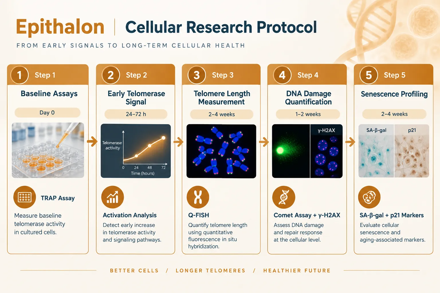
At intake: Log the vial arrival date, inspect packaging integrity, and transfer immediately to the appropriate storage tier. Vials showing signs of moisture ingress or color change should be quarantined.

During the experiment: Track cumulative room-temperature exposure for any reconstituted solution. The 72-hour cumulative limit at room temperature applies even if the solution has been refrigerated between uses.

At data analysis: Flag any data points collected from solutions older than the recommended stability window. Degraded Epithalon may produce attenuated telomerase activity readings, introducing systematic underestimation of effect size.

The GHK-Cu peptide purchase and copper peptide research sourcing guide demonstrates how analogous documentation practices are applied to other research-grade peptides with similar stability sensitivities.

Conclusion

Epithalon peptide formulations present a clear hierarchy of stability: lyophilized powder at minus 20 C is the gold standard for telomere research, offering verified purity above 95% for up to 24 months. Reconstituted solutions are working reagents with a defined shelf life, 28 days in bacteriostatic water at 2 to 8 C, 24 hours in plain sterile water, and no more than 72 cumulative hours at room temperature.

Actionable next steps for research teams:

  • Audit current storage conditions against the three-tier model and reassign vials to the appropriate temperature tier
  • Switch to bacteriostatic water as the default diluent for all reconstituted Epithalon solutions
  • Build a 2 to 7-day sampling window into telomere assay protocols to align with solution stability limits
  • Implement vial intake logging that captures arrival date, storage tier assignment, and first-use date
  • For multi-site studies, standardize on lyophilized shipment with local reconstitution to eliminate inter-site reagent variability

Rigorous formulation management does not add complexity to telomere research, it removes a hidden source of noise that can obscure real biological signals.

https://www.puretestedpeptides.com/wp-content/uploads/2026/08/epithalon-peptide-formulations-how-labs-compare-lyophilized-vs-solution-stabilit.webp 1024 1536 https://www.puretestedpeptides.com/wp-content/uploads/2026/01/buy-peptides-online.jpg 2026-08-17 13:05:172026-08-17 13:05:17Epithalon Peptide Formulations: How Labs Compare Lyophilized vs Solution Stability in Telomere Research
Where to Buy Glow Blend, Klow Blend, and Klow Nasal Peptide Sprays: Lab-Grade vs Cosmetic-Grade Options

Where to Buy Glow Blend, Klow Blend, and Klow Nasal Peptide Sprays: Lab-Grade vs Cosmetic-Grade Options

August 17, 2026/0 Comments/in Uncategorized/by

Fewer than 15% of peptide products sold online in 2026 carry independently verified purity data, yet demand for nasal peptide sprays like Glow Blend and Klow Blend has surged sharply across research and wellness communities. For anyone navigating where to buy Glow Blend, Klow Blend, and Klow Nasal Peptide Sprays: Lab-Grade vs Cosmetic-Grade Options, the distinction between a rigorously tested research vial and an unverified cosmetic spray is not a minor detail. It is the single most important factor in sourcing decisions.

Key Takeaways

  • Glow Blend and Klow Blend are proprietary multi-peptide research formulations, not FDA-approved or peer-reviewed products.
  • Lab-grade versions come with Certificate of Analysis (COA) documentation and HPLC purity data; cosmetic-grade versions typically do not.
  • Klow Blend is often described as "Glow plus KPV with higher GHK-Cu concentration," making it a more complex research stack.
  • US and international vendors standardize Klow Blend at 80 mg lab-grade vials; cosmetic sprays vary widely in concentration.
  • All components in these blends remain unapproved for human therapeutic use and are sold strictly for research purposes.

Understanding Glow Blend and Klow Blend Formulations

Understanding Glow Blend and Klow Blend Formulations

Glow Blend is a multi-peptide formulation centered on skin and recovery-focused peptides, most commonly including GHK-Cu (copper peptide) and BPC-157. It is positioned by research vendors as a compound of interest for tissue repair and dermal research. Klow Blend extends this profile by adding KPV (a tripeptide fragment of alpha-MSH) and increasing the GHK-Cu concentration. Expert commentary from August 2026 consistently frames Klow as "Glow plus KPV, higher GHK-Cu", a more targeted stack for researchers studying inflammatory response and skin-barrier mechanisms.

The Klow Nasal Peptide Spray delivers this four-peptide stack via intranasal administration. Nasal delivery is chosen by researchers because it bypasses first-pass metabolism and allows faster systemic absorption compared to oral routes. Vendors such as Nova Labs, Research Peptides Europe, and PeptidePowerEU (EU-focused suppliers) have standardized their lab-grade Klow Blend offerings at 80 mg vials with full batch documentation.

It is critical to note that "Klow Blend" carries no regulatory recognition and no peer-reviewed clinical classification as of mid-2026. It is a proprietary research concept. Researchers and clinicians must treat it accordingly.

Comparison: Glow Blend vs Klow Blend

Feature Glow Blend Klow Blend
Core peptides GHK-Cu, BPC-157 GHK-Cu (higher), BPC-157, KPV
Primary research focus Skin recovery, tissue repair Inflammation, skin barrier, recovery
Standard vial size Varies by vendor 80 mg (US/international standard)
Nasal spray format Available (cosmetic risk) Yes, lab-grade framing
COA typically included Lab-grade only Lab-grade only

Lab-Grade vs Cosmetic-Grade: The Core Distinction

Lab-Grade vs Cosmetic-Grade: The Core Distinction

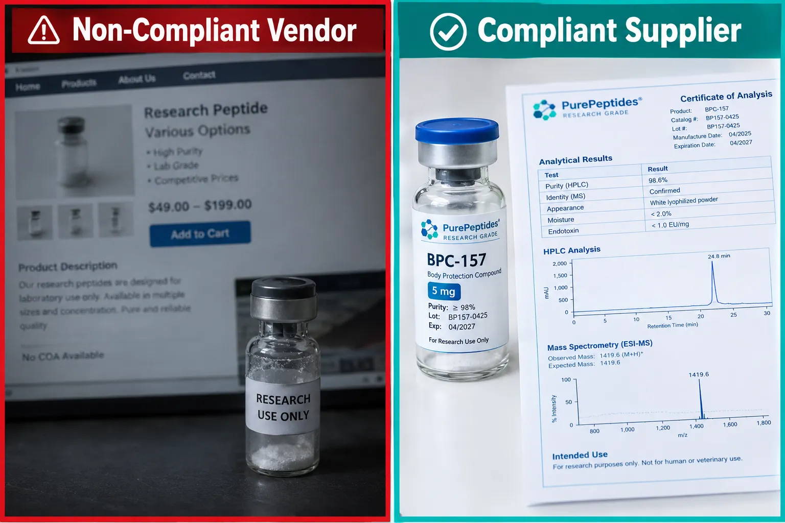
The phrase "lab-grade" in the peptide market refers to products manufactured under controlled conditions, tested by third-party laboratories, and supplied with a peptide COA (Certificate of Analysis). A genuine COA includes HPLC purity data, mass spectrometry confirmation, and batch-specific results. Without this documentation, there is no reliable way to confirm what is actually in the vial.

Cosmetic-grade nasal sprays occupy a legally ambiguous space. Glow nasal sprays marketed for "skin radiance" or "healing recovery" blur the line between research compounds and consumer wellness products. These products may use the same peptide names but offer no COA, no batch traceability, and no standardized concentration. The risk of underdosing, overdosing, or receiving a contaminated product increases significantly.

"A COA is not a marketing badge, it is the minimum evidence standard for any research-grade peptide purchase."

Researchers sourcing Semax, Selank, or complex blends like Glow and Klow should apply the same verification standard across all nasal peptide formats. For context on how rigorous sourcing applies to other peptide categories, the guide to where to buy SS31 and Epithalon online outlines the same COA-first framework.

Key markers of a lab-grade supplier:

  • Third-party HPLC and mass spec data per batch
  • Downloadable COA with lot number
  • "For research use only" labeling
  • Transparent manufacturing location
  • No therapeutic or cosmetic claims

Where to Buy Glow Blend, Klow Blend, and Klow Nasal Peptide Sprays: Lab-Grade vs Cosmetic-Grade Options

Where to Buy Glow Blend, Klow Blend, and Klow Nasal Peptide Sprays: Lab-Grade vs Cosmetic-Grade Options

Sourcing these compounds responsibly requires understanding where to buy Glow Blend, Klow Blend, and Klow Nasal Peptide Sprays across lab-grade vs cosmetic-grade options, and knowing which vendor categories to prioritize or avoid.

Lab-Grade Research Vendors (Recommended for Researchers)

US-based research peptide suppliers and EU-focused vendors including Nova Labs, Research Peptides Europe, and PeptidePowerEU have emerged as primary sources for verified Klow Blend in 2026. These vendors provide:

  • 80 mg standardized vials for Klow Blend
  • Full peptide COA verification with downloadable batch data
  • Research-only labeling with no therapeutic claims
  • Lyophilized powder format for stability

For researchers already familiar with growth hormone-related peptide blends, vendors offering products like the Tesamorelin CJC1295 Ipamorelin 12mg Blend typically apply the same documentation standards to Glow and Klow formulations. This consistency in quality control is a positive signal when evaluating a new supplier. Those seeking higher-dose configurations may also review the Tesamorelin CJC1295 Ipamorelin 12mg Blend Dosage140 as a benchmark for how reputable vendors structure multi-peptide research products.

Cosmetic and Wellness Channels (Use with Caution)

Cosmetic-grade Glow and Klow nasal sprays appear on wellness marketplaces, beauty retailers, and some compounding pharmacy-adjacent platforms. These products are often marketed as "radiance recovery" or "healing peptide therapy." They lack the documentation standards of lab-grade sources and should not be used in formal research contexts.

Nasal Peptide Sprays: Semax and Selank Context

Researchers comparing Glow and Klow to established nasal peptide formats should review the comparative research on Semax and Selank peptides, neurogenesis, and synaptic plasticity for a benchmark on how intranasal peptide delivery is studied. Semax and Selank have a longer research history and provide a useful reference point for evaluating newer nasal spray formulations.

For broader context on multi-peptide research sourcing, the top 5 research peptides for metabolic health buyer's guide covers vendor evaluation criteria applicable across peptide categories.

Regulatory and Safety Positioning

All components in Glow Blend, Klow Blend, and Klow Nasal Peptide Sprays remain unapproved by the FDA and equivalent regulatory bodies as of August 2026. They are not approved for human therapeutic use, diagnosis, or treatment. Every legitimate lab-grade supplier labels these products strictly for in vitro or preclinical research use only. Any vendor making health claims or omitting this labeling is a red flag.

Conclusion

Navigating where to buy Glow Blend, Klow Blend, and Klow Nasal Peptide Sprays: Lab-Grade vs Cosmetic-Grade Options comes down to one non-negotiable standard: documentation. Lab-grade suppliers provide COA data, batch traceability, and research-only labeling. Cosmetic-grade channels offer convenience but sacrifice the verification that research integrity demands.
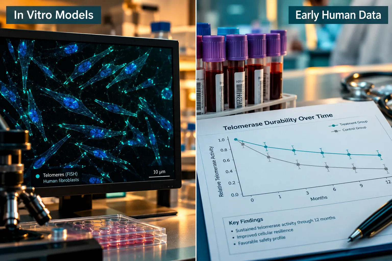
Actionable next steps for researchers in 2026:

  1. Request a downloadable COA with HPLC data before purchasing any Glow or Klow formulation.
  2. Confirm the vendor uses "research use only" labeling, not cosmetic or therapeutic claims.
  3. Cross-reference batch numbers against the supplier's published documentation.
  4. Treat Klow Blend as a four-peptide research stack requiring the same rigor as any complex multi-peptide formulation.
  5. Avoid any nasal peptide spray that cannot provide independent third-party purity verification.

The peptide research space moves quickly, but quality standards do not change. Verified sourcing is the foundation of credible research outcomes.

https://www.puretestedpeptides.com/wp-content/uploads/2026/08/where-to-buy-glow-blend-klow-blend-and-klow-nasal-peptide-sprays-lab-grade-vs-co-1.webp 1024 1536 https://www.puretestedpeptides.com/wp-content/uploads/2026/01/buy-peptides-online.jpg 2026-08-17 13:05:172026-08-17 13:05:17Where to Buy Glow Blend, Klow Blend, and Klow Nasal Peptide Sprays: Lab-Grade vs Cosmetic-Grade Options
Tesofensine Mechanism in Appetite Research: Where Noradrenergic Modulation Fits Alongside GLP Peptides

Tesofensine Mechanism in Appetite Research: Where Noradrenergic Modulation Fits Alongside GLP Peptides

August 16, 2026/0 Comments/in Uncategorized/by

Obesity affects more than one billion people globally, yet fewer than five pharmacological mechanisms have been validated as durable appetite suppressants in controlled human trials. The tesofensine mechanism in appetite research stands out as one of the most instructive examples of how noradrenergic modulation fits alongside GLP peptides, not as a competitor, but as a mechanistically distinct layer that operates through different neural circuits to achieve overlapping metabolic goals.

Key Takeaways

  • Tesofensine inhibits reuptake of norepinephrine, dopamine, and serotonin simultaneously, with noradrenergic action playing a central role in appetite suppression.
  • Its primary weight-loss effect in research models is driven by reduced caloric intake rather than increased energy expenditure.
  • A 2024 mechanistic finding identified silencing of lateral hypothalamic GABAergic feeding neurons as a key downstream effect.
  • GLP-1 receptor pathways and central noradrenergic circuits act on distinct but converging appetite nodes, making combination research strategies scientifically plausible.
  • Cardiovascular effects remain a key variable that separates tesofensine's anti-obesity mechanism from its hemodynamic profile.

How Tesofensine Inhibits Three Monoamine Transporters

Tesofensine is a triple monoamine reuptake inhibitor. It blocks the norepinephrine transporter (NET), the dopamine transporter (DAT), and the serotonin transporter (SERT) simultaneously. Among these three targets, the noradrenergic component carries the greatest weight in appetite suppression.

How Tesofensine Inhibits Three Monoamine Transporters

When norepinephrine reuptake is blocked, synaptic norepinephrine levels rise. This activates alpha-adrenoceptors in the hypothalamus, particularly in the paraventricular nucleus, triggering a hypophagic response, meaning the drive to eat is reduced. This alpha-adrenoceptor-mediated hypophagia is well-characterized in preclinical models and aligns with human appetite sensation data showing increased satiety and fullness scores without meaningful changes in total energy expenditure.

The dopamine component adds a second layer. Elevated dopamine in mesolimbic circuits reduces food reward salience, the craving dimension of appetite, rather than purely homeostatic hunger. The serotonin component reinforces satiety through 5-HT2C receptor engagement in the hypothalamus, a pathway also targeted by earlier anti-obesity agents.

What makes tesofensine distinct is not any single transporter block, but the simultaneous elevation of all three monoamines, which produces a broader appetite-suppression profile than selective agents alone.

A notable 2024 mechanistic advance identified that tesofensine silences lateral hypothalamic GABAergic feeding neurons. These neurons normally disinhibit feeding behavior. When tesofensine suppresses their activity, the net result is a sustained reduction in meal initiation, a finding that positions the compound within modern circuit-level appetite neuroscience rather than older receptor-pharmacology frameworks.

Noradrenergic Modulation and GLP-1 Receptor Pathways: Where the Circuits Converge

Understanding the tesofensine mechanism in appetite research requires mapping how noradrenergic modulation fits alongside GLP peptides at the circuit level. GLP-1 receptor agonists, a class that includes compounds actively studied in obesity and MASLD research, work primarily through peripheral and central GLP-1 receptors. Their appetite-suppressing signal travels from gut enteroendocrine cells via the vagus nerve to the nucleus tractus solitarius (NTS), then projects to the hypothalamus and limbic system.

Noradrenergic Modulation and GLP-1 Receptor Pathways: Where the Circuits Converge

Noradrenergic modulation, by contrast, originates centrally. Tesofensine elevates norepinephrine directly within hypothalamic synapses, bypassing the gut-brain axis that GLP-1 agonists depend on. This distinction matters for experimental design.

Researchers exploring GLP-1 peptides in obesity models are increasingly interested in whether adding a central monoamine component amplifies outcomes. The hypothalamic GABA circuits affected by tesofensine overlap anatomically with regions that express GLP-1 receptors, suggesting the two mechanisms could act synergistically rather than redundantly.

For those researching metabolic compounds, the top research peptides for metabolic health resource provides useful context on how multiple peptide classes are being evaluated alongside small-molecule agents in 2026 research designs.

A key distinction also emerges around energy expenditure. GLP-1 agonists produce modest increases in energy expenditure alongside appetite suppression. Tesofensine's weight loss in clinical data is attributed almost entirely to reduced caloric intake, not thermogenesis. This means the two approaches address appetite through different effector mechanisms even when they converge on the same hypothalamic output.

Research Insight: When noradrenergic modulation and GLP-1 receptor activation are studied in parallel models, their appetite-suppressing effects appear additive rather than redundant, a finding that supports multi-mechanism experimental designs.

Positioning Tesofensine Within Multi-Mechanism Obesity Research

The tesofensine mechanism in appetite research becomes most strategically relevant when placed alongside GLP peptides in multi-target experimental models. Research on triple-agonist compounds like retatrutide, detailed in this Retatrutide and MASLD analysis, has demonstrated that engaging multiple receptor systems simultaneously produces greater metabolic benefits than single-target approaches. Tesofensine offers a central monoamine dimension that peptide-based GLP agents do not cover.

Positioning Tesofensine Within Multi-Mechanism Obesity Research

Cardiovascular effects remain a critical variable. Norepinephrine elevation raises heart rate and blood pressure, which creates a hemodynamic profile that must be separated from the anti-obesity mechanism in research designs. This is not unique to tesofensine, adrenergic agents broadly carry this challenge, but it does mean that dosing strategies and co-administration with GLP-1 agents require careful titration in preclinical and early clinical models.

For researchers sourcing validated compounds for such studies, understanding where to buy peptides from quality-controlled suppliers is a practical starting point. Purity and documentation standards are especially important when combining small-molecule agents with peptide compounds in the same experimental protocol.

Speculative future directions, and these should be clearly framed as predictions rather than established science, point toward combined central monoamine and GLP-1 strategies as a next frontier. If lateral hypothalamic GABA silencing by tesofensine and GLP-1 receptor-mediated NTS activation both converge on paraventricular nucleus output, a rationally designed combination could produce durable appetite suppression with lower individual doses of each agent, potentially reducing cardiovascular and gastrointestinal side-effect burden. This hypothesis remains to be tested in controlled trials.

Researchers interested in the broader landscape of hormone research compounds will find that the noradrenergic-GLP-1 intersection is one of several active areas where mechanistic diversity is being deliberately engineered into next-generation obesity protocols.

For additional context on how GLP-2 and related peptide variants are being studied alongside appetite-modulating agents, the GLP-2 peptide research tag provides relevant compound documentation.

Conclusion

The tesofensine mechanism in appetite research offers a precise, centrally acting noradrenergic tool that fills a mechanistic gap that GLP peptides do not address. By blocking NET, DAT, and SERT simultaneously, tesofensine elevates hypothalamic norepinephrine, silences lateral hypothalamic GABAergic feeding neurons, and reduces caloric intake through satiety enhancement rather than energy expenditure changes.

Actionable next steps for researchers in 2026:

  • Map experimental designs to include both central monoamine endpoints and peripheral GLP-1 receptor endpoints when studying appetite suppression in obesity or MASLD models.
  • Account for cardiovascular variables separately from anti-obesity outcomes when interpreting noradrenergic data.
  • Prioritize compounds sourced with verified purity documentation when combining peptide and small-molecule agents in the same protocol.
  • Monitor emerging trial data on combination central monoamine and GLP-1 strategies as the most likely near-term advance in multi-mechanism obesity pharmacology.

The noradrenergic and incretin pathways are not rivals. They are complementary axes in a complex appetite circuit, and understanding where each one acts is the foundation for designing more effective metabolic research in the years ahead.

https://www.puretestedpeptides.com/wp-content/uploads/2026/08/tesofensine-mechanism-in-appetite-research-where-noradrenergic-modulation-fits-a.webp 1024 1536 https://www.puretestedpeptides.com/wp-content/uploads/2026/01/buy-peptides-online.jpg 2026-08-16 13:05:212026-08-16 13:05:21Tesofensine Mechanism in Appetite Research: Where Noradrenergic Modulation Fits Alongside GLP Peptides
Complement-Dependent Cytotoxicity: What Peptide Researchers Need to Know About Immune Assays and Safety

Complement-Dependent Cytotoxicity: What Peptide Researchers Need to Know About Immune Assays and Safety

August 16, 2026/0 Comments/in Uncategorized/by

Roughly 30% of peptide drug candidates that fail in early preclinical screening do so because of unanticipated immune activation, not poor receptor binding. For labs working with compounds like BPC-157, GHK-Cu, or GLP-class peptides, understanding complement-dependent cytotoxicity (CDC) is no longer optional background knowledge. It is a core safety competency. This guide on complement-dependent cytotoxicity: what peptide researchers need to know about immune assays and safety covers the assay fundamentals, immunogenicity risk factors, and practical lab protocols that matter most in 2026.

Key Takeaways

  • CDC is a serum-mediated immune mechanism that can destroy cells coated with antibodies, and certain peptide structures can trigger or modulate this pathway.
  • Peptide length, charge, and aggregation state are the primary structural variables that influence complement activation risk.
  • A well-designed CDC assay requires fresh complement source, validated controls, and a consistent readout method.
  • Heat-inactivated serum is the standard negative control; omitting it is one of the most common protocol errors in peptide labs.
  • Emerging peptide-based complement inhibitors are reshaping how researchers think about CDC modulation as a therapeutic strategy.

How Complement-Dependent Cytotoxicity Works

How Complement-Dependent Cytotoxicity Works

The complement system is a cascade of plasma proteins that amplifies immune responses. In CDC, the sequence begins when antibodies bind to a target cell surface. This antibody coating recruits the C1q protein, which triggers a chain reaction through the classical pathway. The cascade culminates in the formation of the membrane attack complex (MAC), a pore-like structure that punctures the cell membrane and causes lysis.

Three pathways can initiate complement activation:

  • Classical pathway, triggered by antigen-antibody complexes (most relevant to CDC assays)
  • Lectin pathway, activated by carbohydrate patterns on cell surfaces
  • Alternative pathway, spontaneous, low-level activation amplified by foreign surfaces

For peptide researchers, the classical pathway is the primary concern. A peptide that elicits even a modest antibody response in a preclinical model can become a CDC trigger if those antibodies bind with sufficient density.

"The complement system does not distinguish between a pathogen and a therapeutic peptide, it responds to the antibody signal, not the molecule itself."

Why peptide structure matters: Short peptides under 10 amino acids rarely activate complement directly. However, longer polypeptides, cyclic structures, and aggregated peptide assemblies can interact with complement proteins non-specifically. Researchers exploring cyclic peptides should treat complement screening as a standard preclinical step, not an afterthought.

Complement-Dependent Cytotoxicity: What Peptide Researchers Need to Know About Immune Assays and Safety, Assay Design

Complement-Dependent Cytotoxicity: What Peptide Researchers Need to Know About Immune Assays and Safety, Assay Design

A standard CDC assay measures the percentage of target cells lysed when exposed to antibody-coated cells and a complement source. The core components are:

Component Standard Specification
Target cells Relevant cell line expressing the antigen
Antibody Peptide-specific IgG or IgM at defined concentration
Complement source Fresh rabbit or human serum (not heat-inactivated)
Serum concentration Typically 10-25% v/v final
Incubation 37°C, 60-120 minutes
Readout LDH release, propidium iodide uptake, or luminescence

Complement source selection is critical. Rabbit serum is the most widely used source because it produces robust CDC activity and is commercially reproducible. Human serum introduces donor variability. Regardless of source, serum must be used fresh or stored at -80°C in single-use aliquots. Freeze-thaw cycles degrade complement activity rapidly.

Controls every peptide lab must include:

  1. Maximum lysis control, detergent-treated cells establish the 100% lysis benchmark
  2. Spontaneous lysis control, cells in buffer only, no antibody or complement
  3. Heat-inactivated serum control, serum heated to 56°C for 30 minutes destroys complement activity; this confirms that any observed lysis is complement-dependent
  4. No-antibody control, complement plus cells without antibody, to detect non-specific activation

The percentage specific lysis is calculated as:

% Specific Lysis = [(Experimental Lysis − Spontaneous Lysis) / (Maximum Lysis − Spontaneous Lysis)] × 100

This formula, aligned with current USP guidance, allows direct comparison across experiments and laboratories.

For researchers working with mitochondria-targeted peptides such as SS-31 research peptide considerations, CDC profiling is especially relevant because cationic peptides can interact non-specifically with negatively charged cell membranes, potentially confounding lysis readouts.

Complement-Dependent Cytotoxicity: What Peptide Researchers Need to Know About Immune Assays and Safety, Peptide-Specific Risks and Protocols

Complement-Dependent Cytotoxicity: What Peptide Researchers Need to Know About Immune Assays and Safety, Peptide-Specific Ris

Not all peptides carry equal CDC risk. The following structural and formulation factors elevate concern:

  • Aggregation, peptide aggregates mimic particulate antigens and can activate complement non-specifically
  • High cationic charge, positively charged peptides (e.g., Arg-rich sequences) bind cell membranes and may generate false-positive lysis signals
  • Conjugation, peptides linked to carrier proteins or nanoparticles dramatically increase immunogenicity
  • Route of delivery, mucosal and nasal delivery routes expose peptides to secretory IgA environments where complement interactions differ from systemic exposure

Researchers evaluating GLP-class compounds should review the GLP-1 and GLP-2 peptide family research guide for structural context, as incretin peptides present distinct immunogenicity profiles compared to cationic antimicrobial or mitochondria-targeted sequences.

Emerging area: peptide-based complement inhibitors. A growing class of research compounds is designed not to trigger CDC but to suppress it. Compstatin analogs and short cyclic peptides targeting C3 convertase are under active investigation. For labs studying retatrutide phase 3 and metabolic research, understanding whether a compound modulates complement adds an important layer to its safety profile.

Practical safety steps for peptide labs in 2026:

  • Run CDC screening alongside standard cytotoxicity panels, not as a separate late-stage test
  • Use fresh complement serum from a validated, lot-tracked supplier
  • Include a complement inhibitor (e.g., EDTA or compstatin) as an additional mechanistic control
  • Document peptide aggregation state before each assay using dynamic light scattering
  • For PT-141 and similar receptor-targeted peptides, verify that the cell line used in the assay expresses the relevant receptor to avoid false-negative results

Researchers interested in peptide classification frameworks will find that grouping compounds by charge, length, and cyclization status provides a practical triage tool for prioritizing which candidates need full CDC panels versus abbreviated screening.

Conclusion

Complement-dependent cytotoxicity is a mechanistically well-defined immune process with direct relevance to peptide safety evaluation. For labs working across the spectrum from short linear sequences to larger polypeptide constructs, integrating CDC assays into standard preclinical workflows closes a significant gap in immunogenicity data.

Actionable next steps for peptide researchers:

  1. Audit current preclinical protocols to confirm CDC assays are included, not assumed to be unnecessary for small peptides.
  2. Standardize complement source selection and establish lot-to-lot qualification criteria.
  3. Always include a heat-inactivated serum control, it is the single most informative negative control in the assay.
  4. Characterize peptide aggregation state before each CDC experiment to prevent confounded data.
  5. Stay current with USP and regulatory guidance updates, as methodological standards for peptide immunogenicity screening continue to evolve rapidly.

Rigorous CDC profiling protects both research integrity and downstream translational value. Labs that build this competency early will be better positioned as peptide-based therapeutics move through increasingly demanding regulatory review.

https://www.puretestedpeptides.com/wp-content/uploads/2026/08/complement-dependent-cytotoxicity-what-peptide-researchers-need-to-know-about-im-2.webp 1024 1536 https://www.puretestedpeptides.com/wp-content/uploads/2026/01/buy-peptides-online.jpg 2026-08-16 13:05:072026-08-16 13:05:07Complement-Dependent Cytotoxicity: What Peptide Researchers Need to Know About Immune Assays and Safety
5-Amino-1MQ and MOTS-c Synergy in Adiposity Research: How Labs Stack Mitochondrial Peptides

5-Amino-1MQ and MOTS-c Synergy in Adiposity Research: How Labs Stack Mitochondrial Peptides

August 16, 2026/0 Comments/in Uncategorized/by

Visceral fat accumulation drives metabolic disease more aggressively than subcutaneous fat, yet most research compounds target only one pathway at a time. The growing interest in combining 5-Amino-1MQ and MOTS-c synergy in adiposity research reflects a shift in how labs approach mitochondrial peptide stacking, moving from single-target interventions toward coordinated, multi-pathway designs that address the underlying bioenergetic dysfunction behind excess adiposity.

Key Takeaways

  • 5-Amino-1MQ is a small-molecule NNMT inhibitor, not a peptide, but is routinely co-studied with mitochondrial peptides because of its shared NAD+ framework.
  • MOTS-c activates AMPK and improves metabolic homeostasis, with particular relevance to visceral fat reduction in preclinical models.
  • The mechanistic rationale for stacking these two compounds is strong, but all current evidence is preclinical; no approved human indications exist as of 2026.
  • Researchers quantify synergy through specific outcome measures including AMPK phosphorylation, NAD+ levels, and body composition endpoints.
  • Combined stacks including SLUPP332 are emerging, but remain strictly research-use only pending safety and off-target risk clarification.

Understanding the Two Compounds Before Stacking

Understanding the Two Compounds Before Stacking

Before modeling a combined protocol, it is essential to understand what each compound actually does, and where common misconceptions arise.

5-Amino-1MQ is a small-molecule inhibitor of nicotinamide N-methyltransferase (NNMT), the enzyme responsible for consuming SAM (S-adenosylmethionine) and degrading NAD+ precursors in adipose tissue. By blocking NNMT, 5-Amino-1MQ elevates intracellular NAD+ and reduces adipocyte hypertrophy. In diet-induced obesity (DIO) mouse models, it has demonstrated measurable reductions in total adiposity without significant lean mass loss. A critical clarification: 5-Amino-1MQ is frequently mis-grouped as a "mitochondrial peptide" in popular research blogs, but it is a non-peptide small molecule. Its inclusion in peptide stacks is based on functional overlap within the NAD+/mitochondrial axis, not structural similarity.

MOTS-c is a 16-amino-acid peptide encoded within the mitochondrial genome, specifically within the 12S rRNA region. It is a true mitochondrial-derived peptide (MDP). Its primary mechanism involves activation of AMPK (AMP-activated protein kinase), the master metabolic regulator that promotes fatty acid oxidation, suppresses lipogenesis, and improves insulin sensitivity. Industry summaries in 2026 increasingly highlight its visceral-fat-targeting effects as a distinguishing feature among metabolic research peptides. For a broader overview of how MOTS-c is positioned alongside other mitochondrial compounds, see the MOTS-c and Elamipretide research overview.

Feature 5-Amino-1MQ MOTS-c
Compound class Small molecule Mitochondrial peptide
Primary target NNMT enzyme AMPK pathway
Key metabolic effect NAD+ elevation, fat cell reduction Fatty acid oxidation, insulin sensitivity
Evidence base DIO mouse models Preclinical; human pilot data emerging
Route in research Oral Subcutaneous injection

Modeling Research Designs for 5-Amino-1MQ and MOTS-c Synergy in Adiposity Research

Modeling Research Designs for 5-Amino-1MQ and MOTS-c Synergy in Adiposity Research

Most published synergy explainers stop at mechanism. A more useful framing for researchers involves modeling how a dual-compound study would actually be structured, including dose timing, sequencing, and how synergy is quantified rather than assumed.

Dose Timing and Sequencing Rationale

In preclinical adiposity models, the general design logic follows this sequence:

  1. Baseline assessment (Week 0): Body composition via MRI or DEXA, fasting glucose, insulin, and tissue NAD+ levels established in DIO subjects.
  2. MOTS-c administration (Weeks 1-4): Subcutaneous delivery to activate AMPK and prime mitochondrial fatty acid oxidation pathways before introducing the NNMT inhibitor.
  3. 5-Amino-1MQ introduction (Week 3 onward, overlapping): Oral administration begins while MOTS-c continues, allowing NAD+ elevation to amplify the metabolic environment already primed by AMPK activation.
  4. Mid-study checkpoint (Week 4): AMPK phosphorylation assays, plasma NAD+ metabolomics, and adipose tissue biopsy for lipid droplet morphology.
  5. Endpoint analysis (Week 8): Full body composition, visceral vs. subcutaneous fat volume, inflammatory cytokine panels, and methylation markers to monitor SAM/SAH ratios.

This staggered approach is mechanistically justified: MOTS-c's AMPK activation creates a catabolic metabolic state that may enhance the downstream effects of elevated NAD+ produced by NNMT inhibition. The two pathways are complementary rather than redundant.

Quantifying Synergy, Not Just Additive Effects

Researchers distinguish between additive and synergistic effects using the Bliss independence model or Loewe additivity framework. In a well-designed metabolic study, synergy would be demonstrated if the combined reduction in visceral fat volume exceeds the mathematical sum of each compound's individual effect at the same dose. Secondary markers for synergy include:

  • AMPK phosphorylation ratio (pAMPK/total AMPK) in adipose and liver tissue
  • Intracellular NAD+/NADH ratio in white adipose tissue
  • Adiponectin and leptin levels as functional adiposity biomarkers
  • Methylation index (SAM/SAH) to confirm NNMT inhibition without excessive methyl donor depletion

For researchers exploring how metabolic peptides are evaluated across different endpoints, the top 5 research peptides for metabolic health buyer's guide provides useful comparative context.

The Expanding Stack: SLUPP332, Evidence Gaps, and Research Outlook

The Expanding Stack: SLUPP332, Evidence Gaps, and Research Outlook

The concept of the "NAD+/MOTS-c/5-Amino-1MQ mitochondrial longevity stack" has gained traction in 2026 research community discussions, with one notable expansion: SLUPP332, a synthetic REV-ERB agonist that regulates circadian metabolic rhythms, is now being included in advanced stack models alongside MOTS-c and 5-Amino-1MQ. The rationale is that circadian dysregulation compounds adiposity by disrupting the timing of mitochondrial biogenesis, a gap that neither NNMT inhibition nor AMPK activation directly addresses.

"Mechanistic promise is not clinical proof. Every current stack model involving 5-Amino-1MQ and MOTS-c remains explicitly hypothetical until controlled human trial data exists."

This caution is not pessimism, it is the appropriate scientific framing. As of mid-2026, no formal clinical trials have been completed for this compound combination. All stacking guidance circulating in research blogs is derived from mechanistic reasoning, not outcome data. Researchers interested in adjacent mitochondrial peptide comparisons may find the LL-37 versus SS-31 peptide benefits comparison useful for understanding how different mitochondrial-targeting peptides are differentiated in research settings.

Those sourcing MOTS-c for preclinical work should review dedicated sourcing resources such as the buy MOTS-c peptide sourcing page to ensure compound purity and certificate of analysis standards are met.

Key Evidence Gaps Researchers Must Address

  • NAD+/methylation crosstalk risk: NNMT inhibition affects SAM availability; prolonged inhibition could theoretically disrupt methylation-dependent processes. No long-term safety data exists.
  • Off-target AMPK effects: Systemic AMPK activation via MOTS-c may affect cardiac and skeletal muscle tissue in ways not yet characterized at combined doses.
  • Species translation: DIO mouse model results for 5-Amino-1MQ do not automatically translate to human adiposity phenotypes, which are metabolically more heterogeneous.

For researchers working within a broader metabolic peptide framework, the GLP-1 peptide generational research concepts and sourcing notes and the Retatrutide and MASLD triple-agonist research overview offer complementary perspectives on how multi-target metabolic strategies are being evaluated in 2026.

Conclusion

The intersection of 5-Amino-1MQ and MOTS-c synergy in adiposity research represents one of the more mechanistically coherent compound stacking concepts in current metabolic science. The logic is clear: NNMT inhibition elevates NAD+ while AMPK activation drives fat oxidation, and the two pathways reinforce each other within the mitochondrial bioenergetic framework.

Actionable next steps for researchers:

  • Design studies with staggered dosing (MOTS-c preceding 5-Amino-1MQ) to allow AMPK priming before NAD+ elevation.
  • Use Bliss independence or Loewe additivity models to formally test synergy rather than assuming it from mechanism alone.
  • Include methylation index (SAM/SAH) and AMPK phosphorylation assays as mandatory secondary endpoints.
  • Source compounds with verified certificates of analysis and maintain strict research-use-only protocols.
  • Monitor the literature for early human pilot trial data, which industry analysts expect to emerge within the next few years as preclinical evidence matures.

Until controlled human data is available, the stack remains a hypothesis worth testing rigorously, not a protocol ready for translation.

https://www.puretestedpeptides.com/wp-content/uploads/2026/08/5-amino-1mq-and-mots-c-synergy-in-adiposity-research-how-labs-stack-mitochondria.webp 1024 1536 https://www.puretestedpeptides.com/wp-content/uploads/2026/01/buy-peptides-online.jpg 2026-08-16 13:04:092026-08-16 13:04:095-Amino-1MQ and MOTS-c Synergy in Adiposity Research: How Labs Stack Mitochondrial Peptides
Page 1 of 53123›»
×

Helpful Links

  • My account
  • Cart
  • Checkout
  • Refund and Returns Policy
  • Privacy Policy
  • SMS Privacy Policy
  • Login
  • My Account
  • Logout

USA Made Lab Tested Peptides

All products are sold for research, laboratory, or analytical purposes only, and are not for human consumption


Pure Tested Peptides is a chemical supplier. Pure Tested Peptides is not a compounding / chemical compounding facility as defined under 503A of the Federal Food, Drug, and Cosmetic act. Pure Tested Peptides is not an outsourcing facility as defined under 503B of the Federal Food, Drug, and Cosmetic act.

The statements made within this website have not been evaluated by the US Food and Drug Administration. The products we offer are not intended to diagnose, treat, cure or prevent any disease.

Human/Animal Consumption Prohibited. Laboratory/In-Vitro Experimental Use Only

Scroll to top Scroll to top Scroll to top