Call or Text 727-513-9780
  • Shopping Cart Shopping Cart
    0Shopping Cart
Pure Tested Peptides | America's most trusted Peptides for sale online
  • Peptides for sale
    • Oral Peptides for sale
      • Peptide Capsules for sale
      • BPC 157 Capsules 1000mcg
      • SLU-PP-332 Capsules | 1000 mcg
      • 5-Amino-1MQ 50mg Capsules
      • Tesofensine 500mcg
    • All Peptides for sale
    • Peptide Sprays
      • BPC 157 Nasal Spray Kit
      • BPC-157 TB500 Nasal Spray Kit
      • Semax Nasal Spray 10mg
      • Selank – Nasal Spray Kit – 10mg
      • Epithalon 50MG Nasal Spray Kit
      • Ipamorelin 10mg Nasal Spray
      • Klow Nasal Spray (BPC-157 + TB-500 + GHK-Cu + KPV) | 80mg
      • Hulk Nasal Spray Tesa / Ipa Blend 6/3 MG
      • Klow Nasal Spray
      • NAD + 500 mg Nasal Spray
      • PT-141 Nasal Spray Kit
    • GHRH Peptides
      • Ipa Peptides
      • CJC-1295 Peptides
        • CJC-1295 with DAC 5 mg
        • CJC-1295 without DAC 5 mg
        • CJC-1295 Ipa 10mg
      • Tesa Peptides
        • Tesa Peptide
        • Tesa 20 mg
    • GHK-Cu Peptides
      • All GHK-Cu Peptides
      • GHK-Cu 100mg
      • KLOW Peptide Blend – Buy KLOW blend online
    • BPC Peptides
      • All BPC Peptides
      • BPC-157
      • BPC-157 TB-500
      • BPC 157 capsules 1000mcg
    • SLU-PP-332 Peptides
      • All SLU-PP-332 Peptides
      • SLU-PP-332 5mg
    • GLP3 Peptides
    • PT-141 Peptides
      • PT-141 Peptides for sale
      • PT-141 10mg
      • PT-141 Nasal Spray
    • CAG Peptides
      • Lipo-C Peptide Blend
      • CAG 5mg
      • CAG 10mg
    • MOTS-C Peptides
      • MOTS-C Peptides for sale
      • MOTS-c peptide
      • MOTS-c 10mg *6 pack*
    • 5 Amino 1MQ Peptides
      • 5 Amino 1MQ Peptides for sale
      • 5-Amino-1MQ 50mg Capsules
      • 5-Amino-1MQ 5mg
    • Epithalon Peptides
      • Epithalon Peptides for sale
      • Epithalon 10mg
      • Epithalon 50mg
  • Shop
    • GLPs
      • 5-Amino-1MQ 50mg Capsules
      • 5-Amino-1MQ 5mg
      • L-Carnitine 500mg/ml
      • Tesofensine 500mcg
      • SLU-PP-332 5mg
      • MOTS-c 10mg *6 pack*
    • Epithalon & BPC Peptides
      • Epithalon 10mg
      • Epithalon 50mg
      • BPC-157
      • BPC 157 capsules 1000mcg
      • BPC-157 TB-500
      • BPC-157 TB500 Nasal Spray Kit
      • BPC 157 Nasal Spray Kit
    • BPC TB-500 & NAD+ Peptides
      • NAD+ 500 mg
      • KLOW Peptide Blend – Buy KLOW blend online
      • GLOW Peptide Blend
      • TB 500 5mg
      • BPC 157 capsules 1000mcg – Supplement
      • BPC 157 Nasal Spray Kit
      • BPC-157
      • BPC-157 TB500 Nasal Spray Kit
      • BPC-157 TB-500
      • BPC 157 capsules 1000mcg
    • LL-37 Peptide
      • LL-37 10 mg
    • MOTS-C & Selank
      • MOTS-c peptide
      • Selank 10mg
    • GHK Peptides
      • GHK-Cu 100mg
      • GLOW Peptide Blend
      • KLOW Peptide Blend – Buy KLOW blend online
  • COAs
  • Wholesale
    • Wholesale Peptides for sale
  • PTP FAQ
  • Affiliates
    • Selank Peptide: Advanced Pharmacological Mechanisms Underlying Its Anxiolytic and Nootropic Effects in Research
      • 5-Amino-1MQ Peptide: Detailed Mechanisms of NNMT Inhibition and Its Impact on Cellular Metabolism Research
        • MOTS-C Peptide: Unraveling Its Role in Mitochondrial Dynamics and Energy Metabolism Research
          • Epithalon Peptide and Telomerase Regulation: Investigating Its Impact on Cellular Senescence and Lifespan Research Models
            • GLP2-T vs GLP2 Tirz Peptide: Understanding the Naming, Mechanistic Nuances, and Research Implications for Gut Health
      • Peptides and Polypeptides in Modern Pharmacology: What Research on Metoprolol, Prednisone, and Amlodipine Reveals
        • Polypeptide Peptides vs NSAIDs: What Naproxen and Diclofenac Teach Tissue-Repair Researchers About BPC‑157 and TB‑500
          • Peptides and Polypeptides in Endocrine Pharmacology: How GLP-1, GLP-2, and GLP-3 Retatrutide Differ From Classic Drugs Like Prednisone and Amlodipine
          • 5‑Amino‑1MQ Peptide and NNMT Inhibition: How It Compares With Statins Like Atorvastatin in Adipose and Lipid Metabolism Research
      • Enclomiphene, Estrogen Receptor Signaling, and Luteinizing Phase Biology: What Hormone Researchers Should Measure
        • Mesenchymal Stem Cells, BPC‑157, and GHK‑Cu: How Tissue Repair Peptides Compare With Classic NSAIDs Like Naproxen in Injury Models
          • Peptide Calculator Use Cases Beyond Growth Hormone: Working Through GLP‑3 Retatrutide, MOTS‑c, and BPC‑157 Research Dosing
        • Polypeptide Peptides in Endocrine and Metabolic Pharmacology: Lessons From Amlodipine, Prednisone, and Metoprolol
        • Peptides Calculator for Advanced Blends: Worked Examples for Tesamorelin, CJC‑1295, and Ipamorelin Stacks
          • Peptides and Polypeptides in Human Physiology: How Molecular Size Shapes Research Applications
            • Selank Peptide: Advanced Pharmacological Mechanisms Underlying Its Anxiolytic and Nootropic Effects in Research
          • 5-Amino-1MQ Peptide: Detailed Mechanisms of NNMT Inhibition and Its Impact on Cellular Metabolism Research
            • MOTS-C Peptide: Unraveling Its Role in Mitochondrial Dynamics and Energy Metabolism Research
              • Epithalon Peptide and Telomerase Regulation: Investigating Its Impact on Cellular Senescence and Lifespan Research Models
                • GLP2-T vs GLP2 Tirz Peptide: Understanding the Naming, Mechanistic Nuances, and Research Implications for Gut Health
                  • Peptides and Polypeptides in Endocrine Pharmacology: How GLP-1, GLP-2, and GLP-3 Retatrutide Differ From Classic Drugs Like Prednisone and Amlodipine
                    • Polypeptide Peptides vs NSAIDs: What Naproxen and Diclofenac Teach Tissue-Repair Researchers About BPC‑157 and TB‑500
                      • Peptides and Polypeptides in Modern Pharmacology: What Research on Metoprolol, Prednisone, and Amlodipine Reveals
                        • 5‑Amino‑1MQ Peptide and NNMT Inhibition: How It Compares With Statins Like Atorvastatin in Adipose and Lipid Metabolism Research
                        • Enclomiphene, Estrogen Receptor Signaling, and Luteinizing Phase Biology: What Hormone Researchers Should Measure
                        • Mesenchymal Stem Cells, BPC‑157, and GHK‑Cu: How Tissue Repair Peptides Compare With Classic NSAIDs Like Naproxen in Injury Models
                        • Peptide Calculator Use Cases Beyond Growth Hormone: Working Through GLP‑3 Retatrutide, MOTS‑c, and BPC‑157 Research Dosing
                        • Polypeptide Peptides in Endocrine and Metabolic Pharmacology: Lessons From Amlodipine, Prednisone, and Metoprolol
                        • Peptides Calculator for Advanced Blends: Worked Examples for Tesamorelin, CJC‑1295, and Ipamorelin Stacks
                        • Peptides and Polypeptides in Human Physiology: How Molecular Size Shapes Research Applications
                        • Selank Peptide: Advanced Pharmacological Mechanisms Underlying Its Anxiolytic and Nootropic Effects in Research
                        • MOTS-C Peptide: Unraveling Its Role in Mitochondrial Dynamics and Energy Metabolism Research
                        • Peptides and Polypeptides in Modern Pharmacology: What Research on Metoprolol, Prednisone, and Amlodipine Reveals
                        • Peptides and Polypeptides in Endocrine Pharmacology: How GLP-1, GLP-2, and GLP-3 Retatrutide Differ From Classic Drugs Like Prednisone and Amlodipine
                        • 5-Amino-1MQ Peptide: Detailed Mechanisms of NNMT Inhibition and Its Impact on Cellular Metabolism Research
                        • Mesenchymal Stem Cells, BPC‑157, and GHK‑Cu: How Tissue Repair Peptides Compare With Classic NSAIDs Like Naproxen in Injury Models
                        • Peptide Calculator Use Cases Beyond Growth Hormone: Working Through GLP‑3 Retatrutide, MOTS‑c, and BPC‑157 Research Dosing
                        • GLP2-T vs GLP2 Tirz Peptide: Understanding the Naming, Mechanistic Nuances, and Research Implications for Gut Health
                        • Polypeptide Peptides in Endocrine and Metabolic Pharmacology: Lessons From Amlodipine, Prednisone, and Metoprolol
                        • Polypeptide Peptides vs NSAIDs: What Naproxen and Diclofenac Teach Tissue-Repair Researchers About BPC‑157 and TB‑500
                        • Peptides and Polypeptides in Human Physiology: How Molecular Size Shapes Research Applications
                        • Peptides Calculator for Advanced Blends: Worked Examples for Tesamorelin, CJC‑1295, and Ipamorelin Stacks
                        • 5‑Amino‑1MQ Peptide and NNMT Inhibition: How It Compares With Statins Like Atorvastatin in Adipose and Lipid Metabolism Research
                        • Enclomiphene, Estrogen Receptor Signaling, and Luteinizing Phase Biology: What Hormone Researchers Should Measure
                        • Epithalon Peptide and Telomerase Regulation: Investigating Its Impact on Cellular Senescence and Lifespan Research Models
                        • Best research protocol Klow blend
                        • best time to take BPC-157
                        • best time to take DSIP (Delta Sleep Inducing Peptide)
                        • best time to take CJC-1295
                        • best time to take AOD-9604
                        • best time to take Follistatin 344
                        • best time to take Ipamorelin
                        • best time to take MK-677 (Ibutamoren)
                        • best time to take Ligandrol (LGD-4033) — research compound
                        • best time to take Ostarine (MK-2866) — research compound
                        • best time to take GHK-CU
                        • best time to take TB-500
                        • best time to take MOTS-c
                        • best time to take Semax
                        • best time to take RAD-140 (Testolone) — research compound
                        • best time to take Thymosin Alpha-1
                        • best time to take PEG-MGF
                        • Biolife Plasma, Octapharma Plasma, and Research Peptides: How Plasma Donation Labs Differ From Peptide Suppliers
                        • best time to take YK-11 — research compound
                        • best time to take PT-141 (Bremelanotide)
                        • Best research protocol Klow blend
                        • 5-Amino-1MQ and MOTS-C Synergy: Metabolic Signaling, Mitochondria, and Research Design
                        • BPC-157 and TB-500: Investigating Their Combined Effects on Angiogenesis and Cellular Migration in Tissue Repair Models
                        • BPC-157 Peptide: Gut Barrier Function, Inflammation, and Tissue-Recovery Research
                        • 5‑Amino‑1MQ and MOTS‑c Synergy in Metabolic Research: Designing NNMT and Mitochondrial Biogenesis Stacks
                        • CJC-1295 with DAC vs. Without DAC: Half-Life, Release Kinetics, and Research Implications
                        • CJC‑1295 with DAC vs. Without DAC: Expanding on Half‑Life Differences Using Tesamorelin and Ipamorelin Blend Case Studies
                        • Collagen Biology and Copper‑Binding Peptides: How GHK‑Cu, Glow Blend, and Klow Blend Interact with Skin and Connective Tissue
                        • Collagen Biology and Regenerative Peptides: How GHK‑Cu, Glow Blend, and Klow Blend Affect Extracellular Matrix Research
                    • DNA, Telomeres, and Longevity Peptides: Positioning Epithalon and MOTS‑c in Genetic Aging Research
                      • Enclomiphene Citrate: serm Mechanism, Testosterone Research, and Stack Compatibility
                        • Enclomiphene vs Enclomiphene Citrate: Formulation, Bioavailability, and Research Distinctions
                        • Epithalon Peptide Research: Telomerase Activation, Aging, and Pineal Gland Function
                        • Estrogen Receptor Signaling and Enclomiphene: How Selective Modulators Compare with Classic Polypeptide Hormones
                        • GHK-Cu Peptide: Advanced Mechanisms in Extracellular Matrix Remodeling and Wound Healing Research
                        • GHK-Cu Peptide: Collagen Synthesis, Wound Repair, and Skin-Barrier Research Models
                        • GLP-1 vs GLP-3 vs GLP-2: Peptide Classification and Research Applications
                        • GLP-2 Peptide Research Guide: Gut Barrier Function, Nutrient Absorption, and Intestinal Recovery Models
                        • GLP-3 Retatrutide vs. GLP-1 Drugs: What Triple-Agonist Biology Changes in Research Models
                        • Ipamorelin and Tesamorelin Combination: Synergistic GH Secretagogue Research and Dosing Protocols
                        • GLP2 Tirz Peptide: What It Is, Why the Name Exists, and How Researchers Should Interpret It
                        • Klow Blend Peptide Nasal Spray: What the Formulation Is Trying to Do in Cognitive Research
                        • Mitochondria, NNMT Inhibition, and Peptide Modulators: Where MOTS‑c and 5‑Amino‑1MQ Fit in Cellular Energy Research
                        • MOTS-c Peptide: Mitochondrial Function, Energy Metabolism, and What Researchers Measure
                        • MOTS-c vs. 5-Amino-1MQ: Which Metabolic Research Questions Each Compound Actually Answers
                        • Nasal Spray Peptides: Bioavailability, Administration, and Semax/Selank Research Applications
                        • PT-141 Peptide Research: Mechanism of Action and Melanocortin Receptor Signaling
                        • Retatrutide for Research: Mechanism, Structure, and GLP-1/GLP-3 Dual Action
                        • Retatrutide for Obesity and Type 2 Diabetes: What the Latest Trial Data Suggest
                        • Tesofensine Peptide Research: Mechanism, Appetite Suppression, and Neuropeptide Y Pathways
  • Contact
    • Contact Customer Service
    • Text Customer Support
  • About US
  • Shop all peptides
  • Affiliate Program
    • Affiliate Signup
  • Login / Register Login / Register Page Link Login / Register Page Link
  • Click to open the search input field Click to open the search input field Search
  • Menu Menu
Selank vs Semax Peptide Research: Choosing Behavioral, Inflammatory, and Neurotrophic Readouts

Selank vs Semax Peptide Research: Choosing Behavioral, Inflammatory, and Neurotrophic Readouts

October 11, 2026/0 Comments/in Uncategorized/by

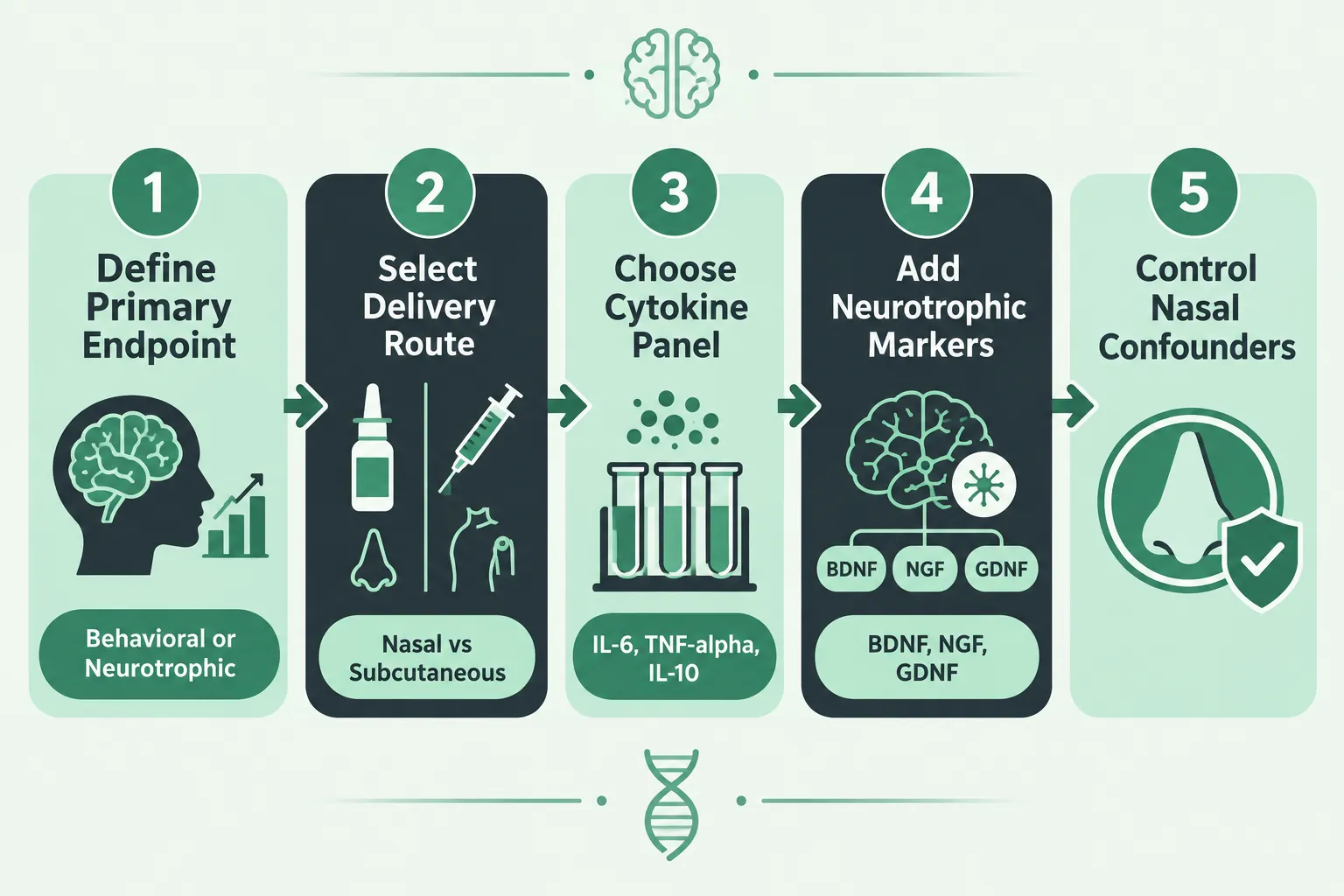
Two neuropeptides derived from endogenous sequences, one from tuftsin, one from ACTH, have generated a steady stream of preclinical and limited clinical data for nearly three decades, yet most researchers still treat them as interchangeable nootropics. That conflation creates real study-design problems. Selank vs Semax peptide research: choosing behavioral, inflammatory, and neurotrophic readouts is not a branding question; it is a methodological one that determines which endpoints are valid, which confounders must be controlled, and whether a research kit should contain one compound or both.

Key Takeaways

  • Semax shows stronger, more consistent neurotrophic effects (BDNF, NGF, GDNF), making it the better anchor for neurotrophic readout studies.
  • Selank's tuftsin lineage makes it the more defensible choice for cytokine and immune-modulation panels.
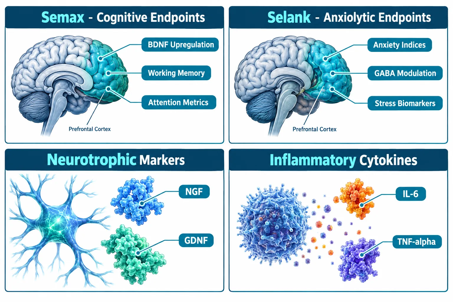
  • Behavioral readouts diverge sharply: Semax maps to cognitive and attention endpoints; Selank maps to anxiolytic and stress-response endpoints.
  • Nasal delivery introduces olfactory confounders that must be controlled in any behavioral assay for either peptide.
  • Dual-axis research kits combining both peptides are an emerging 2026 trend, but require pre-specified primary endpoints to avoid inflated false-discovery rates.

Mapping Peptide Origins to Study-Design Decisions
Mapping Peptide Origins to Study-Design Decisions

Understanding the structural lineage of each peptide is the fastest way to justify endpoint selection to a review committee.

Semax is a synthetic heptapeptide analog of the ACTH(4-10) fragment. Its sequence preserves the melanocortin receptor-binding core while adding a C-terminal Pro-Gly-Pro extension that slows enzymatic degradation. This origin predicts its primary research utility: cognitive performance, attention, and neurotrophic signaling in corticofrontal circuits.

Selank is a synthetic hexapeptide derived from tuftsin (Thr-Lys-Pro-Arg), an endogenous tetrapeptide produced by IgG cleavage in the spleen. Tuftsin is a well-characterized immune modulator. Selank extends this scaffold with a Gly-Glu-Pro tail, conferring anxiolytic-like properties and a more pronounced immune-regulatory profile.

Feature Semax Selank
Endogenous parent ACTH(4-10) Tuftsin
Primary behavioral signal Cognitive enhancement Anxiolysis
Neurotrophic evidence Strong (BDNF, NGF, GDNF) Weak to moderate
Cytokine evidence Moderate (cerebrovascular focus) Strong (IL-6, TNF-alpha, IL-10)
Regulatory status (Russia) Approved nasal spray Approved nasal spray

"Choosing the wrong primary endpoint for either peptide does not just waste resources, it produces null results that obscure genuine signals."

Researchers sourcing compounds for nasal administration should review nasal Semax spray specifications, including purity certificates and excipient profiles, before finalizing a delivery protocol.


Neurotrophic and Inflammatory Readouts: Where the Evidence Actually Points
Neurotrophic and Inflammatory Readouts: Where the Evidence Actually Points

Neurotrophic Markers: Semax as the Stronger Anchor

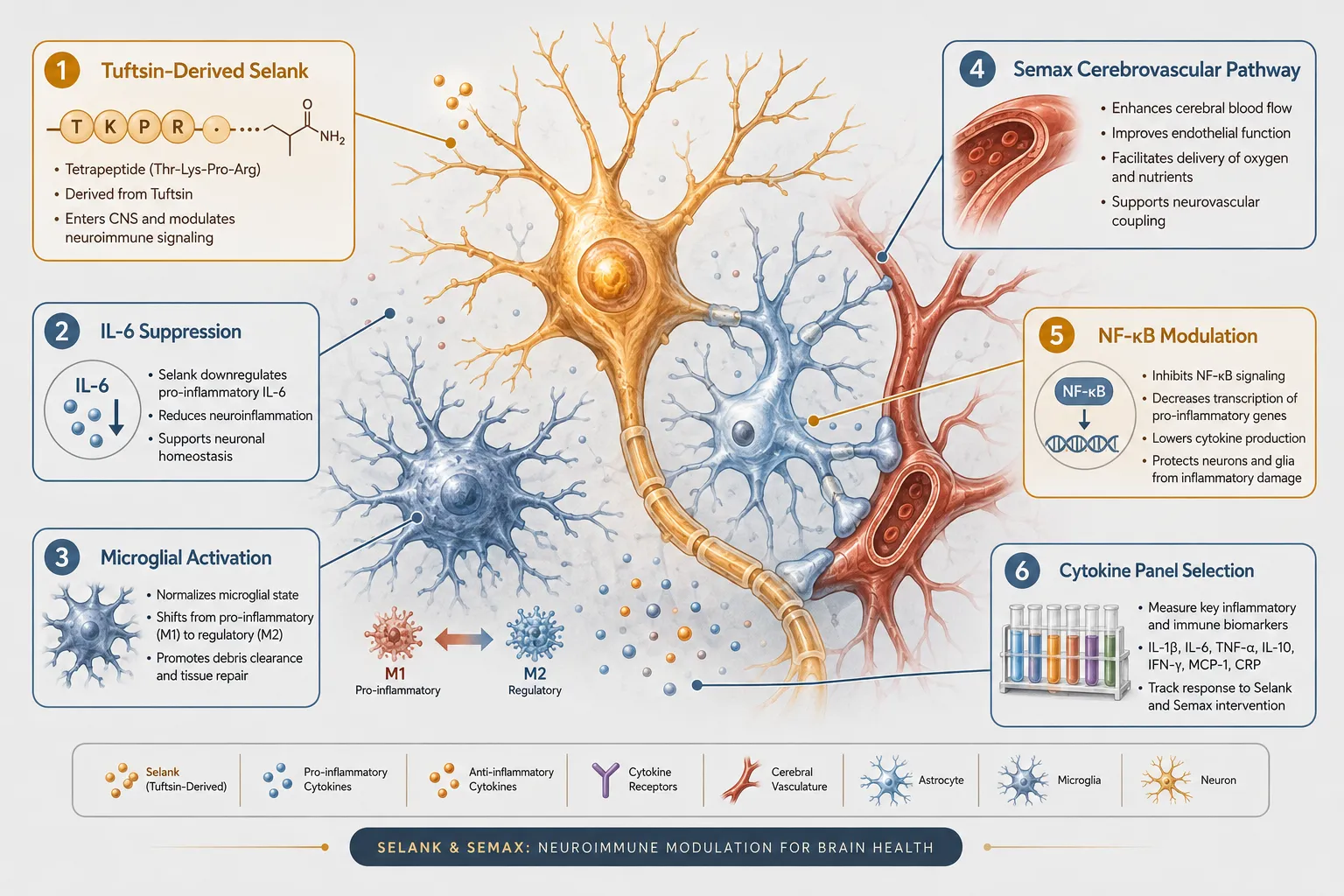
Semax's most replicated finding across rodent and limited human studies is upregulation of brain-derived neurotrophic factor (BDNF). Effects on nerve growth factor (NGF) and glial cell line-derived neurotrophic factor (GDNF) have also been reported, particularly in hippocampal and cortical tissue. These findings support using Semax as the primary compound when a study's pre-specified outcome is a neurotrophic marker panel.

Selank shows some BDNF-related activity, but the data are less consistent and the effect sizes smaller. Selecting Selank as the sole compound in a BDNF-focused study risks underpowering the assay.

Recommended neurotrophic readout panel for Semax studies:

  • BDNF (serum and tissue, time-matched)
  • NGF (hippocampal homogenate preferred)
  • GDNF (if cerebellar or dopaminergic circuits are in scope)
  • TrkB phosphorylation as a downstream confirmation marker

Inflammatory and Immune Readouts: Selank's Structural Advantage

Selank's tuftsin lineage makes it the more defensible anchor for cytokine panels. Published data show suppression of IL-6 and TNF-alpha alongside upregulation of IL-10, a pattern consistent with a shift toward anti-inflammatory macrophage polarization. Enkephalin system interactions have also been reported, adding a neuroimmune dimension.

Semax does carry anti-inflammatory signals, but these are more tightly associated with cerebrovascular protection models, ischemia-reperfusion paradigms, for example, rather than systemic immune modulation. Mixing Semax into a cytokine panel designed around Selank's immune profile introduces mechanistic noise.

Recommended cytokine panel for Selank studies:

  • IL-6, TNF-alpha, IL-1beta (pro-inflammatory axis)
  • IL-10, TGF-beta (regulatory axis)
  • Enkephalin metabolites if neuroimmune crosstalk is a secondary aim

For researchers building broader neuroprotective protocols, the SS-31 10mg research peptide considerations page offers useful context on mitochondrial-targeted peptides that can complement inflammatory readout designs.


Behavioral Endpoints, Nasal Delivery Confounders, and Dual-Axis Strategies
Behavioral Endpoints, Nasal Delivery Confounders, and Dual-Axis Strategies

Behavioral Readout Selection

Behavioral endpoints must match the peptide's mechanistic profile. Using an anxiety assay as the primary outcome for a Semax study, or a cognitive battery as the primary outcome for a Selank study, produces endpoints misaligned with the compound's most robust signals.

Semax behavioral endpoints (supported):

  • Morris water maze and radial arm maze (spatial memory)
  • Novel object recognition (recognition memory)
  • Sustained attention tasks (5-choice serial reaction time in rodents)
  • ADHD-adjacent attention metrics (exploratory; evidence is speculative in 2026)

Selank behavioral endpoints (supported):

  • Elevated plus maze and open field test (anxiety indices)
  • Forced swim test (stress-coping behavior)
  • Social interaction assays (social anxiety models)
  • Conditioned fear extinction protocols

Controlling Nasal Delivery Confounders

Both compounds are most commonly administered intranasally in research settings, mirroring their approved clinical formulations. This creates a specific confounder class: olfactory stimulation artifacts. Any behavioral assay sensitive to olfactory cues, which includes most rodent anxiety and memory paradigms, requires a vehicle-matched nasal control group that receives the same excipient volume and osmolarity without the active peptide.

Failure to include this control group is the single most common methodological weakness in published Selank and Semax behavioral studies. Reviewers in 2026 increasingly flag this omission as a reason for rejection.

Dual-Axis Research Kits: A 2026 Trend

Combined Selank-plus-Semax research kits have grown in popularity as investigators seek to interrogate both neurotrophic and immune axes within a single study. This approach is valid under specific conditions:

  1. Pre-specify a single primary endpoint before data collection begins.
  2. Assign secondary status to all cross-axis markers (e.g., cytokines in a BDNF-primary study).
  3. Apply appropriate corrections for multiple comparisons across the full readout panel.
  4. Include a four-arm design: vehicle, Semax alone, Selank alone, and combination.

Without these guardrails, dual-axis kits inflate false-discovery rates and produce results that are difficult to interpret or reproduce.

Researchers building multi-peptide protocols may also find the MOTS-C peptide and GLP-3 peptide signaling pathway resources useful for understanding how mitochondrial and metabolic peptides interact with neurotrophic readout systems.

Purity verification remains non-negotiable. Studies using sub-95% purity material cannot reliably attribute effects to the target compound. Third-party HPLC and mass spectrometry certificates should be obtained before any behavioral or biomarker assay begins.


Conclusion

Selank vs Semax peptide research: choosing behavioral, inflammatory, and neurotrophic readouts comes down to three decisions made before a single animal is dosed or a single sample is collected.

Actionable next steps for researchers:

  1. Lock the primary endpoint first. Neurotrophic focus defaults to Semax; immune-cytokine focus defaults to Selank. Mixed designs require a four-arm layout and pre-registered analysis plans.
  2. Audit the delivery protocol. Nasal administration demands a vehicle-matched control group in every behavioral assay. Document excipient composition and volume in the methods section.
  3. Verify compound purity independently. Request certificates of analysis showing HPLC purity above 99% and mass spectrometry confirmation before procurement.
  4. Align the behavioral battery with mechanistic expectations. Cognitive and attention assays for Semax; anxiety and stress-coping assays for Selank.
  5. Label speculative endpoints clearly. ADHD applications, neuroimmune crosstalk in humans, and long-term neurodevelopmental effects remain exploratory in 2026 and should be framed as hypothesis-generating, not confirmatory.

Rigorous endpoint selection is what separates publishable data from inconclusive noise in this research space.

https://www.puretestedpeptides.com/wp-content/uploads/2026/10/selank-vs-semax-peptide-research-choosing-behavioral-inflammatory-and-neurotroph.webp 1024 1536 https://www.puretestedpeptides.com/wp-content/uploads/2026/01/buy-peptides-online.jpg 2026-10-11 13:04:402026-10-11 13:04:40Selank vs Semax Peptide Research: Choosing Behavioral, Inflammatory, and Neurotrophic Readouts
What Does MCHC Mean in Peptide Research? Interpreting Hemoglobin Indices When Evaluating GLP-Class and Tissue-Repair Models

What Does MCHC Mean in Peptide Research? Interpreting Hemoglobin Indices When Evaluating GLP-Class and Tissue-Repair Models

October 11, 2026/0 Comments/in Uncategorized/by

Fewer than 15% of published preclinical peptide studies list MCHC as a primary endpoint, yet researchers routinely flag unexpected shifts in this index and struggle to interpret what they mean. Understanding what does MCHC mean in peptide research, and how to interpret hemoglobin indices when evaluating GLP-class and tissue-repair models, is a practical skill that separates rigorous data review from guesswork.

Key Takeaways

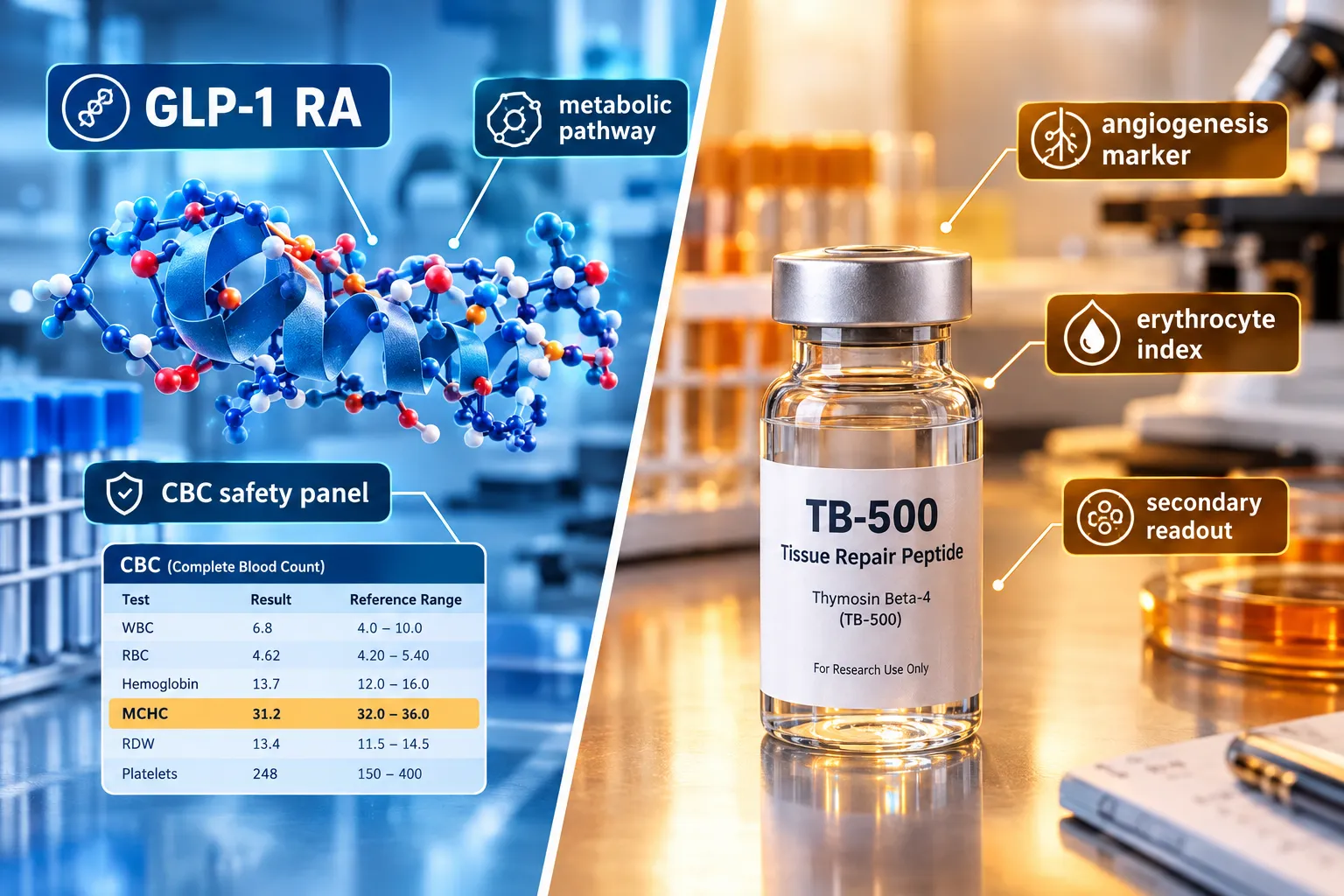
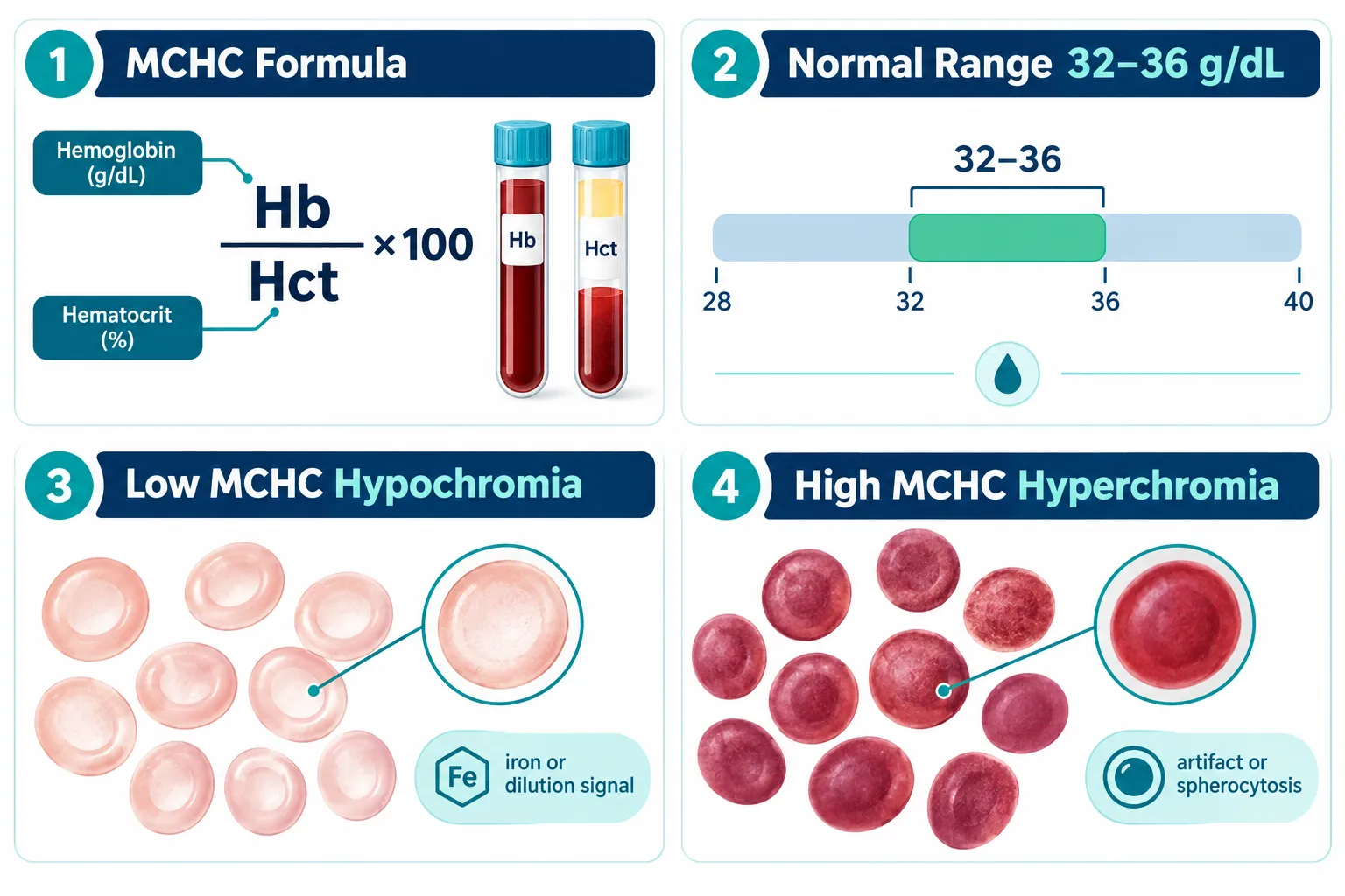
  • MCHC (Mean Corpuscular Hemoglobin Concentration) measures hemoglobin density inside red blood cells and is calculated as hemoglobin divided by hematocrit, expressed in g/dL.
  • In peptide research, MCHC functions as a secondary safety and physiology marker, not a primary efficacy endpoint.
  • Low MCHC (hypochromia) most often signals iron deficiency, chronic inflammation, or sample dilution rather than a direct peptide effect.
  • High MCHC (hyperchromia) in preclinical panels frequently reflects laboratory artifact, particularly hemolysis from poor sample handling.
  • MCHC should always be interpreted alongside MCV, RDW, and full study context before attributing changes to any peptide compound.

Defining MCHC: Calculation, Normal Ranges, and the Erythrocyte Index Hierarchy

MCHC stands for Mean Corpuscular Hemoglobin Concentration. It quantifies how much hemoglobin is packed into a given volume of red blood cells. The formula is straightforward:

MCHC (g/dL) = (Hemoglobin / Hematocrit) x 100

In most mammalian species used in preclinical research, the reference range sits between 32 and 36 g/dL. Values below this threshold indicate hypochromia (pale, hemoglobin-poor cells). Values above suggest hyperchromia, though true biological hyperchromia is rare.

Defining MCHC: Calculation, Normal Ranges, and the Erythrocyte Index Hierarchy

Within the broader erythrocyte index hierarchy, MCHC ranks below MCV (Mean Corpuscular Volume) and RDW (Red Cell Distribution Width) in diagnostic weight. Here is how the indices compare:

Index What It Measures Research Priority
MCV Average red cell size High
RDW Size variation across cells High
MCH Average hemoglobin per cell Moderate
MCHC Hemoglobin concentration per cell volume Supportive

MCHC is most useful when read as part of this full panel, not in isolation. Researchers sourcing compounds through platforms that emphasize building robust peptide benchmarks will recognize that baseline CBC panels, including MCHC, are part of responsible quality-control workflows.


Why MCHC Matters When Interpreting Hemoglobin Indices in GLP-Class Models

GLP-1 receptor agonist (GLP-RA) compounds have become central to metabolic research. When reviewing CBC safety data from GLP-class studies, MCHC appears as a secondary safety lab rather than a target biomarker. Its role is to confirm that metabolic changes driven by energy restriction, body composition shifts, or altered nutrient absorption are not producing clinically meaningful hematologic stress.

Researchers working with GLP-1 peptide sourcing and generational research concepts should note that caloric restriction models, common in GLP-RA efficacy studies, can mildly suppress MCHC through reduced dietary iron intake. This is a model-design artifact, not a compound toxicity signal.

Key considerations for GLP-class CBC interpretation:

  • Weight loss velocity can transiently alter plasma volume, diluting hemoglobin concentration and lowering apparent MCHC.
  • Visceral fat mobilization studies, such as those explored in visceral fat research models, may produce mild hemodilution as fluid compartments shift.
  • GLP-2-class compounds, which target intestinal repair and nutrient absorption, carry a more direct theoretical link to iron and B12 uptake, making MCHC monitoring more relevant in those specific models.

For researchers evaluating next-generation triple-agonist compounds, resources on GLP-3 for sale and triple agonist research planning provide useful context on how expanded receptor profiles may affect systemic physiology, including hematologic readouts.

Why MCHC Matters When Interpreting Hemoglobin Indices in GLP-Class Models


MCHC in Tissue-Repair Peptide Models: A Practical Interpretation Framework

Tissue-repair peptides, including TB-500 analogs and mitochondria-targeting compounds, introduce different considerations for hemoglobin index interpretation. Researchers following TB-500 controlled experimental models and QC workflow already understand that angiogenesis-promoting compounds can alter red cell dynamics through vascular remodeling rather than direct erythropoietic effects.

For mitochondria-targeted peptides, the relationship between cellular energy metabolism and red cell lifespan is an active area of interest. Work in SS-31 mitochondrial research and related SS-31 kidney health research highlights how oxidative stress reduction may indirectly support erythrocyte membrane integrity, a variable that influences MCHC readings over longer study durations.

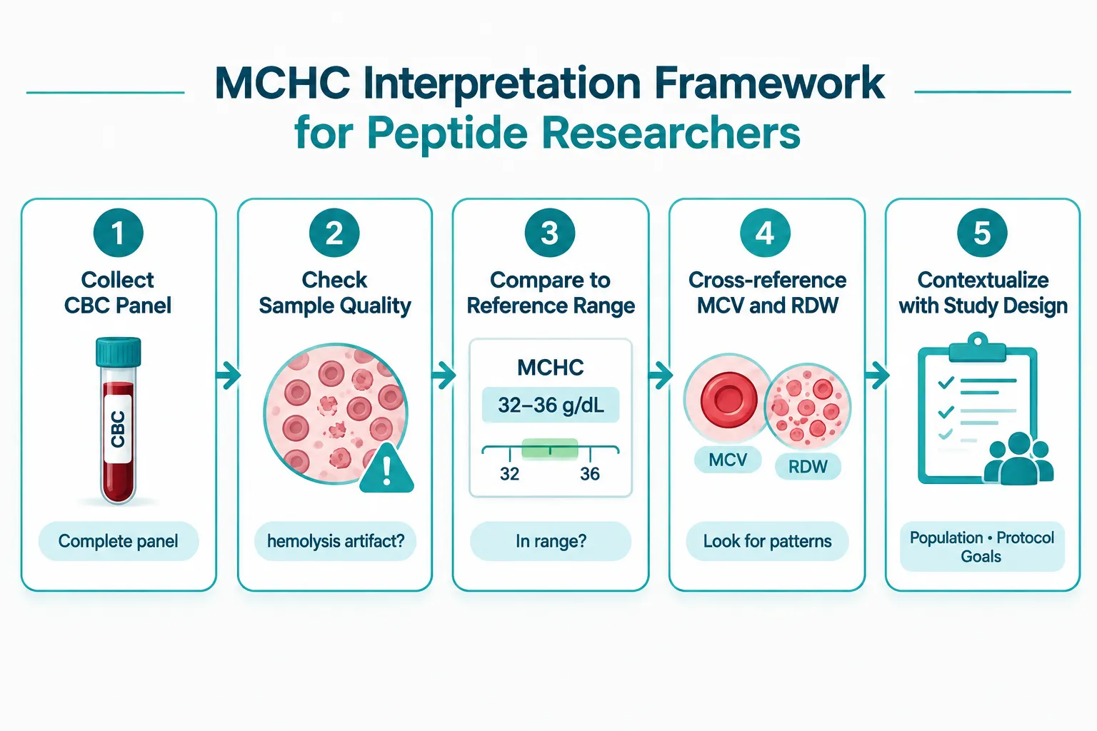
A practical five-step MCHC interpretation framework for peptide researchers:

  1. Verify sample quality first. Hemolysis from poor venipuncture or freeze-thaw cycling is the single most common cause of falsely elevated MCHC. Confirm sample integrity before drawing conclusions.
  2. Check the full CBC panel. An isolated MCHC shift without corresponding MCV or RDW changes is more likely an artifact than a biological signal.
  3. Compare against species-specific reference ranges. Murine, rat, and canine reference ranges differ meaningfully from human norms.
  4. Cross-reference study design variables. Diet, hydration status, and blood collection timing all affect MCHC independently of any peptide compound.
  5. Assign interpretive weight proportionally. MCHC is a supporting character in the CBC story, not the lead.

MCHC in Tissue-Repair Peptide Models: A Practical Interpretation Framework

As of late 2026, the published literature contains very few peptide-specific MCHC studies. Most available data comes from broader hematology safety panels embedded in metabolic or regenerative medicine trials. This gap means researchers must apply general hematology principles carefully rather than relying on peptide-specific MCHC benchmarks.


Conclusion

Understanding what does MCHC mean in peptide research, and how to interpret hemoglobin indices when evaluating GLP-class and tissue-repair models, ultimately comes down to context. MCHC is a reliable supporting marker when used correctly, but it is frequently misread when treated as a standalone signal.

Actionable next steps for peptide researchers in 2026:

  • Always collect a full CBC panel, not individual indices, as part of baseline and endpoint safety monitoring.
  • Document sample collection conditions meticulously to rule out hemolysis artifacts before flagging MCHC shifts.
  • When MCHC deviates from reference range, cross-check MCV, RDW, and reticulocyte count before attributing the change to a peptide compound.
  • For GLP-class models, account for caloric restriction and fluid redistribution as confounding variables in hematologic data.
  • For tissue-repair and mitochondria-targeting models, monitor MCHC trends over time rather than at single time points to distinguish transient from sustained changes.

Rigorous interpretation of hemoglobin indices strengthens the overall quality of peptide research data and supports more reliable conclusions across all model types.

https://www.puretestedpeptides.com/wp-content/uploads/2026/10/what-does-mchc-mean-in-peptide-research-interpreting-hemoglobin-indices-when-eva.webp 1024 1536 https://www.puretestedpeptides.com/wp-content/uploads/2026/01/buy-peptides-online.jpg 2026-10-11 13:04:122026-10-11 13:04:12What Does MCHC Mean in Peptide Research? Interpreting Hemoglobin Indices When Evaluating GLP-Class and Tissue-Repair Models
Enclomiphene Research in Male Hormone Models: Choosing Testosterone, LH, FSH, Estradiol, and Fertility Endpoints

Enclomiphene Research in Male Hormone Models: Choosing Testosterone, LH, FSH, Estradiol, and Fertility Endpoints

October 11, 2026/0 Comments/in Uncategorized/by

{"cover":"Professional landscape format (1536×1024) hero image with bold text overlay 'Enclomiphene Research in Male Hormone' in crisp white modern sans-serif on a deep navy semi-transparent overlay panel, centered with 8% safe margins from every edge. Background: macro editorial photograph of a laboratory hormone assay panel showing glass vials labeled testosterone, LH, FSH, and estradiol arranged on a clean white lab bench with soft cool studio lighting, teal and navy color palette, medical research magazine cover aesthetic, high contrast, editorial quality.","content":["Numbered four-step annotated mechanism diagram illustrating the hypothalamic-pituitary-gonadal axis response to enclomiphene in a male hormone model. Step 1 labeled 'Hypothalamus: GnRH Release' with callout arrow, Step 2 labeled 'Pituitary: LH and FSH Rise' with upward arrow, Step 3 labeled 'Testes: Testosterone Synthesis' with callout, Step 4 labeled 'Estradiol Monitoring Point' with a side callout box. Clean white background, teal and charcoal palette, thin callout lines, bold step numbers, editorial medical-illustration style, landscape 1536×1024.","Split-screen landscape comparison (1536×1024) infographic: left panel labeled 'Testosterone Gel' showing a downward-pointing arrow for LH and FSH with callout 'Gonadotropin Suppression' and a sperm icon with label 'Spermatogenesis Impaired'; right panel labeled 'Enclomiphene' showing upward arrows for LH, FSH, Testosterone with callout 'Gonadotropin Preserved' and a sperm icon labeled 'Spermatogenesis Maintained'. Bold panel headings, teal-and-amber palette, thin dividing line, clean white background, editorial clinical-comparison style.","Annotated timeline infographic landscape (1536×1024) showing a horizontal research protocol timeline for enclomiphene male hormone studies: five labeled milestone cards, Baseline (Week 0): Testosterone, LH, FSH, Estradiol, Sperm Count; Early Response (Week 3): LH and FSH surge callout; Mid-Trial (Week 6): Testosterone normalization callout; Extended (Week 12-16): Sperm concentration endpoint callout; Final (Week 16+): Composite endpoint card labeled Testosterone plus Sperm greater than 10M/mL. Teal and slate color scheme, short callout labels, bold card headings, clean editorial infographic style."]

Professional landscape hero image () with a reading "Enclomiphene Research in Male Hormone". CRITICAL TYPOGRAPHY RULES:

Fewer than half of men with secondary hypogonadism who receive conventional testosterone therapy retain adequate sperm production, a clinical trade-off that has pushed researchers toward selective estrogen receptor modulators (serms) as a mechanistically distinct alternative. Enclomiphene research in male hormone models: choosing testosterone, LH, FSH, estradiol, and fertility endpoints is now a structured discipline, with phase II and III trial data, systematic meta-analyses, and 2026 clinical guidance all converging on a defined set of biomarkers and measurement protocols. This article provides a practical endpoint-selection guide for investigators designing or evaluating enclomiphene studies in male endocrine models.


Key Takeaways

  • Enclomiphene raises total testosterone by stimulating LH and FSH rather than suppressing the hypothalamic-pituitary-gonadal (HPG) axis, making gonadotropin measurement essential in any study design.
  • Estradiol monitoring is a critical safety endpoint; enclomiphene produces significantly smaller estradiol increases than clomiphene, and this distinction matters for adverse-effect interpretation.
  • Composite endpoints combining testosterone normalization (300-1,040 ng/dL) with sperm concentration above 10×10^6/mL are now considered the regulatory standard for fertility-preserving research.
  • Pregnancy and live-birth data remain absent from existing randomized trials; "fertility-preserving potential" is the appropriate framing until conception endpoints are reported.
  • Temporal sampling strategy, particularly early gonadotropin measurement at weeks 3-6, is as important as endpoint selection itself.

The HPG Axis as the Primary Research Framework

The HPG Axis as the Primary Research Framework

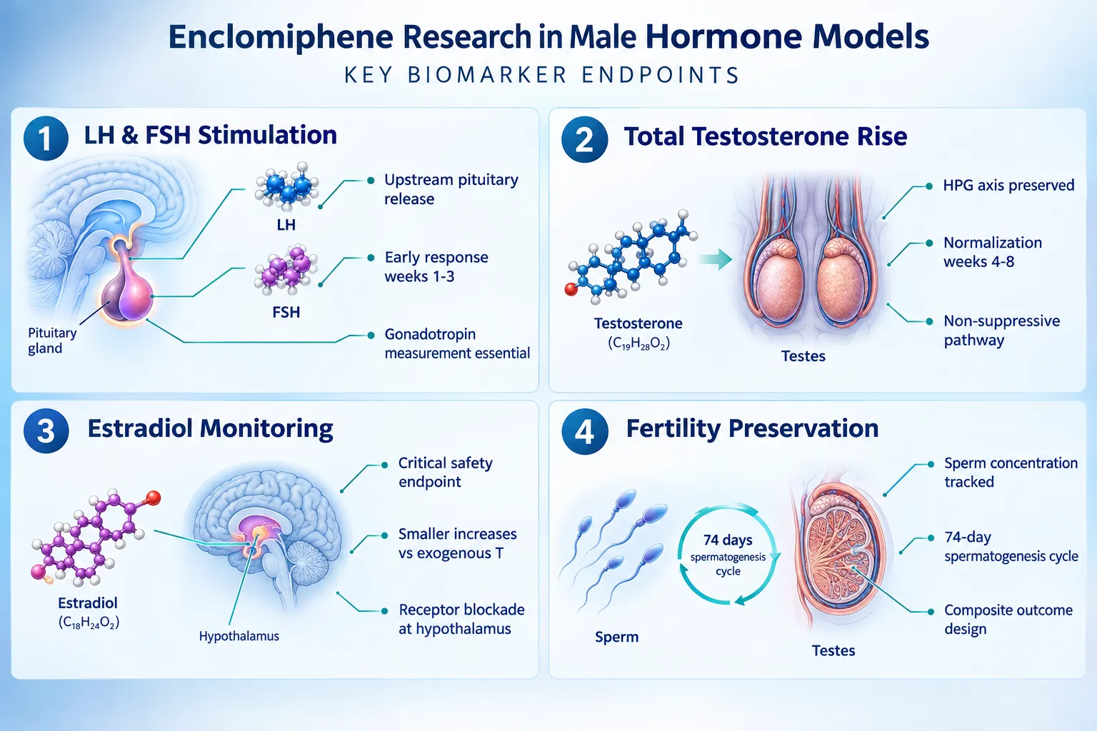
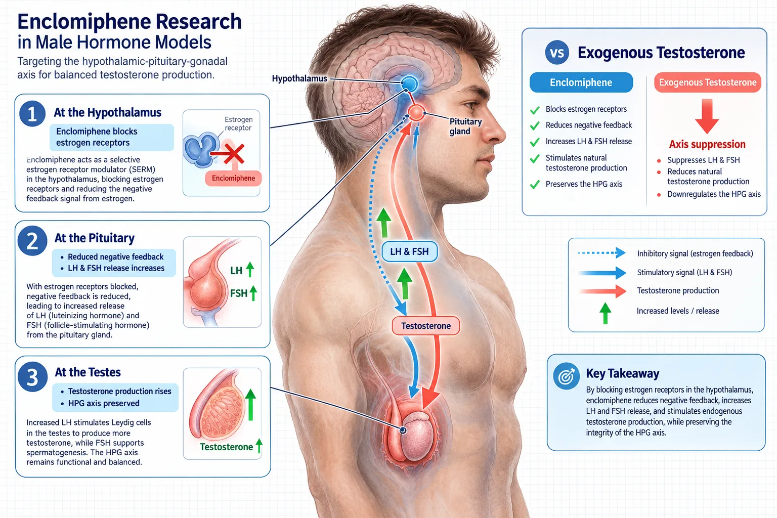
Enclomiphene blocks estrogen receptors at the hypothalamus, reducing negative feedback and prompting the pituitary to release more LH and FSH. This upstream mechanism is what separates it from exogenous testosterone, which suppresses the same axis. Any male hormone model that omits gonadotropin measurement is therefore incomplete.

Phase III data in secondary hypogonadism (men with baseline testosterone at or below 300 ng/dL) show that a 25 mg daily dose raised mean LH from approximately 5.3 to 11.9 mIU/mL and FSH from 9.4 to 14.9 mIU/mL over six weeks, while mean testosterone climbed to roughly 604 ng/dL. These numbers establish the expected magnitude of response and serve as calibration benchmarks when designing new studies.

Recommended baseline panel before any enclomiphene intervention:

Biomarker Rationale
Total testosterone Primary efficacy endpoint
LH Confirms secondary hypogonadism; tracks HPG activation
FSH Spermatogenesis support; distinguishes primary from secondary deficiency
Estradiol (E2) Safety monitoring; tracks serm-related estrogenic activity
Sperm concentration Fertility-related endpoint; composite outcome anchor
Hematocrit Adverse-effect screen

For researchers exploring related peptide-based hormonal research, the IPA Sermorelin Stack Research resource offers useful context on multi-compound endocrine protocols.


Enclomiphene Research in Male Hormone Models: Temporal Sampling and Gonadotropin Interpretation

Enclomiphene Research in Male Hormone Models: Temporal Sampling and Gonadotropin Interpretation

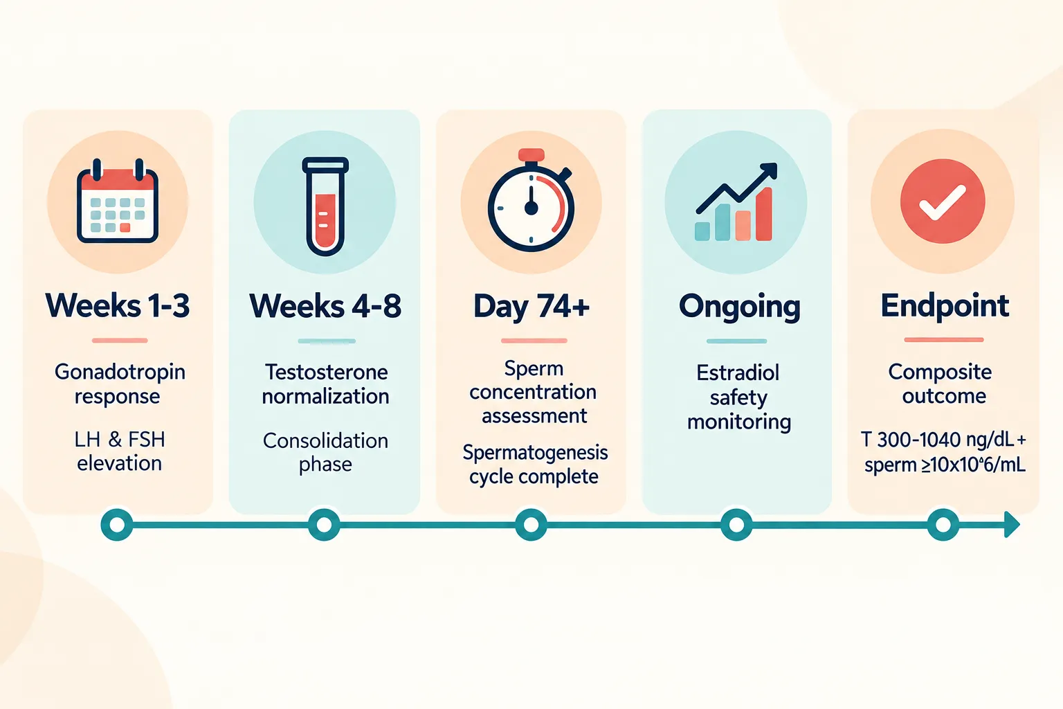
Timing is where many study designs underperform. Gonadotropin responses appear early, often within the first two to three weeks, while testosterone normalization consolidates between weeks four and eight. Sperm concentration, which reflects spermatogenesis cycles of approximately 74 days, requires sampling at three months or later to be interpretable.

A 2025 systematic review and meta-analysis of ten randomized controlled trials (819 men with functional hypogonadism) found that serm therapy including enclomiphene raised total testosterone by a mean of 273.76 ng/dL versus placebo. Critically, the same analysis showed no significant difference in total testosterone between enclomiphene and testosterone gel, yet enclomiphene preserved or improved sperm concentration while testosterone gel suppressed gonadotropins below normal range. This contrast is only detectable when LH and FSH are measured at both early (week 3-6) and late (week 12-16) timepoints.

Recommended sampling schedule:

  • Week 0: Full baseline panel (see table above)
  • Week 3-6: LH, FSH, total testosterone, captures early gonadotropin surge
  • Week 12: Total testosterone, estradiol, sperm concentration, mid-trial safety and efficacy check
  • Week 16+: Composite endpoint assessment, testosterone normalization plus sperm concentration

"Gonadotropin suppression in the testosterone gel group versus preservation in the enclomiphene group is only visible when both arms are sampled at the same early timepoint, this design detail determines whether the study can answer its core question."

A 2024 comparative study of 66 men reinforces the importance of estradiol timing. Enclomiphene produced a median testosterone increase of 166 ng/dL versus 98 ng/dL with clomiphene, but the estradiol change was markedly different: -5.92 pg/mL with enclomiphene versus +17.50 pg/mL with clomiphene (p=0.001). This gap is a product-identity distinction, not just a safety signal, it confirms that enclomiphene and clomiphene are not interchangeable in research models.

Investigators working with broader research-grade compound sourcing may find the Peptides 101 for Research-Use Only Buyers guide helpful for understanding purity and identity verification standards that apply across endocrine research compounds.


Enclomiphene Research in Male Hormone Models: Fertility Endpoints and Composite Outcome Design

Enclomiphene Research in Male Hormone Models: Fertility Endpoints and Composite Outcome Design

The regulatory standard for fertility-preserving therapy now centers on a composite primary endpoint: total testosterone within the normal range (300-1,040 ng/dL) and sperm concentration at or above 10×10^6/mL. This dual requirement reflects the clinical reality that testosterone normalization alone does not confirm preserved reproductive function.

Phase III pivotal trial data (256 overweight men, aged 18-60) show enclomiphene raised mean testosterone from approximately 205 ng/dL to 413-446 ng/dL at 16 weeks versus placebo, while maintaining sperm concentration. Across the broader serm meta-analysis, LH rose by approximately 4.66 IU/L and FSH by 4.59 IU/L, both statistically significant versus placebo.

Key fertility endpoint categories for enclomiphene studies:

  • Semen parameters: Sperm concentration, motility, morphology (minimum 3-month sampling window)
  • Gonadotropin activity: LH and FSH as proxies for spermatogenesis support
  • Hormonal composite: Testosterone normalization concurrent with gonadotropin preservation
  • Downstream outcomes: Conception and live-birth rates (currently absent from all published RCTs)

The absence of pregnancy and live-birth data is the field's most significant gap. Current evidence supports "fertility-preserving potential", a mechanistically grounded but clinically unconfirmed claim. Future trial designs should incorporate conception endpoints wherever feasible.

The British Society for Sexual Medicine's 2026 position statement endorses testosterone, LH, FSH, and semen parameters as central endpoints, while also flagging routine monitoring of estradiol, hematocrit, libido changes, and fatigue as standard safety measures.

For researchers investigating metabolic co-variables that may interact with HPG axis function, the Visceral Fat Research resource addresses adiposity-driven hormonal suppression, a relevant confounder in overweight male cohorts.


Conclusion

Enclomiphene research in male hormone models: choosing testosterone, LH, FSH, estradiol, and fertility endpoints requires a structured, multi-biomarker approach built around the HPG axis mechanism. The following steps represent the current best-practice framework for 2026 study design:

  1. Establish a full baseline panel including total testosterone, LH, FSH, estradiol, sperm concentration, and hematocrit before any intervention.
  2. Sample gonadotropins early (weeks 3-6) to capture the LH and FSH surge that distinguishes enclomiphene from testosterone replacement.
  3. Use a composite primary endpoint that combines testosterone normalization with sperm concentration above 10×10^6/mL for any fertility-focused model.
  4. Monitor estradiol as a safety and product-identity endpoint, not merely an afterthought, the enclomiphene-versus-clomiphene estradiol differential is a mechanistically meaningful distinction.
  5. Plan for 12-16 week minimum duration to allow spermatogenesis endpoints to reach interpretable values.
  6. Acknowledge the live-birth gap in any fertility claim and design future trials to include conception endpoints.

Rigorous endpoint selection is what converts a hormone-level study into actionable clinical evidence. The data infrastructure now exists; the next priority is applying it with the temporal precision and composite outcome design that the field requires.

https://www.puretestedpeptides.com/wp-content/uploads/2026/10/enclomiphene-research-in-male-hormone-models-choosing-testosterone-lh-fsh-estrad.webp 1024 1536 https://www.puretestedpeptides.com/wp-content/uploads/2026/01/buy-peptides-online.jpg 2026-10-11 13:03:492026-10-11 13:03:49Enclomiphene Research in Male Hormone Models: Choosing Testosterone, LH, FSH, Estradiol, and Fertility Endpoints
Klow Blend Versus Single-Agent Intranasal Peptides: Designing Permeability, Deposition, and Brain-Exposure Controls

Klow Blend Versus Single-Agent Intranasal Peptides: Designing Permeability, Deposition, and Brain-Exposure Controls

October 11, 2026/0 Comments/in Uncategorized/by

A four-peptide vial containing 80 mg of lyophilized material raises an immediate research question: does its apparent activity reflect the sum of its components, or does combining GHK-Cu, BPC-157, TB-500, and KPV in a single intranasal formulation change how each peptide behaves at the mucosal surface and beyond? The comparison of Klow Blend versus single-agent intranasal peptides, specifically around designing permeability, deposition, and brain-exposure controls, demands more than a simple ingredient audit. It requires a structured framework that separates concentration effects, excipient interactions, mucosal residence time, and combined receptor signaling.

Key Takeaways

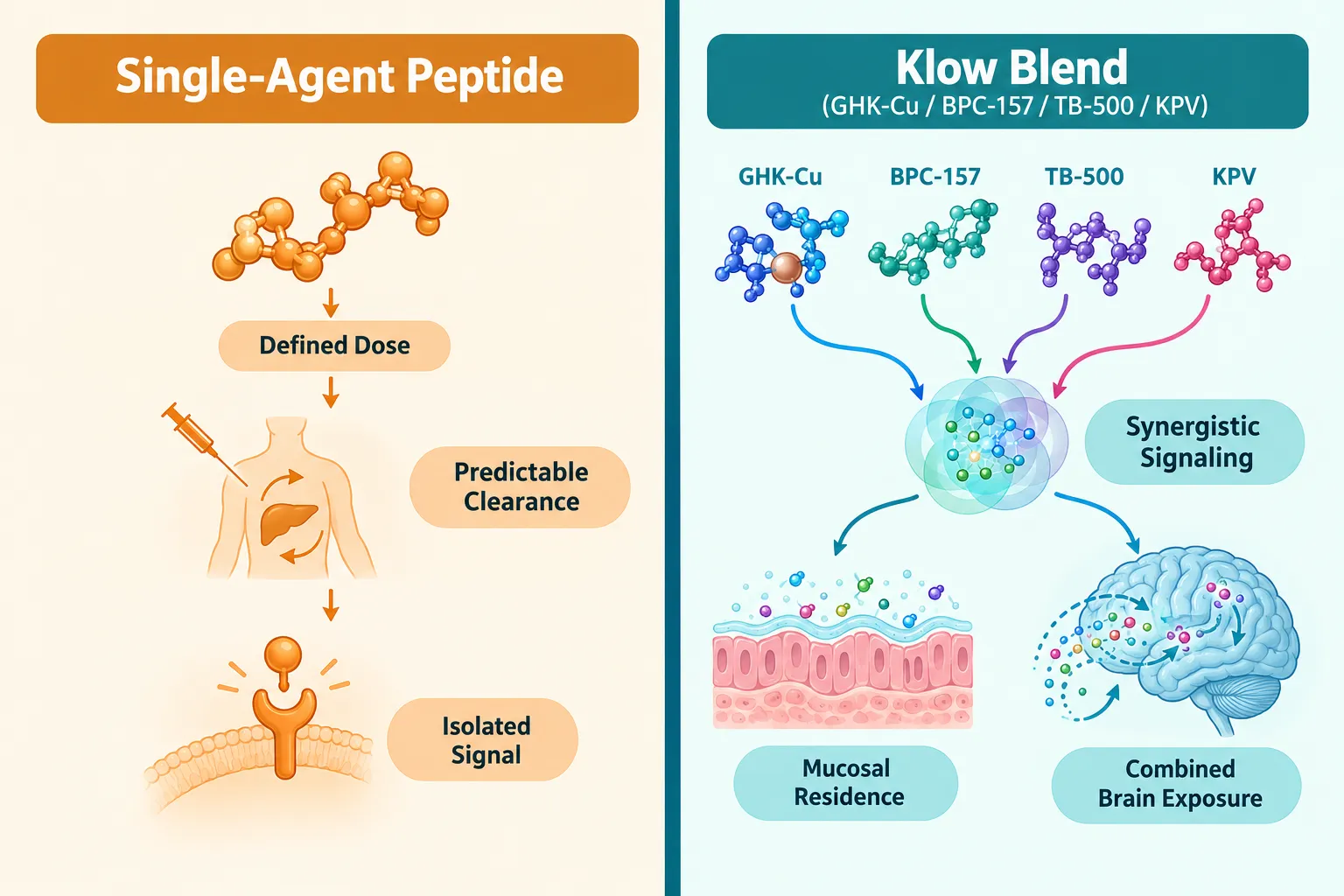
  • The Klow Blend is a research-only, four-peptide formulation (GHK-Cu 50 mg, BPC-157 10 mg, TB-500 10 mg, KPV 10 mg) with no recognized pharmacopeial or regulatory drug identity.
  • Intranasal delivery bypasses first-pass metabolism, but deposition patterns and mucociliary clearance differ meaningfully between single-agent peptides and multi-component blends.
  • Concentration, excipients, and mucosal residence time can each independently drive apparent differences between a blend and its individual components.
  • A rigorous comparison requires matched in vitro permeability assays and ex vivo nasal tissue models run at equivalent molar concentrations.
  • Brain-exposure controls must account for olfactory and trigeminal nerve routes, not just systemic absorption.

Understanding the Klow Blend Formulation

The most widely cited Klow Blend formula contains 50 mg GHK-Cu, 10 mg BPC-157, 10 mg TB-500, and 10 mg KPV in a single lyophilized 80 mg vial. Providers also offer it as a ready-mixed intranasal solution, typically in 10 mL bottles delivering approximately 57 metered sprays per bottle. No scientific literature or regulatory body currently lists Klow Blend as a recognized drug entity; the four peptides share a vial rather than forming a single chemical compound.

Understanding the Klow Blend Formulation

This distinction matters for experimental design. When comparing Klow Blend versus single-agent intranasal peptides across permeability, deposition, and brain-exposure controls, researchers must decide whether the blend is being tested as a fixed-ratio combination or whether each component is being evaluated at its blend-equivalent concentration. Conflating these two designs produces uninterpretable data.

For researchers exploring related intranasal peptide formats, Semax nasal spray represents a well-characterized single-agent reference standard useful for benchmarking deposition behavior. Understanding peptide half-life is equally critical, since shorter half-lives amplify the importance of mucosal residence time in any blend-versus-single-agent comparison.


Designing Permeability Controls: Blend Versus Single Agent

Intranasal peptide permeability depends on molecular weight, charge, lipophilicity, and the presence of tight-junction modulators in the formulation. GHK-Cu, a copper-chelating tripeptide, has a low molecular weight and reasonable membrane affinity. BPC-157 and TB-500 are larger peptides with distinct solubility profiles. KPV is a tripeptide fragment with anti-inflammatory properties.

Key permeability variables to control:

Variable Single-Agent Consideration Blend Consideration
Molecular weight Defined per agent Mixed population in solution
Excipient load Minimal Potentially higher
Tight-junction effects Agent-specific Possible additive modulation
pH of solution Optimized per peptide Compromise pH required

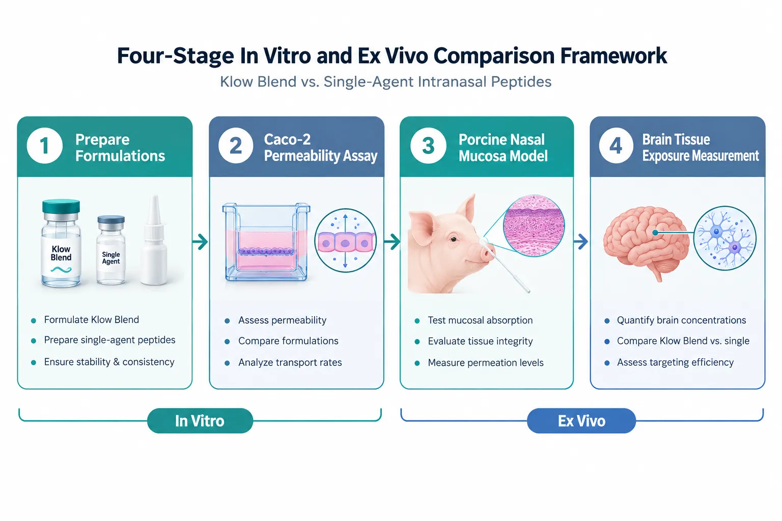
When running Caco-2 monolayer or RPMI 2650 nasal epithelial assays, each peptide in the blend should first be tested at its blend-equivalent concentration as a standalone. Only after establishing individual apparent permeability coefficients (Papp) can researchers attribute any blend-specific deviation to formulation interactions rather than simple concentration effects.

"The most common error in blend-versus-single-agent research is testing the blend at total protein concentration rather than at per-component molar equivalence."

Researchers working with lab tested peptides that carry verified purity documentation gain a significant advantage here, since impurity profiles in lower-grade materials can independently alter permeability readings.


Deposition and Mucosal Residence Time in Multi-Peptide Formulations

Intranasal deposition is governed by droplet size, spray velocity, nasal anatomy, and solution viscosity. A blend formulation may have a different viscosity profile than any of its individual components in solution, which directly affects where in the nasal cavity material deposits and how long it remains in contact with the epithelium.

Deposition and Mucosal Residence Time in Multi-Peptide Formulations

Three factors that independently alter residence time in blends:

  1. Viscosity modifiers, Excipients added to stabilize one peptide may increase overall solution viscosity, slowing mucociliary clearance for all components.
  2. Mucoadhesive interactions, Certain peptide sequences bind mucin glycoproteins, extending contact time beyond what the individual agent would achieve alone.
  3. Competitive absorption, Multiple peptides competing for the same transporter or paracellular pathway may reduce net flux for each component compared to single-agent delivery.

Ex vivo porcine nasal mucosa models offer the most practical system for isolating these effects without full animal studies. By mounting excised tissue in Franz diffusion cells and applying the blend versus individual components at matched concentrations, researchers can measure both permeated mass and retained mucosal mass, directly quantifying residence time differences.

For context on how multi-peptide blends handle reconstitution and dosing logistics, the Tesamorelin CJC-1295 Ipamorelin 12mg blend reconstitution guide provides a practical reference for multi-component peptide handling. Reviewing BPC-157 core peptides documentation also clarifies the individual stability and solubility characteristics of one of the Klow Blend's key components.


Brain-Exposure Controls: Olfactory and Trigeminal Routes

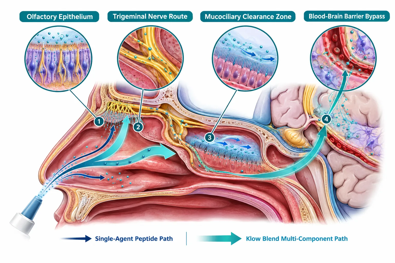
Brain exposure from intranasal peptides does not rely solely on systemic absorption followed by blood-brain barrier crossing. Two direct neural pathways, the olfactory nerve route and the trigeminal nerve route, allow peptides deposited in the upper nasal cavity to reach the olfactory bulb and brainstem, respectively, without entering systemic circulation.

Brain-Exposure Controls: Olfactory and Trigeminal Routes

Designing valid brain-exposure controls requires:

  • Separate deposition targeting: upper nasal (olfactory region) versus lower nasal (respiratory region) delivery
  • Matched time-point sampling in olfactory bulb, cortex, and brainstem tissue
  • Systemic plasma sampling to distinguish direct neural transport from systemic re-entry

When comparing Klow Blend versus single-agent intranasal peptides for brain-exposure controls, a critical confound is that GHK-Cu's copper chelation may alter tight-junction integrity in olfactory epithelium, potentially enhancing paracellular access for co-administered BPC-157 or KPV. This interaction cannot be detected without running the blend and each component separately under identical deposition conditions.

Researchers interested in nasal peptide formats designed for central nervous system research should also review available nasal Semax spray documentation, as Semax has one of the better-characterized intranasal-to-brain transport profiles among research peptides and serves as a useful positive control.


Conclusion

Comparing Klow Blend versus single-agent intranasal peptides across permeability, deposition, and brain-exposure controls is not a single experiment, it is a layered experimental program. The actionable steps for researchers are clear:

  • Establish individual Papp values for each Klow component at blend-equivalent concentrations before running any blend assay.
  • Use ex vivo porcine nasal mucosa in Franz diffusion cells to separate viscosity, mucoadhesion, and competitive transport effects.
  • Design brain-exposure studies with both olfactory-targeted and systemic-route controls to avoid misattributing neural transport to systemic absorption.
  • Document excipient composition for both blend and single-agent preparations, since excipient differences alone can produce false-positive blend effects.
  • Ensure all peptide materials carry verified purity documentation before any permeability or deposition data is collected.

The Klow Blend remains a research-only preparation with no approved clinical indication. Any observed differences between the blend and its individual components should be interpreted strictly within a research context, with full transparency about concentration design, excipient controls, and deposition methodology.

https://www.puretestedpeptides.com/wp-content/uploads/2026/10/klow-blend-versus-single-agent-intranasal-peptides-designing-permeability-deposi.webp 1024 1536 https://www.puretestedpeptides.com/wp-content/uploads/2026/01/buy-peptides-online.jpg 2026-10-11 13:03:342026-10-11 13:03:34Klow Blend Versus Single-Agent Intranasal Peptides: Designing Permeability, Deposition, and Brain-Exposure Controls
GLP-2-T and GLP2 Tirz Peptide Research: Distinguishing Epithelial Repair From Changes in Motility and Intake

GLP-2-T and GLP2 Tirz Peptide Research: Distinguishing Epithelial Repair From Changes in Motility and Intake

October 11, 2026/0 Comments/in Uncategorized/by

Researchers designing intestinal studies in 2026 face a specific confounding problem: two peptide categories share nearly identical naming conventions but act through entirely different receptor systems. Misassigning GLP-2-T and GLP2 Tirz Peptide Research outcomes to the wrong biological pathway can invalidate an entire experimental dataset. Understanding which compound drives epithelial repair versus which one shifts motility and food intake is no longer optional, it is a foundational requirement for rigorous experimental design.

Key Takeaways

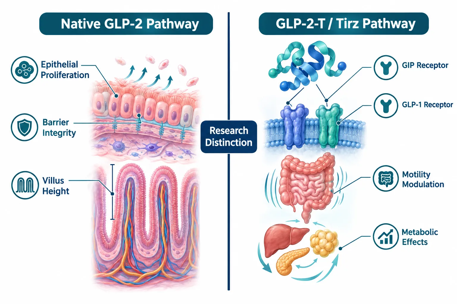
  • Native GLP-2 remains the reference standard for epithelial repair, mucosal growth, and barrier integrity research.
  • Commercial "GLP-2-T" materials are tirzepatide-class dual GIP/GLP-1 agonists, not GLP-2 receptor agonists.
  • GLP2-TZ and Tirz compounds are research tools for motility and metabolic studies, not epithelial proliferation.
  • Experimental designs must separate barrier integrity, epithelial proliferation, gastric transit, food intake, and systemic metabolic effects into distinct readout groups.
  • All GLP-2-T, GLP2-TZ, and GLP2 Tirz materials remain strictly research-use only as of 2026.

What Native GLP-2 Actually Does in Intestinal Research

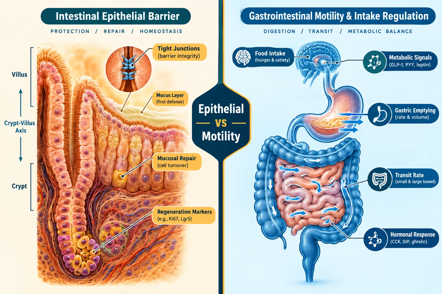
GLP-2 (glucagon-like peptide-2) is a 33-amino acid peptide secreted by intestinal L-cells. Its receptor, GLP-2R, is expressed on enteric neurons, subepithelial myofibroblasts, and enteroendocrine cells. When GLP-2R is activated, the downstream effects are well-characterized: increased crypt-cell proliferation, reduced enterocyte apoptosis, greater villus height, and strengthened tight-junction integrity.

These effects make native GLP-2 the reference compound for epithelial repair biology. Researchers measuring mucosal healing, barrier permeability, or intestinal adaptation use GLP-2 as the positive control precisely because its mechanism is receptor-specific and its epithelial outcomes are reproducible.

What Native GLP-2 Actually Does in Intestinal Research

Key epithelial readouts in GLP-2 studies include:

  • Villus height-to-crypt depth ratio
  • Tight-junction protein expression (claudin-3, occludin)
  • Crypt-cell proliferation index (Ki-67 staining)
  • Intestinal permeability (lactulose/mannitol ratio)
  • Enterocyte apoptosis rate

For researchers exploring related incretin biology, the GLP-1 peptide buy generational research concepts and sourcing notes resource provides useful context on how GLP-1 and GLP-2 differ in their gut-specific roles.


GLP-2-T and GLP2 Tirz Peptide Research: The Naming Confusion That Distorts Results

The commercial category labeled "GLP-2-T" does not activate the GLP-2 receptor. These materials are tirzepatide-class dual agonists targeting GIP (glucose-dependent insulinotropic polypeptide) receptors and GLP-1 receptors simultaneously. The "GLP-2" prefix in the product name refers to a research classification framework, not receptor pharmacology.

This distinction is critical. A researcher who selects a GLP-2-T compound expecting epithelial proliferation data will instead collect motility, gastric emptying, and metabolic endpoints. The experimental output will be real, but it will answer the wrong question.

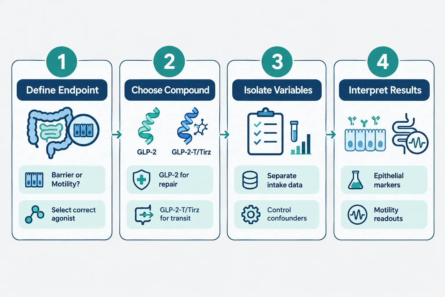
The core rule: If the research question involves mucosal healing or barrier repair, the compound must be a native GLP-2 analog. If the question involves gastric transit, intestinal motility, or energy intake regulation, GLP-2-T or GLP2 Tirz compounds are appropriate tools.

GLP2-TZ and Tirz compounds work through a "dual axis" mechanism:

Receptor Target Primary Research Endpoint
GIP receptor Insulin secretion, fat metabolism
GLP-1 receptor Gastric emptying, satiety signaling
GLP-2 receptor (NOT targeted) Epithelial repair (not applicable here)

The 2026 expansion of GLP-2-T catalog offerings reflects growing interest in motility and metabolic research, not a new application in epithelial biology. For a broader view of how multi-agonist peptides are categorized, the Peptides 101 for research-use only buyers: structure, mechanisms, and where GLP-3, MOTS-C, and 5-amino-1MQ fit in guide is a strong reference point.

Researchers planning studies that incorporate triple-agonist designs may also find the GLP3 for sale triple agonist research planning and catalog navigation resource relevant for understanding how GLP-2-T fits within a broader agonist framework.


Separating Confounders: Experimental Design for GLP-2-T and GLP2 Tirz Peptide Research

The most common methodological error in GLP-2-T and GLP2 Tirz Peptide Research is failing to isolate the five primary variables that can shift simultaneously when a dual agonist is administered:

  1. Barrier integrity (tight-junction protein levels, permeability assays)
  2. Epithelial proliferation (crypt-villus morphometry, mitotic index)
  3. Gastric and intestinal transit (bead transit time, scintigraphy)
  4. Food intake (caloric consumption, meal frequency)
  5. Systemic metabolic effects (insulin, glucose, lipid panels)

When a GLP2 Tirz compound reduces food intake, it secondarily alters nutrient exposure to the intestinal epithelium. This can produce apparent changes in villus morphology that are intake-dependent, not compound-dependent. Without a pair-fed control group, researchers cannot determine whether observed epithelial changes result from direct receptor action or from reduced luminal nutrient load.

Separating Confounders: Experimental Design for GLP-2-T and GLP2 Tirz Peptide Research

Recommended control structure for GLP-2-T motility studies:

  • Ad libitum control group (vehicle only)
  • GLP-2-T treatment group (ad libitum feeding)
  • Pair-fed control group (matched to GLP-2-T intake)
  • Native GLP-2 positive control group (epithelial repair benchmark)

This four-arm design allows researchers to attribute motility changes to direct compound action while ruling out intake-mediated epithelial artifacts. Sourcing verified reference-grade materials matters here; the Bachem and reference standards: building robust peptide benchmarks article outlines how benchmark-grade compounds strengthen experimental validity.

For researchers evaluating supplier quality before finalizing their compound selection, the peptide supplier comparisons: interpreting PeptideTech and PeptideSC guide provides practical sourcing criteria.


Speculative Outlook: Combined GLP-2 and Tirz Strategies

Looking ahead, and this represents a forward-looking prediction, not established science, some researchers are exploring whether combined GLP-2 plus Tirz protocols could address complex gut-metabolic disorders where both mucosal repair and motility dysregulation occur together. The hypothesis is that native GLP-2 could anchor epithelial restoration while a Tirz compound manages transit and intake simultaneously.

This "triple axis" framework (gut mucosa, motility, metabolism) remains speculative as of 2026. No peer-reviewed evidence yet supports combined dosing protocols in controlled research settings. Until that evidence exists, the most defensible approach is to keep the two compound classes in separate experimental arms.

Speculative Outlook: Combined GLP-2 and Tirz Strategies

Regulatory status is clear: GLP-2-T, GLP2-TZ, and GLP2 Tirz materials are strictly research-use only with no approved clinical applications as of October 2026.


Conclusion

GLP-2-T and GLP2 Tirz Peptide Research demands precise separation of biological endpoints before a single experiment is designed. Native GLP-2 owns the epithelial repair pathway. GLP-2-T and Tirz compounds belong to motility and metabolic research. Conflating the two produces data that cannot be interpreted cleanly.

Actionable next steps for research teams:

  • Audit existing protocols to confirm compound-to-endpoint alignment before initiating new studies.
  • Add pair-fed control arms to any GLP-2-T study measuring intestinal morphology.
  • Use native GLP-2 as the positive control whenever epithelial proliferation or barrier integrity is a primary endpoint.
  • Source compounds from suppliers that provide third-party purity data and reference-standard documentation.
  • Treat any combined GLP-2 plus Tirz design as exploratory and hypothesis-generating only.

Rigorous confound control is what separates publishable mechanistic data from ambiguous results. Keeping the receptor pharmacology of each compound class clearly mapped to its experimental purpose is the single most important step any research team can take in 2026.

https://www.puretestedpeptides.com/wp-content/uploads/2026/10/glp-2-t-and-glp2-tirz-peptide-research-distinguishing-epithelial-repair-from-cha.webp 1024 1536 https://www.puretestedpeptides.com/wp-content/uploads/2026/01/buy-peptides-online.jpg 2026-10-11 13:03:332026-10-11 13:03:33GLP-2-T and GLP2 Tirz Peptide Research: Distinguishing Epithelial Repair From Changes in Motility and Intake
Retatrutide Research vs Conventional Obesity Drugs: A Receptor- and Endpoint-Based Comparison

Retatrutide Research vs Conventional Obesity Drugs: A Receptor- and Endpoint-Based Comparison

October 10, 2026/0 Comments/in Uncategorized/by

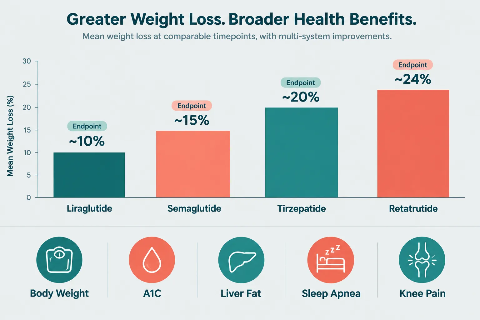
Obesity pharmacotherapy crossed a measurable threshold in 2026: a single investigational molecule achieved mean weight reductions of roughly 24 percent at 48 weeks in phase 2 trials, and up to 25 percent over 80 weeks in phase 3 data, numbers that no approved obesity drug had reached before. That molecule is retatrutide (LY3437943), and understanding why it performs differently from established agents requires a close look at receptor biology, not just headline weight numbers. This article provides a structured Retatrutide Research vs Conventional Obesity Drugs: A Receptor- and Endpoint-Based Comparison, examining how triple agonism changes the pharmacological landscape across food intake, energy expenditure, glycemic control, and tolerability endpoints.

Key Takeaways

  • Retatrutide simultaneously activates GLP-1, GIP, and glucagon receptors, a mechanism that goes beyond the single or dual receptor targeting of all currently approved obesity drugs.
  • Phase 3 TRIUMPH-1 data show mean weight losses of up to 25 percent at 80 weeks, exceeding typical results for semaglutide and approaching or surpassing tirzepatide benchmarks.
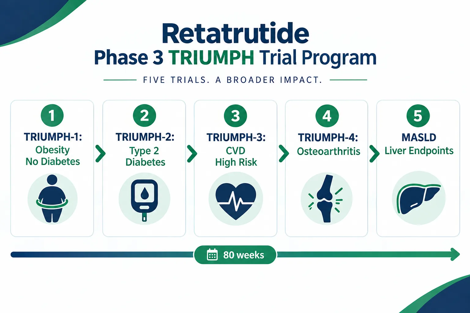
  • Beyond weight, retatrutide trials measure endpoints including liver fat, sleep apnea indices, knee osteoarthritis pain, and A1C, reflecting a multi-system strategy.
  • A 2025 meta-analysis found no statistically significant difference in overall adverse event rates between retatrutide and comparator groups, suggesting the efficacy gains do not come with a proportional safety cost.
  • As of late 2026, retatrutide remains investigational; multiple phase 3 readouts have been reported, and a regulatory filing window is anticipated in late 2026 to 2027.

Receptor Biology: Where the Comparison Begins

Receptor Biology: Where the Comparison Begins

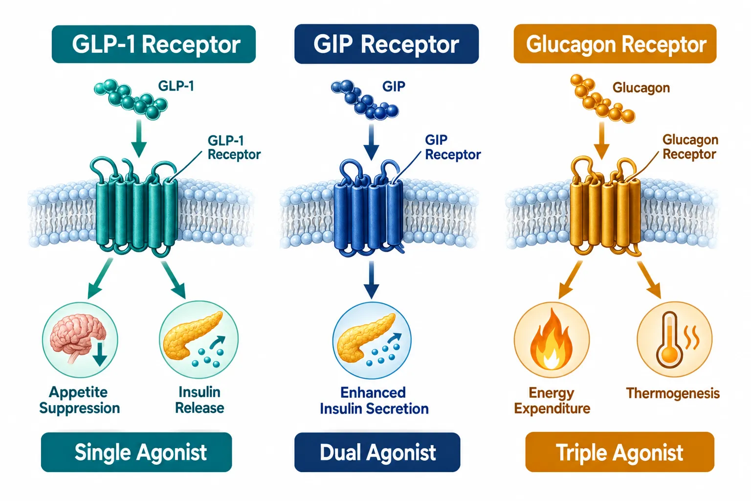
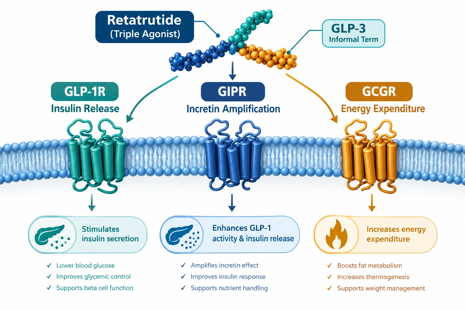
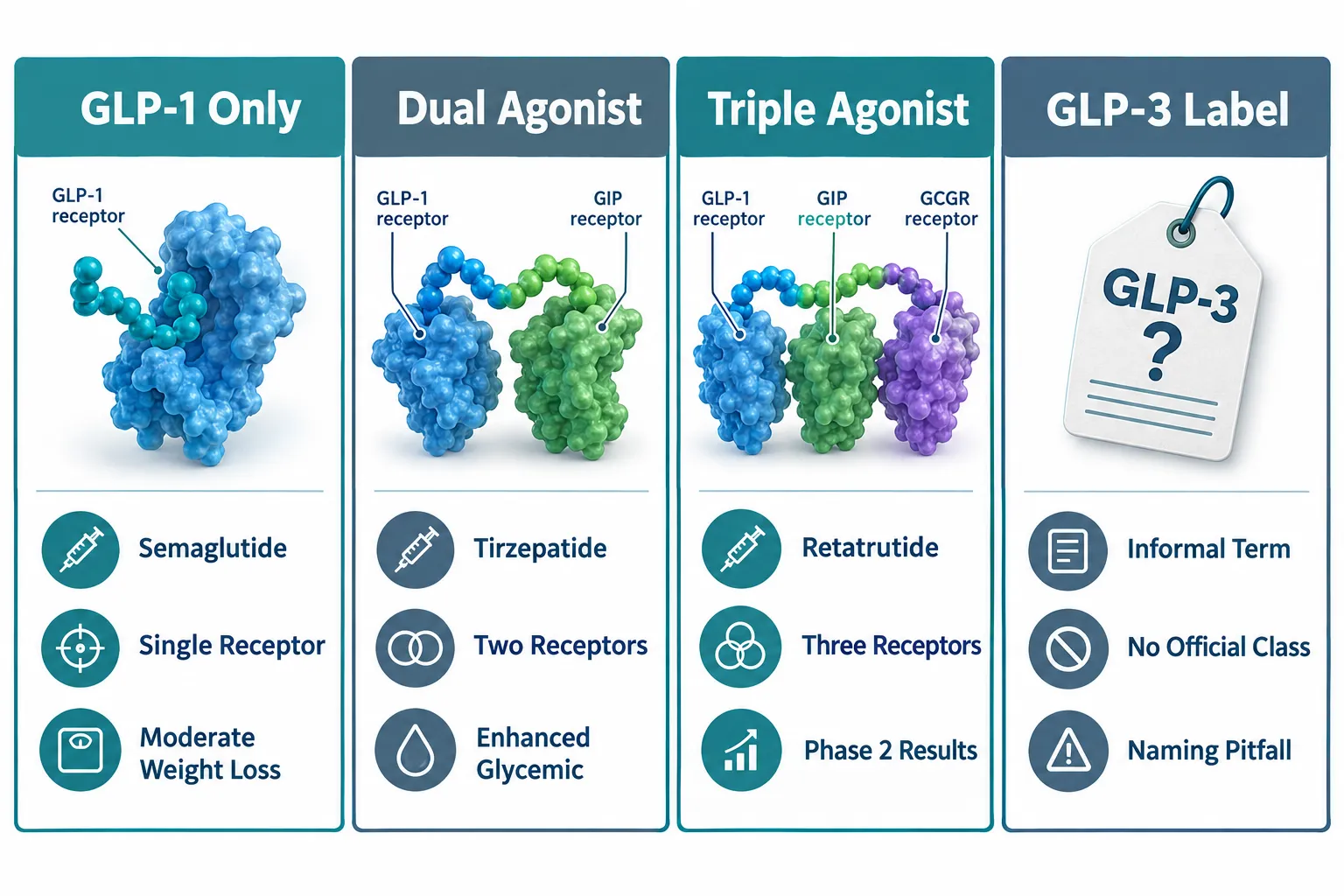
Every approved obesity drug in current clinical use targets at most two hormone receptors. Liraglutide and semaglutide are selective GLP-1 receptor agonists. Tirzepatide, the most recent approval, adds GIP receptor co-agonism. Retatrutide extends this architecture by adding full glucagon receptor activation, making it a triple hormone receptor agonist.

Each receptor contributes a distinct physiological role:

  • GLP-1 receptor activation slows gastric emptying, suppresses appetite through central and peripheral pathways, and stimulates glucose-dependent insulin release.
  • GIP receptor activation amplifies insulin secretion and may reduce nausea associated with GLP-1 agonism, partly explaining why dual agonists are often better tolerated at high doses than single GLP-1 agonists.
  • Glucagon receptor activation increases hepatic glucose output in a fasting state and, more importantly for obesity therapy, raises energy expenditure through thermogenic mechanisms.

This third receptor is the key differentiator. No approved obesity drug currently engages glucagon receptor signaling in a meaningful therapeutic way. By doing so, retatrutide addresses energy expenditure directly, a pathway that GLP-1-only drugs do not substantially activate. For a deeper look at how GLP-1 receptor biology intersects with retatrutide's design, see the GLP-1 Retatrutide overview and the dedicated Retatrutide Clinical Trials resource.

"Triple agonism is not simply an additive effect, it is a coordinated engagement of appetite, insulin sensitivity, and caloric burn that single-pathway drugs cannot replicate by design."


Efficacy Endpoints: Weight Loss, Glycemic Control, and Beyond

Efficacy Endpoints: Weight Loss, Glycemic Control, and Beyond

A rigorous Retatrutide Research vs Conventional Obesity Drugs: A Receptor- and Endpoint-Based Comparison must go beyond weight percentages and examine the full endpoint profile.

Weight Loss Benchmarks Across Drug Classes

The table below summarizes approximate mean weight reductions from pivotal trials across drug classes:

Agent Receptor Target Approx. Mean Weight Loss Trial Duration
Liraglutide GLP-1 ~8-10% 56 weeks
Semaglutide GLP-1 ~15% 68 weeks
Tirzepatide GIP + GLP-1 ~20-22% 72 weeks
Retatrutide GIP + GLP-1 + Glucagon ~23-25% 48-80 weeks

Phase 3 TRIUMPH-1 data in adults with obesity but without diabetes reported weight changes of -17.6 percent at 4 mg, -23.7 percent at 9 mg, and -25.0 percent at 12 mg, compared with -3.9 percent for placebo. These results represent a statistically and clinically meaningful separation from all approved comparators.

Glycemic Endpoints: TRIUMPH-2 and the Diabetes Population

TRIUMPH-2 evaluated retatrutide in adults with type 2 diabetes and obesity over 80 weeks. All three studied doses (4, 9, and 12 mg) delivered substantial body weight reduction alongside significant A1C reduction versus placebo. A 2025 meta-analysis confirmed mean A1C reductions of approximately 0.91 percent and fasting plasma glucose reductions of roughly 23.5 mg/dL across pooled retatrutide studies. This dual-endpoint performance is relevant when comparing retatrutide to GLP-1 agonists, which already improve both parameters but generally achieve smaller relative weight reductions at comparable timepoints.

Multi-System Endpoints: The Receptor-Driven Advantage

This is where the comparison becomes most striking. TRIUMPH-1 phase 3 data showed that retatrutide also reduced knee osteoarthritis pain scores and decreased apnea-hypopnea events in participants with obstructive sleep apnea. A phase 2a trial in metabolic dysfunction-associated steatotic liver disease (MASLD) demonstrated meaningful reductions in hepatic fat content and liver enzyme levels. A 2025 meta-analysis also documented systolic blood pressure reductions of approximately 9.9 mm Hg.

Earlier GLP-1-only programs rarely incorporated these endpoints within a single trial design. The breadth of retatrutide's endpoint coverage, spanning liver fat, sleep architecture, joint pain, glycemic control, and cardiovascular risk markers, reflects a receptor-driven strategy that treats obesity as a systemic disease rather than a weight number. For related research on combined peptide approaches, the Cagrilintide and Retatrutide comparison offers additional context. Readers interested in how noradrenergic mechanisms compare to incretin-based pathways may also find the Tesofensine mechanism vs incretin-based GLP-1 pathways analysis useful.


Tolerability, Dosing, and the Phase 3 Program

Tolerability, Dosing, and the Phase 3 Program

A complete Retatrutide Research vs Conventional Obesity Drugs: A Receptor- and Endpoint-Based Comparison must address safety alongside efficacy.

Gastrointestinal Tolerability

Like all GLP-1-based drugs, retatrutide produces gastrointestinal adverse events, primarily nausea, vomiting, and diarrhea. However, the 2025 meta-analysis found no statistically significant difference in overall adverse event rates between retatrutide and comparator groups (relative risk 1.11, p=0.24). This finding is notable: the additional glucagon receptor engagement does not appear to substantially increase the total adverse event burden compared with single or dual agonists.

Lilly has designed a dedicated phase 3 trial specifically to evaluate different dose-escalation regimens over approximately 113 weeks and 26 visits, directly addressing the practical challenge of tolerability at higher doses.

Once-Weekly Dosing and the Broader TRIUMPH Program

Retatrutide is administered as a once-weekly subcutaneous injection at doses ranging from 1 mg to 12 mg, with gradual titration. This matches the dosing frequency of semaglutide and tirzepatide, which simplifies head-to-head comparisons and supports potential clinical adoption.

The TRIUMPH phase 3 program extends across at least seven additional trials covering:

  • TRIUMPH-3: Severe obesity (BMI 35+) with established cardiovascular disease
  • TRIUMPH-4: Knee osteoarthritis
  • Moderate-to-severe obstructive sleep apnea
  • Chronic low back pain
  • MASLD / steatotic liver disease
  • Cardiorenal outcomes

Most of these trials are expected to complete in 2026, with a potential regulatory filing anticipated in late 2026 or 2027 if safety data remain consistent. As of late 2026, retatrutide remains investigational with no approved indication. Research-grade formulations referenced in resources such as GLP-3 Reta Cag 10mg and Reta 20mg are available for preclinical and laboratory research contexts only.


Conclusion

The receptor-and-endpoint lens makes the comparison clear. Conventional GLP-1 agonists work primarily through appetite suppression and slowed gastric emptying. Dual GIP/GLP-1 agonists add insulin sensitization and improved tolerability at higher doses. Retatrutide's triple agonism introduces glucagon receptor-driven energy expenditure as a third mechanism, and the clinical data show this translates into measurably greater weight loss and a broader multi-system endpoint profile than any currently approved agent.

Actionable next steps for researchers and clinicians:

  1. Review the full TRIUMPH phase 3 dataset as readouts continue through late 2026, paying particular attention to cardiovascular and MASLD endpoints.
  2. Compare tolerability profiles at equivalent dose-escalation schedules, not just peak doses, when evaluating retatrutide against tirzepatide or semaglutide.
  3. Monitor regulatory communications from Lilly for a potential New Drug Application filing window, currently anticipated in late 2026 to 2027.
  4. For preclinical research contexts, consult current literature on glucagon receptor co-agonism to understand the mechanistic basis for energy expenditure findings before extrapolating to clinical populations.

The data available in 2026 position retatrutide as the most efficacious pharmacological weight-loss agent studied to date, with a multi-system endpoint strategy that reframes obesity treatment as metabolic disease management rather than weight management alone.

https://www.puretestedpeptides.com/wp-content/uploads/2026/10/retatrutide-research-vs-conventional-obesity-drugs-a-receptor-and-endpoint-based.webp 1024 1536 https://www.puretestedpeptides.com/wp-content/uploads/2026/01/buy-peptides-online.jpg 2026-10-10 13:07:232026-10-10 13:07:23Retatrutide Research vs Conventional Obesity Drugs: A Receptor- and Endpoint-Based Comparison
What Is a GLP-3 Peptide? Retatrutide Receptor Activity, Terminology, and Research-Model Selection

What Is a GLP-3 Peptide? Retatrutide Receptor Activity, Terminology, and Research-Model Selection

October 10, 2026/0 Comments/in Uncategorized/by

Researchers searching for "GLP-3 peptide" often land in a terminology gap: no officially recognized peptide class carries that name, yet the phrase circulates widely in metabolic research circles and supplement forums alike. Understanding why that gap exists, and what retatrutide actually does at the receptor level, is essential before selecting assay formats or animal models for triple-agonist studies.

Key Takeaways

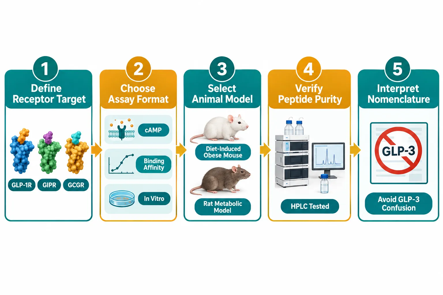
  • "GLP-3 peptide" is an informal, colloquial label, not an established pharmacological class; retatrutide is formally described as a GLP-1R/GIPR/GCGR triple agonist.
  • Retatrutide engages three distinct G-protein-coupled receptors simultaneously, producing additive metabolic effects that exceed those of GLP-1 monotherapy.
  • Phase 2 clinical data showed average body weight reductions approaching 24% over 48 weeks, among the largest reported for any pharmacological obesity intervention.
  • Naming confusion between GLP-1, GLP-2, and informal "GLP-3" labels can compromise assay design and literature searches if not addressed upfront.
  • Research-model selection for triple agonism requires receptor-specific binding assays and metabolically relevant animal models, not standard GLP-1 protocols.

Where Does the "GLP-3 Peptide" Label Come From?

The glucagon-like peptide family is derived from the proglucagon gene. Proglucagon processing yields GLP-1 and GLP-2 as well-characterized peptides with defined receptors. No endogenous peptide named GLP-3 has been isolated or assigned an official receptor in peer-reviewed pharmacology. The informal "GLP-3" label appears to have emerged from two sources: a loose shorthand for third-generation GLP-1-class drugs, and occasional misreading of triple-agonist pipeline terminology.

Retatrutide is the compound most frequently linked to this informal label. Eli Lilly developed it as a single synthetic peptide capable of activating three receptors, GLP-1R, GIPR, and GCGR, in a carefully balanced ratio. Calling it a "GLP-3 peptide" conflates the number of receptors targeted with a nonexistent peptide subfamily. Researchers and procurement teams should note this distinction before designing experiments or sourcing materials.

For those exploring GLP-3 peptide signaling pathway data, understanding this naming context prevents misattribution of results to the wrong mechanistic framework.

Where Does the "GLP-3 Peptide" Label Come From?


Retatrutide Receptor Activity: Triple Agonism Explained

Understanding what is a GLP-3 peptide in practical terms requires a close look at retatrutide's receptor pharmacology.

GLP-1R (Glucagon-Like Peptide-1 Receptor)
Activation suppresses appetite, slows gastric emptying, and stimulates glucose-dependent insulin secretion. This is the same receptor targeted by semaglutide and liraglutide.

GIPR (Glucose-Dependent Insulinotropic Polypeptide Receptor)
GIP amplifies the incretin effect and has been shown to enhance GLP-1R-mediated weight loss when combined, as demonstrated by tirzepatide's dual-agonist mechanism.

GCGR (Glucagon Receptor)
Glucagon receptor agonism increases energy expenditure and promotes hepatic fat oxidation. This is the distinguishing feature that separates retatrutide from dual GLP-1/GIP agonists.

"Triple agonism at GLP-1R, GIPR, and GCGR produces a pharmacological synergy that neither receptor alone nor any dual combination fully replicates."

The phase 2 obesity trial published in the New England Journal of Medicine reported mean weight reductions of approximately 17% at the 4 mg dose and up to 24% at the 12 mg dose over 48 weeks, figures that exceeded prior benchmarks for pharmacological interventions. Cardiometabolic markers, including triglycerides, fasting glucose, and blood pressure, also improved across dose cohorts.

Those interested in sourcing research-grade material can buy Retatrutide peptide through verified suppliers, or review available GLP-3 Retatrutide buy online USA options for laboratory use.

Retatrutide Receptor Activity: Triple Agonism Explained


Terminology Pitfalls and How They Affect Research Design

The naming confusion around what is a GLP-3 peptide creates three concrete problems for researchers.

1. Literature Search Errors
Searching databases for "GLP-3" returns inconsistent results. Some papers use it as shorthand for third-generation agents; others use it in the context of proglucagon processing fragments with no receptor activity. Filtering by receptor target (GLP-1R + GIPR + GCGR) yields cleaner, more reproducible literature sets.

2. Assay Misspecification
Standard GLP-1 receptor binding assays measure only GLP-1R affinity. A triple-agonist compound like retatrutide requires three parallel cAMP accumulation assays, one per receptor, to characterize its full pharmacological profile. Using a single-receptor protocol underestimates potency and misrepresents the mechanism.

3. Procurement Confusion
Vendors sometimes list retatrutide under "GLP-3" product codes. Confirming the sequence, purity by HPLC, and receptor-binding data before purchase is non-negotiable. Products such as GLP-3R 30mg peptide and GLP-3R 10mg peptide GA10 use vendor-specific nomenclature that refers to retatrutide-class compounds, not an officially classified GLP-3 receptor ligand.

For broader context on how these compounds compare with other metabolic peptides, the article on polypeptide peptides in cardiometabolic models offers a useful structural comparison.


Research-Model Selection for Retatrutide and Triple-Agonist Studies

Choosing the right model is as important as understanding what is a GLP-3 peptide in the first place. The table below outlines common options.
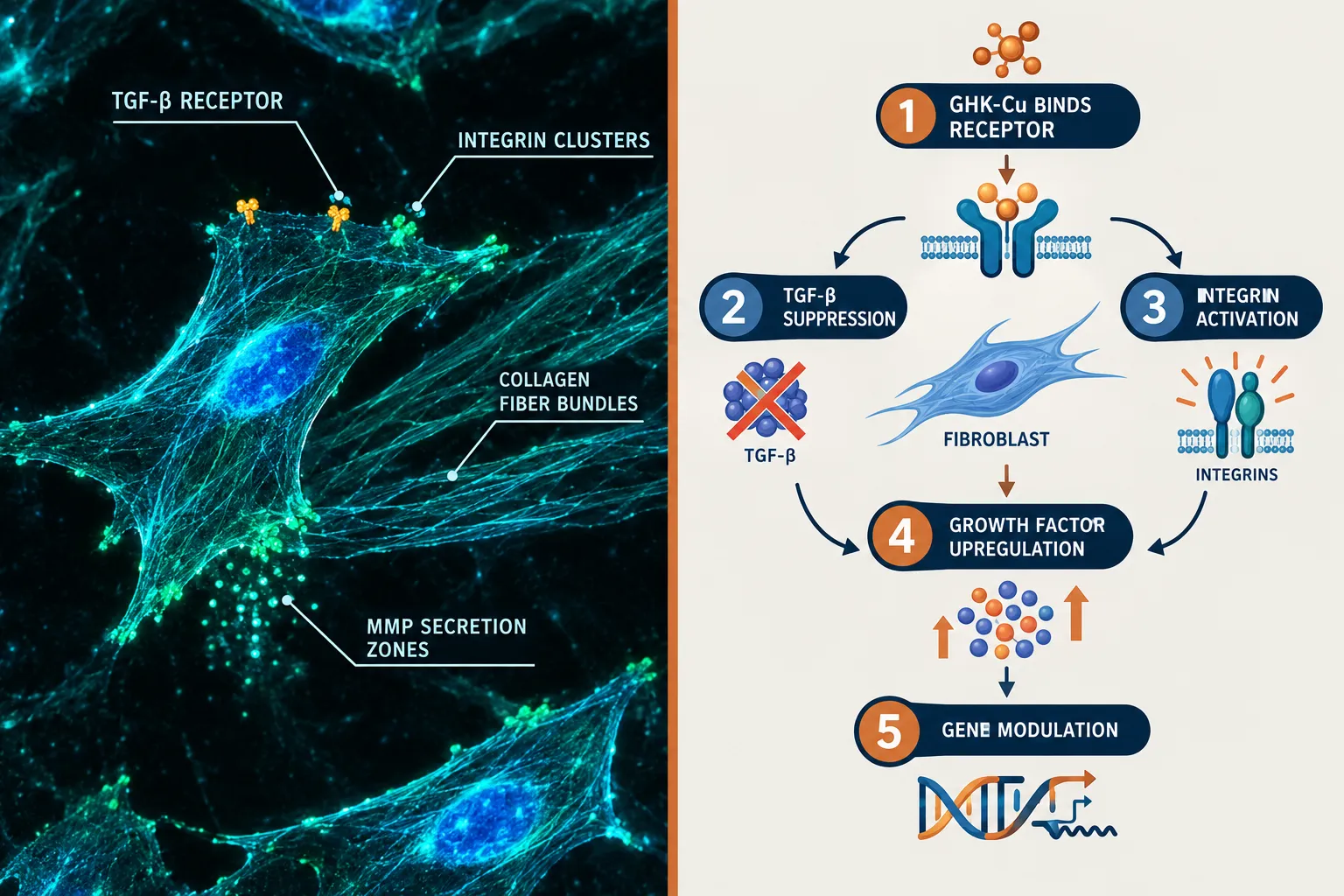
Model Type Best For Key Limitation
Diet-Induced Obese (DIO) Mouse Weight loss, adiposity, metabolic panels GCGR expression differs from humans
Zucker Diabetic Fatty Rat Glycemic control, insulin sensitivity Limited cardiovascular readouts
Non-Human Primate (NHP) Translational cardiometabolic endpoints High cost, regulatory complexity
In Vitro cAMP Assay (HEK293) Receptor-specific potency ranking No systemic pharmacokinetics

For in vivo work, the DIO mouse model remains the most widely used starting point. Researchers should confirm that their animal colony expresses all three target receptors at physiologically relevant levels before interpreting weight or glucose data as triple-agonist effects.

Ongoing Retatrutide clinical trial data from phase 3 programs (initiated 2024-2025) are expected to clarify dose-response relationships and cardiovascular outcomes through 2026 and beyond. Analysts broadly expect regulatory submissions to follow positive phase 3 readouts, though timelines remain speculative and subject to trial outcomes.

Research-Model Selection for Retatrutide and Triple-Agonist Studies


Conclusion

The question of what is a GLP-3 peptide resolves to a clear answer: it is an informal term, not a recognized pharmacological class. Retatrutide is a GLP-1R/GIPR/GCGR triple agonist whose clinical weight-loss results and cardiometabolic benefits place it among the most closely watched compounds in metabolic medicine in 2026. For researchers, the actionable steps are straightforward:

  1. Replace "GLP-3" with "GLP-1R/GIPR/GCGR triple agonist" in all literature searches and assay documentation.
  2. Run three parallel receptor-binding or cAMP assays rather than a single GLP-1R protocol.
  3. Select animal models with confirmed expression of all three target receptors.
  4. Verify peptide purity and sequence data from suppliers before initiating any study.
  5. Monitor phase 3 trial publications for updated dose-response and safety benchmarks.

Those sourcing material for preclinical work can explore options including GLP-3 Reta 30mg for sale and confirm specifications align with their receptor-activity requirements before procurement.

https://www.puretestedpeptides.com/wp-content/uploads/2026/10/what-is-a-glp-3-peptide-retatrutide-receptor-activity-terminology-and-research-m.webp 1024 1536 https://www.puretestedpeptides.com/wp-content/uploads/2026/01/buy-peptides-online.jpg 2026-10-10 13:04:552026-10-10 13:04:55What Is a GLP-3 Peptide? Retatrutide Receptor Activity, Terminology, and Research-Model Selection
BPC-157 Peptide Research: Separating Angiogenesis, Barrier Repair, and Inflammatory Signaling

BPC-157 Peptide Research: Separating Angiogenesis, Barrier Repair, and Inflammatory Signaling

October 10, 2026/0 Comments/in Uncategorized/by

Fewer than 15 amino acids long, BPC-157 generates more mechanistic debate per residue than almost any other peptide in preclinical science. As 2026 research programs expand across vascular, epithelial, and immune models, the central challenge in BPC-157 peptide research: separating angiogenesis, barrier repair, and inflammatory signaling is no longer whether effects exist, it is how to measure each pathway cleanly without confounding the others.
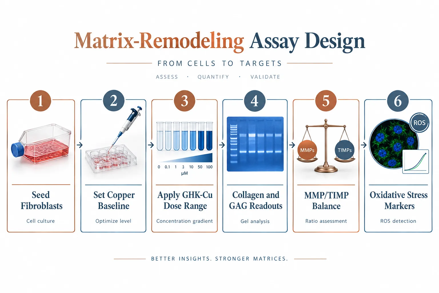
Key Takeaways

  • BPC-157 activates at least three distinct biological readouts: vascular remodeling, epithelial barrier restoration, and cytokine-level immune modulation.
  • New 2026 mechanistic work links the FBXO22-BACH1 axis to BPC-157-driven angiogenesis, shifting the debate from "stimulating" to "organizing" blood vessel growth.
  • Intestinal tight-junction repair is the most replicated barrier-repair finding, but corneal and mucosal models are extending the picture.
  • NF-kB suppression and downstream cytokine reduction represent the clearest inflammatory signaling readout, though pathway cross-talk complicates interpretation.
  • Rigorous experimental design, orthogonal assays, appropriate controls, and staged time points, is essential for drawing pathway-specific conclusions.

The Angiogenesis Question: Stimulating or Organizing?

The Angiogenesis Question: Stimulating or Organizing?

The oldest debate in BPC-157 peptide research centers on what the compound actually does to blood vessels. Early models consistently showed accelerated capillary formation in wound beds, which led to the label "pro-angiogenic." More precise 2026 mechanistic work complicates that framing significantly.

The FBXO22-BACH1 signaling axis has emerged as a key upstream node. FBXO22, an E3 ubiquitin ligase, targets BACH1 for proteasomal degradation. BACH1 normally represses a suite of pro-vascular genes. When BPC-157 appears to facilitate FBXO22 activity, BACH1 levels drop, and genes governing vessel sprouting and stabilization become more accessible. Critically, this is a regulatory event, not a raw amplification of VEGF output.

The VEGF-nitric oxide (NO) axis remains central to the vascular story. BPC-157 consistently upregulates endothelial nitric oxide synthase (eNOS) in preclinical models, driving local NO production that relaxes vessel walls and supports lumen formation. However, the effect appears dose-sensitive and context-dependent, disordered vasculature in ischemic tissue tends to become more organized, not simply more abundant.

Key distinction: "Angiogenesis-organizing" describes a process that improves vessel architecture and perfusion efficiency. "Angiogenesis-stimulating" implies net new vessel mass regardless of context. The evidence in 2026 leans toward the former.

Recommended assay approach for vascular readouts:

Readout Preferred Assay Critical Control
Capillary density CD31 immunostaining Vehicle-matched sham
Vessel maturation Alpha-SMA co-staining Time-matched baseline
NO production DAF-FM diacetate fluorescence eNOS inhibitor (L-NAME)
BACH1 degradation Western blot / co-IP FBXO22 knockdown line

Researchers exploring related vascular peptide mechanisms may find useful context in IPA Sermorelin Stack Research, which addresses growth-factor-driven vascular signaling in adjacent model systems.


Barrier Repair: Gut, Cornea, and Mucosal Models

Barrier Repair: Gut, Cornea, and Mucosal Models

Intestinal barrier restoration is the most replicated finding in BPC-157 peptide research: separating angiogenesis, barrier repair, and inflammatory signaling requires treating this pathway as mechanistically independent from the vascular work, even though both involve tissue remodeling.

In gut models, BPC-157 consistently accelerates tight-junction reassembly following chemically induced permeability damage. The proteins most clearly affected are claudin family members and occludin, which form the paracellular seal between enterocytes. Upregulation of these proteins reduces translocation of luminal antigens into submucosal tissue, which in turn lowers the inflammatory stimulus that would otherwise perpetuate barrier disruption.

This creates an important experimental pitfall: if a study measures inflammation reduction after BPC-157 administration in a leaky-gut model, the result may reflect barrier repair rather than direct immune modulation. Separating these effects requires:

  1. Measuring tight-junction protein expression at early time points (6-12 hours) before cytokine changes peak
  2. Using a barrier-intact inflammatory model as a parallel arm
  3. Confirming permeability with FITC-dextran flux assays independent of histology

Beyond the gut, barrier-repair activity has been demonstrated in corneal epithelial models and oral mucosal tissue. In corneal studies, BPC-157 accelerates re-epithelialization after abrasion, with effects on goblet cell density and submucosal vascularity that parallel gut findings. This cross-tissue consistency suggests a conserved epithelial repair mechanism rather than organ-specific pharmacology.

For researchers sourcing peptides with documented purity for barrier-model work, the discussion at Where to Buy Research-Grade Glow Blend Peptide covers purity standards and skin-model compatibility considerations relevant to epithelial assay design.

Thymosin Alpha 4 Peptide research offers a useful comparative framework, as thymosin beta-4 also promotes actin cytoskeleton remodeling in epithelial repair, providing an orthogonal positive control for barrier-reassembly studies.


Inflammatory Signaling: NF-kB, Cytokines, and Pathway Cross-Talk

Inflammatory Signaling: NF-kB, Cytokines, and Pathway Cross-Talk

The inflammatory signaling arm of BPC-157 peptide research: separating angiogenesis, barrier repair, and inflammatory signaling demands the most careful experimental architecture, because NF-kB sits at the intersection of all three pathways.

BPC-157 consistently suppresses NF-kB nuclear translocation in preclinical inflammatory models. Downstream, this reduces transcription of TNF-alpha, IL-6, and IL-1-beta, the primary cytokines driving both acute tissue damage and chronic low-grade inflammation. In orthopedic and sports-medicine models, this anti-inflammatory profile has generated significant interest for tendon and ligament repair applications, though clinical data in humans remains minimal as of 2026.

The cross-talk problem is real. Consider this cascade:

  • BPC-157 repairs the gut barrier
  • Reduced antigen translocation lowers systemic LPS burden
  • Lower LPS burden reduces TLR4-NF-kB activation
  • Measured cytokine reduction appears to be a direct anti-inflammatory effect

Without a barrier-intact arm in the study design, the cytokine reduction is easily misattributed. Orthogonal assay strategies that isolate each pathway are not optional, they are methodologically required for publishable mechanistic claims.

Recommended time-point staging for inflammatory readouts:

  • 0-6 hours: Measure NF-kB translocation and immediate cytokine mRNA
  • 12-24 hours: Assess protein-level cytokine secretion (ELISA)
  • 48-72 hours: Evaluate tissue histology and immune cell infiltration
  • Day 7+: Confirm resolution versus suppression using resolution-phase markers (e.g., lipoxin A4)

Cross-system applications, including neurodegeneration models where NF-kB-driven neuroinflammation is a primary target, and liver fibrosis models where stellate cell activation is NF-kB-dependent, are expanding the research footprint. SS-31 Mitochondrial Research Themes provides relevant context for researchers examining mitochondrial-inflammatory crosstalk in these overlapping disease models.

Safety and regulatory framing remains an open issue. BPC-157 is not approved for human use in any jurisdiction as of 2026, and regulatory agencies have flagged uncertainty around systemic vascular effects at higher doses. The "regeneration vs. risk" framing that dominates industry discussion should not substitute for controlled dose-response data in validated models.


Conclusion

Advancing BPC-157 peptide research means resisting the temptation to report a single unified "healing effect." The vascular, epithelial, and immune readouts are real, but they are distinct, they interact, and they require purpose-built experimental designs to separate cleanly.

Actionable next steps for research teams in 2026:

  • Design studies with three parallel arms: vascular-only, barrier-intact inflammatory, and full-model, to isolate pathway contributions
  • Use FBXO22-BACH1 Western blots and eNOS fluorescence assays as orthogonal vascular controls alongside CD31 staining
  • Stage time points explicitly: tight-junction protein expression before cytokine peaks, and resolution markers after the acute phase
  • Include barrier-flux assays (FITC-dextran) in any gut-inflammation model to prevent misattribution of cytokine changes
  • Cross-reference findings against structurally related repair peptides, including Thymosin Alpha 4 Peptide, to build mechanistic confidence through orthogonal validation

The peptide's small size belies its signaling complexity. Rigorous pathway separation is not a methodological luxury, it is the foundation on which any translational claim must eventually stand.

https://www.puretestedpeptides.com/wp-content/uploads/2026/10/bpc-157-peptide-research-separating-angiogenesis-barrier-repair-and-inflammatory.webp 1024 1536 https://www.puretestedpeptides.com/wp-content/uploads/2026/01/buy-peptides-online.jpg 2026-10-10 13:04:492026-10-10 13:04:49BPC-157 Peptide Research: Separating Angiogenesis, Barrier Repair, and Inflammatory Signaling
Epithalon Peptide Research: Telomere-Length Measurement Pitfalls and Better Senescence Controls

Epithalon Peptide Research: Telomere-Length Measurement Pitfalls and Better Senescence Controls

October 10, 2026/0 Comments/in Uncategorized/by

Fewer than a dozen completed human trials have ever prospectively measured telomere length before and after any peptide intervention, and epitalon is not yet among them. That gap between compelling cell-culture data and verified human outcomes sits at the heart of the current debate in Epithalon Peptide Research: Telomere-Length Measurement Pitfalls and Better Senescence Controls, where the quality of the assay used to measure telomeres can determine whether a study's conclusion holds up or quietly collapses under replication pressure.

Key Takeaways

  • Epitalon has demonstrated dose-dependent telomere elongation and hTERT upregulation in normal human fibroblasts and mammary epithelial cells in controlled culture studies, with some lines showing roughly 12-fold increases in hTERT expression at 1 ug/ml.
  • The same peptide elongated telomeres in breast cancer cell lines through an Alternative Lengthening of Telomeres (ALT) mechanism, raising important questions about oncogenic risk in telomerase-targeting research.
  • qPCR, the most common telomere assay in epitalon studies, reports only average telomere length and suffers from significant inter-laboratory variability, making it insufficient as a standalone measure.
  • No completed, well-controlled human clinical trial has yet linked epitalon administration to measurable telomere extension in living people.
  • Robust epitalon research requires multi-method telomere validation paired with functional senescence markers, not telomere length data alone.

What the Cell-Culture Evidence Actually Shows

A 2025 study from Brunel University London published in Biogerontology produced some of the most detailed quantitative data on epitalon's telomere effects to date. Using both qPCR and telomere immunofluorescence, researchers documented telomere elongation in normal human fibroblasts and mammary epithelial cells that was both dose-dependent and time-dependent. In certain normal cell lines, 1 ug/ml epitalon drove approximately 12-fold upregulation of hTERT, the catalytic subunit of telomerase, while 0.5 ug/ml produced roughly 5-fold upregulation. Overall telomere length in some cultures reached about 2.4 times the baseline measurement.

What the Cell-Culture Evidence Actually Shows

A correction published later in 2025 refined the mechanistic interpretation of that study without overturning the core quantitative findings. The key clarification involved distinguishing between telomerase-driven elongation in normal cells and ALT-mediated elongation in cancer cell lines. In breast cancer lines (21NT and BT474), epitalon did extend telomeres, but via ALT rather than canonical telomerase activation. That distinction matters enormously for safety evaluation.

The takeaway is not that epitalon fails in cancer cells, it is that the mechanism differs, and the implications for oncogenic risk require separate, careful assessment.

A 2026 review on therapeutic peptides in gerontology confirmed that epitalon's telomerase effects in normal somatic cells have been replicated across independent laboratories, strengthening the mechanistic case. Additional 2025 experimental work extended these findings to bovine cumulus cells and cumulus-oocyte complexes, suggesting the peptide's influence on telomerase is not limited to a single cell type.

For researchers sourcing compounds for controlled studies, understanding purity and batch consistency is critical. Resources on lab tested peptides and guidance on choosing a best peptide manufacturer are relevant starting points for experimental design.


Telomere-Length Measurement Pitfalls in Epithalon Peptide Research

Telomere-Length Measurement Pitfalls in Epithalon Peptide Research

The most significant methodological vulnerability in current Epithalon Peptide Research: Telomere-Length Measurement Pitfalls and Better Senescence Controls literature is over-reliance on a single assay type, particularly qPCR.

Why qPCR Alone Is Not Enough

qPCR-based telomere measurement is fast and scalable, which explains its dominance. However, it carries serious limitations:

Limitation Why It Matters for Epitalon Studies
Reports average telomere length only Misses critically short telomeres that drive senescence
High inter-laboratory variability Results may not replicate across research groups
Sensitive to DNA quality and normalization Small technical errors produce large apparent differences
Cannot resolve telomere length distributions A few very long telomeres can mask many short ones

Critically short telomeres, not average telomere length, are the primary drivers of genomic instability and replicative senescence. A treatment that lengthens average telomeres while leaving critically short ones intact may show impressive qPCR numbers without meaningfully reducing senescence burden.

Alternative Methods and Their Trade-Offs

Fluorescence in situ hybridization (FISH) and quantitative FISH (Q-FISH) allow spatial visualization of individual telomeres and can detect length distributions rather than averages. However, they require meticulous calibration with standards of known telomeric repeat content and are low throughput. Terminal Restriction Fragment (TRF) analysis offers greater precision but is labor-intensive and difficult to scale.

The expert consensus from 2025-2026 methodology reviews is clear: multi-method validation combining qPCR with FISH/Q-FISH or TRF is the minimum standard for interventional telomere research. Cross-laboratory comparisons without harmonized protocols should be avoided.

Research teams exploring synergistic peptide combinations, such as those examining Epithalon and MOTS-c synergy, face compounded assay complexity, since each compound may affect telomere biology through distinct pathways that interact unpredictably in mixed-assay conditions.


Better Senescence Controls: Moving Beyond Telomere Length

Better Senescence Controls: Moving Beyond Telomere Length

The most important shift recommended by 2026 gerontology and methodology reviews is reframing the research question. Telomere elongation is a molecular event. Cellular senescence is a functional state. The two are related but not identical, and Epithalon Peptide Research: Telomere-Length Measurement Pitfalls and Better Senescence Controls must address both to produce actionable conclusions.

Recommended Senescence Control Strategies

Leading experts propose the following framework for future epitalon studies:

  • Include reference samples with independently verified telomere lengths in every assay run
  • Report assay variability explicitly, including coefficient of variation across replicates
  • Use blinded replicates to reduce observer bias in both measurement and interpretation
  • Characterize telomere length distributions, not just means, to identify critically short telomere populations
  • Add functional senescence markers: senescence-associated beta-galactosidase (SA-beta-gal), p21 expression, DNA damage foci (gamma-H2AX), and senescence-associated secretory phenotype (SASP) components
  • Measure replicative capacity: does epitalon-treated cell culture actually divide more times before reaching the Hayflick limit?
  • Assess mitochondrial function and reactive oxygen species as independent indicators of cellular health

This multi-endpoint approach transforms a telomere chemistry study into a genuine senescence biology study. Researchers interested in comparing mitochondrial-targeting peptides alongside epitalon may find the MOTS-c peptide dosage calculator and information on buying MOTS-c peptide useful for designing comparative experimental arms.

The Human Translation Gap

Despite strong in vitro data, no completed, well-controlled human clinical trial has prospectively linked epitalon administration to telomere length changes in blood or tissue samples. Existing human data, primarily from older Khavinson-era studies, are limited in sample size and methodologically heterogeneous. This gap does not invalidate the cell-culture findings, but it does mean that claims about epitalon extending human lifespan or reversing human aging remain scientifically unsupported as of 2026.

Telomerase activation also carries a theoretical oncogenic risk. The ALT-pathway findings in cancer cell lines are not evidence that epitalon causes cancer, but they are a clear signal that telomerase-modulating interventions require rigorous long-term safety monitoring in any future human studies.

For researchers building out broader peptide research programs, resources on wholesale peptides and buying bulk peptide can support the scale needed for properly powered, multi-endpoint studies.


Conclusion

The cell-culture evidence for epitalon's telomere-extending and telomerase-activating effects is reproducible and quantitatively significant. However, the field's current reliance on average telomere length measured by qPCR alone is a structural weakness that undermines the translational value of many published findings.

Actionable next steps for researchers:

  1. Adopt multi-method telomere measurement protocols (qPCR plus Q-FISH or TRF) as a minimum standard for any new epitalon study.
  2. Add functional senescence endpoints, SA-beta-gal, p21, SASP markers, and replicative capacity, to every telomere-focused experimental design.
  3. Distinguish clearly between telomerase-driven and ALT-mediated elongation, especially when working with any cancer-derived cell lines.
  4. Prioritize harmonized protocols before conducting cross-laboratory comparisons.
  5. Treat the human translation gap as an open research question requiring prospective, controlled clinical study design, not an assumption that can be bridged by extrapolating cell-culture data.

The science of epitalon is genuinely promising. Making that promise actionable requires the field to hold itself to the same measurement standards it would demand of any other longevity intervention.

https://www.puretestedpeptides.com/wp-content/uploads/2026/10/epithalon-peptide-research-telomere-length-measurement-pitfalls-and-better-senes.webp 1024 1536 https://www.puretestedpeptides.com/wp-content/uploads/2026/01/buy-peptides-online.jpg 2026-10-10 13:04:472026-10-10 13:04:47Epithalon Peptide Research: Telomere-Length Measurement Pitfalls and Better Senescence Controls
GHK-Cu Peptide Research: Copper Availability, Fibroblast Signaling, and Matrix-Remodeling Assay Design

GHK-Cu Peptide Research: Copper Availability, Fibroblast Signaling, and Matrix-Remodeling Assay Design

October 10, 2026/0 Comments/in Uncategorized/by

Fewer than three amino acids separate GHK-Cu from one of the most studied copper-binding peptides in modern connective-tissue research. The tripeptide glycine-histidine-lysine, when complexed with a cupric ion, creates a bioactive metallopeptide that has attracted serious laboratory attention for its roles in extracellular matrix remodeling, fibroblast gene regulation, and wound biology. As 2026 brings a new wave of clinician-guided and research-only applications, rigorous assay design for GHK-Cu peptide research, covering copper availability, fibroblast signaling, and matrix-remodeling endpoints, has never been more important.

Key Takeaways

  • GHK-Cu is a copper-chelating tripeptide that acts as a copper shuttle to the extracellular matrix and antioxidant enzymes, not an approved therapeutic drug.
  • Fibroblast signaling downstream of GHK-Cu involves broad gene modulation, TGF-beta pathway suppression, integrin activation, and growth factor upregulation.
  • Copper availability in culture media is a critical variable that must be controlled before any dose-response experiment is interpreted.
  • Matrix-remodeling endpoints include collagen synthesis, glycosaminoglycan deposition, MMP/TIMP ratios, and mechanical stiffness assays.
  • Oxidative-stress markers and gene-expression profiling provide systems-level context that single-endpoint assays cannot capture alone.

Copper Availability: The Variable Most Researchers Underestimate

Copper Availability: The Variable Most Researchers Underestimate

The biological activity of GHK-Cu depends entirely on the copper ion it carries. Standard cell culture media, including DMEM and RPMI, contain trace copper levels that vary by lot, manufacturer, and storage conditions. Before any GHK-Cu peptide research experiment begins, the baseline copper concentration in the working medium must be measured and standardized.

Why this matters: If ambient copper is already saturating cellular uptake pathways, adding GHK-Cu may produce a blunted response that researchers misinterpret as peptide inactivity. Conversely, copper-depleted media can amplify responses in ways that do not reflect physiological conditions.

Practical controls to include:

  • A copper-only control (CuSO4 or CuCl2 at equimolar concentration to the GHK-Cu treatment)
  • A GHK-only (apo-peptide) control to separate copper-independent peptide effects
  • Inductively coupled plasma mass spectrometry (ICP-MS) measurement of basal copper in each media lot

GHK-Cu functions as a copper shuttle, delivering Cu2+ to extracellular matrix enzymes such as lysyl oxidase and to intracellular antioxidant enzymes including superoxide dismutase (SOD). This dual delivery role means that copper bioavailability affects both structural matrix outcomes and oxidative-stress readouts simultaneously. Researchers designing assays around only one of these endpoints risk missing the full picture.

"Controlling for ambient copper is not optional, it is the foundational step that determines whether every downstream result is interpretable."


Fibroblast Signaling: What GHK-Cu Actually Does Inside the Cell

Fibroblast Signaling: What GHK-Cu Actually Does Inside the Cell

Dermal fibroblasts are the primary cell model used in GHK-Cu peptide research because they are the main producers of collagen, elastin, and glycosaminoglycans (GAGs) in connective tissue. The signaling landscape downstream of GHK-Cu is broad and context-dependent.

Key signaling nodes documented in fibroblast studies:

Pathway Observed Effect Assay Relevance
TGF-beta1 Suppressed in keloid fibroblasts Scar and fibrosis models
Integrins (alpha-v, beta-1) Upregulated Adhesion and migration assays
Growth factors (FGF, VEGF) Increased expression Angiogenesis and repair models
MMP-2 / MMP-9 Modulated bidirectionally Matrix turnover readouts
TIMP-1 / TIMP-2 Counterbalanced with MMPs Net remodeling index

The TGF-beta1 suppression effect is particularly relevant for keloid and hypertrophic scar research. In these models, GHK-Cu appears to normalize the overactive collagen deposition driven by TGF-beta1 signaling, shifting fibroblasts toward a more balanced remodeling phenotype rather than a fibrotic one.

For researchers interested in how peptide signaling intersects with broader metabolic pathways, the work on MOTS-C peptide and mitochondrial signaling offers a useful parallel for understanding how small peptides can drive large gene-expression programs.

Concentration-response considerations:

GHK-Cu displays a non-linear, often biphasic dose-response curve in fibroblast cultures. Low nanomolar concentrations (1-10 nM) tend to stimulate collagen synthesis, while higher concentrations (above 1 microM) can shift toward matrix degradation by upregulating MMPs. This biphasic behavior makes single-concentration studies unreliable. A minimum of five concentration points spanning at least four orders of magnitude is recommended for any definitive fibroblast signaling experiment.

Proper peptide dosing strategy, even in a research context, requires this kind of systematic range-finding before drawing mechanistic conclusions.


Matrix-Remodeling Assay Design: Endpoints, Controls, and Interpretation

Matrix-Remodeling Assay Design: Endpoints, Controls, and Interpretation

Designing a robust matrix-remodeling assay for GHK-Cu peptide research requires selecting endpoints that reflect both synthetic and degradative arms of ECM turnover. No single endpoint is sufficient.

Recommended endpoint panel:

  1. Collagen synthesis, Sircol collagen assay or hydroxyproline quantification from cell-layer lysates
  2. Elastin deposition, Fastin elastin assay on conditioned media and matrix fractions
  3. Glycosaminoglycan content, DMMB (dimethylmethylene blue) assay for sulfated GAGs
  4. MMP/TIMP ratio, ELISA or multiplex bead-based assay for MMP-1, MMP-2, MMP-9, TIMP-1, TIMP-2
  5. Oxidative-stress markers, Intracellular ROS (DCFH-DA fluorescence), 8-OHdG for oxidative DNA damage, and SOD activity assay
  6. Mechanical properties, Atomic force microscopy (AFM) or collagen gel contraction assay for matrix stiffness

Oxidative-stress markers deserve special attention. Because GHK-Cu delivers copper to SOD, a well-designed assay should show reduced ROS alongside increased SOD activity at physiologically relevant concentrations. If ROS increases rather than decreases, it may indicate copper overload or media incompatibility, a result worth investigating rather than discarding.

For gene-expression profiling, RNA-seq or a targeted wound-healing gene panel (covering COL1A1, COL3A1, ELN, FN1, MMP1, TIMP1, TGFB1, CTGF) provides systems-level context. This approach has revealed that GHK-Cu can upregulate over 30 genes involved in ECM organization while simultaneously downregulating inflammatory mediators, a pattern that single-endpoint assays cannot detect.

Common assay pitfalls to avoid:

  • Using serum-containing media without dialysis to remove endogenous copper
  • Failing to passage-match fibroblast donors (passage 3-6 is standard)
  • Measuring only one time point (24 hours misses matrix deposition peaks at 72-96 hours)
  • Ignoring cell viability alongside matrix endpoints (MTT or live/dead staining should run in parallel)

Researchers working with other tissue-remodeling peptides such as BPC-157 and TB-500 will recognize many of these assay design principles, as matrix remodeling and fibroblast activation are shared mechanistic themes across repair-focused peptide research.

For those sourcing research-grade materials, working with a lab-tested peptide supplier with documented purity certificates is essential for reproducible results. Copper-peptide complexes are particularly sensitive to oxidation and degradation during storage, so certificate of analysis data for both peptide identity and copper content should be requested before use.

It is also worth noting that GHK-Cu remains a research agent and cosmetic ingredient, not an approved drug. Systemic injection protocols are considered ahead of the available clinical evidence, and researchers should frame their work within appropriate in-vitro or preclinical boundaries. For broader context on how peptide research compounds are evaluated, the discussion of SS-31 peptide research considerations offers a useful methodological parallel.


Conclusion

Productive GHK-Cu peptide research in 2026 depends on three disciplined foundations: controlling copper availability before any treatment begins, mapping fibroblast signaling across a full concentration range rather than a single dose, and selecting a multi-endpoint matrix-remodeling panel that captures both synthetic and degradative ECM activity alongside oxidative-stress markers.

Actionable next steps for researchers:

  • Measure and standardize copper content in all culture media lots using ICP-MS before starting experiments
  • Include apo-GHK and copper-only controls in every experiment to isolate peptide-specific effects
  • Run at least five concentration points spanning 1 nM to 10 microM to capture the full dose-response curve
  • Add SOD activity and ROS assays alongside collagen and MMP endpoints to capture the antioxidant arm of GHK-Cu biology
  • Use gene-expression profiling on a wound-healing panel to contextualize single-endpoint findings

When these controls are in place, GHK-Cu research yields interpretable, reproducible data that genuinely advances understanding of copper-dependent matrix remodeling.

https://www.puretestedpeptides.com/wp-content/uploads/2026/10/ghk-cu-peptide-research-copper-availability-fibroblast-signaling-and-matrix-remo.webp 1024 1536 https://www.puretestedpeptides.com/wp-content/uploads/2026/01/buy-peptides-online.jpg 2026-10-10 13:04:452026-10-10 13:04:45GHK-Cu Peptide Research: Copper Availability, Fibroblast Signaling, and Matrix-Remodeling Assay Design
Page 3 of 80‹12345›»
×

Helpful Links

  • My account
  • Cart
  • Checkout
  • Refund and Returns Policy
  • Privacy Policy
  • SMS Privacy Policy
  • Login
  • My Account
  • Logout

USA Made Lab Tested Peptides

All products are sold for research, laboratory, or analytical purposes only, and are not for human consumption


Pure Tested Peptides is a chemical supplier. Pure Tested Peptides is not a compounding / chemical compounding facility as defined under 503A of the Federal Food, Drug, and Cosmetic act. Pure Tested Peptides is not an outsourcing facility as defined under 503B of the Federal Food, Drug, and Cosmetic act.

The statements made within this website have not been evaluated by the US Food and Drug Administration. The products we offer are not intended to diagnose, treat, cure or prevent any disease.

Human/Animal Consumption Prohibited. Laboratory/In-Vitro Experimental Use Only

Scroll to top Scroll to top Scroll to top