Call or Text 727-513-9780
  • Shopping Cart Shopping Cart
    0Shopping Cart
Pure Tested Peptides | America's most trusted Peptides for sale online
  • Peptides for sale
    • Oral Peptides for sale
      • Peptide Capsules for sale
      • BPC 157 Capsules 1000mcg
      • SLU-PP-332 Capsules | 1000 mcg
      • 5-Amino-1MQ 50mg Capsules
      • Tesofensine 500mcg
    • All Peptides for sale
    • Peptide Sprays
      • BPC 157 Nasal Spray Kit
      • BPC-157 TB500 Nasal Spray Kit
      • Semax Nasal Spray 10mg
      • Selank – Nasal Spray Kit – 10mg
      • Epithalon 50MG Nasal Spray Kit
      • Ipamorelin 10mg Nasal Spray
      • Klow Nasal Spray (BPC-157 + TB-500 + GHK-Cu + KPV) | 80mg
      • Hulk Nasal Spray Tesa / Ipa Blend 6/3 MG
      • Klow Nasal Spray
      • NAD + 500 mg Nasal Spray
      • PT-141 Nasal Spray Kit
    • GHRH Peptides
      • Ipa Peptides
      • CJC-1295 Peptides
        • CJC-1295 with DAC 5 mg
        • CJC-1295 without DAC 5 mg
        • CJC-1295 Ipa 10mg
      • Tesa Peptides
        • Tesa Peptide
        • Tesa 20 mg
    • GHK-Cu Peptides
      • All GHK-Cu Peptides
      • GHK-Cu 100mg
      • KLOW Peptide Blend – Buy KLOW blend online
    • BPC Peptides
      • All BPC Peptides
      • BPC-157
      • BPC-157 TB-500
      • BPC 157 capsules 1000mcg
    • SLU-PP-332 Peptides
      • All SLU-PP-332 Peptides
      • SLU-PP-332 5mg
    • GLP3 Peptides
    • PT-141 Peptides
      • PT-141 Peptides for sale
      • PT-141 10mg
      • PT-141 Nasal Spray
    • CAG Peptides
      • Lipo-C Peptide Blend
      • CAG 5mg
      • CAG 10mg
    • MOTS-C Peptides
      • MOTS-C Peptides for sale
      • MOTS-c peptide
      • MOTS-c 10mg *6 pack*
    • 5 Amino 1MQ Peptides
      • 5 Amino 1MQ Peptides for sale
      • 5-Amino-1MQ 50mg Capsules
      • 5-Amino-1MQ 5mg
    • Epithalon Peptides
      • Epithalon Peptides for sale
      • Epithalon 10mg
      • Epithalon 50mg
  • Shop
    • GLPs
      • 5-Amino-1MQ 50mg Capsules
      • 5-Amino-1MQ 5mg
      • L-Carnitine 500mg/ml
      • Tesofensine 500mcg
      • SLU-PP-332 5mg
      • MOTS-c 10mg *6 pack*
    • Epithalon & BPC Peptides
      • Epithalon 10mg
      • Epithalon 50mg
      • BPC-157
      • BPC 157 capsules 1000mcg
      • BPC-157 TB-500
      • BPC-157 TB500 Nasal Spray Kit
      • BPC 157 Nasal Spray Kit
    • BPC TB-500 & NAD+ Peptides
      • NAD+ 500 mg
      • KLOW Peptide Blend – Buy KLOW blend online
      • GLOW Peptide Blend
      • TB 500 5mg
      • BPC 157 capsules 1000mcg – Supplement
      • BPC 157 Nasal Spray Kit
      • BPC-157
      • BPC-157 TB500 Nasal Spray Kit
      • BPC-157 TB-500
      • BPC 157 capsules 1000mcg
    • LL-37 Peptide
      • LL-37 10 mg
    • MOTS-C & Selank
      • MOTS-c peptide
      • Selank 10mg
    • GHK Peptides
      • GHK-Cu 100mg
      • GLOW Peptide Blend
      • KLOW Peptide Blend – Buy KLOW blend online
  • COAs
  • Wholesale
    • Wholesale Peptides for sale
  • PTP FAQ
  • Affiliates
    • Selank Peptide: Advanced Pharmacological Mechanisms Underlying Its Anxiolytic and Nootropic Effects in Research
      • 5-Amino-1MQ Peptide: Detailed Mechanisms of NNMT Inhibition and Its Impact on Cellular Metabolism Research
        • MOTS-C Peptide: Unraveling Its Role in Mitochondrial Dynamics and Energy Metabolism Research
          • Epithalon Peptide and Telomerase Regulation: Investigating Its Impact on Cellular Senescence and Lifespan Research Models
            • GLP2-T vs GLP2 Tirz Peptide: Understanding the Naming, Mechanistic Nuances, and Research Implications for Gut Health
      • Peptides and Polypeptides in Modern Pharmacology: What Research on Metoprolol, Prednisone, and Amlodipine Reveals
        • Polypeptide Peptides vs NSAIDs: What Naproxen and Diclofenac Teach Tissue-Repair Researchers About BPC‑157 and TB‑500
          • Peptides and Polypeptides in Endocrine Pharmacology: How GLP-1, GLP-2, and GLP-3 Retatrutide Differ From Classic Drugs Like Prednisone and Amlodipine
          • 5‑Amino‑1MQ Peptide and NNMT Inhibition: How It Compares With Statins Like Atorvastatin in Adipose and Lipid Metabolism Research
      • Enclomiphene, Estrogen Receptor Signaling, and Luteinizing Phase Biology: What Hormone Researchers Should Measure
        • Mesenchymal Stem Cells, BPC‑157, and GHK‑Cu: How Tissue Repair Peptides Compare With Classic NSAIDs Like Naproxen in Injury Models
          • Peptide Calculator Use Cases Beyond Growth Hormone: Working Through GLP‑3 Retatrutide, MOTS‑c, and BPC‑157 Research Dosing
        • Polypeptide Peptides in Endocrine and Metabolic Pharmacology: Lessons From Amlodipine, Prednisone, and Metoprolol
        • Peptides Calculator for Advanced Blends: Worked Examples for Tesamorelin, CJC‑1295, and Ipamorelin Stacks
          • Peptides and Polypeptides in Human Physiology: How Molecular Size Shapes Research Applications
            • Selank Peptide: Advanced Pharmacological Mechanisms Underlying Its Anxiolytic and Nootropic Effects in Research
          • 5-Amino-1MQ Peptide: Detailed Mechanisms of NNMT Inhibition and Its Impact on Cellular Metabolism Research
            • MOTS-C Peptide: Unraveling Its Role in Mitochondrial Dynamics and Energy Metabolism Research
              • Epithalon Peptide and Telomerase Regulation: Investigating Its Impact on Cellular Senescence and Lifespan Research Models
                • GLP2-T vs GLP2 Tirz Peptide: Understanding the Naming, Mechanistic Nuances, and Research Implications for Gut Health
                  • Peptides and Polypeptides in Endocrine Pharmacology: How GLP-1, GLP-2, and GLP-3 Retatrutide Differ From Classic Drugs Like Prednisone and Amlodipine
                    • Polypeptide Peptides vs NSAIDs: What Naproxen and Diclofenac Teach Tissue-Repair Researchers About BPC‑157 and TB‑500
                      • Peptides and Polypeptides in Modern Pharmacology: What Research on Metoprolol, Prednisone, and Amlodipine Reveals
                        • 5‑Amino‑1MQ Peptide and NNMT Inhibition: How It Compares With Statins Like Atorvastatin in Adipose and Lipid Metabolism Research
                        • Enclomiphene, Estrogen Receptor Signaling, and Luteinizing Phase Biology: What Hormone Researchers Should Measure
                        • Mesenchymal Stem Cells, BPC‑157, and GHK‑Cu: How Tissue Repair Peptides Compare With Classic NSAIDs Like Naproxen in Injury Models
                        • Peptide Calculator Use Cases Beyond Growth Hormone: Working Through GLP‑3 Retatrutide, MOTS‑c, and BPC‑157 Research Dosing
                        • Polypeptide Peptides in Endocrine and Metabolic Pharmacology: Lessons From Amlodipine, Prednisone, and Metoprolol
                        • Peptides Calculator for Advanced Blends: Worked Examples for Tesamorelin, CJC‑1295, and Ipamorelin Stacks
                        • Peptides and Polypeptides in Human Physiology: How Molecular Size Shapes Research Applications
                        • Selank Peptide: Advanced Pharmacological Mechanisms Underlying Its Anxiolytic and Nootropic Effects in Research
                        • MOTS-C Peptide: Unraveling Its Role in Mitochondrial Dynamics and Energy Metabolism Research
                        • Peptides and Polypeptides in Modern Pharmacology: What Research on Metoprolol, Prednisone, and Amlodipine Reveals
                        • Peptides and Polypeptides in Endocrine Pharmacology: How GLP-1, GLP-2, and GLP-3 Retatrutide Differ From Classic Drugs Like Prednisone and Amlodipine
                        • 5-Amino-1MQ Peptide: Detailed Mechanisms of NNMT Inhibition and Its Impact on Cellular Metabolism Research
                        • Mesenchymal Stem Cells, BPC‑157, and GHK‑Cu: How Tissue Repair Peptides Compare With Classic NSAIDs Like Naproxen in Injury Models
                        • Peptide Calculator Use Cases Beyond Growth Hormone: Working Through GLP‑3 Retatrutide, MOTS‑c, and BPC‑157 Research Dosing
                        • GLP2-T vs GLP2 Tirz Peptide: Understanding the Naming, Mechanistic Nuances, and Research Implications for Gut Health
                        • Polypeptide Peptides in Endocrine and Metabolic Pharmacology: Lessons From Amlodipine, Prednisone, and Metoprolol
                        • Polypeptide Peptides vs NSAIDs: What Naproxen and Diclofenac Teach Tissue-Repair Researchers About BPC‑157 and TB‑500
                        • Peptides and Polypeptides in Human Physiology: How Molecular Size Shapes Research Applications
                        • Peptides Calculator for Advanced Blends: Worked Examples for Tesamorelin, CJC‑1295, and Ipamorelin Stacks
                        • 5‑Amino‑1MQ Peptide and NNMT Inhibition: How It Compares With Statins Like Atorvastatin in Adipose and Lipid Metabolism Research
                        • Enclomiphene, Estrogen Receptor Signaling, and Luteinizing Phase Biology: What Hormone Researchers Should Measure
                        • Epithalon Peptide and Telomerase Regulation: Investigating Its Impact on Cellular Senescence and Lifespan Research Models
                        • Best research protocol Klow blend
                        • best time to take BPC-157
                        • best time to take DSIP (Delta Sleep Inducing Peptide)
                        • best time to take CJC-1295
                        • best time to take AOD-9604
                        • best time to take Follistatin 344
                        • best time to take Ipamorelin
                        • best time to take MK-677 (Ibutamoren)
                        • best time to take Ligandrol (LGD-4033) — research compound
                        • best time to take Ostarine (MK-2866) — research compound
                        • best time to take GHK-CU
                        • best time to take TB-500
                        • best time to take MOTS-c
                        • best time to take Semax
                        • best time to take RAD-140 (Testolone) — research compound
                        • best time to take Thymosin Alpha-1
                        • best time to take PEG-MGF
                        • Biolife Plasma, Octapharma Plasma, and Research Peptides: How Plasma Donation Labs Differ From Peptide Suppliers
                        • best time to take YK-11 — research compound
                        • best time to take PT-141 (Bremelanotide)
                        • Best research protocol Klow blend
                        • 5-Amino-1MQ and MOTS-C Synergy: Metabolic Signaling, Mitochondria, and Research Design
                        • BPC-157 and TB-500: Investigating Their Combined Effects on Angiogenesis and Cellular Migration in Tissue Repair Models
                        • BPC-157 Peptide: Gut Barrier Function, Inflammation, and Tissue-Recovery Research
                        • 5‑Amino‑1MQ and MOTS‑c Synergy in Metabolic Research: Designing NNMT and Mitochondrial Biogenesis Stacks
                        • CJC-1295 with DAC vs. Without DAC: Half-Life, Release Kinetics, and Research Implications
                        • CJC‑1295 with DAC vs. Without DAC: Expanding on Half‑Life Differences Using Tesamorelin and Ipamorelin Blend Case Studies
                        • Collagen Biology and Copper‑Binding Peptides: How GHK‑Cu, Glow Blend, and Klow Blend Interact with Skin and Connective Tissue
                        • Collagen Biology and Regenerative Peptides: How GHK‑Cu, Glow Blend, and Klow Blend Affect Extracellular Matrix Research
                    • DNA, Telomeres, and Longevity Peptides: Positioning Epithalon and MOTS‑c in Genetic Aging Research
                      • Enclomiphene Citrate: serm Mechanism, Testosterone Research, and Stack Compatibility
                        • Enclomiphene vs Enclomiphene Citrate: Formulation, Bioavailability, and Research Distinctions
                        • Epithalon Peptide Research: Telomerase Activation, Aging, and Pineal Gland Function
                        • Estrogen Receptor Signaling and Enclomiphene: How Selective Modulators Compare with Classic Polypeptide Hormones
                        • GHK-Cu Peptide: Advanced Mechanisms in Extracellular Matrix Remodeling and Wound Healing Research
                        • GHK-Cu Peptide: Collagen Synthesis, Wound Repair, and Skin-Barrier Research Models
                        • GLP-1 vs GLP-3 vs GLP-2: Peptide Classification and Research Applications
                        • GLP-2 Peptide Research Guide: Gut Barrier Function, Nutrient Absorption, and Intestinal Recovery Models
                        • GLP-3 Retatrutide vs. GLP-1 Drugs: What Triple-Agonist Biology Changes in Research Models
                        • Ipamorelin and Tesamorelin Combination: Synergistic GH Secretagogue Research and Dosing Protocols
                        • GLP2 Tirz Peptide: What It Is, Why the Name Exists, and How Researchers Should Interpret It
                        • Klow Blend Peptide Nasal Spray: What the Formulation Is Trying to Do in Cognitive Research
                        • Mitochondria, NNMT Inhibition, and Peptide Modulators: Where MOTS‑c and 5‑Amino‑1MQ Fit in Cellular Energy Research
                        • MOTS-c Peptide: Mitochondrial Function, Energy Metabolism, and What Researchers Measure
                        • MOTS-c vs. 5-Amino-1MQ: Which Metabolic Research Questions Each Compound Actually Answers
                        • Nasal Spray Peptides: Bioavailability, Administration, and Semax/Selank Research Applications
                        • PT-141 Peptide Research: Mechanism of Action and Melanocortin Receptor Signaling
                        • Retatrutide for Research: Mechanism, Structure, and GLP-1/GLP-3 Dual Action
                        • Retatrutide for Obesity and Type 2 Diabetes: What the Latest Trial Data Suggest
                        • Tesofensine Peptide Research: Mechanism, Appetite Suppression, and Neuropeptide Y Pathways
  • Contact
    • Contact Customer Service
    • Text Customer Support
  • About US
  • Shop all peptides
  • Affiliate Program
    • Affiliate Signup
  • Login / Register Login / Register Page Link Login / Register Page Link
  • Click to open the search input field Click to open the search input field Search
  • Menu Menu
Famotidine, Omeprazole, and GLP-2 Research: Choosing Acid-Suppression Controls for Intestinal Models

Famotidine, Omeprazole, and GLP-2 Research: Choosing Acid-Suppression Controls for Intestinal Models

October 7, 2026/0 Comments/in Uncategorized/by

Gastric acid suppression is rarely the variable of interest in GLP-2 research, yet it may be quietly shaping every epithelial readout an experiment produces. When researchers combine acid-suppressive drugs with GLP-2 analogs in intestinal models, they add a second antisecretory layer on top of a peptide that already reduces gastric acid output on its own. The result is a compound intervention that can obscure whether observed mucosal changes stem from GLP-2 signaling, altered luminal pH, or both. Navigating famotidine, omeprazole, and GLP-2 research, and choosing the right acid-suppression controls for intestinal models, demands a clear understanding of how each agent works, when each is appropriate, and what confounders each introduces.

Key Takeaways

  • GLP-2 itself reduces gastric acid secretion, meaning any added acid-suppressive drug layers a second antisecretory mechanism onto the model.
  • Famotidine (H2 blocker) produces partial, titratable acid suppression, making it a more controlled background agent in GLP-2 intestinal experiments.
  • Omeprazole (PPI) achieves near-complete suppression of stimulated acid, which can alter luminal pH, microbiota composition, and mucosal adaptation beyond the target variable.
  • No published consensus guideline currently defines the optimal acid-suppression control for GLP-2 intestinal models, this remains an active research gap in 2026.
  • Reporting luminal pH, motility metrics, and epithelial proliferation data alongside the acid-suppression regimen is essential for reproducible, interpretable results.

GLP-2 Biology and Its Built-In Acid-Suppressive Effects

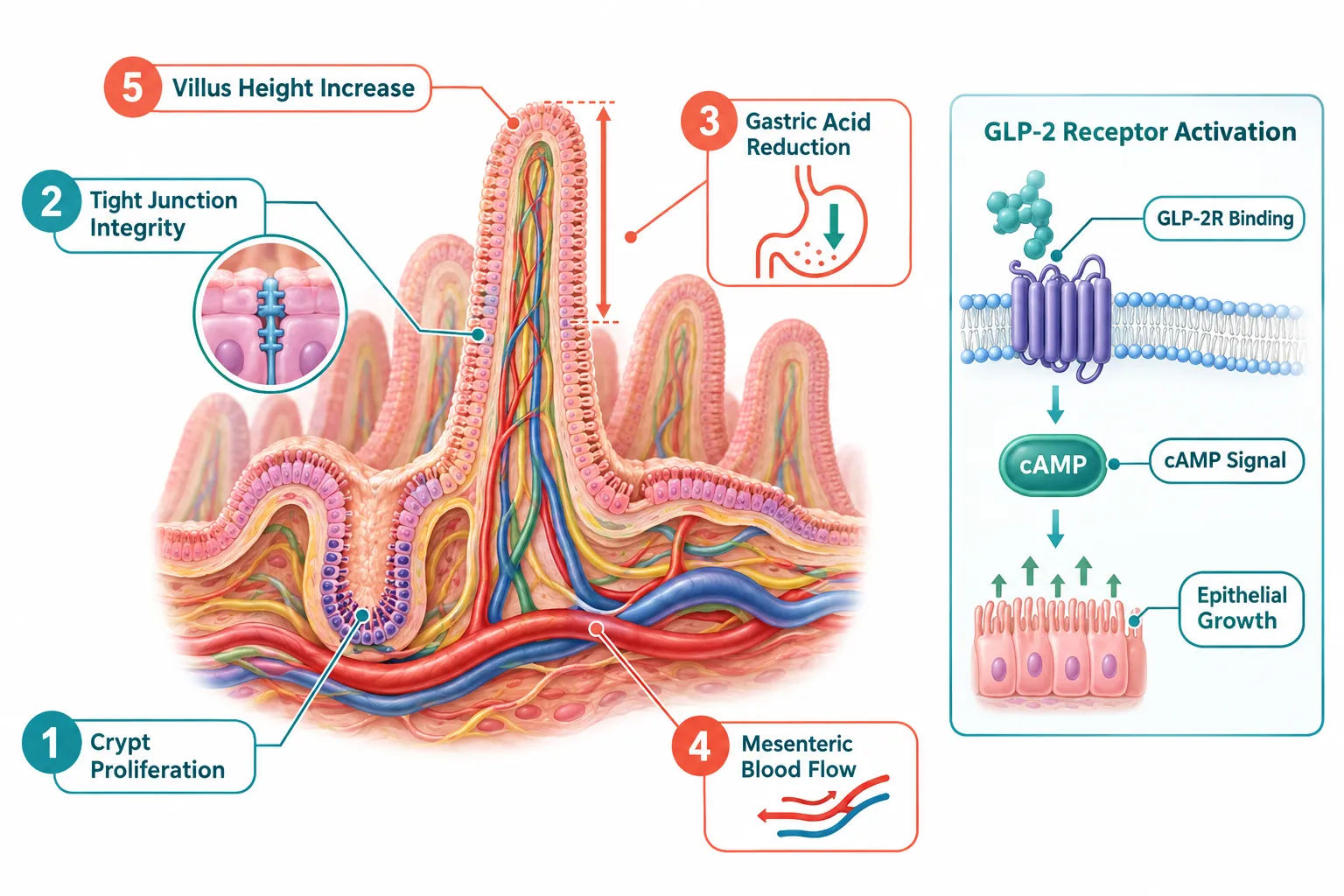
GLP-2 is an intestinotrophic hormone secreted by L-cells of the distal gut. Its core pharmacodynamic profile includes stimulation of intestinal epithelial proliferation, expansion of intestinal stem cells, increased villus height, enhanced nutrient and fluid absorption, and strengthened tight-junction integrity. Critically for experimental design, GLP-2 also slows gastrointestinal motility, increases mesenteric blood flow, and, most relevant here, reduces gastric acid secretion.
GLP-2 Biology and Its Built-In Acid-Suppressive Effects

This built-in antisecretory effect means that any intestinal model already treated with a GLP-2 analog is operating under modified luminal pH conditions before a single dose of famotidine or omeprazole is introduced. GLP-2 analogs such as teduglutide, developed for short-bowel syndrome and Crohn's disease, drive mucosal growth and barrier repair primarily through receptor-mediated epithelial signaling rather than through direct acid suppression. Still, the incidental acid-lowering effect is real and measurable.

Researchers exploring GLP-2's role in gut permeability, NLRP3 inflammasome inhibition, or lipopolysaccharide translocation must account for this baseline shift. For those interested in broader peptide mechanisms, the Peptides 101 for Research-Use Only Buyers resource provides useful context on how structurally distinct peptides engage different receptor systems.


Famotidine vs. Omeprazole: Mechanism, Timing, and pH Profile

The choice between famotidine and omeprazole as a control agent is not simply a matter of potency. It involves timing, reversibility, pH ceiling, and downstream effects on the intestinal environment.
Famotidine vs. Omeprazole: Mechanism, Timing, and pH Profile

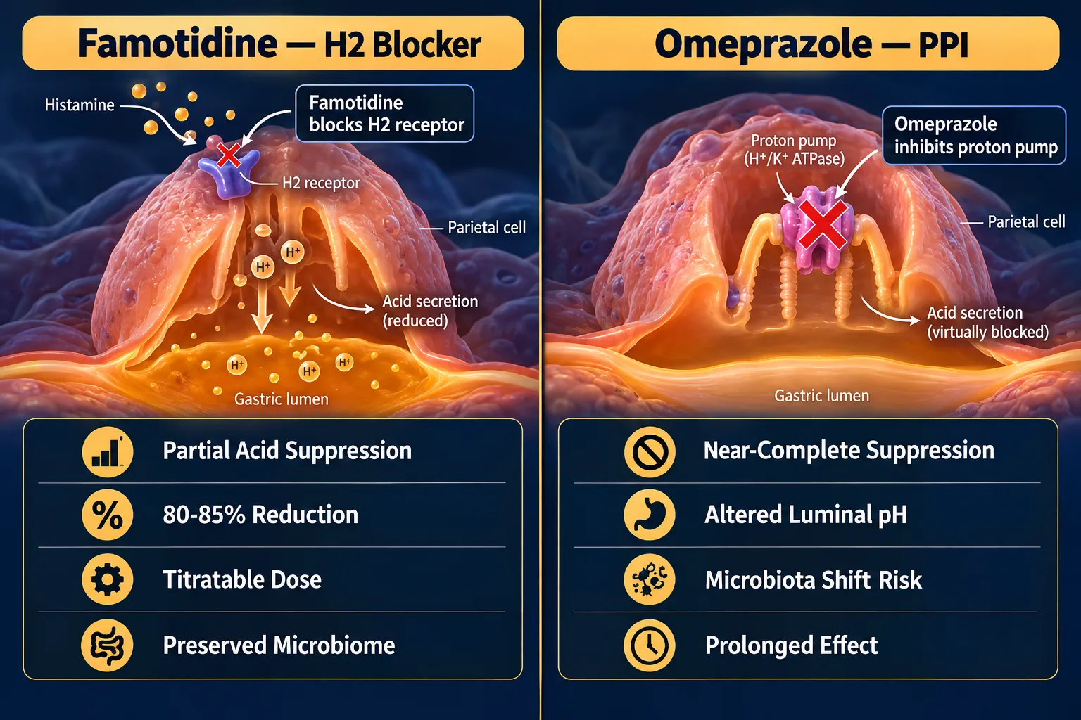
Famotidine is a competitive histamine H2-receptor antagonist. It inhibits basal and nocturnal gastric acid secretion and reduces gastric volume, acidity, and pepsin content, including secretion triggered by food, caffeine, insulin, and pentagastrin. Crucially, famotidine does not completely abolish acid production. It reduces stimulated output by roughly 80-85%, leaving a residual acid environment that more closely approximates physiological conditions. This partial suppression is titratable and relatively predictable, which is a significant advantage when the goal is to isolate GLP-2-driven epithelial effects.

Omeprazole, a proton-pump inhibitor (PPI), irreversibly blocks the H+/K+-ATPase enzyme on parietal cells. It can nearly abolish stimulated acid secretion, driving intragastric pH substantially higher than famotidine achieves. This more profound suppression introduces several confounders in GLP-2 intestinal models:

  • Luminal pH shift: Near-achlorhydric conditions alter protein digestion, nutrient solubility, and drug absorption kinetics.
  • Microbiota composition: Sustained high gastric pH permits bacterial overgrowth in the proximal gut, modifying the microbial environment that GLP-2's barrier-protective effects are being measured against.
  • Mucosal adaptation: Prolonged PPI use can independently stimulate enterochromaffin-like cell hyperplasia and alter mucosal architecture, adding trophic noise to GLP-2 proliferation readouts.
  • Timing mismatch: Because omeprazole requires active proton-pump cycling for full effect, its onset and offset kinetics differ meaningfully from the more immediate H2-blockade of famotidine, complicating washout periods in crossover designs.
Key distinction: Famotidine suppresses acid without completely eliminating it, preserving a more physiological luminal environment. Omeprazole’s near-complete suppression adds confounders, microbiota shifts, altered nutrient absorption, and independent mucosal effects, that can obscure GLP-2-specific epithelial signals.

For researchers also working with related GLP-class peptides, the GLP-3 peptide explained simply page offers accessible background on receptor-level distinctions across the GLP family.


Applying Famotidine, Omeprazole, and GLP-2 Research Principles to Intestinal Model Design

Translating these mechanistic differences into practical experimental decisions requires matching the acid-suppression agent to the specific endpoint being measured. The table below outlines how each agent performs across key experimental variables.

Variable Famotidine (H2 Blocker) Omeprazole (PPI)
Acid suppression depth Partial (~80-85%) Near-complete
Onset/offset Rapid, reversible Delayed, irreversible (pump-cycle dependent)
Microbiota impact Minimal at standard doses Risk of proximal bacterial overgrowth
Trophic mucosal effects Not reported Possible enterochromaffin-like cell effects
Interaction with GLP-2 acid suppression Additive but controlled Layered suppression, harder to deconvolute
Best suited for Isolating GLP-2 epithelial proliferation and barrier effects Models explicitly studying profound acid reduction

Applying Famotidine, Omeprazole, and GLP-2 Research Principles to Intestinal Model Design

Practical Protocol Recommendations

When designing a GLP-2 intestinal model that requires an acid-suppression control in 2026, the following steps reflect current best practice based on available mechanistic evidence:

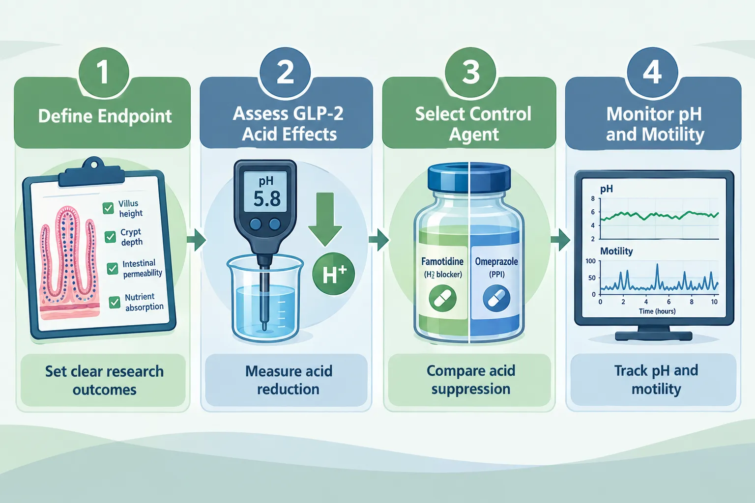
  1. Define the primary endpoint first. If the goal is to measure GLP-2-driven villus height, crypt proliferation, or tight-junction protein expression, choose the agent that introduces the fewest independent mucosal variables, which points to famotidine.
  2. Monitor luminal pH throughout. Because GLP-2 already shifts gastric acid output, baseline pH should be established before any acid-suppressive agent is added. Continuous or interval pH monitoring allows researchers to distinguish GLP-2-mediated acid changes from drug-mediated ones.
  3. Report motility data alongside acid-suppression regimen. GLP-2 slows gut transit; famotidine and omeprazole can independently alter motility. Reporting both variables together is essential for reproducibility.
  4. Reserve omeprazole for acid-focused designs. If the research question specifically involves near-achlorhydric conditions, for example, studying how profound pH elevation modifies GLP-2 receptor expression, omeprazole is appropriate. In all other GLP-2 intestinal models, its additional confounders outweigh its benefits.
  5. Account for renal function in dosing. Both famotidine and omeprazole require dose adjustment in animal models with renal impairment, and famotidine in particular has well-characterized renal clearance kinetics that affect systemic exposure.

Researchers working with GLP-class peptide blends may also find value in reviewing the GLP-3 Retatrutide buy online USA page for compound sourcing context, and the Peptides and Polypeptides in Endocrine Pharmacology article for broader receptor biology background.

The Current Research Gap

As of 2026, no published consensus statement or experimental guideline specifically addresses optimal acid-suppression control selection for GLP-2 intestinal organoid or animal models. Available literature provides authoritative descriptions of GLP-2 physiology and famotidine's pharmacodynamic profile, but the comparative control-selection question remains open. This gap is practically significant: without standardized control protocols, cross-study comparisons of GLP-2 epithelial effects are complicated by uncontrolled variation in luminal pH, microbiota composition, and motility.

For researchers interested in related intestinal peptide research, the BPC-157 and TB-500 blend category and the Glp3-Reta-Cag 10mg product page offer additional compound context relevant to gut-repair research models.


Conclusion

Choosing between famotidine and omeprazole as acid-suppression controls in GLP-2 intestinal models is not a trivial methodological footnote, it directly shapes the interpretability of every epithelial readout the experiment produces. Because GLP-2 already reduces gastric acid secretion as part of its core pharmacodynamic profile, adding a second antisecretory agent creates a compounded intervention. Famotidine's partial, titratable H2-receptor blockade introduces fewer independent variables and is better suited to experiments where GLP-2-driven mucosal growth and barrier function are the primary endpoints. Omeprazole's near-complete proton-pump inhibition is appropriate only when profound acid reduction is itself the experimental variable.

Actionable next steps for researchers:

  • Establish luminal pH baselines before introducing any acid-suppressive agent in GLP-2 models.
  • Default to famotidine as the background control unless the study design explicitly requires near-achlorhydric conditions.
  • Report acid-suppression regimen, luminal pH, and motility data together in every publication to enable meaningful cross-study comparison.
  • Advocate for field-wide consensus guidelines on acid-suppression control selection in GLP-2 intestinal research, this gap is addressable and worth closing.
https://www.puretestedpeptides.com/wp-content/uploads/2026/10/famotidine-omeprazole-and-glp-2-research-choosing-acid-suppression-controls-for.webp 1024 1536 https://www.puretestedpeptides.com/wp-content/uploads/2026/01/buy-peptides-online.jpg 2026-10-07 13:05:052026-10-07 13:05:05Famotidine, Omeprazole, and GLP-2 Research: Choosing Acid-Suppression Controls for Intestinal Models
Ondansetron in GLP-Class Research: Separating Antiemetic Effects From Gastric-Emptying and Intake Measurements

Ondansetron in GLP-Class Research: Separating Antiemetic Effects From Gastric-Emptying and Intake Measurements

October 7, 2026/0 Comments/in Uncategorized/by

Nearly half of all participants in major semaglutide trials report nausea, yet weight loss outcomes remain robust even in those who never feel sick. That gap between symptom prevalence and therapeutic outcome sits at the heart of a critical methodological challenge: when ondansetron is co-prescribed to manage GLP-1-associated nausea, do researchers risk blurring the very endpoints they are trying to measure?

Ondansetron in GLP-Class Research: Separating Antiemetic Effects From Gastric-Emptying and Intake Measurements is not just a pharmacology question. It is a study-design question that shapes how reliably investigators can attribute changes in food intake, body weight, and gastric motility to the GLP-class agent itself.

Key Takeaways

  • GLP-1 receptor agonists slow gastric emptying through peripheral and central mechanisms that are distinct from the pathways that cause nausea.
  • Ondansetron reduces subjective nausea by blocking 5-HT3 receptors at vagal afferents but does not meaningfully reverse GLP-1-induced gastric stasis.
  • Nausea rates in GLP-1 trials range from roughly 17% to 44%, yet all dose groups produce substantial weight loss, showing that nausea alone does not drive intake reduction.
  • In research settings, ondansetron should be treated as a time-varying covariate, not a confounder of GLP-1 pharmacodynamics on motility or intake.
  • Objective measures such as acetaminophen absorption curves and gastric scintigraphy remain valid even when ondansetron is co-administered.

How GLP-1 Agents Alter Gastric Emptying and Food Intake

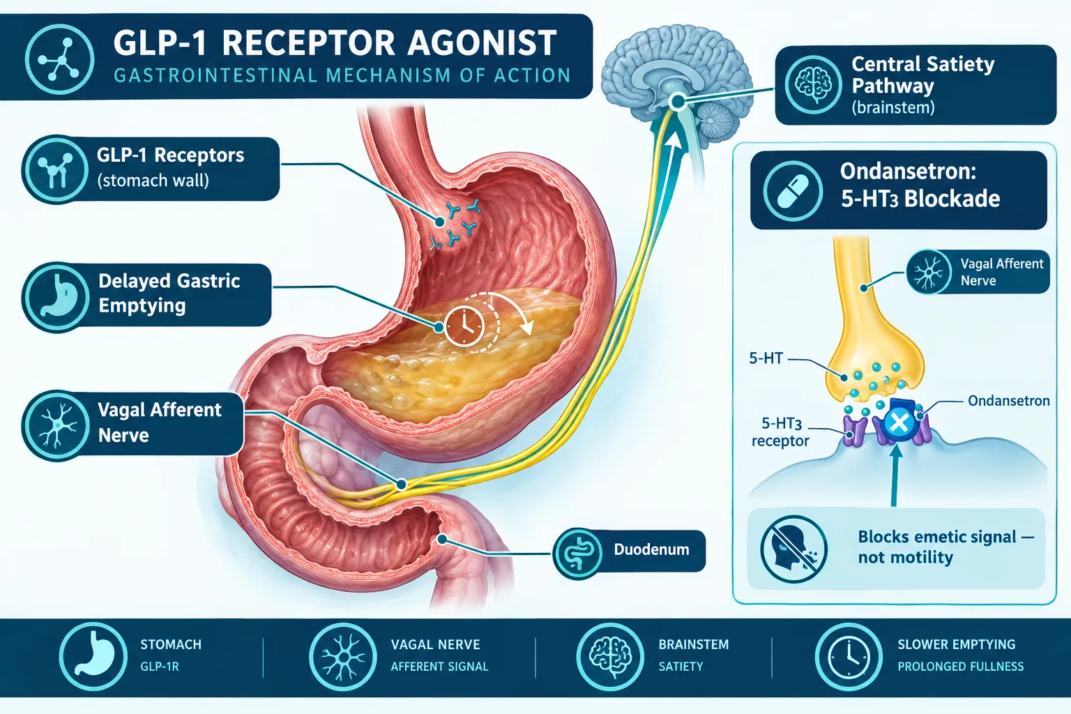
GLP-1 receptor agonists act on receptors located in the stomach wall, the duodenum, and the brainstem. The result is a well-documented slowing of gastric emptying that is independent of whether a patient feels nauseated. Semaglutide, for example, delays gastric emptying by roughly 30-40% in the first weeks of therapy. Even after 12 weeks of chronic use, gastric retention remains 10-33% longer than baseline. Gastric scintigraphy and acetaminophen absorption studies consistently confirm this effect.

This motility change matters for intake because food stays in the stomach longer, prolonging satiety signals and reducing how much a person eats at the next meal. Central appetite pathways reinforce that effect. The practical result is that reduced caloric intake on GLP-class drugs reflects both peripheral gastric slowing and central satiety signaling, not nausea-driven food avoidance.

For researchers exploring the GLP-3 peptide signaling pathway and related incretin biology, understanding this mechanistic separation is foundational before any antiemetic covariate is introduced.

How GLP-1 Agents Alter Gastric Emptying and Food Intake


Nausea Prevalence Does Not Equal Mechanism: What Trial Data Show

The pooled STEP trials reported nausea in approximately 42-44% of participants on semaglutide 2.4 mg, compared with roughly 17% on placebo. Tirzepatide trials in the SURPASS and SURMOUNT programs showed nausea rates in the 17-33% range, with a clear dose-response: 24.6% at the lowest dose, rising to 33.3% at mid-dose, then plateauing near 31% at the highest dose. Critically, all dose groups produced substantial and comparable weight loss.

This pattern supports a key conclusion: delayed gastric emptying and central appetite suppression, not nausea per se, are the primary drivers of reduced intake and weight outcomes on GLP-class drugs.

Trial Program Agent Nausea Rate Weight Outcome
STEP (pooled) Semaglutide 2.4 mg ~42-44% Substantial across all groups
SURMOUNT-1 Tirzepatide 5 mg ~24.6% Significant
SURMOUNT-1 Tirzepatide 10 mg ~33.3% Significant
SURMOUNT-1 Tirzepatide 15 mg ~31.0% Significant

Nausea is a real side effect, but it is a parallel signal, not the causal pathway. Researchers reviewing GLP-1 third-generation agents should keep this distinction in mind when designing endpoints.

Nausea Prevalence Does Not Equal Mechanism: What Trial Data Show


Ondansetron in GLP-Class Research: Separating Antiemetic Effects From Gastric-Emptying and Intake Measurements in Practice

Ondansetron is now the most commonly prescribed antiemetic for GLP-1-associated nausea in clinical practice, typically at 4-8 mg as needed. It is used in roughly 15-20% of patients during early titration or after dose escalations. Expert reviews consistently frame it as a short-term tolerability tool, not a modifier of the pharmacologic effects on gastric emptying or weight.

Mechanistically, the separation is clean:

  • GLP-1 agents slow gastric emptying via peripheral GLP-1 receptors and central satiety circuits.
  • Ondansetron blocks 5-HT3 receptors at vagal afferents and in the brainstem chemoreceptor trigger zone.
  • These are anatomically and pharmacologically distinct targets.

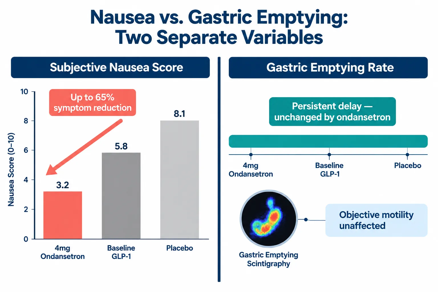
Clinical data back this up. Small retrospective analyses report roughly 40-65% reductions in nausea scores with 4-8 mg ondansetron in GLP-1-treated patients, yet objective gastric-emptying measures remain persistently delayed. Case reports of semaglutide-induced gastroparesis managed with intravenous ondansetron describe improved symptom tolerance but continued motility impairment on imaging.

Preclinical work adds nuance: in a musk shrew model, exendin-4-induced emesis was not blocked by ondansetron, suggesting that GLP-1-triggered vomiting does not rely exclusively on 5-HT3 pathways. Human patients still benefit clinically, but this finding reinforces the conceptual gap between GLP-1 emetic circuitry and the peripheral motility changes that drive intake reduction.

"Ondansetron addresses the symptom without counteracting the therapeutic mechanism", a principle that should anchor how researchers handle antiemetic co-exposure in GLP-class studies.

For broader context on how peptide mechanisms interact across systems, the Peptides 101 resource for research-use buyers provides useful background on receptor-level distinctions.


Research Design: Treating Ondansetron as a Covariate, Not a Confounder

Ondansetron in GLP-Class Research: Separating Antiemetic Effects From Gastric-Emptying and Intake Measurements demands a clear methodological response. As of 2026, no large randomized trial has tested ondansetron for GLP-1 nausea with prespecified gastric-emptying or caloric intake endpoints. Most current evidence comes from retrospective analyses and practice guidelines. That gap creates practical obligations for investigators.

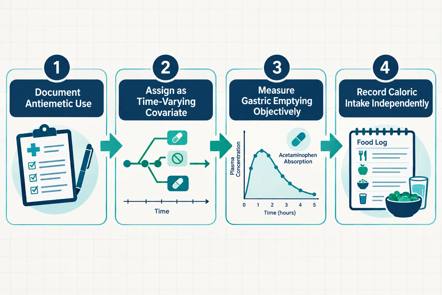
Recommended research-design steps:

  1. Document antiemetic use prospectively, record dose, timing, and duration for every participant.
  2. Assign ondansetron as a time-varying covariate in statistical models, not a fixed group variable.
  3. Use objective gastric-emptying measures, acetaminophen absorption curves or scintigraphy, rather than relying on self-reported fullness.
  4. Record caloric intake independently using validated dietary recall or direct observation, not nausea-behavior proxies.
  5. Monitor GI motility endpoints separately, ondansetron can worsen constipation in patients already prone to bowel slowing on GLP-1 agents, making stool frequency a necessary secondary endpoint.

That last point deserves emphasis. A 2026 clinical commentary specifically cautions that ondansetron may exacerbate GLP-1-related constipation, recommending non-pharmacologic alternatives such as hydration, fiber, and ginger when bowel slowing threatens adherence. For any study tracking broader GI motility, ondansetron use becomes a meaningful covariate, not just for nausea, but for the full motility picture.

Researchers also interested in how peptide combinations affect tolerability profiles can review data on cagrilintide and retatrutide as examples of multi-mechanism agents where covariate management becomes even more complex.

No clinically significant pharmacokinetic interaction between ondansetron and semaglutide or other major GLP-1 agents has been identified through mid-2026, and weight-loss or glucose-lowering efficacy appears unchanged when ondansetron is co-prescribed. This strengthens the case for treating it as a symptom-control covariate rather than a variable that alters GLP-1 pharmacodynamics.

Research Design: Treating Ondansetron as a Covariate, Not a Confounder


Looking Ahead: What Future Studies Need to Resolve

As of 2026, no GLP-1-specific antiemetic has reached phase 3 trials. Growing clinical use of GLP-class agents, including newer agents discussed in resources on GLP-1 reta formulations, is expected to drive development of nausea-targeted adjuncts that modulate GLP-1 emetic pathways without touching gastric emptying or appetite suppression.

Future trials are anticipated to randomize participants to antiemetic versus placebo arms while measuring gastric-emptying times, caloric intake, and weight outcomes in parallel. That design would finally provide definitive data on whether ondansetron or its successors can be used in GLP-class research without confounding mechanistic endpoints.

Until then, the available evidence strongly supports a working framework: ondansetron modifies symptom perception, not gastric-emptying kinetics. Researchers who document its use carefully and apply appropriate statistical controls can preserve the integrity of their motility and intake measurements.


Conclusion

The core finding is straightforward: ondansetron reduces nausea through 5-HT3 receptor blockade at vagal afferents, while GLP-1 agents slow gastric emptying through a separate set of peripheral and central receptors. These mechanisms do not meaningfully overlap, and current evidence confirms that gastric stasis persists even when nausea is well controlled.

Actionable next steps for researchers and clinicians in 2026:

  • Pre-register antiemetic use as a time-varying covariate in any GLP-class study that includes gastric-emptying or caloric intake endpoints.
  • Use objective motility measures, not patient-reported nausea, as primary evidence of GLP-1 pharmacodynamic activity.
  • Track constipation and broader GI motility separately, since ondansetron can compound bowel slowing already induced by GLP-1 agents.
  • Advocate for randomized antiemetic sub-studies embedded within larger GLP-class trials to close the current evidence gap.

Separating what ondansetron does from what GLP-class agents do is not merely an academic exercise. It is the methodological foundation that makes gastric-emptying and intake data trustworthy, and that makes the science of GLP-class research credible.

https://www.puretestedpeptides.com/wp-content/uploads/2026/10/ondansetron-in-glp-class-research-separating-antiemetic-effects-from-gastric-emp.webp 1024 1536 https://www.puretestedpeptides.com/wp-content/uploads/2026/01/buy-peptides-online.jpg 2026-10-07 13:04:192026-10-07 13:04:19Ondansetron in GLP-Class Research: Separating Antiemetic Effects From Gastric-Emptying and Intake Measurements
Enclomiphene Research Materials: Base, Citrate, and Analytical Identity Checks for Hormone-Model Experiments

Enclomiphene Research Materials: Base, Citrate, and Analytical Identity Checks for Hormone-Model Experiments

October 7, 2026/0 Comments/in Uncategorized/by

Fewer than one in five commercial enclomiphene lots tested by independent analytical laboratories in recent years have carried full counterion confirmation alongside standard purity data, a gap that creates real problems when researchers try to compare results across batches or institutions. For anyone sourcing Enclomiphene Research Materials: Base, Citrate, and Analytical Identity Checks for Hormone-Model Experiments, that single oversight can invalidate dosing calculations, skew receptor-binding data, and make batch-to-batch reproducibility impossible.

This guide moves beyond the basic base-versus-citrate comparison to give researchers a structured purchasing and verification checklist covering salt identity, assay method, residual solvents, counterion confirmation, and batch consistency.

Key Takeaways

  • Enclomiphene is supplied as either the free base or the citrate salt; the two forms carry different molecular weights, and failing to apply the correct salt factor corrupts dosing math in hormone-model experiments.
  • A complete Certificate of Analysis (COA) for research-grade material should include HPLC purity, NMR salt identity, ESI-MS confirmation, residual solvent data (GC headspace), and water content.
  • Counterion confirmation via NMR or ion chromatography is the most commonly missing data point in commercial COAs as of 2026.
  • Batch consistency checks, comparing COA values across at least two consecutive lots, are essential before committing to a long experimental series.
  • Enclomiphene is classified as a research-use-only compound in most jurisdictions; it is not approved for human use outside specific regulatory pathways.

Understanding the Two Chemical Forms: Base and Citrate

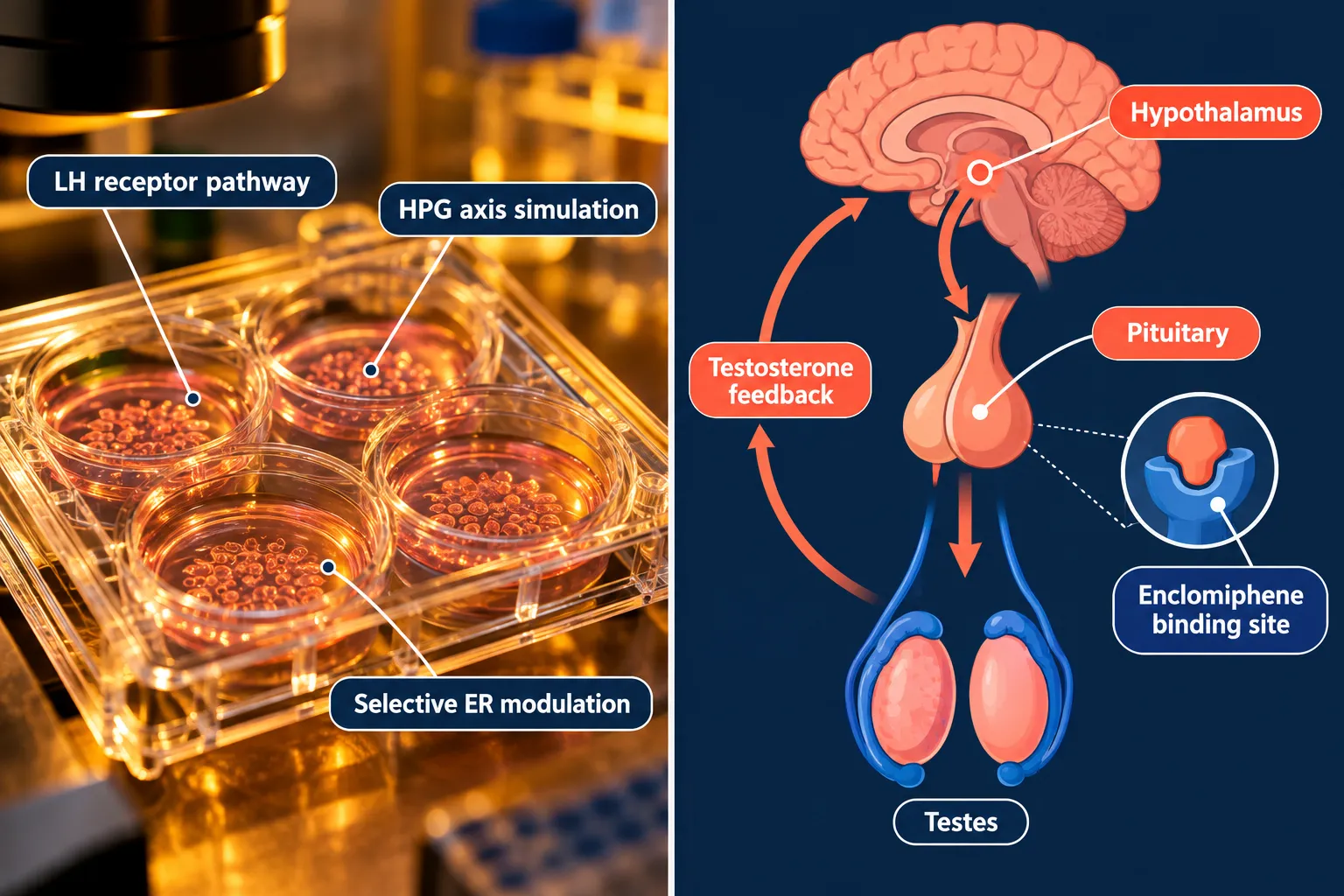
Enclomiphene is the trans-isomer of clomiphene, a selective estrogen receptor modulator (serm) that acts at hypothalamic estrogen receptors to reduce negative feedback on gonadotropin-releasing hormone. It reaches the market in two primary salt forms, and the distinction matters far more than many procurement workflows acknowledge.

Understanding the Two Chemical Forms: Base and Citrate

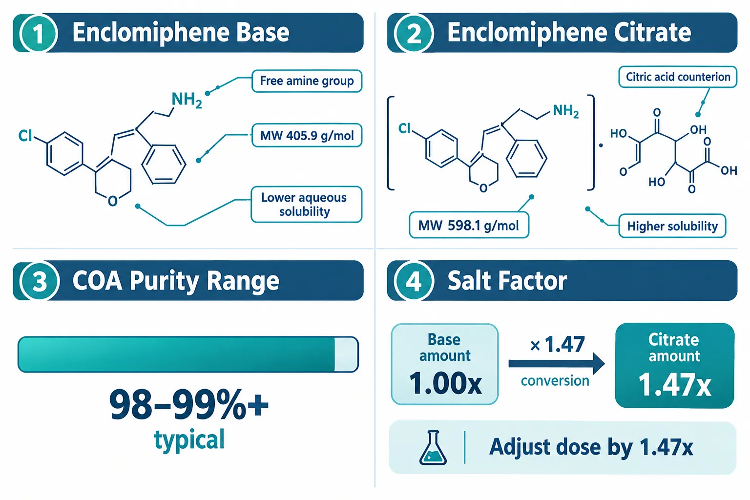
Enclomiphene free base carries a molecular weight of approximately 405.9 g/mol. It is less soluble in aqueous buffers, which makes it better suited to organic-solvent stock preparations (DMSO, ethanol) commonly used in cell-based assays. Enclomiphene citrate pairs the active molecule with citric acid as a counterion, raising the molecular weight to roughly 598.1 g/mol. The citrate form dissolves more readily in water, which is advantageous for in vitro hormone-model systems that require aqueous dosing vehicles.

The practical consequence: a researcher who assumes citrate when the material is actually the free base will underdose by approximately 32% relative to the active moiety. The conversion factor, roughly 1.47, must be applied whenever switching between forms or when a supplier's labeling is ambiguous.

Quick reference: key differences

Property Free Base Citrate Salt
Molecular weight ~405.9 g/mol ~598.1 g/mol
Aqueous solubility Low Moderate, high
Preferred vehicle DMSO / ethanol Aqueous buffer
Salt factor vs base 1.00 ~1.47
Counterion None Citric acid

Researchers building hormone-model systems, particularly hypothalamic-pituitary-gonadal (HPG) axis simulations, should document the chosen form in every experimental protocol. This is the same level of rigor applied when sourcing other structurally complex research agents; see the discussion of structural identity documentation in this overview of peptides 101 for research-use only buyers.


The Analytical Identity Checklist for Enclomiphene Research Materials

The core of responsible sourcing for Enclomiphene Research Materials: Base, Citrate, and Analytical Identity Checks for Hormone-Model Experiments is a structured verification checklist applied to every lot before it enters an experiment. The following five-point framework covers the most critical analytical dimensions.

The Analytical Identity Checklist for Enclomiphene Research Materials

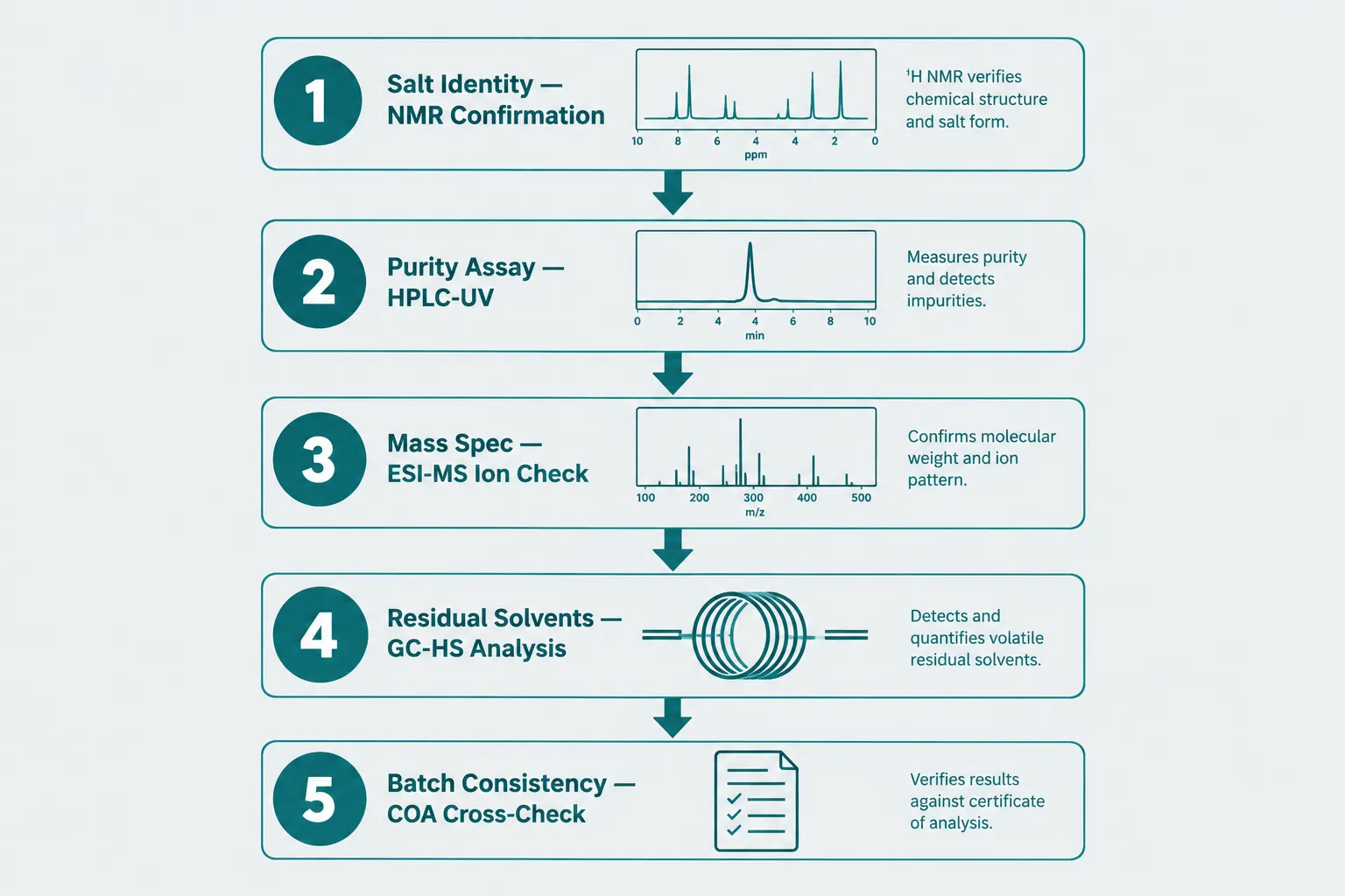
1. Salt Identity via NMR

Proton NMR (1H-NMR) is the gold-standard method for confirming whether a lot is the free base or a specific salt form. For the citrate, characteristic citric acid proton signals appear in the 2.5-2.8 ppm region. A COA that reports only HPLC purity without NMR data cannot confirm salt identity. Suppliers who provide NMR spectra as part of their documentation package should be prioritized.

2. Purity Assay via HPLC-UV

High-performance liquid chromatography with UV detection (typically at 254 nm) remains the standard purity assay. Research-grade material in 2026 is expected to meet 98% or higher purity by HPLC. The COA should specify the column type, mobile phase, and integration method. A single broad purity figure without chromatographic conditions is insufficient for experimental validation.

3. Mass Spectrometry Confirmation (ESI-MS)

Electrospray ionization mass spectrometry (ESI-MS) provides molecular ion confirmation independent of chromatographic separation. For enclomiphene free base, the expected [M+H]+ ion appears at m/z 406.2; for the citrate, the spectrum should show both the enclomiphene cation and the citrate anion. ESI-MS data on a COA rules out structural analogs and degradation products that can co-elute on HPLC.

4. Residual Solvents via GC Headspace

Synthesis and recrystallization of enclomiphene typically involves chlorinated solvents or alcohols. ICH Q3C residual solvent limits apply to pharmaceutical-grade material, and research-grade suppliers increasingly adopt these thresholds as a quality benchmark. Gas chromatography headspace (GC-HS) analysis should be requested for any lot used in cell-viability or receptor-binding assays where solvent contamination could confound results.

5. Counterion Confirmation via Ion Chromatography

This is the most commonly absent data point. Ion chromatography (IC) or capillary electrophoresis can quantify the citrate counterion directly, confirming the correct stoichiometry of the salt. Without it, a lot labeled "citrate" may be a partial salt or a mixed form. Batch-to-batch counterion ratio variation is a known source of irreproducibility in serm research.

Checklist summary:

  • NMR spectrum confirming salt form
  • HPLC-UV purity (98%+ with stated conditions)
  • ESI-MS molecular ion confirmation
  • GC-HS residual solvent panel
  • IC or CE counterion quantification
  • Karl Fischer water content (relevant for hygroscopic citrate lots)

Applying Verified Materials in Hormone-Model Experiments

Once analytical identity is confirmed, the experimental design for HPG-axis hormone models can proceed with confidence. Enclomiphene's mechanism centers on competitive antagonism at hypothalamic estrogen receptors, which reduces estrogenic negative feedback and elevates luteinizing hormone (LH) and follicle-stimulating hormone (FSH) output. This makes it a useful tool in cell-based and ex vivo models studying gonadotropin regulation, male hypogonadism mechanisms, and fertility signaling.

Applying Verified Materials in Hormone-Model Experiments

For researchers working with aqueous dosing systems, the citrate form is generally preferred due to its superior solubility. For lipid-based or organic-vehicle assay systems, the free base dissolves more cleanly. In either case, stock solutions should be prepared fresh or stored under inert gas at -20°C to prevent oxidative degradation of the stilbene backbone.

Batch consistency across a multi-experiment series requires comparing COA values, purity, water content, and counterion ratio, across at least two consecutive lots before committing. A purity drop of even 1-2% between lots can translate to meaningful receptor-occupancy differences at low nanomolar concentrations typical of serm experiments.

The approach mirrors best practices seen in other research-grade compound categories. For example, the purity evaluation standards applied when sourcing research-grade Glow Blend Peptide materials follow the same logic: identity first, purity second, batch consistency third.

Researchers interested in broader hormone-axis modeling may also find value in reviewing growth hormone secretagogue mechanisms, such as the detailed pharmacokinetic profile covered in this CJC-1295 with and without DAC mechanism comparison, as multi-axis hormone models increasingly combine serm and GH-pathway tools.

For procurement teams evaluating supplier quality programs, the standards applied to wholesale peptides for sale at reputable suppliers, including third-party COA availability and batch traceability, provide a useful quality benchmark applicable to small-molecule research materials like enclomiphene as well.

"The counterion is not a footnote, it is half the molecule. Treating salt identity as an afterthought is one of the most preventable sources of irreproducibility in serm research."

As of mid-2026, regulatory positioning for enclomiphene remains research-use only in most jurisdictions outside specific approved clinical pathways. Researchers should confirm local regulatory status before procurement and ensure all use is confined to in vitro or appropriately approved in vivo model systems.


Conclusion

Sourcing Enclomiphene Research Materials: Base, Citrate, and Analytical Identity Checks for Hormone-Model Experiments responsibly requires more than checking a purity percentage. The five-point analytical checklist, NMR salt identity, HPLC-UV purity, ESI-MS molecular confirmation, GC-HS residual solvents, and IC counterion quantification, gives research teams the data needed to dose accurately, compare results across batches, and publish reproducible findings.

Actionable next steps for research teams in 2026:

  1. Request full COA documentation including NMR and ESI-MS before approving any new lot.
  2. Apply the 1.47 salt factor correction whenever switching between base and citrate forms.
  3. Implement a batch-consistency log comparing at least two consecutive lot COAs before starting a new experimental series.
  4. Specify the dosing vehicle (aqueous vs. organic) in the protocol and select the appropriate salt form accordingly.
  5. Confirm the regulatory classification of enclomiphene in your jurisdiction before procurement.

Analytical rigor at the purchasing stage is the cheapest insurance a research program can buy.

https://www.puretestedpeptides.com/wp-content/uploads/2026/10/enclomiphene-research-materials-base-citrate-and-analytical-identity-checks-for.webp 1024 1536 https://www.puretestedpeptides.com/wp-content/uploads/2026/01/buy-peptides-online.jpg 2026-10-07 13:04:082026-10-07 13:04:08Enclomiphene Research Materials: Base, Citrate, and Analytical Identity Checks for Hormone-Model Experiments
Semax Peptide Nasal Spray Reviews for Research Use: A Structured Method for Comparing Products

Semax Peptide Nasal Spray Reviews for Research Use: A Structured Method for Comparing Products

October 7, 2026/0 Comments/in Uncategorized/by

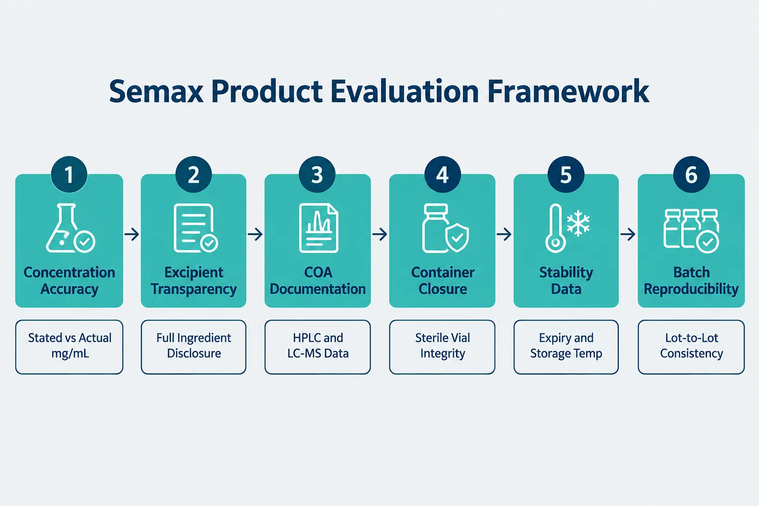
Most published commentary on Semax nasal spray focuses on subjective user experiences rather than the analytical criteria that actually determine whether a product is suitable for controlled research. That gap matters enormously. When a laboratory study produces inconsistent results, the culprit is often a poorly characterized formulation, not the peptide itself. This article presents a structured review rubric for evaluating Semax peptide nasal spray products for research use, covering concentration accuracy, excipient transparency, analytical documentation, container closure, stability data, and batch reproducibility.

Key Takeaways

  • Semax nasal spray products sold for research use must carry a "research use only" label and are not approved for human therapeutic use in most Western jurisdictions.
  • Analytical documentation, specifically HPLC and LC-MS Certificates of Analysis, is the single most important differentiator between research-grade and substandard products.
  • Concentration accuracy and excipient transparency directly affect reproducibility in preclinical models.
  • Intranasal delivery provides a rationale for bypassing the blood-brain barrier, making formulation quality especially critical.
  • A six-point evaluation rubric gives researchers an objective method for comparing Semax nasal spray products without relying on anecdotal testimonials.

Understanding Semax and Its Research Context

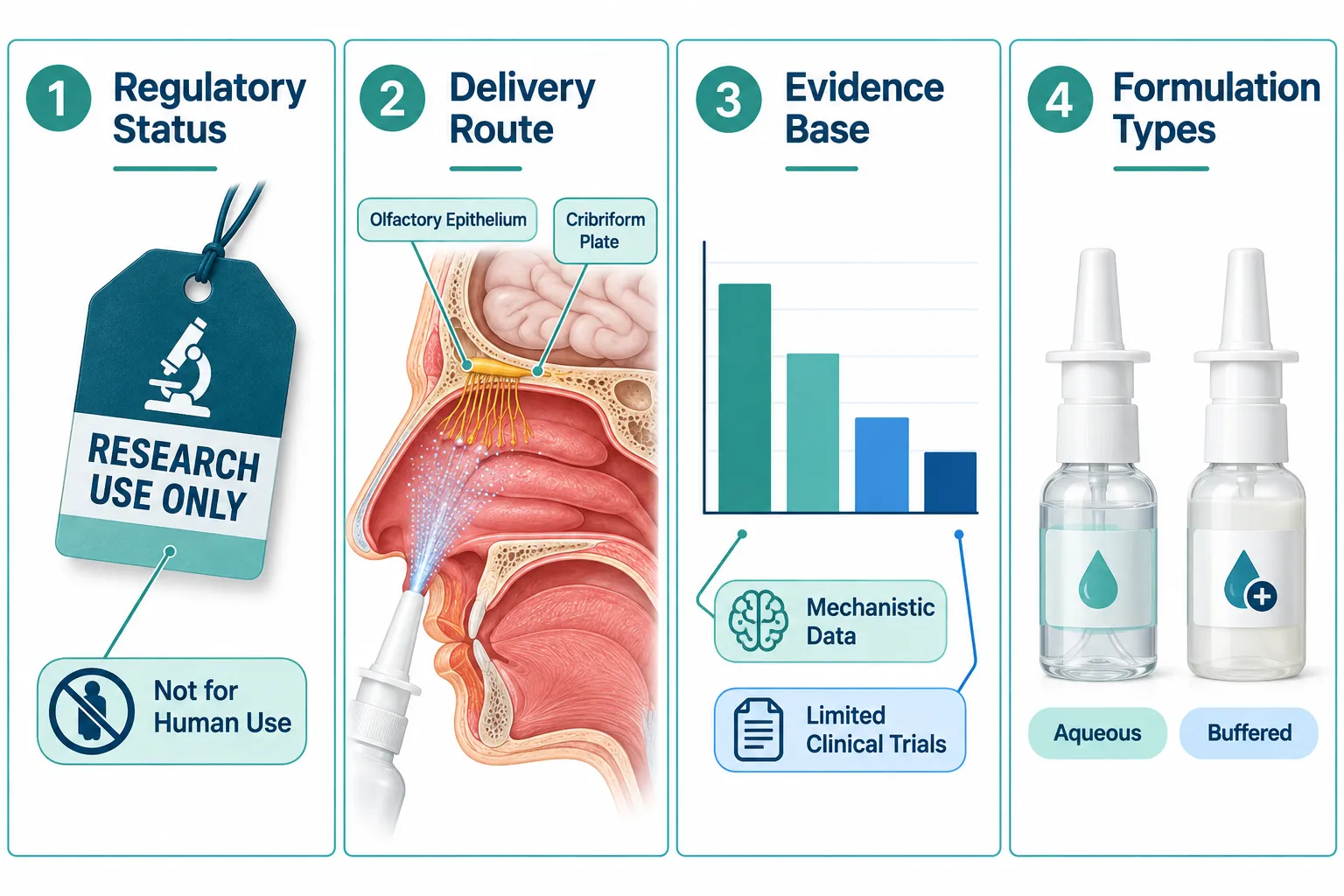
Semax is a synthetic heptapeptide analog of the adrenocorticotropic hormone (ACTH) fragment 4-10, originally developed in Russia during the 1980s. It has been used clinically in Russia and Ukraine for neurological indications, but it holds no regulatory approval from the FDA, EMA, or equivalent Western agencies for therapeutic use. In 2026, products sold in the United States, United Kingdom, and most of Europe are labeled strictly for research use only and must not be administered to humans outside of formally approved clinical protocols.

The mechanistic rationale for Semax is well-supported. Research demonstrates interactions with brain-derived neurotrophic factor (BDNF) pathways, serotonergic and dopaminergic systems, and emerging data point to copper-chelation activity with potential anti-amyloid properties. Intranasal delivery is the preferred route in most research models because the olfactory epithelium and cribriform plate provide a pathway that can bypass first-pass hepatic metabolism, making formulation quality directly relevant to experimental outcomes.

Researchers comparing Semax products can also benefit from reviewing related peptide evaluation frameworks, such as those used for research-grade Glow Blend peptide purity and copper complexes, which share overlapping quality criteria.

Understanding Semax and Its Research Context


A Structured Method for Comparing Semax Peptide Nasal Spray Products for Research Use

Applying a consistent rubric removes subjectivity from the evaluation process. The six criteria below form the core of any defensible product comparison.

1. Concentration Accuracy

The stated peptide concentration (typically expressed in mg/mL) must be verifiable against independent analytical data. A product labeled at 0.1% Semax (1 mg/mL) should reflect that figure in the accompanying Certificate of Analysis (COA). Discrepancies between labeled and actual concentration directly undermine dose-response calculations in research models.

"Concentration accuracy is not a quality-of-life feature, it is a scientific validity requirement."

2. Excipient Transparency

Nasal spray formulations require excipients: preservatives, buffering agents, and tonicity adjusters. Common components include benzalkonium chloride, sodium phosphate buffers, and sodium chloride. A reputable supplier discloses the full excipient list. Undisclosed preservatives can confound results, particularly in neurological cell models where cytotoxicity thresholds are low.

3. Analytical Documentation (COA, HPLC, LC-MS)

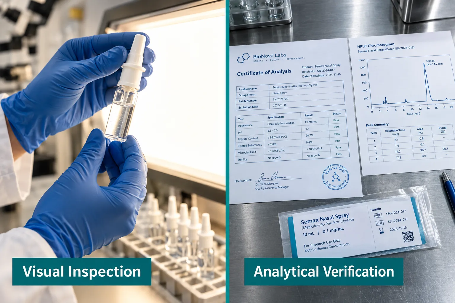
This is the most critical differentiator. A credible research-grade product provides:

  • HPLC purity data confirming peptide identity and ruling out major impurities
  • LC-MS mass confirmation verifying molecular weight matches the Semax sequence
  • Endotoxin testing results (LAL assay or equivalent) for any product intended for cell-based or animal research
  • Lot-specific COA tied to the batch number on the product label

Suppliers who provide only generic or undated COAs should be treated with caution. Researchers evaluating SS-31 10mg research peptide considerations face identical documentation standards, illustrating that this criterion applies across peptide classes.

4. Container Closure Integrity

Nasal spray vials must maintain sterility and prevent oxidative degradation. Key features to evaluate include:

Feature Research-Grade Standard
Vial material Borosilicate glass or pharmaceutical-grade polymer
Closure type Crimp-sealed or luer-lock with tamper evidence
Headspace Nitrogen or argon purge preferred
Volume accuracy Actuator-calibrated spray volume stated

Poor container closure accelerates peptide degradation and introduces contamination risk that invalidates experimental data.

5. Stability Data and Storage Conditions

Semax is sensitive to temperature and light. Suppliers should provide stability data indicating shelf life under recommended storage conditions (typically 2-8°C refrigerated, protected from light). Products lacking stated expiry dates or stability studies present an unquantifiable variable in research design. This consideration is equally relevant when sourcing other sensitive peptides, such as those discussed in peptide dosing guidance.

6. Batch Reproducibility

Single-lot purity data is insufficient for ongoing research programs. Researchers should request multi-lot COA data or ask suppliers directly about lot-to-lot variability metrics. High reproducibility, defined as less than 2% variance in purity across consecutive lots, is the standard that serious research suppliers should be able to demonstrate.

6. Batch Reproducibility


Applying the Rubric: Practical Comparison of Semax Nasal Spray Products for Research Use

When applying the structured method above to available Semax nasal spray products for research use, several patterns emerge from 2025-2026 supplier analyses.

Products with strong analytical packages typically come from suppliers who operate ISO-certified or cGMP-adjacent facilities, provide lot-specific HPLC and LC-MS data on their product pages, and disclose full excipient lists. These products command a premium but reduce experimental confounders substantially.

Products with weak documentation often rely on third-party review aggregators and user testimonials rather than analytical data. While anecdotal feedback can suggest tolerability in informal research contexts, it provides no information about concentration accuracy or impurity profiles.

Researchers can explore Semax nasal spray product listings alongside comparisons such as Selank vs Semax to understand how structurally related peptides differ in formulation requirements. For broader peptide research context, reviewing how other nasal or injectable peptides are evaluated, for example, the CJC-1295 with and without DAC mechanism and pharmacokinetic comparison, reinforces how formulation variables affect research outcomes across compound classes.

A practical checklist before purchasing:

  • Request the lot-specific COA before placing an order
  • Confirm HPLC purity is stated as a percentage (not just "high purity")
  • Verify the excipient list matches the intended research model's compatibility requirements
  • Check that storage and expiry information appears on the product label, not just the website
  • Confirm the supplier's return or replacement policy for failed QC verification

Applying the Rubric: Practical Comparison of Semax Nasal Spray Products for Research Use


Conclusion

Semax peptide nasal spray reviews for research use are only as useful as the criteria applied to generate them. Subjective testimonials cannot substitute for concentration accuracy data, excipient disclosure, lot-specific HPLC and LC-MS documentation, verified container closure integrity, stability studies, and demonstrated batch reproducibility. Researchers who apply the six-point rubric outlined here will make more defensible procurement decisions and produce more reproducible experimental outcomes.

Actionable next steps:

  1. Download and review the COA for any Semax nasal spray product before purchase, reject any supplier that cannot provide a lot-specific document.
  2. Cross-reference excipient lists against the cytotoxicity thresholds of the specific cell or animal model being used.
  3. Establish a baseline lot comparison by requesting data from at least two consecutive batches before committing to a supplier for a multi-experiment research program.
  4. Revisit supplier documentation annually, as manufacturing processes and testing standards evolve.

Rigorous product selection is not bureaucratic overhead, it is the foundation of valid research.

https://www.puretestedpeptides.com/wp-content/uploads/2026/10/semax-peptide-nasal-spray-reviews-for-research-use-a-structured-method-for-compa.webp 1024 1536 https://www.puretestedpeptides.com/wp-content/uploads/2026/01/buy-peptides-online.jpg 2026-10-07 13:03:532026-10-07 13:03:53Semax Peptide Nasal Spray Reviews for Research Use: A Structured Method for Comparing Products
Spironolactone and Peptide Research: Interpreting Potassium, Renal, Hormonal, and Fluid-Balance Variables

Spironolactone and Peptide Research: Interpreting Potassium, Renal, Hormonal, and Fluid-Balance Variables

October 7, 2026/0 Comments/in Uncategorized/by

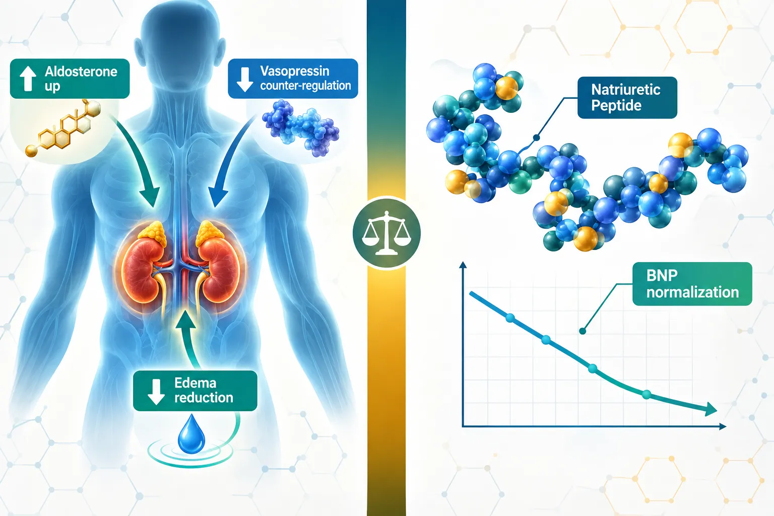
Aldosterone blockade does not simply pull water out of the body, it reshapes the entire hormonal landscape that peptide researchers depend on for clean endpoint interpretation. Understanding Spironolactone and Peptide Research: Interpreting Potassium, Renal, Hormonal, and Fluid-Balance Variables is therefore essential for anyone designing or analyzing studies where mineralocorticoid receptor (MR) antagonism intersects with natriuretic peptides, renal biomarkers, and fluid status. As of 2026, updated pharmacovigilance datasets confirm that hyperkalaemia and renal function shifts remain the most clinically significant safety signals with spironolactone, yet the drug's disease-modifying effects on hormonal and peptide endpoints make it indispensable in cardiovascular and renal research.

Key Takeaways

  • Spironolactone blocks aldosterone at the mineralocorticoid receptor, causing predictable potassium retention and modest sodium excretion that must be tracked as confounders in peptide studies.
  • Natriuretic peptide levels, including BNP and NT-proBNP, fall with spironolactone use even as RAAS feedback drives aldosterone and angiotensin II upward.
  • Creatinine and eGFR shifts are small but consistent; monitoring thresholds in 2026 guidance remain strict, especially in CKD stages 3b, 5.
  • Fluid-balance and volume metrics improve alongside natriuretic peptide normalization in resistant hypertension cohorts.
  • Accurate interpretation of peptide research data requires separating spironolactone's direct MR-antagonist effects from secondary hormonal feedback loops.

How Mineralocorticoid Receptor Antagonism Shapes Peptide Research Variables

How Mineralocorticoid Receptor Antagonism Shapes Peptide Research Variables

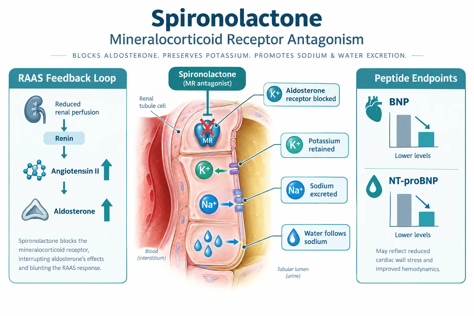
Spironolactone competitively binds the mineralocorticoid receptor in the distal nephron, blocking aldosterone's instruction to retain sodium and excrete potassium. The downstream consequences are not limited to electrolytes. When aldosterone signaling is suppressed at the receptor level, the body compensates through RAAS feedback, aldosterone and angiotensin II levels rise in plasma even as their end-organ effects are blunted. This dual signal creates a confounding environment for any peptide assay that uses aldosterone or angiotensin-related markers as primary endpoints.

Three core variables shift predictably with spironolactone:

Variable Direction of Change Research Implication
Serum potassium Increases Risk of hyperkalaemia; confounds cardiac peptide safety data
Serum creatinine Modest increase Apparent renal function decline; not always true GFR loss
Natriuretic peptides (BNP/NT-proBNP) Decreases Reflects reduced cardiac wall stress, not peptide catabolism

For researchers working with SS-31 peptide or other mitochondria-targeted compounds that influence renal tubular function, spironolactone's effect on tubular sodium transport is a critical baseline variable. Any study failing to account for concurrent MR antagonism risks misattributing renal transporter changes to the investigational peptide.

"Spironolactone is disease-modifying beyond simple diuresis, but potassium and renal markers remain the critical guardrails for peptide-driven research.", 2026 expert consensus


Potassium and Renal Thresholds: The Non-Negotiable Guardrails

Potassium and Renal Thresholds: The Non-Negotiable Guardrails

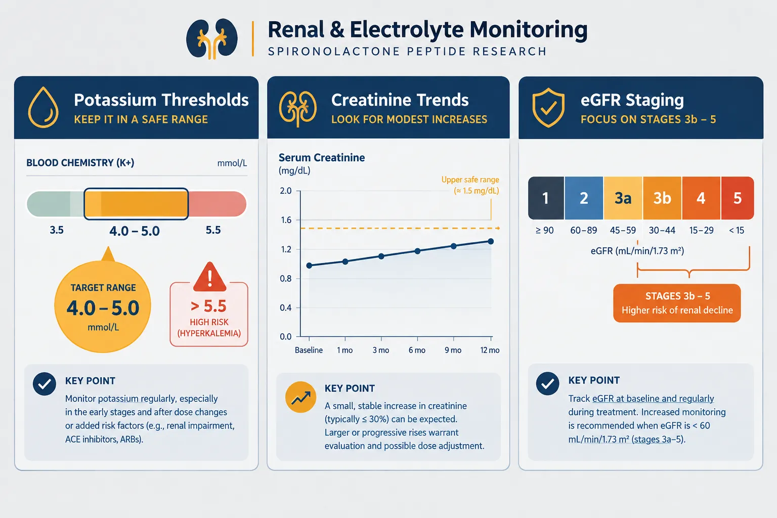
Current 2026 clinical guidance maintains strict thresholds for spironolactone initiation and continuation. Serum potassium above 5.0 mEq/L typically triggers dose reduction or discontinuation, and eGFR below 30 mL/min/1.73m² generally contraindicates use in standard clinical settings. However, emerging 2025 data from advanced CKD cohorts (stages 3b, 5) show that low-dose spironolactone combined with ACE inhibitor or ARB therapy can deliver cardiovascular benefit without excess hyperkalaemia or accelerated renal failure when monitoring is rigorous.

For peptide researchers, these thresholds matter because:

  • Potassium elevation can independently alter cardiac electrophysiology, confounding any peptide study with cardiac safety endpoints.
  • Creatinine rise within the first 2-4 weeks of spironolactone use reflects haemodynamic adjustment, not structural nephron loss, a distinction critical when interpreting renal peptide biomarkers.
  • eGFR staging determines which renal transporter pathways are active, directly affecting the pharmacokinetics of co-administered peptides.

Researchers exploring peptide dosing protocols alongside mineralocorticoid antagonists must pre-specify potassium and creatinine monitoring windows. Multi-year heart failure cohort data confirm that renal and electrolyte shifts with spironolactone are small but consistent, meaning cumulative drift can cross safety thresholds over extended study periods.

Recommended monitoring intervals in research settings:

  1. Baseline potassium and creatinine before spironolactone introduction
  2. Repeat at 1 week and 4 weeks post-initiation
  3. Every 3 months during stable dosing phases
  4. Immediately after any dose change or addition of RAAS-active co-therapy

Hormonal and Fluid-Balance Endpoints in Spironolactone and Peptide Research

Hormonal and Fluid-Balance Endpoints in Spironolactone and Peptide Research

The hormonal picture in Spironolactone and Peptide Research: Interpreting Potassium, Renal, Hormonal, and Fluid-Balance Variables is layered. Aldosterone and vasopressin are the primary counter-regulators of water and sodium balance, and both respond to MR antagonism in ways that directly affect peptide endpoint interpretation.

Natriuretic peptide dynamics are particularly instructive. Spironolactone lowers BNP and NT-proBNP levels in heart failure and resistant hypertension cohorts, reflecting reduced ventricular wall stress as volume overload resolves. This reduction is not caused by peptide degradation or receptor downregulation, it is a genuine haemodynamic improvement. Researchers studying GLP-3 peptide compounds or other metabolic peptides that influence fluid retention must therefore distinguish spironolactone-driven natriuretic peptide changes from those attributable to the investigational agent.

Vasopressin remains an underappreciated variable. As spironolactone reduces volume overload, vasopressin secretion adjusts, altering free water clearance. Studies on peptides with antidiuretic properties, including vasoactive intestinal peptide analogs such as those found in VIP-5 peptide research, need to account for this vasopressin modulation as a confounder.

In primary aldosteronism, meta-analyses position spironolactone as first-line medical therapy, with potassium outcomes comparable to newer selective MRAs. This makes it a relevant background medication in any hormonal peptide study where aldosterone excess is a design variable. Researchers examining peptides and polypeptides in endocrine pharmacology will recognize that RAAS-peptide crosstalk extends well beyond simple sodium balance.

Ongoing trials in 2026 are titrating spironolactone to renin and blood pressure responses in low-renin hypertension, with integrated hormonal endpoints that include natriuretic peptides, aldosterone-to-renin ratios, and plasma angiotensin II. These trials provide a methodological template for any peptide study that incorporates MR antagonism as a background or co-intervention variable.

For researchers also examining mitochondrial or cytoprotective peptides, the SS-31 peptide benefits literature offers a useful parallel: renal tubular protection endpoints in that body of work face similar confounding from background diuretic and RAAS-active therapies.


Conclusion

Spironolactone and Peptide Research: Interpreting Potassium, Renal, Hormonal, and Fluid-Balance Variables demands a structured, mechanistically grounded approach. The drug's MR-antagonist action creates predictable shifts in potassium, creatinine, natriuretic peptides, aldosterone feedback, and vasopressin tone, each of which can masquerade as a peptide-specific effect if not properly controlled.

Actionable next steps for researchers:

  • Pre-specify potassium and eGFR thresholds as stopping rules in any protocol that includes spironolactone.
  • Separate haemodynamic natriuretic peptide changes from direct peptide-receptor effects using time-matched biomarker panels.
  • Account for RAAS feedback elevation (aldosterone, angiotensin II) when interpreting hormonal peptide endpoints.
  • Apply 2026 monitoring intervals, baseline, week 1, week 4, then quarterly, as the minimum standard for renal and electrolyte safety.
  • Cross-reference fluid-balance metrics with vasopressin and osmolality data to isolate volume effects from peptide-specific actions.

Rigorous variable interpretation is what separates publishable peptide research from ambiguous data. Spironolactone's hormonal reach is wide; accounting for it fully is the foundation of credible results.

https://www.puretestedpeptides.com/wp-content/uploads/2026/10/spironolactone-and-peptide-research-interpreting-potassium-renal-hormonal-and-fl.webp 1024 1536 https://www.puretestedpeptides.com/wp-content/uploads/2026/01/buy-peptides-online.jpg 2026-10-07 13:03:532026-10-07 13:03:53Spironolactone and Peptide Research: Interpreting Potassium, Renal, Hormonal, and Fluid-Balance Variables
Atorvastatin and Research Peptides: Designing Lipid and Liver-Control Groups for Metabolic Studies

Atorvastatin and Research Peptides: Designing Lipid and Liver-Control Groups for Metabolic Studies

October 6, 2026/0 Comments/in Uncategorized/by

Cardiovascular disease remains the leading cause of death worldwide, yet fewer than half of high-risk research subjects in preclinical metabolic models reach target lipid endpoints without a standardized control framework. Designing rigorous control groups is where most metabolic studies succeed or fail, and the intersection of Atorvastatin and Research Peptides: Designing Lipid and Liver-Control Groups for Metabolic Studies sits at the center of that challenge. As novel peptides such as retatrutide, MOTS-c, and 5-amino-1MQ enter metabolic research pipelines, pairing them against a well-characterized statin benchmark like atorvastatin is no longer optional, it is methodologically essential.

Key Takeaways

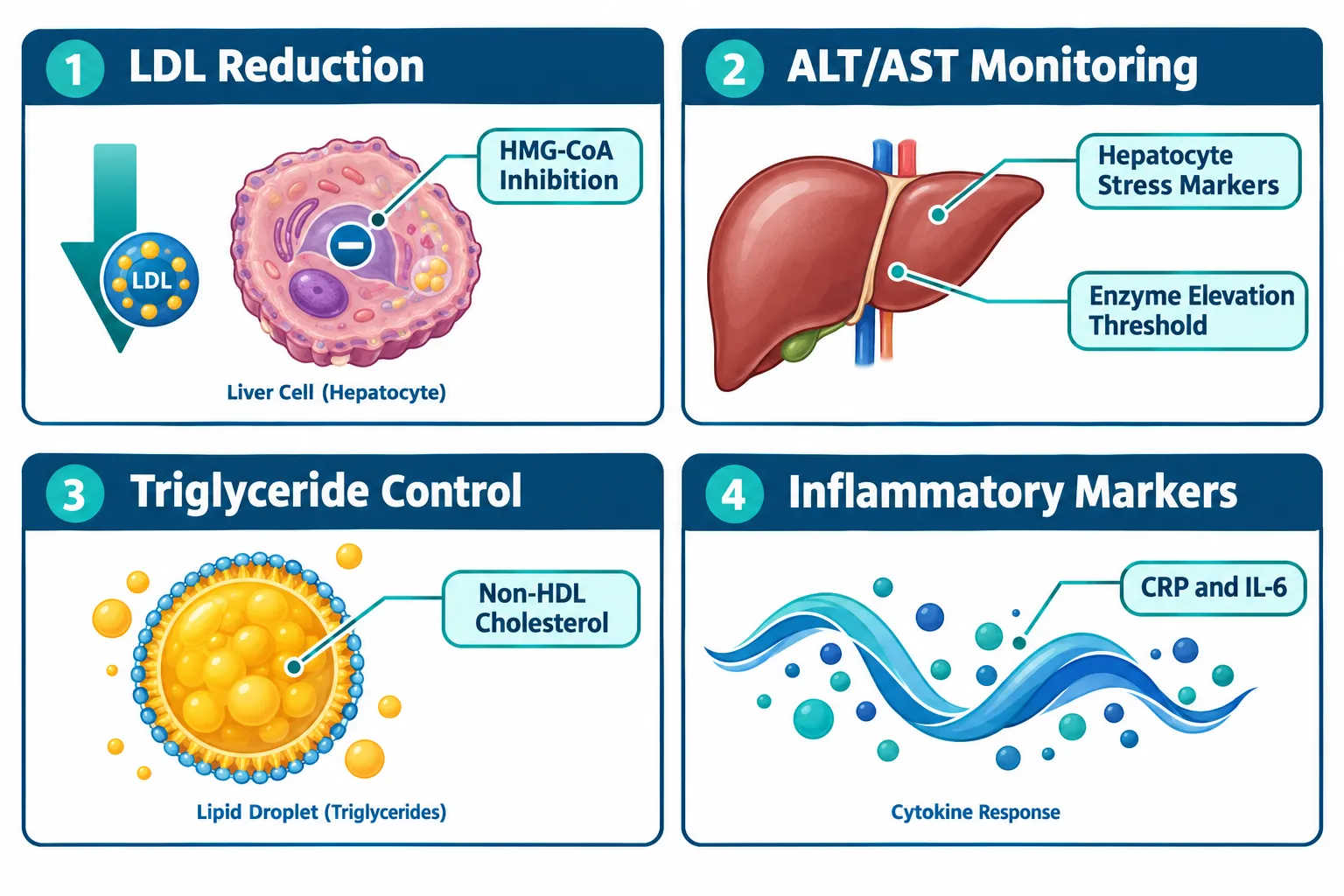
  • Atorvastatin reliably lowers LDL, triglycerides, and non-HDL cholesterol, making it the gold-standard lipid control comparator in metabolic studies.
  • Liver safety endpoints, particularly ALT, AST, and histological steatosis scores, must be tracked in every atorvastatin-containing control arm.
  • Peptide arms (retatrutide, MOTS-c, 5-amino-1MQ) require a statin-exposed control group to isolate peptide-specific metabolic contributions.
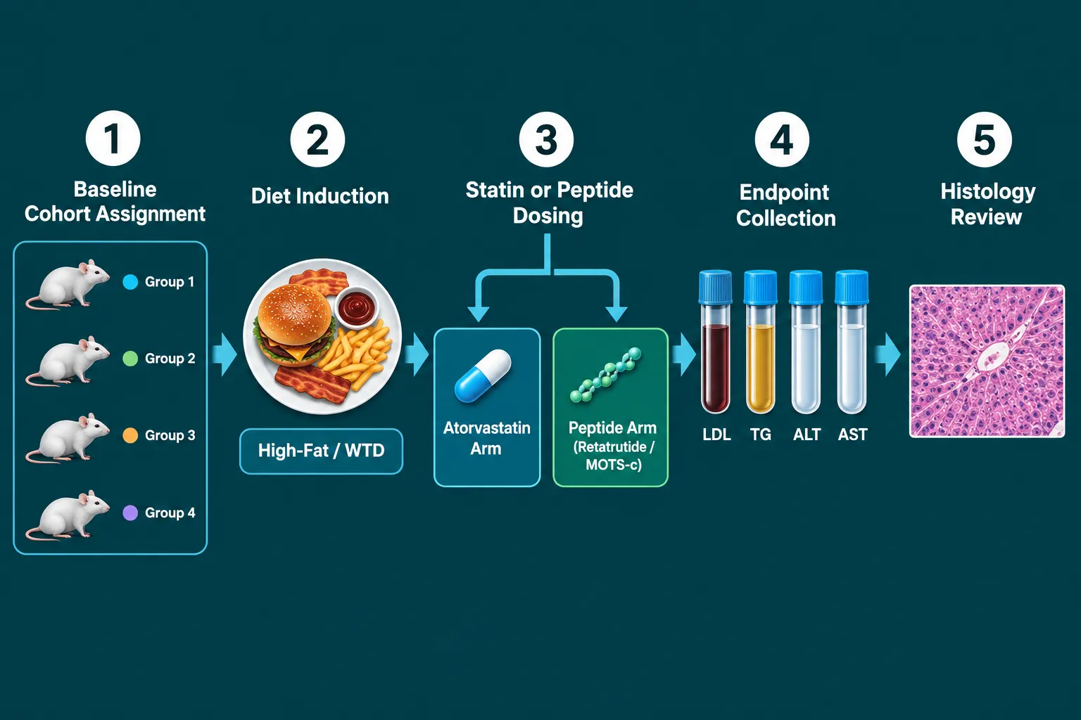
  • Western-type diet (WTD) models with and without atorvastatin remain the canonical structure for NAFLD and hyperlipidemia research designs.
  • Multi-arm study designs that combine diet, statin exposure, and peptide co-therapy now represent the evolving standard in 2026 metabolic research.

Why Atorvastatin Anchors Lipid and Liver-Control Group Design

Why Atorvastatin Anchors Lipid and Liver-Control Group Design

Atorvastatin is a high-intensity HMG-CoA reductase inhibitor with a well-mapped pharmacological profile. In metabolic research, that predictability is an asset. When a study introduces a novel peptide, researchers need a control arm whose lipid and liver effects are already understood, atorvastatin provides exactly that anchor.

Key lipid biomarkers affected by atorvastatin exposure include:

  • LDL cholesterol, typically reduced 40-55% at standard research doses
  • Triglycerides, modestly lowered, relevant in high-fat diet models
  • Non-HDL cholesterol, a composite marker that captures atherogenic particle burden
  • HDL cholesterol, marginally elevated, though not the primary endpoint

On the liver side, atorvastatin is metabolized extensively through CYP3A4 hepatic pathways. This makes ALT (alanine aminotransferase) and AST (aspartate aminotransferase) critical safety and efficacy readouts in every control arm. Current evidence from 2026 confirms that atorvastatin is safe even in subjects with advanced chronic liver disease, though transient enzyme elevations can occur and must be tracked systematically.

"A control group without liver enzyme monitoring is not a control group, it is an incomplete experiment."

For NAFLD and NASH models, atorvastatin also reduces hepatic steatosis scores and portal pressure markers. This means the "normal + atorvastatin" control arm carries meaningful liver biology, not just a lipid signal.


Structuring Multi-Arm Designs for Retatrutide, MOTS-c, and 5-Amino-1MQ Studies

Structuring Multi-Arm Designs for Retatrutide, MOTS-c, and 5-Amino-1MQ Studies

When Atorvastatin and Research Peptides: Designing Lipid and Liver-Control Groups for Metabolic Studies are combined in a multi-arm preclinical model, the architecture of the study determines what conclusions are actually possible. A poorly structured design cannot separate statin effects from peptide effects, and that ambiguity destroys interpretive value.

Recommended Multi-Arm Framework

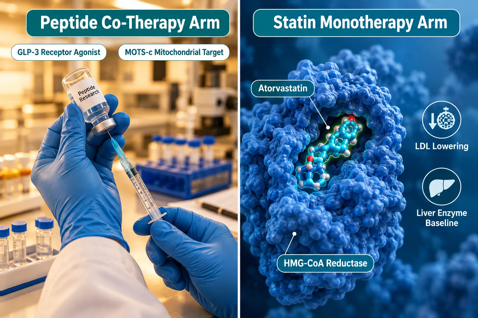
Arm Diet Statin Peptide Primary Readouts
Vehicle Control Standard chow None None Baseline LDL, ALT, AST
Lipid Control WTD Atorvastatin None LDL, TG, non-HDL, ALT
Peptide Arm A WTD None Retatrutide GLP receptor activity, lipids
Peptide Arm B WTD None MOTS-c Mitochondrial markers, AST
Combination Arm WTD Atorvastatin Retatrutide or MOTS-c All lipid + liver endpoints

Retatrutide targets GLP-1, GIP, and glucagon receptors simultaneously, producing meaningful reductions in body weight and triglycerides. For researchers exploring this compound, the Buy Retatrutide Peptide resource provides relevant formulation context. When placed against an atorvastatin lipid-control arm, the incremental triglyceride and non-HDL reduction attributable to retatrutide becomes statistically isolatable.

MOTS-c operates through mitochondrial AMPK activation, influencing glucose metabolism and fatty acid oxidation. Its liver effects, particularly on hepatic lipid accumulation, overlap with atorvastatin's steatosis-reducing properties. Researchers sourcing this compound can reference the Buy MOTS-c Peptide page for purity and dosing specifications. The MOTS-c Peptide Dosage Calculator is also a practical tool for designing consistent dosing protocols.

5-amino-1MQ inhibits NNMT (nicotinamide N-methyltransferase), shifting cellular metabolism toward fat oxidation. Its inflammatory marker profile, including CRP and IL-6, makes it particularly important to track alongside ALT and AST in the atorvastatin control arm, since both compounds affect hepatic inflammatory tone.

For broader context on how these peptides compare mechanistically to small-molecule drugs, the article on Polypeptide Peptides in Cardiometabolic Models provides a strong conceptual foundation.


Liver-Centric Endpoints and Inflammatory Marker Integration

Liver-Centric Endpoints and Inflammatory Marker Integration

The most common failure in Atorvastatin and Research Peptides: Designing Lipid and Liver-Control Groups for Metabolic Studies is treating liver safety as secondary. In 2026, the evidence base has shifted firmly: liver endpoints are co-primary, not supplementary.

Liver-centric endpoints to include in every control arm:

  1. ALT and AST, tracked at baseline, mid-study, and endpoint; atorvastatin can cause transient elevations that must be distinguished from peptide-induced changes
  2. Hepatic steatosis score, histological grading from liver biopsy or MRI-based fat fraction
  3. Portal pressure markers, relevant in advanced NAFLD/NASH models
  4. Liver-to-body weight ratio, a rapid structural readout in rodent models

Inflammatory markers add a second interpretive layer. CRP, IL-6, and TNF-alpha are all modulated by atorvastatin through pleiotropic anti-inflammatory mechanisms. When a peptide like MOTS-c or GLP-3 peptide also reduces inflammatory load, the atorvastatin control arm provides the benchmark against which that reduction is measured.

Combination therapy designs, such as "phosphatidylcholine (PPC) alone" versus "PPC + atorvastatin", demonstrate how isolating statin-specific contributions requires a clean single-agent control. The same logic applies when adding SS-31 peptides to a metabolic model: the mitochondria-targeted antioxidant effects of SS-31 must be separated from atorvastatin's lipid-lowering signal through deliberate arm structure.

Monitoring schedule best practices:

  • Collect lipid panels at weeks 0, 4, 8, and 12 minimum
  • Run liver enzyme panels at the same intervals
  • Reserve histology for endpoint only (terminal in rodent models)
  • Include fasting insulin and HOMA-IR as metabolic co-endpoints

Conclusion

Designing rigorous control groups for metabolic peptide research is not a procedural formality, it is the scientific foundation on which every meaningful comparison rests. Atorvastatin's well-characterized effects on LDL, triglycerides, non-HDL cholesterol, ALT, and AST make it the most defensible lipid and liver-control comparator available for studies involving retatrutide, MOTS-c, or 5-amino-1MQ.

Actionable next steps for researchers in 2026:

  • Build a minimum five-arm design that separates vehicle, statin, peptide, and combination conditions
  • Pre-specify all liver enzyme thresholds as stopping rules and safety endpoints
  • Use Western-type diet induction as the metabolic stressor to activate both lipid and hepatic pathways
  • Align inflammatory marker panels (CRP, IL-6) with lipid and liver readouts for mechanistic depth
  • Consult formulation-specific resources for peptide purity verification before dosing begins

A well-structured control group does not slow research down, it makes the data worth publishing.

https://www.puretestedpeptides.com/wp-content/uploads/2026/10/atorvastatin-and-research-peptides-designing-lipid-and-liver-control-groups-for.webp 672 1008 https://www.puretestedpeptides.com/wp-content/uploads/2026/01/buy-peptides-online.jpg 2026-10-06 13:10:132026-10-06 13:10:13Atorvastatin and Research Peptides: Designing Lipid and Liver-Control Groups for Metabolic Studies
Metoprolol in Peptide Experiments: Controlling Heart Rate, Blood Pressure, and Adrenergic Confounding

Metoprolol in Peptide Experiments: Controlling Heart Rate, Blood Pressure, and Adrenergic Confounding

October 6, 2026/0 Comments/in Uncategorized/by

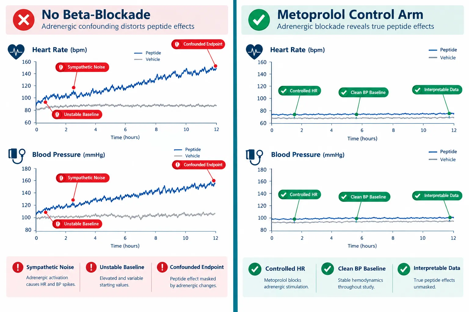
Cardiovascular endpoints fail silently when sympathetic noise is not controlled. Across retatrutide trials, growth-hormone-secretagogue models, and metabolic peptide studies, unmanaged adrenergic tone can shift heart rate by five to ten beats per minute and systolic blood pressure by several millimeters of mercury, differences large enough to obscure or mimic a genuine peptide effect. Understanding the role of metoprolol in peptide experiments for controlling heart rate, blood pressure, and adrenergic confounding is therefore not a minor methodological footnote; it is a foundational design decision.

Key Takeaways

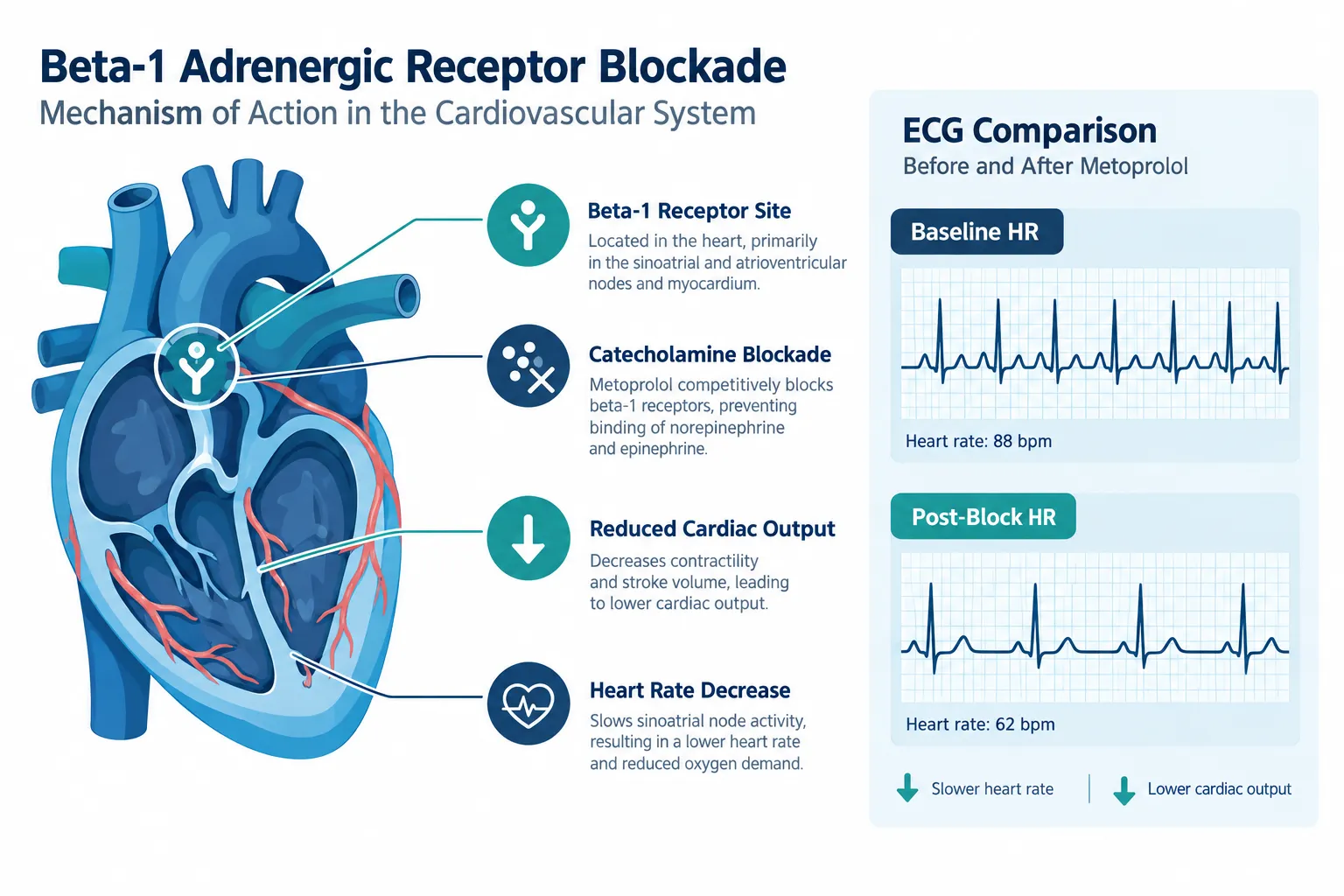
  • Metoprolol is a beta-1 selective blocker that reduces heart rate, systolic blood pressure, and cardiac output without raising peripheral resistance, making it a precise tool for quieting adrenergic noise.
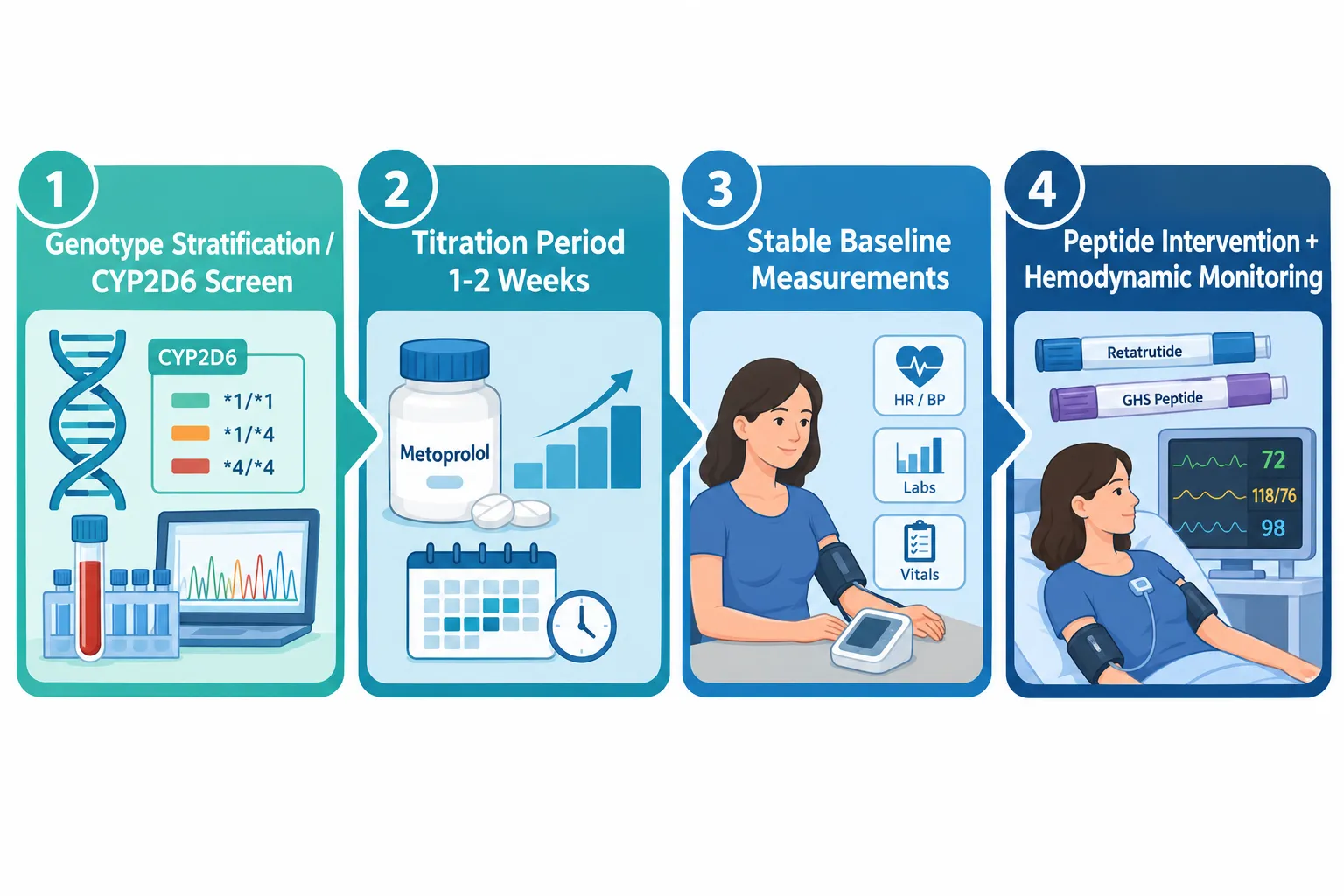
  • Placebo-corrected reductions of 3-6 mmHg systolic and 5-7 beats per minute are typical, but inter-subject variability, partly driven by CYP2D6 metabolizer status, can be substantial.
  • Stable beta-blockade takes approximately one week of oral dosing; acute sympathetic blockade begins within 15 minutes of a single dose, so experimental timing must match the intended control strategy.
  • Metoprolol controls adrenergic tone but does not normalize all advanced structural or functional endpoints, meaning it cannot substitute for a peptide's mechanism of action.
  • Proper use requires genotype stratification, defined titration periods, and both resting and exercise-state baseline measurements before any peptide intervention begins.

Why Adrenergic Tone Confounds Peptide Cardiovascular Readouts

Why Adrenergic Tone Confounds Peptide Cardiovascular Readouts

Many peptides under active investigation, including metabolic agents like retatrutide and GLP-3 class compounds, exert direct or indirect effects on the cardiovascular system. Growth-hormone secretagogues, vasoactive intestinal peptides such as VIP-5, and mitochondria-targeted peptides like SS-31 all operate in physiological environments where sympathetic tone fluctuates with stress, fasting state, exercise, and even the act of venipuncture.

Catecholamines, adrenaline and noradrenaline, bind beta-1 receptors on the myocardium and raise heart rate, contractility, and blood pressure. If a peptide also raises or lowers these parameters through a separate pathway, the two signals overlap. Without a pharmacological anchor, investigators cannot cleanly attribute observed hemodynamic changes to the peptide alone.

This is the core problem metoprolol solves. By selectively blocking beta-1 adrenergic receptors, it attenuates catecholamine-driven cardiovascular fluctuations, providing a more stable hemodynamic backdrop against which peptide-specific effects can be measured.

"Metoprolol does not eliminate cardiovascular physiology, it standardizes the sympathetic component of it, allowing other signals to emerge more clearly."

Key adrenergic phenomena that metoprolol suppresses include:

  • Resting tachycardia driven by anxiety or metabolic stimulation
  • Exercise-induced heart rate overshoot
  • Isoproterenol-provoked tachycardia used in challenge protocols
  • Reflex orthostatic tachycardia during postural changes

Each of these can appear as a false-positive or false-negative peptide effect if left uncontrolled.


Practical Application of Metoprolol in Peptide Experiments: Controlling Heart Rate, Blood Pressure, and Adrenergic Confounding

Practical Application of Metoprolol in Peptide Experiments: Controlling Heart Rate, Blood Pressure, and Adrenergic Confoundin

Dose Range and Titration Timing

Oral metoprolol doses in hypertension and heart-rate control protocols typically range from 100 to 450 mg per day, titrated at weekly intervals. A single 50-80 mg dose begins reducing exercise systolic blood pressure within approximately 15 minutes, but stable, chronic antihypertensive effect requires roughly one week of consistent dosing.
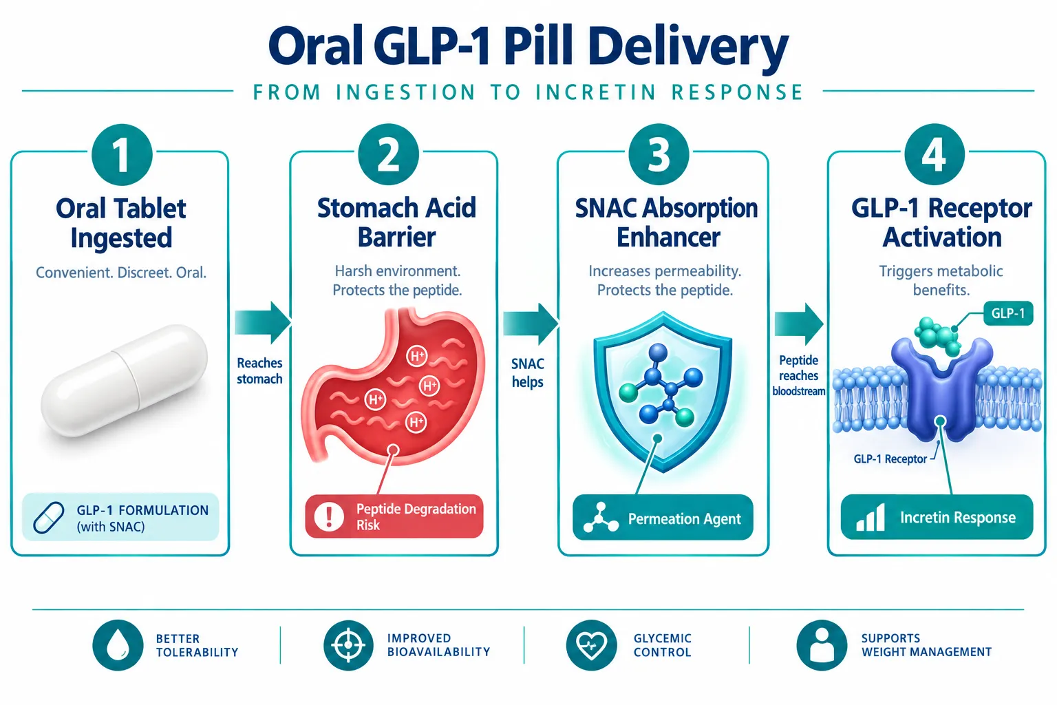
This distinction matters enormously for experimental design:

Control Strategy Onset Use Case
Acute single dose ~15 minutes Rapid sympathetic stabilization before a one-time challenge
Chronic titration (1 week+) ~7 days Stable baseline for multi-day peptide dosing protocols

For most mechanistic peptide trials, including those assessing cardiometabolic polypeptide models, chronic titration is preferred because it removes day-to-day adrenergic variability rather than just blunting a single measurement window.

Accounting for CYP2D6 Metabolizer Status

A critical and often overlooked source of confounding within the metoprolol control arm itself is pharmacogenomic variability. Poor CYP2D6 metabolizers accumulate significantly higher serum metoprolol concentrations, producing greater and more consistent reductions in heart rate, diastolic blood pressure, and mean arterial pressure compared to extensive metabolizers at the same dose.

If subjects are not stratified by CYP2D6 status, the beta-blockade "control" becomes a new source of variance. Practical steps include:

  1. Pre-screen subjects for CYP2D6 poor-metabolizer status
  2. Stratify randomization accordingly
  3. Monitor resting and exercise heart rate at baseline, after titration, and at each peptide dosing interval

Baseline Measurement Protocol

Baseline measurements must capture both resting and dynamic (exercise) cardiovascular state. Metoprolol reduces heart rate and systolic blood pressure at rest and during exercise, these are separate physiological states that peptides may affect differently. Recording only resting values misses exercise-induced adrenergic surges that some metabolic peptides, including growth-hormone secretagogues, actively modulate.


Interpretation Cautions: What Metoprolol Cannot Control

Interpretation Cautions: What Metoprolol Cannot Control

Metoprolol's selectivity is both its strength and its limitation. As a beta-1 selective agent, it does not block alpha-1 receptors or beta-2 receptors. Contrast this with carvedilol, a non-selective blocker with additional alpha-1 antagonism, which suppresses adrenergic activity through a broader mechanism. The choice between these agents changes the nature of residual adrenergic confounding, a distinction highlighted in comparative cardiovascular outcome protocols.

For researchers working with peptides in endocrine pharmacology contexts or GH-analogue peptides, additional considerations include:

  • Structural endpoints: Beta-blockade does not normalize advanced structural or functional cardiac endpoints. Recent data from myosin inhibitor trials confirm that metoprolol, despite controlling heart rate and blood pressure, did not replicate the specific functional improvements seen with next-generation myosin-targeted agents. Peptide trials must not assume that hemodynamic control equals functional equivalence.
  • Peripheral vascular effects: Metoprolol does not increase peripheral resistance, but it also does not reduce it. Peptides with vasodilatory mechanisms, such as VIP-class agents, will still show vascular effects that metoprolol leaves unmasked.
  • Metabolic interactions: Some metabolic peptides affect insulin sensitivity and glucose metabolism in ways that secondarily alter sympathetic tone. Metoprolol blunts the cardiovascular expression of this interaction but does not eliminate the underlying metabolic signal.

Conclusion

Metoprolol in peptide experiments for controlling heart rate, blood pressure, and adrenergic confounding represents one of the most practical pharmacological tools available to cardiovascular researchers in 2026. Its beta-1 selectivity, well-characterized dose-response profile, and rapid onset make it well suited for both acute challenge designs and chronic multi-week protocols.

Actionable next steps for research teams:

  • Determine whether acute or chronic beta-blockade better matches the experimental timeline before dosing begins
  • Screen and stratify subjects by CYP2D6 metabolizer status to prevent pharmacogenomic variance from contaminating the control arm
  • Collect resting and exercise-state hemodynamic baselines after titration, not before
  • Document which adrenergic phenomena remain unblocked (alpha-1, beta-2 pathways) and account for them in statistical models
  • Avoid interpreting stable hemodynamic readings as evidence that all cardiovascular endpoints are normalized, structural and functional parameters require independent assessment

Selecting high-purity, well-characterized peptides from a reliable peptide supplier is equally important; compound variability introduces its own confounding that no pharmacological control can fully correct.

https://www.puretestedpeptides.com/wp-content/uploads/2026/10/metoprolol-in-peptide-experiments-controlling-heart-rate-blood-pressure-and-adre.webp 672 1008 https://www.puretestedpeptides.com/wp-content/uploads/2026/01/buy-peptides-online.jpg 2026-10-06 13:10:002026-10-06 13:10:00Metoprolol in Peptide Experiments: Controlling Heart Rate, Blood Pressure, and Adrenergic Confounding
Prednisone and Research-Use Peptides: How Glucocorticoid Exposure Can Distort Metabolic, Immune, and Tissue-Repair Readouts

Prednisone and Research-Use Peptides: How Glucocorticoid Exposure Can Distort Metabolic, Immune, and Tissue-Repair Readouts

October 6, 2026/0 Comments/in Uncategorized/by

Roughly one in five research subjects enrolled in metabolic or immune-modulating studies carries an active or recent glucocorticoid prescription, yet fewer than half of published peptide protocols formally account for this exposure. The problem of Prednisone and Research-Use Peptides: How Glucocorticoid Exposure Can Distort Metabolic, Immune, and Tissue-Repair Readouts is not theoretical. When a subject is taking prednisone, the drug reshapes glucose handling, cytokine profiles, body composition, and wound biology simultaneously, making it nearly impossible to attribute changes in those endpoints to a peptide compound without deliberate experimental controls.

This guide is written for researchers, protocol designers, and informed buyers of research-use peptide compounds who need a clear, actionable map of where glucocorticoid confounding is most severe and how to design around it.


Key Takeaways

  • Prednisone induces a "pseudo-metabolic syndrome" that directly mimics or masks outcomes targeted by metabolic peptides.
  • Standard inflammatory biomarkers, CRP, leukocyte count, neutrophil ratio, are dose-dependently suppressed by glucocorticoids, distorting immune readouts.
  • Tissue-repair and wound-healing endpoints are particularly vulnerable because glucocorticoids both dampen acute inflammation and impair collagen remodeling.
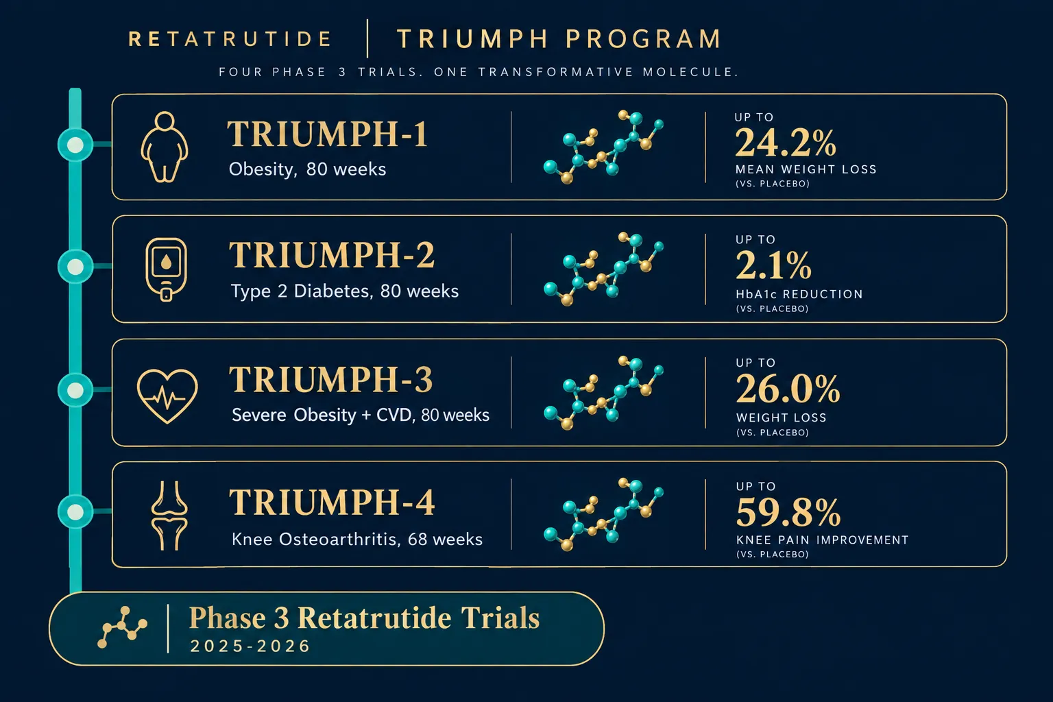
  • Anabolic and sarcopenia-targeted peptides face the strongest confounding from glucocorticoid-driven muscle catabolism.
  • Glucocorticoid exposure should be treated as a core design variable, not background noise, in any peptide research protocol.

How Glucocorticoids Create a Pseudo-Metabolic Syndrome

How Glucocorticoids Create a Pseudo-Metabolic Syndrome

Prednisone does not simply suppress inflammation. At doses commonly used in clinical and research-adjacent settings, it triggers a cascade of metabolic changes that closely resemble metabolic syndrome: elevated fasting glucose, increased visceral adiposity, dyslipidemia, and insulin resistance. For researchers studying cardiometabolic peptide compounds, this is a direct collision.

The core metabolic confounders introduced by prednisone include:

Endpoint Glucocorticoid Effect Research Risk
Fasting glucose Elevated (hepatic gluconeogenesis) Masks GLP-class peptide efficacy
Visceral fat Increased via lipid redistribution Distorts body-composition endpoints
Insulin sensitivity Reduced Confounds metabolic peptide response
Appetite / caloric intake Increased Alters weight-loss peptide readouts
Cortisol / HPA axis Suppressed endogenous output Disrupts hormonal biomarker panels

The HPA-axis suppression point deserves particular attention. When exogenous glucocorticoids are present, endogenous cortisol production falls. This changes the hormonal baseline against which peptide pharmacodynamics are measured. For GH-analogue peptides and compounds that interact with the growth hormone axis, this hormonal shift can produce entirely misleading dose-response curves.

Researchers studying visceral fat redistribution should also note that glucocorticoid-driven adiposity preferentially accumulates centrally, the same compartment tracked in visceral fat research models. Without a glucocorticoid-naive control arm, it becomes impossible to separate peptide-driven changes from steroid-driven ones.


Immune Biomarker Distortion: What Prednisone Does to CRP, Cytokines, and Leukocytes

Immune Biomarker Distortion: What Prednisone Does to CRP, Cytokines, and Leukocytes

The immune-modulating effects of prednisone are both well-documented and deeply problematic for peptide research. The drug produces strong, dose-dependent suppression of nearly every standard inflammatory biomarker used in research protocols.

Biomarkers most severely distorted by glucocorticoid exposure:

  • C-reactive protein (CRP): Hepatic CRP synthesis is directly suppressed, producing artificially low readings that can mimic anti-inflammatory peptide effects.
  • Interleukin-6 (IL-6) and TNF-alpha: Both are blunted by glucocorticoid receptor activation, masking inflammatory resolution that a peptide may or may not be driving.
  • Leukocyte and neutrophil counts: Prednisone causes a characteristic leukocytosis with neutrophilia and lymphopenia, a pattern that can be mistaken for an immune-activation signal from a peptide compound.
  • Tissue-resident immune cells: Recent work confirms that glucocorticoids selectively modulate tissue-resident macrophages and mast cells in ways that differ from circulating immune cells. This matters enormously for regenerative peptides like BPC-157 and TB-500, where local tissue immune activity is the primary readout.

"When prednisone is present, the immune landscape a peptide encounters is fundamentally different from the one it was designed to modulate. Treating that as a minor covariate is a protocol error."

The practical consequence is that an immune-modulating peptide may appear highly effective, completely ineffective, or even counterproductive depending entirely on the glucocorticoid dose the subject carries. Without stratifying subjects by steroid exposure, or excluding active users entirely, the data cannot be interpreted with confidence.


Tissue-Repair and Anabolic Readouts: Where Glucocorticoid Confounding Is Most Severe

Tissue-Repair and Anabolic Readouts: Where Glucocorticoid Confounding Is Most Severe

Wound healing and tissue repair represent perhaps the most complex confounding scenario in Prednisone and Research-Use Peptides: How Glucocorticoid Exposure Can Distort Metabolic, Immune, and Tissue-Repair Readouts. Glucocorticoids produce a paradoxical two-phase effect on repair biology.

Phase 1 (acute): Inflammation is suppressed, which can make a wound-healing peptide appear to accelerate early healing simply because the steroid has already cleared the inflammatory field.

Phase 2 (remodeling): Glucocorticoids impair fibroblast proliferation, reduce collagen synthesis, and suppress angiogenesis. A peptide that promotes collagen remodeling may show blunted or absent efficacy, not because it failed, but because the steroid has suppressed the very cellular machinery the peptide targets.

For anabolic and sarcopenia-targeted peptides, the problem is equally stark. Glucocorticoids drive skeletal muscle catabolism through multiple mechanisms: they upregulate muscle-specific ubiquitin ligases, suppress IGF-1 signaling, and reduce protein synthesis rates. Any peptide designed to promote lean mass retention or muscle repair is working against a pharmacologically imposed catabolic state. The net readout will underestimate peptide efficacy, sometimes dramatically.

Mitochondrial function is also affected. Glucocorticoids impair mitochondrial biogenesis in muscle tissue, which is directly relevant for compounds studied in SS-31 mitochondrial research contexts.

Designing Around Glucocorticoid Confounding: Practical Protocol Controls

Researchers in 2026 have several validated strategies to reduce or detect glucocorticoid confounding:

  1. Screen and stratify at enrollment. Collect steroid use history, current dose, and duration. Stratify rather than exclude where possible to preserve sample size.
  2. Use glucocorticoid-resistant biomarkers where available. Some cytokines and repair markers are less sensitive to steroid suppression and can serve as secondary endpoints.
  3. Build in washout windows. For subjects who can safely discontinue steroids, a documented washout period before peptide exposure begins reduces confounding substantially.
  4. Run parallel control arms. A glucocorticoid-exposed control arm receiving vehicle only allows direct quantification of steroid-driven endpoint changes.
  5. Report steroid exposure as a primary covariate. Statistical models should treat glucocorticoid dose as a continuous variable, not a binary yes/no flag.

Conclusion

The relationship between Prednisone and Research-Use Peptides: How Glucocorticoid Exposure Can Distort Metabolic, Immune, and Tissue-Repair Readouts is now well enough characterized that ignoring it constitutes a preventable design flaw. Across glucose metabolism, inflammatory cytokines, collagen remodeling, body composition, and immune-cell measurements, prednisone introduces quantifiable, directional biases that can make an effective peptide look inert, or an inert one look effective.

Actionable next steps for research protocol designers:

  • Add glucocorticoid screening to all enrollment checklists for metabolic, immune, or repair-focused peptide studies.
  • Pre-specify steroid dose as a covariate in the statistical analysis plan before data collection begins.
  • Consult updated 2026 biomarker guidance on glucocorticoid-resistant immune endpoints when designing immune-modulating peptide studies.
  • Review compound-specific confounding risk, peptides targeting GH axis, collagen synthesis, or mitochondrial function carry the highest vulnerability.

Treating glucocorticoid exposure as a core design variable rather than background noise is the single highest-leverage methodological improvement available to most peptide research teams today.

https://www.puretestedpeptides.com/wp-content/uploads/2026/10/prednisone-and-research-use-peptides-how-glucocorticoid-exposure-can-distort-met.webp 1024 1536 https://www.puretestedpeptides.com/wp-content/uploads/2026/01/buy-peptides-online.jpg 2026-10-06 13:06:132026-10-06 13:06:13Prednisone and Research-Use Peptides: How Glucocorticoid Exposure Can Distort Metabolic, Immune, and Tissue-Repair Readouts
Amlodipine in Peptide Research: Separating Calcium-Channel Effects From Blood-Pressure and Vascular Readouts

Amlodipine in Peptide Research: Separating Calcium-Channel Effects From Blood-Pressure and Vascular Readouts

October 6, 2026/0 Comments/in Uncategorized/by

Fewer than one in five preclinical peptide studies that co-administer a calcium-channel blocker formally account for the drug's independent vascular effects, a methodological gap that can quietly invalidate entire datasets. When amlodipine in peptide research enters the experimental picture, the challenge is not simply adjusting for lower blood pressure. It is disentangling at least three overlapping biological layers: direct L-type calcium-channel blockade, blood-pressure-dependent vascular changes, and blood-pressure-independent vascular remodeling. Getting these layers wrong means attributing amlodipine's known anti-proliferative and anti-atherosclerotic actions to the peptide under study, or, just as problematically, masking a real peptide signal behind the drug's dominant pharmacology.

Key Takeaways

  • Amlodipine blocks L-type calcium channels in vascular smooth muscle, reducing intracellular Ca²⁺ and causing arterial relaxation independently of any peptide co-administered in a study.
  • The drug produces vascular benefits, including inhibited smooth-muscle cell proliferation and slowed atherosclerosis, that are separate from its blood-pressure-lowering effect.
  • At clinically relevant concentrations, amlodipine does not activate CRAC channels and does not interfere with fluorescence-based Ca²⁺ imaging dyes such as Cal-520, Fluo-4, or Fura-Red.
  • Researchers should pre-specify which readouts are calcium-channel-mediated, blood-pressure-dependent, or peptide-specific before data collection begins.
  • Orthogonal biomarkers, endothelial nitric oxide, arterial compliance, and vascular remodeling indices, can help isolate a peptide's contribution from amlodipine's background pharmacology.

How Amlodipine Works: The Calcium-Channel Mechanism Researchers Must Account For

How Amlodipine Works: The Calcium-Channel Mechanism Researchers Must Account For

Amlodipine is a dihydropyridine calcium antagonist. Its primary action is inhibiting the transmembrane influx of calcium ions through L-type voltage-gated channels, predominantly in vascular smooth muscle cells. The result is reduced intracellular Ca²⁺, diminished actin-myosin interaction, smooth-muscle relaxation, and arterial vasodilation. Its effect on vascular smooth muscle is substantially greater than its effect on cardiac muscle, a selectivity profile that matters when interpreting any cardiovascular or vascular readout in a peptide experiment.

This mechanism is not trivial background noise. It is a dominant pharmacological signal. Any peptide co-administered with amlodipine that also influences vascular tone, intracellular calcium, or smooth-muscle behavior will generate data that reflects both agents simultaneously unless the study design explicitly separates them.

What this means practically:

  • Vascular tone measurements (e.g., wire myography, pressure myography) will reflect amlodipine's baseline relaxation effect.
  • Intracellular Ca²⁺ flux assays will show a suppressed resting Ca²⁺ level before the peptide is even introduced.
  • Any peptide with vasodilatory properties may appear to have a blunted or absent effect simply because the vascular bed is already partially relaxed.

Researchers working with mitochondria-targeted peptides such as those explored in SS-31 10mg research peptide considerations should note that oxidative-stress and membrane-potential readouts can also be indirectly affected when intracellular calcium homeostasis is altered by a co-administered channel blocker.


Separating Calcium-Channel Effects From Blood-Pressure and Vascular Readouts: A Practical Framework

Separating Calcium-Channel Effects From Blood-Pressure and Vascular Readouts: A Practical Framework

The central challenge in amlodipine in peptide research, separating calcium-channel effects from blood-pressure and vascular readouts, requires a tiered classification of every endpoint before data collection begins.

Tier 1: Blood-Pressure-Dependent Readouts

These are endpoints that change because amlodipine lowers arterial pressure. They include:

  • Brachial or carotid blood pressure values
  • Cardiac afterload indices
  • Glomerular filtration rate changes driven by reduced perfusion pressure

Practical step: Include a vehicle-treated, amlodipine-only control group at the same dose. Any change in these readouts in that group represents the amlodipine baseline, not a peptide effect.

Tier 2: Blood-Pressure-Independent Vascular Effects

Contemporary cardiovascular literature consistently identifies a second category of amlodipine actions that persist even when blood pressure is matched to comparator agents. These include:

Effect Mechanism Research Implication
Smooth-muscle cell anti-proliferation Direct Ca²⁺ signaling suppression May confound peptide anti-proliferative claims
Anti-atherosclerotic remodeling Reduced oxidative stress in vessel wall Arterial stiffness data needs amlodipine-only control
Improved arterial compliance Structural vascular changes Central pulse-wave velocity requires correction

For peptide studies examining vascular remodeling, such as those involving VIP peptide mechanisms or vasoactive signaling pathways, these BP-independent effects represent the most likely source of confounding.

Tier 3: Calcium-Channel-Specific Molecular Readouts

This tier covers direct molecular measurements of L-type channel activity and downstream Ca²⁺ signaling. A key methodological advantage here: at therapeutic plasma concentrations of approximately 0.5 µM, amlodipine does not activate calcium-release-activated calcium (CRAC) channels. This means CRAC-mediated signaling events attributed to a peptide are not being driven by amlodipine itself.

Additionally, fluorescence-based Ca²⁺ imaging using dyes such as Cal-520, Fluo-4, and Fura-Red is not confounded by amlodipine fluorescence at these concentrations. This is a meaningful green light for researchers using live-cell calcium imaging alongside peptide stimulation assays.

"Knowing which calcium compartment amlodipine does and does not touch is the single most useful piece of information a peptide researcher can carry into experimental design."

Researchers examining peptide signaling pathways, including those studying GLP-3 peptide signaling pathway interactions, benefit from this CRAC-channel clarity because receptor-operated calcium entry can then be attributed with greater confidence to the peptide's receptor engagement rather than to the channel blocker.


Designing the Experiment: Isolating Peptide-Specific Signals

Designing the Experiment: Isolating Peptide-Specific Signals

A rigorous experimental framework for amlodipine in peptide research, one that genuinely separates calcium-channel effects from blood-pressure and vascular readouts, involves five concrete design decisions.

Step 1, Define the calcium-channel baseline. Run amlodipine-only controls at the intended study concentration. Measure intracellular Ca²⁺, vascular tone, and any remodeling markers. This establishes the floor the peptide must be measured against.

Step 2, Select orthogonal vascular markers. Choose at least two endpoints that amlodipine does not directly regulate. Endothelial nitric oxide synthase (eNOS) activity, circulating endothelin-1 levels, and specific peptide receptor phosphorylation states are examples. These serve as peptide-specific signal channels.

Step 3, Apply fluorescence Ca²⁺ imaging with confidence. Given that amlodipine does not interfere with Cal-520, Fluo-4, or Fura-Red at relevant concentrations, live-cell imaging remains a valid tool. Pair it with a CRAC-channel inhibitor control to confirm that any observed Ca²⁺ flux is L-type-independent when the peptide is active.

Step 4, Separate central from peripheral blood pressure. Peripheral brachial pressure can be identical across treatment groups while central aortic pressure and pulse-wave velocity differ. Use central hemodynamic measurements to capture amlodipine's BP-independent arterial effects separately from peptide-driven changes.

Step 5, Validate with receptor-blocking controls. If the peptide of interest acts through a specific receptor, as is the case with many research peptides including those reviewed in LL-37 versus SS-31 benefits of each peptide comparisons, include a receptor-antagonist arm. Any vascular effect that disappears with receptor blockade but persists with amlodipine alone is attributable to the peptide.

Proper peptide dosing protocols also matter here: dose-response curves for the peptide should be generated in both the presence and absence of amlodipine to detect any shift in potency or efficacy caused by the background calcium-channel blockade.

For researchers sourcing compounds for controlled in vitro work, ensuring purity and verified composition is non-negotiable. Resources such as guidance on where to buy research-grade peptides evaluating purity underscore why compound quality directly affects the reliability of mechanistic conclusions.


Conclusion

Amlodipine is a well-characterized L-type calcium-channel blocker with a pharmacological footprint that extends well beyond simple blood-pressure reduction. In any peptide research context, failing to account for its anti-proliferative, anti-atherosclerotic, and Ca²⁺-suppressive actions risks misattributing drug effects to the peptide, or obscuring genuine peptide biology beneath a dominant pharmacological background.

Actionable next steps for researchers in 2026:

  1. Pre-classify every planned endpoint into one of the three tiers: blood-pressure-dependent, BP-independent vascular, or calcium-channel-specific molecular.
  2. Include an amlodipine-only control group at the study concentration in every experimental arm.
  3. Use fluorescence-based Ca²⁺ imaging with confidence at therapeutic concentrations, but pair it with CRAC-channel controls to confirm the signal source.
  4. Add orthogonal endothelial and receptor-specific biomarkers that amlodipine does not directly regulate.
  5. Run full dose-response curves for the peptide both with and without amlodipine present to detect potency shifts.

Rigorous separation of these layers does not complicate peptide research, it protects it. The result is data that can be defended, reproduced, and built upon.


https://www.puretestedpeptides.com/wp-content/uploads/2026/10/amlodipine-in-peptide-research-separating-calcium-channel-effects-from-blood-pre.webp 1024 1536 https://www.puretestedpeptides.com/wp-content/uploads/2026/01/buy-peptides-online.jpg 2026-10-06 13:05:532026-10-06 13:05:53Amlodipine in Peptide Research: Separating Calcium-Channel Effects From Blood-Pressure and Vascular Readouts
What Is a GLP-1 Pill? Comparing Oral Delivery Concepts With Research-Use GLP-3 Retatrutide Models

What Is a GLP-1 Pill? Comparing Oral Delivery Concepts With Research-Use GLP-3 Retatrutide Models

October 6, 2026/0 Comments/in Uncategorized/by

An oral tablet approved for weight loss sounds like a major leap forward, and in 2026, that leap has officially happened. But the question researchers, clinicians, and informed consumers are now asking is whether a GLP-1 pill and an injectable triple agonist like retatrutide are even playing the same game. Understanding what is a GLP-1 pill, comparing oral delivery concepts with research-use GLP-3 retatrutide models, reveals a surprisingly wide gap between two approaches that share a name but differ sharply in mechanism, scope, and research application.

Key Takeaways

  • GLP-1 pills are oral small-molecule drugs that activate the GLP-1 receptor; two were approved in the U.S. in 2026 for obesity.
  • Oral delivery faces significant biochemical barriers, requiring absorption enhancers to survive the digestive tract.
  • Retatrutide is a triple agonist targeting GLP-1, GIP, and glucagon receptors simultaneously, making it mechanistically distinct from any single-agonist pill.
  • The TRIUMPH Phase 3 program is testing retatrutide across obesity, type 2 diabetes, cardiovascular disease, and osteoarthritis populations.
  • An oral GLP-1 pill is not a substitute for a triple-agonist injectable in research or clinical contexts.

How a GLP-1 Pill Actually Works

The glucagon-like peptide-1 (GLP-1) receptor is a well-validated target for metabolic disease. When activated, it slows gastric emptying, reduces appetite, and stimulates insulin secretion in a glucose-dependent manner. Injectable GLP-1 receptor agonists have dominated this space for years, but 2026 marks a turning point for oral delivery.

How a GLP-1 Pill Actually Works

Two oral GLP-1 agents reached U.S. approval in 2026. Eli Lilly's orforglipron (brand name Foundayo) became the first approved oral-only GLP-1 pill specifically indicated for obesity. Novo Nordisk's semaglutide pill, already available for type 2 diabetes, also received approval for weight loss in adults in both the U.S. and EU markets. These are not peptide-based tablets, they are small-molecule non-peptide compounds or peptide formulations engineered to survive the hostile environment of the gastrointestinal tract.

The core challenge with oral peptide delivery is enzymatic degradation. Stomach acid and proteases break down peptide chains before they can reach systemic circulation. Semaglutide tablets overcome this using a co-formulation with sodium N-(8-[2-hydroxybenzoyl] amino) caprylate (SNAC), an absorption enhancer that temporarily raises local gastric pH and facilitates mucosal permeation. Orforglipron sidesteps this problem entirely by being a small-molecule compound that does not require an absorption enhancer.

Key oral delivery constraints include:

  • Low bioavailability compared to subcutaneous injection
  • Strict fasting requirements before dosing (for semaglutide tablets)
  • Narrower therapeutic window due to absorption variability
  • No flexibility in dose titration beyond what the formulation allows

For those exploring the broader landscape of peptides used in metabolic and weight-loss research, what is the strongest peptide for weight loss provides useful context on how different compound classes compare.


Retatrutide: The Triple-Agonist Model That Changes the Comparison

Calling retatrutide a "GLP-3" is a colloquial shorthand that has gained traction in research communities, but it is worth clarifying what that label actually means. Retatrutide is a single peptide molecule that simultaneously activates three receptors: GLP-1, glucose-dependent insulinotropic polypeptide (GIP), and glucagon. This triple agonism is why it is sometimes distinguished from standard GLP-1 agents with the informal "GLP-3" designation.

Retatrutide: The Triple-Agonist Model That Changes the Comparison

This mechanistic distinction is critical when comparing oral delivery concepts with research-use GLP-3 retatrutide models. A GLP-1 pill activates one receptor pathway. Retatrutide activates three, producing overlapping and synergistic metabolic effects that include:

  • GLP-1 activity: appetite suppression, insulin secretion, slowed gastric emptying
  • GIP activity: enhanced insulin response, potential adipose tissue effects
  • Glucagon activity: increased energy expenditure, hepatic fat mobilization

Phase 2 data for retatrutide showed weight reductions exceeding 24% of body weight at the highest dose over 48 weeks, a benchmark that no single GLP-1 agonist, oral or injectable, has matched in head-to-head comparisons at similar timepoints.

For more background on how retatrutide fits within the GLP-1 research landscape, see the GLP-1 retatrutide overview. Researchers seeking access to standardized research-grade material can also review GLP3 Reta 10mg and GLP-3 Reta 30mg for sale options for preclinical study use.


The TRIUMPH Phase 3 Program and What It Tells Researchers

The most comprehensive data set currently being generated on retatrutide comes from the TRIUMPH Phase 3 program, a suite of large-scale trials running across 2025 and 2026. These trials are designed to test retatrutide across multiple disease populations, not just general obesity.

The TRIUMPH Phase 3 Program and What It Tells Researchers

Trial Population Duration Primary Focus
TRIUMPH-1 Obesity or overweight (no T2D) 80 weeks Weight loss efficacy and safety
TRIUMPH-2 Type 2 diabetes + obesity 80 weeks Weight loss and glycemic control
TRIUMPH-3 Severe obesity + CVD (BMI 35+) 80 weeks Cardiovascular and weight outcomes
TRIUMPH-4 Obesity + knee osteoarthritis 68 weeks Weight loss and knee pain reduction

Topline 2026 results from the TRIUMPH program and the parallel TRANSCEND-T2D-1 diabetes trial have shown meaningful reductions in A1C, body weight, and comorbidity burden. These outcomes extend well beyond what a single-receptor oral pill is designed to achieve.

For researchers tracking the evolution of this compound, the retatrutide clinical trials resource aggregates ongoing study data in one place.

"Retatrutide's triple-receptor mechanism produces metabolic effects that are qualitatively different from those of any approved oral GLP-1 agent, not just quantitatively larger."

It is also worth noting that retatrutide remains an injectable peptide in all clinical and research settings. There is no oral formulation in development at this stage, which means the oral vs. injectable distinction is not merely a delivery preference, it reflects a fundamental difference in what each compound can do.

For researchers interested in combinatorial peptide approaches in metabolic contexts, the article on combining Tesamorelin with CDC Ipamorelin offers relevant mechanistic parallels.


Conclusion

Understanding what is a GLP-1 pill, comparing oral delivery concepts with research-use GLP-3 retatrutide models, makes one conclusion unavoidable: these are fundamentally different tools. The 2026 approvals of orforglipron and oral semaglutide for obesity represent a genuine advance in patient convenience and access. However, their single-receptor mechanism, oral bioavailability constraints, and narrower metabolic scope place them in a separate category from retatrutide's triple-agonist profile.

Actionable next steps for researchers and informed readers:

  1. Review the TRIUMPH trial registry entries to follow Phase 3 retatrutide data as it becomes available.
  2. Distinguish between oral GLP-1 agents (small-molecule or SNAC-formulated peptide) and injectable triple agonists before drawing efficacy comparisons.
  3. If evaluating retatrutide for preclinical research, source only verified, tested-grade material and consult current retatrutide clinical trial literature before designing protocols.
  4. Do not assume that oral availability implies equivalent biological activity, receptor breadth, bioavailability, and dosing pharmacokinetics all differ substantially between compound classes.

The GLP-1 pill is a meaningful milestone. Retatrutide is a different kind of milestone entirely.

https://www.puretestedpeptides.com/wp-content/uploads/2026/10/what-is-a-glp-1-pill-comparing-oral-delivery-concepts-with-research-use-glp-3-re.webp 1024 1536 https://www.puretestedpeptides.com/wp-content/uploads/2026/01/buy-peptides-online.jpg 2026-10-06 13:05:492026-10-06 13:05:49What Is a GLP-1 Pill? Comparing Oral Delivery Concepts With Research-Use GLP-3 Retatrutide Models
Page 5 of 80«‹34567›»
×

Helpful Links

  • My account
  • Cart
  • Checkout
  • Refund and Returns Policy
  • Privacy Policy
  • SMS Privacy Policy
  • Login
  • My Account
  • Logout

USA Made Lab Tested Peptides

All products are sold for research, laboratory, or analytical purposes only, and are not for human consumption


Pure Tested Peptides is a chemical supplier. Pure Tested Peptides is not a compounding / chemical compounding facility as defined under 503A of the Federal Food, Drug, and Cosmetic act. Pure Tested Peptides is not an outsourcing facility as defined under 503B of the Federal Food, Drug, and Cosmetic act.

The statements made within this website have not been evaluated by the US Food and Drug Administration. The products we offer are not intended to diagnose, treat, cure or prevent any disease.

Human/Animal Consumption Prohibited. Laboratory/In-Vitro Experimental Use Only

Scroll to top Scroll to top Scroll to top