Call or Text 727-513-9780
  • Shopping Cart Shopping Cart
    0Shopping Cart
Pure Tested Peptides | America's most trusted Peptides for sale online
  • Peptides for sale
    • Oral Peptides for sale
      • Peptide Capsules for sale
      • BPC 157 Capsules 1000mcg
      • SLU-PP-332 Capsules | 1000 mcg
      • 5-Amino-1MQ 50mg Capsules
      • Tesofensine 500mcg
    • All Peptides for sale
    • Peptide Sprays
      • BPC 157 Nasal Spray Kit
      • BPC-157 TB500 Nasal Spray Kit
      • Semax Nasal Spray 10mg
      • Selank – Nasal Spray Kit – 10mg
      • Epithalon 50MG Nasal Spray Kit
      • Ipamorelin 10mg Nasal Spray
      • Klow Nasal Spray (BPC-157 + TB-500 + GHK-Cu + KPV) | 80mg
      • Hulk Nasal Spray Tesa / Ipa Blend 6/3 MG
      • Klow Nasal Spray
      • NAD + 500 mg Nasal Spray
      • PT-141 Nasal Spray Kit
    • GHRH Peptides
      • Ipa Peptides
      • CJC-1295 Peptides
        • CJC-1295 with DAC 5 mg
        • CJC-1295 without DAC 5 mg
        • CJC-1295 Ipa 10mg
      • Tesa Peptides
        • Tesa Peptide
        • Tesa 20 mg
    • GHK-Cu Peptides
      • All GHK-Cu Peptides
      • GHK-Cu 100mg
      • KLOW Peptide Blend – Buy KLOW blend online
    • BPC Peptides
      • All BPC Peptides
      • BPC-157
      • BPC-157 TB-500
      • BPC 157 capsules 1000mcg
    • SLU-PP-332 Peptides
      • All SLU-PP-332 Peptides
      • SLU-PP-332 5mg
    • GLP3 Peptides
    • PT-141 Peptides
      • PT-141 Peptides for sale
      • PT-141 10mg
      • PT-141 Nasal Spray
    • CAG Peptides
      • Lipo-C Peptide Blend
      • CAG 5mg
      • CAG 10mg
    • MOTS-C Peptides
      • MOTS-C Peptides for sale
      • MOTS-c peptide
      • MOTS-c 10mg *6 pack*
    • 5 Amino 1MQ Peptides
      • 5 Amino 1MQ Peptides for sale
      • 5-Amino-1MQ 50mg Capsules
      • 5-Amino-1MQ 5mg
    • Epithalon Peptides
      • Epithalon Peptides for sale
      • Epithalon 10mg
      • Epithalon 50mg
  • Shop
    • GLPs
      • 5-Amino-1MQ 50mg Capsules
      • 5-Amino-1MQ 5mg
      • L-Carnitine 500mg/ml
      • Tesofensine 500mcg
      • SLU-PP-332 5mg
      • MOTS-c 10mg *6 pack*
    • Epithalon & BPC Peptides
      • Epithalon 10mg
      • Epithalon 50mg
      • BPC-157
      • BPC 157 capsules 1000mcg
      • BPC-157 TB-500
      • BPC-157 TB500 Nasal Spray Kit
      • BPC 157 Nasal Spray Kit
    • BPC TB-500 & NAD+ Peptides
      • NAD+ 500 mg
      • KLOW Peptide Blend – Buy KLOW blend online
      • GLOW Peptide Blend
      • TB 500 5mg
      • BPC 157 capsules 1000mcg – Supplement
      • BPC 157 Nasal Spray Kit
      • BPC-157
      • BPC-157 TB500 Nasal Spray Kit
      • BPC-157 TB-500
      • BPC 157 capsules 1000mcg
    • LL-37 Peptide
      • LL-37 10 mg
    • MOTS-C & Selank
      • MOTS-c peptide
      • Selank 10mg
    • GHK Peptides
      • GHK-Cu 100mg
      • GLOW Peptide Blend
      • KLOW Peptide Blend – Buy KLOW blend online
  • COAs
  • Wholesale
    • Wholesale Peptides for sale
  • PTP FAQ
  • Affiliates
    • Selank Peptide: Advanced Pharmacological Mechanisms Underlying Its Anxiolytic and Nootropic Effects in Research
      • 5-Amino-1MQ Peptide: Detailed Mechanisms of NNMT Inhibition and Its Impact on Cellular Metabolism Research
        • MOTS-C Peptide: Unraveling Its Role in Mitochondrial Dynamics and Energy Metabolism Research
          • Epithalon Peptide and Telomerase Regulation: Investigating Its Impact on Cellular Senescence and Lifespan Research Models
            • GLP2-T vs GLP2 Tirz Peptide: Understanding the Naming, Mechanistic Nuances, and Research Implications for Gut Health
      • Peptides and Polypeptides in Modern Pharmacology: What Research on Metoprolol, Prednisone, and Amlodipine Reveals
        • Polypeptide Peptides vs NSAIDs: What Naproxen and Diclofenac Teach Tissue-Repair Researchers About BPC‑157 and TB‑500
          • Peptides and Polypeptides in Endocrine Pharmacology: How GLP-1, GLP-2, and GLP-3 Retatrutide Differ From Classic Drugs Like Prednisone and Amlodipine
          • 5‑Amino‑1MQ Peptide and NNMT Inhibition: How It Compares With Statins Like Atorvastatin in Adipose and Lipid Metabolism Research
      • Enclomiphene, Estrogen Receptor Signaling, and Luteinizing Phase Biology: What Hormone Researchers Should Measure
        • Mesenchymal Stem Cells, BPC‑157, and GHK‑Cu: How Tissue Repair Peptides Compare With Classic NSAIDs Like Naproxen in Injury Models
          • Peptide Calculator Use Cases Beyond Growth Hormone: Working Through GLP‑3 Retatrutide, MOTS‑c, and BPC‑157 Research Dosing
        • Polypeptide Peptides in Endocrine and Metabolic Pharmacology: Lessons From Amlodipine, Prednisone, and Metoprolol
        • Peptides Calculator for Advanced Blends: Worked Examples for Tesamorelin, CJC‑1295, and Ipamorelin Stacks
          • Peptides and Polypeptides in Human Physiology: How Molecular Size Shapes Research Applications
            • Selank Peptide: Advanced Pharmacological Mechanisms Underlying Its Anxiolytic and Nootropic Effects in Research
          • 5-Amino-1MQ Peptide: Detailed Mechanisms of NNMT Inhibition and Its Impact on Cellular Metabolism Research
            • MOTS-C Peptide: Unraveling Its Role in Mitochondrial Dynamics and Energy Metabolism Research
              • Epithalon Peptide and Telomerase Regulation: Investigating Its Impact on Cellular Senescence and Lifespan Research Models
                • GLP2-T vs GLP2 Tirz Peptide: Understanding the Naming, Mechanistic Nuances, and Research Implications for Gut Health
                  • Peptides and Polypeptides in Endocrine Pharmacology: How GLP-1, GLP-2, and GLP-3 Retatrutide Differ From Classic Drugs Like Prednisone and Amlodipine
                    • Polypeptide Peptides vs NSAIDs: What Naproxen and Diclofenac Teach Tissue-Repair Researchers About BPC‑157 and TB‑500
                      • Peptides and Polypeptides in Modern Pharmacology: What Research on Metoprolol, Prednisone, and Amlodipine Reveals
                        • 5‑Amino‑1MQ Peptide and NNMT Inhibition: How It Compares With Statins Like Atorvastatin in Adipose and Lipid Metabolism Research
                        • Enclomiphene, Estrogen Receptor Signaling, and Luteinizing Phase Biology: What Hormone Researchers Should Measure
                        • Mesenchymal Stem Cells, BPC‑157, and GHK‑Cu: How Tissue Repair Peptides Compare With Classic NSAIDs Like Naproxen in Injury Models
                        • Peptide Calculator Use Cases Beyond Growth Hormone: Working Through GLP‑3 Retatrutide, MOTS‑c, and BPC‑157 Research Dosing
                        • Polypeptide Peptides in Endocrine and Metabolic Pharmacology: Lessons From Amlodipine, Prednisone, and Metoprolol
                        • Peptides Calculator for Advanced Blends: Worked Examples for Tesamorelin, CJC‑1295, and Ipamorelin Stacks
                        • Peptides and Polypeptides in Human Physiology: How Molecular Size Shapes Research Applications
                        • Selank Peptide: Advanced Pharmacological Mechanisms Underlying Its Anxiolytic and Nootropic Effects in Research
                        • MOTS-C Peptide: Unraveling Its Role in Mitochondrial Dynamics and Energy Metabolism Research
                        • Peptides and Polypeptides in Modern Pharmacology: What Research on Metoprolol, Prednisone, and Amlodipine Reveals
                        • Peptides and Polypeptides in Endocrine Pharmacology: How GLP-1, GLP-2, and GLP-3 Retatrutide Differ From Classic Drugs Like Prednisone and Amlodipine
                        • 5-Amino-1MQ Peptide: Detailed Mechanisms of NNMT Inhibition and Its Impact on Cellular Metabolism Research
                        • Mesenchymal Stem Cells, BPC‑157, and GHK‑Cu: How Tissue Repair Peptides Compare With Classic NSAIDs Like Naproxen in Injury Models
                        • Peptide Calculator Use Cases Beyond Growth Hormone: Working Through GLP‑3 Retatrutide, MOTS‑c, and BPC‑157 Research Dosing
                        • GLP2-T vs GLP2 Tirz Peptide: Understanding the Naming, Mechanistic Nuances, and Research Implications for Gut Health
                        • Polypeptide Peptides in Endocrine and Metabolic Pharmacology: Lessons From Amlodipine, Prednisone, and Metoprolol
                        • Polypeptide Peptides vs NSAIDs: What Naproxen and Diclofenac Teach Tissue-Repair Researchers About BPC‑157 and TB‑500
                        • Peptides and Polypeptides in Human Physiology: How Molecular Size Shapes Research Applications
                        • Peptides Calculator for Advanced Blends: Worked Examples for Tesamorelin, CJC‑1295, and Ipamorelin Stacks
                        • 5‑Amino‑1MQ Peptide and NNMT Inhibition: How It Compares With Statins Like Atorvastatin in Adipose and Lipid Metabolism Research
                        • Enclomiphene, Estrogen Receptor Signaling, and Luteinizing Phase Biology: What Hormone Researchers Should Measure
                        • Epithalon Peptide and Telomerase Regulation: Investigating Its Impact on Cellular Senescence and Lifespan Research Models
                        • Best research protocol Klow blend
                        • best time to take BPC-157
                        • best time to take DSIP (Delta Sleep Inducing Peptide)
                        • best time to take CJC-1295
                        • best time to take AOD-9604
                        • best time to take Follistatin 344
                        • best time to take Ipamorelin
                        • best time to take MK-677 (Ibutamoren)
                        • best time to take Ligandrol (LGD-4033) — research compound
                        • best time to take Ostarine (MK-2866) — research compound
                        • best time to take GHK-CU
                        • best time to take TB-500
                        • best time to take MOTS-c
                        • best time to take Semax
                        • best time to take RAD-140 (Testolone) — research compound
                        • best time to take Thymosin Alpha-1
                        • best time to take PEG-MGF
                        • Biolife Plasma, Octapharma Plasma, and Research Peptides: How Plasma Donation Labs Differ From Peptide Suppliers
                        • best time to take YK-11 — research compound
                        • best time to take PT-141 (Bremelanotide)
                        • Best research protocol Klow blend
                        • 5-Amino-1MQ and MOTS-C Synergy: Metabolic Signaling, Mitochondria, and Research Design
                        • BPC-157 and TB-500: Investigating Their Combined Effects on Angiogenesis and Cellular Migration in Tissue Repair Models
                        • BPC-157 Peptide: Gut Barrier Function, Inflammation, and Tissue-Recovery Research
                        • 5‑Amino‑1MQ and MOTS‑c Synergy in Metabolic Research: Designing NNMT and Mitochondrial Biogenesis Stacks
                        • CJC-1295 with DAC vs. Without DAC: Half-Life, Release Kinetics, and Research Implications
                        • CJC‑1295 with DAC vs. Without DAC: Expanding on Half‑Life Differences Using Tesamorelin and Ipamorelin Blend Case Studies
                        • Collagen Biology and Copper‑Binding Peptides: How GHK‑Cu, Glow Blend, and Klow Blend Interact with Skin and Connective Tissue
                        • Collagen Biology and Regenerative Peptides: How GHK‑Cu, Glow Blend, and Klow Blend Affect Extracellular Matrix Research
                    • DNA, Telomeres, and Longevity Peptides: Positioning Epithalon and MOTS‑c in Genetic Aging Research
                      • Enclomiphene Citrate: serm Mechanism, Testosterone Research, and Stack Compatibility
                        • Enclomiphene vs Enclomiphene Citrate: Formulation, Bioavailability, and Research Distinctions
                        • Epithalon Peptide Research: Telomerase Activation, Aging, and Pineal Gland Function
                        • Estrogen Receptor Signaling and Enclomiphene: How Selective Modulators Compare with Classic Polypeptide Hormones
                        • GHK-Cu Peptide: Advanced Mechanisms in Extracellular Matrix Remodeling and Wound Healing Research
                        • GHK-Cu Peptide: Collagen Synthesis, Wound Repair, and Skin-Barrier Research Models
                        • GLP-1 vs GLP-3 vs GLP-2: Peptide Classification and Research Applications
                        • GLP-2 Peptide Research Guide: Gut Barrier Function, Nutrient Absorption, and Intestinal Recovery Models
                        • GLP-3 Retatrutide vs. GLP-1 Drugs: What Triple-Agonist Biology Changes in Research Models
                        • Ipamorelin and Tesamorelin Combination: Synergistic GH Secretagogue Research and Dosing Protocols
                        • GLP2 Tirz Peptide: What It Is, Why the Name Exists, and How Researchers Should Interpret It
                        • Klow Blend Peptide Nasal Spray: What the Formulation Is Trying to Do in Cognitive Research
                        • Mitochondria, NNMT Inhibition, and Peptide Modulators: Where MOTS‑c and 5‑Amino‑1MQ Fit in Cellular Energy Research
                        • MOTS-c Peptide: Mitochondrial Function, Energy Metabolism, and What Researchers Measure
                        • MOTS-c vs. 5-Amino-1MQ: Which Metabolic Research Questions Each Compound Actually Answers
                        • Nasal Spray Peptides: Bioavailability, Administration, and Semax/Selank Research Applications
                        • PT-141 Peptide Research: Mechanism of Action and Melanocortin Receptor Signaling
                        • Retatrutide for Research: Mechanism, Structure, and GLP-1/GLP-3 Dual Action
                        • Retatrutide for Obesity and Type 2 Diabetes: What the Latest Trial Data Suggest
                        • Tesofensine Peptide Research: Mechanism, Appetite Suppression, and Neuropeptide Y Pathways
  • Contact
    • Contact Customer Service
    • Text Customer Support
  • About US
  • Shop all peptides
  • Affiliate Program
    • Affiliate Signup
  • Login / Register Login / Register Page Link Login / Register Page Link
  • Click to open the search input field Click to open the search input field Search
  • Menu Menu
Amlodipine and Research Peptides: How Calcium-Channel Blockade Can Confound Blood-Pressure Experiments

Amlodipine and Research Peptides: How Calcium-Channel Blockade Can Confound Blood-Pressure Experiments

October 4, 2026/0 Comments/in Uncategorized/by

Fewer than 10% of preclinical cardiovascular studies explicitly report whether test subjects were receiving background antihypertensive therapy, yet a single oral dose of amlodipine can drop systolic blood pressure by 18 to 45 mmHg for six hours or longer. That gap between standard practice and methodological rigor sits at the heart of the problem explored here. Understanding Amlodipine and Research Peptides: How Calcium-Channel Blockade Can Confound Blood-Pressure Experiments is not a niche concern; it is a prerequisite for any researcher using GLP-class peptides, growth-hormone secretagogues, or tissue-repair compounds in models where blood pressure, vascular tone, or intracellular calcium is a primary endpoint.

Key Takeaways

  • Amlodipine produces dose-dependent, long-lasting blood-pressure reductions that can dominate hemodynamic readouts across typical experimental windows.
  • Calcium-channel blockade can physically interfere with fura-based fluorescent calcium imaging, producing false signals that mimic peptide activity.
  • Circadian blood-pressure patterns, critical for ambulatory monitoring studies, can be altered by amlodipine independently of any peptide intervention.
  • Confounding by indication and dose is a recognized methodological hazard in pharmacoepidemiology that applies directly to peptide trials.
  • Researchers can control for these effects through explicit CCB documentation, adequate washout periods, group stratification, and adjusted statistical models.

How Amlodipine Changes Vascular Tone and Blood-Pressure Baselines

Amlodipine is a dihydropyridine calcium-channel blocker (CCB) that selectively inhibits voltage-gated L-type calcium channels in vascular smooth muscle. By preventing calcium influx, it reduces the contractile force of arterial walls, lowers peripheral vascular resistance, and decreases blood pressure without producing large chronic changes in resting heart rate. This peripheral vasodilatory action is highly relevant when studying any peptide that targets the cardiovascular system.
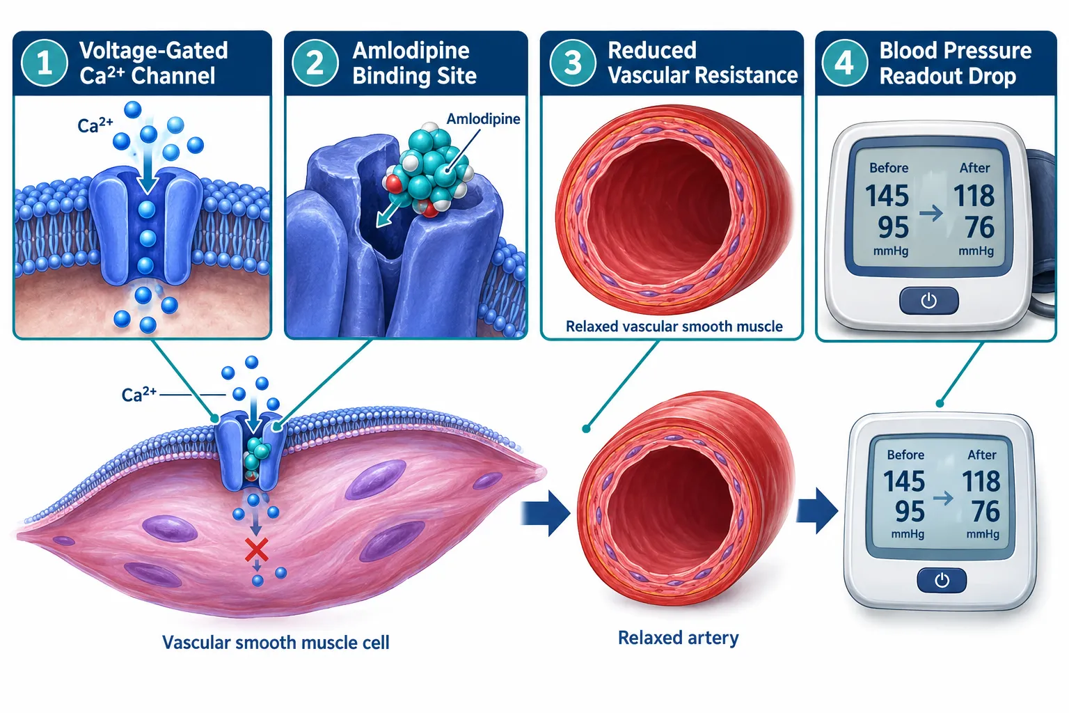
How Amlodipine Changes Vascular Tone and Blood-Pressure Baselines

In spontaneously hypertensive rat (SHR) models, the most widely used preclinical platform for antihypertensive research, oral doses between 1 and 10 mg/kg reduce blood pressure for at least six hours after administration. At higher maintenance doses of around 8 mg/kg per day, marked systolic blood-pressure reductions persist for 16 to 20 hours, effectively spanning an entire experimental window. This matters enormously for researchers studying peptides such as SS-31, which targets mitochondrial cardiolipin and influences cardiac energetics in pressure-overload models. If amlodipine is present in the background, observed improvements in afterload and wall stress may reflect CCB-induced resistance changes rather than peptide activity.

Key hemodynamic facts about amlodipine:

Parameter Typical Effect
Systolic BP reduction (SHR, acute) 18 to 45 mmHg
Duration per oral dose 6 to 20+ hours
Heart rate change (stable angina) Minimal chronic change
Peripheral vascular resistance Significantly reduced

The drug's long half-life, roughly 35 to 50 hours in humans, means that "overnight discontinuation" is wholly inadequate as a washout strategy. Researchers designing experiments around lab-tested peptides must plan washout periods of several days, not hours, to eliminate residual CCB influence on hemodynamic baselines.


Why Calcium-Channel Blockade Specifically Confounds Peptide Research

The confounding problem in Amlodipine and Research Peptides: How Calcium-Channel Blockade Can Confound Blood-Pressure Experiments operates through at least three distinct mechanisms, each capable of invalidating a study on its own.
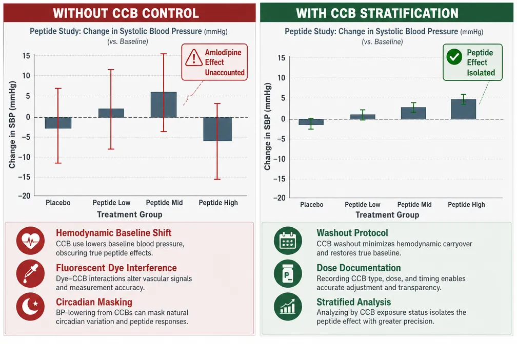
Why Calcium-Channel Blockade Specifically Confounds Peptide Research

Hemodynamic Baseline Masking

Cardioprotective peptides are typically evaluated against endpoints such as infarct size, contractility, fibrosis, and cardiomyocyte apoptosis. All of these endpoints are highly sensitive to baseline hemodynamics. A 2023 clinical analysis found that 10 mg/day of amlodipine lowered systolic blood pressure by 14.2 mmHg and diastolic blood pressure by 13.6 mmHg over eight weeks, while converting approximately 40% of patients from a "non-dipper" to a "dipper" nocturnal profile. Any concurrent peptide therapy evaluated via 24-hour ambulatory monitoring could have its chronobiologic effects masked or mimicked by these CCB-driven shifts.

For researchers studying SS-31 kidney health or cardiac remodeling, this means a peptide that genuinely reduces nocturnal blood pressure might appear no better than placebo if amlodipine has already normalized the dipper profile.

Fluorescent Calcium Imaging Interference

This is perhaps the most underappreciated confound. Amlodipine besylate accumulates in the cytoplasm and overlaps spectrally with fura-2, the most widely used fluorescent dye for measuring intracellular calcium. The drug dominates the fluorescence signal, mimicking calcium transients that do not exist. Any peptide claimed to modulate excitation-contraction coupling, including growth-hormone secretagogues or vasoactive intestinal peptides, could appear falsely effective or ineffective in fura-loaded cells exposed to amlodipine.

"Amlodipine's spectral overlap with fura-2 means that calcium imaging data collected in its presence cannot be reliably attributed to any co-administered peptide."

Confounding by Indication and Dose

Pharmacoepidemiologic research has established that both the clinical reason for prescribing a CCB and the dose level act as independent confounders. In observational peptide trials, unequal distribution of background CCB therapy across study arms, even with randomization, can systematically bias blood-pressure and remodeling outcomes. This problem is compounded by precision-dosing approaches: one trial demonstrated that app-guided amlodipine titration in 205 adults reduced mean blood pressure from 142/87 to 131/81 mmHg over 14 weeks, a moving target that would be nearly impossible to control for without explicit documentation.


Practical Design Strategies for CCB-Aware Peptide Experiments

Addressing Amlodipine and Research Peptides: How Calcium-Channel Blockade Can Confound Blood-Pressure Experiments requires deliberate protocol choices at the design stage, not post-hoc corrections.
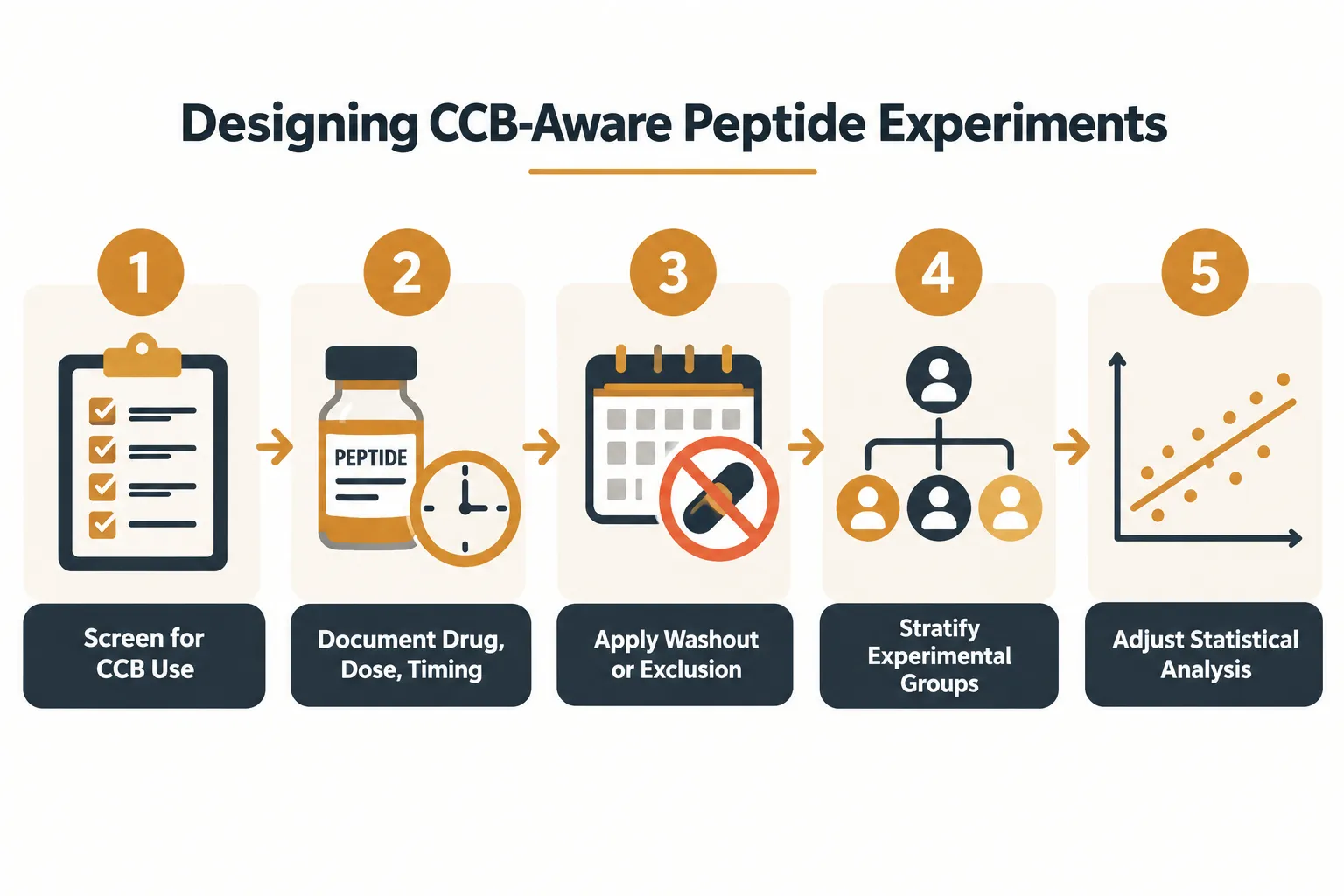
Practical Design Strategies for CCB-Aware Peptide Experiments

Five actionable steps for researchers:

  1. Screen and document all CCB exposure, Record drug name, dose, formulation, and timing for every subject or animal before enrollment.
  2. Apply adequate washout periods, Given amlodipine's half-life, washout of at least five half-lives (seven or more days in humans) is the minimum acceptable standard.
  3. Exclude or stratify CCB-treated subjects, In studies using SS-31 mitochondrial research endpoints or similar oxidative-stress markers, exclusion is preferable; if exclusion is not feasible, stratify and adjust quantitatively.
  4. Avoid fura-based calcium imaging in amlodipine-exposed tissue, Switch to alternative calcium indicators without spectral overlap, or use CCB-naive preparations.
  5. Adjust statistical models for CCB covariates, Include CCB type, dose, and duration as covariates in regression models for blood-pressure and remodeling outcomes.

These principles apply equally to researchers working with MOTS-C peptide in metabolic-cardiovascular models, Thymosin Alpha-4 in cardiac repair studies, or VIP peptide in vascular tone experiments. Each of these compounds interacts with pathways that are directly altered by sustained calcium-channel blockade.

Checklist summary:

  • Document CCB use at baseline
  • Plan washout of at least 5 half-lives
  • Stratify groups by CCB status
  • Avoid fura-2 imaging in CCB-exposed cells
  • Report CCB covariates in all statistical models
  • Pre-register exclusion criteria related to antihypertensive therapy

Conclusion

The intersection of Amlodipine and Research Peptides: How Calcium-Channel Blockade Can Confound Blood-Pressure Experiments is a methodological blind spot with real consequences for data validity. Amlodipine's long half-life, potent hemodynamic effects, and spectral interference with calcium-imaging tools create three separate pathways through which it can distort peptide study results, masking genuine effects, generating false signals, or introducing systematic bias across study arms.

Actionable next steps for research teams in 2026:

  • Audit existing protocols for undocumented CCB exposure before data collection begins.
  • Add CCB screening as a mandatory enrollment step in any blood-pressure or calcium-signaling study.
  • Consult pharmacoepidemiology guidance on confounding by indication when designing observational peptide trials.
  • Source lab-tested peptides with verified purity certificates to eliminate compound-quality confounds alongside pharmacological ones.

Rigorous control of background antihypertensive therapy is not optional, it is the foundation on which credible peptide cardiovascular research is built.

https://www.puretestedpeptides.com/wp-content/uploads/2026/10/amlodipine-and-research-peptides-how-calcium-channel-blockade-can-confound-blood.webp 1024 1536 https://www.puretestedpeptides.com/wp-content/uploads/2026/01/buy-peptides-online.jpg 2026-10-04 13:05:052026-10-04 13:05:05Amlodipine and Research Peptides: How Calcium-Channel Blockade Can Confound Blood-Pressure Experiments
Metoprolol in Incretin and Growth-Hormone Studies: Separating Heart-Rate Effects From Peptide Responses

Metoprolol in Incretin and Growth-Hormone Studies: Separating Heart-Rate Effects From Peptide Responses

October 4, 2026/0 Comments/in Uncategorized/by

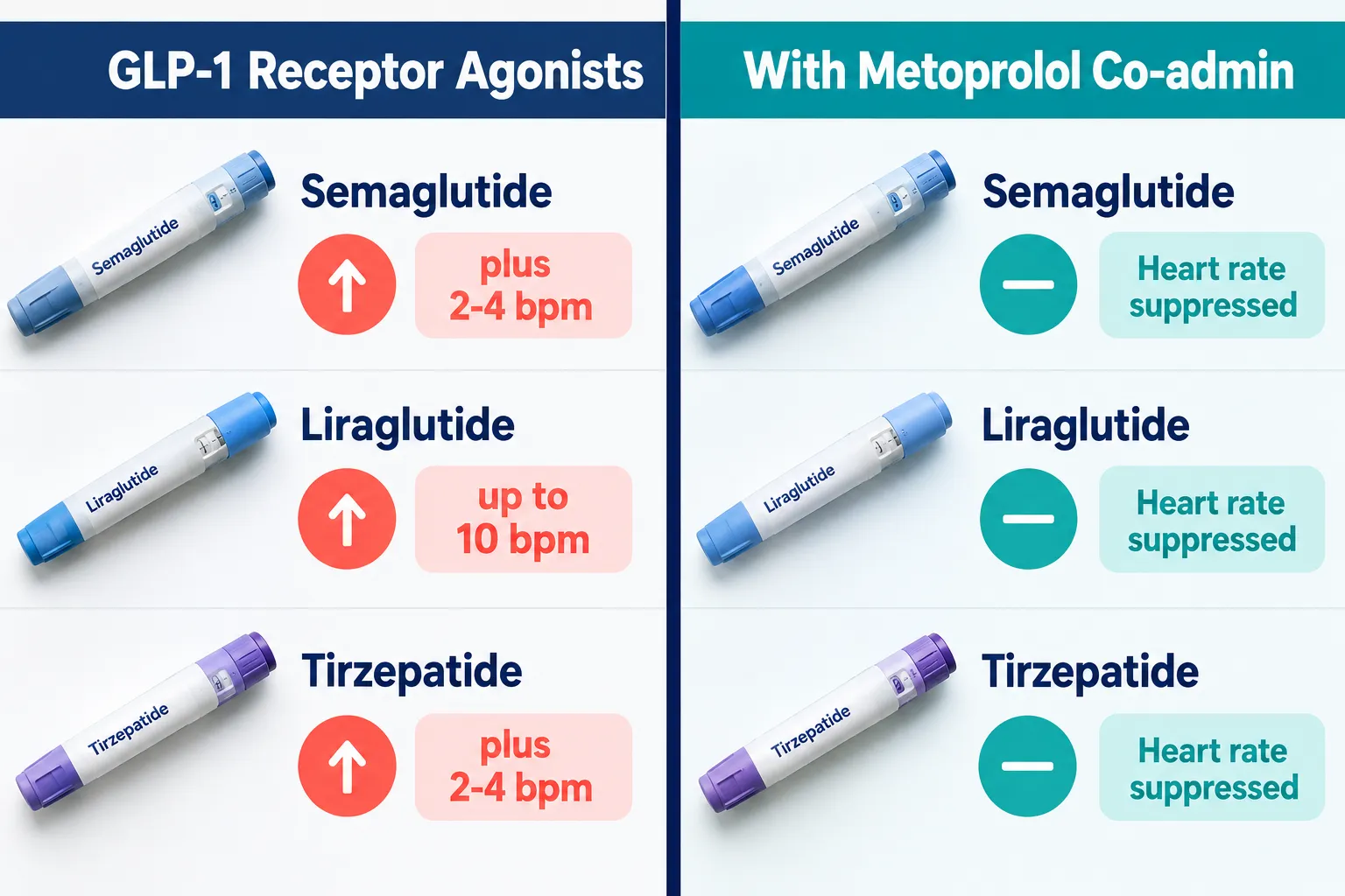
Beta-blockers have long been used as tools to control cardiovascular variables in research, but their role in isolating peptide-specific signals is underappreciated. When GLP-1 receptor agonists can raise resting heart rate by as much as 10 beats per minute in some patients, and when growth-hormone release is modulated by adrenergic tone, the question of how to cleanly attribute any observed response to the peptide itself becomes urgent. This is exactly where metoprolol in incretin and growth-hormone studies: separating heart-rate effects from peptide responses becomes a precise and practical strategy.


Key Takeaways

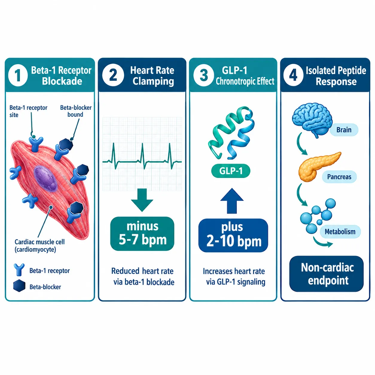
  • Metoprolol lowers resting heart rate by approximately 5-7 bpm on average, making it a reliable pharmacological tool to "clamp" heart rate in mechanistic studies.
  • GLP-1 receptor agonists raise heart rate by 2-10 bpm depending on the agent, creating a confounding variable that beta-blockade can suppress.
  • Beta-adrenergic tone exerts a tonic inhibitory effect on growth-hormone release, meaning metoprolol itself can unmask GH responses.
  • Careful baseline and repeated-measure design is essential when using metoprolol as an experimental variable.
  • Co-administration of metoprolol with incretin or GH-related peptides is clinically feasible with appropriate monitoring.

Why Beta-Blockade Matters as an Experimental Variable

Why Beta-Blockade Matters as an Experimental Variable

Metoprolol is a beta-1 selective adrenergic blocker. At standard doses, it reduces heart rate, myocardial contractility, and cardiac output both at rest and during exercise. Extended-release metoprolol succinate produces mean resting heart-rate reductions of roughly 5-7 bpm, with some individuals showing larger drops. This predictable, dose-dependent, and reversible effect is exactly what researchers need when designing studies where heart rate must be held steady.

The problem in peptide research is that many bioactive compounds have direct or indirect chronotropic effects. If a study measures glucose metabolism, vascular tone, or endocrine secretion after administering an incretin peptide, an uncontrolled rise in heart rate will alter blood pressure, cardiac output, and tissue perfusion simultaneously. Those changes then confound the metabolic or hormonal readout.

By using metoprolol to suppress the chronotropic component, investigators can attribute downstream effects more confidently to the peptide's direct receptor-mediated actions.

This is not a theoretical exercise. Animal studies have used combined beta-blockade and muscarinic blockade to establish intrinsic heart rate, then shown that peptides like oxyntomodulin can still raise intrinsic heart rate through pathways independent of GLP-1 receptors. That finding only became visible because beta-adrenergic confounding was removed first.

For researchers exploring GH analogue peptides or incretin-based compounds, understanding this pharmacological separation is foundational.


Incretin Peptides and the Chronotropic Problem

Incretin Peptides and the Chronotropic Problem

GLP-1 receptor agonists are now central to obesity and diabetes care. Their cardiovascular effects are well documented, but the magnitude of heart-rate elevation varies considerably by agent.

Agent Approximate HR Increase
Liraglutide Up to ~10 bpm
Semaglutide ~2-4 bpm
Dulaglutide ~2-4 bpm
Tirzepatide ~2-4 bpm
24-hour average (class) ~7 bpm

Real-world data from patients with implantable cardioverter-defibrillators show that starting a GLP-1 receptor agonist raises daily heart rate by a median 1.5 bpm and nighttime heart rate by about 2.0 bpm, with roughly 2.5 bpm additional nighttime increases per dose uptitration. These are small numbers in clinical practice, but in a mechanistic study designed to detect a 3-4 bpm signal, they represent meaningful noise.

"When the peptide-induced heart-rate change is the same order of magnitude as the outcome you are trying to measure, you cannot separate signal from confound without pharmacological control."

This is the core argument for metoprolol in incretin and growth-hormone studies: separating heart-rate effects from peptide responses. Beta-blockade suppresses the chronotropic signal, allowing the researcher to focus on non-cardiac endpoints such as insulin secretion, glucose disposal, or endocrine axis activity.

Researchers interested in weight-loss peptide mechanisms should also review what is the strongest peptide for weight loss for context on how incretin-class compounds compare on metabolic endpoints.

For those studying tesa peptide benefits, the same principle applies: any growth-hormone-releasing peptide that alters autonomic tone will require heart-rate control to isolate the GH-specific signal.


Growth Hormone, Adrenergic Tone, and Metoprolol's Dual Role

Historical endocrine research in animal models demonstrated that central administration of beta-adrenergic antagonists, including metoprolol, produced a gradual elevation in plasma growth hormone, while beta-agonists reduced GH. The conclusion was that beta-adrenergic tone exerts a tonic inhibitory effect on GH release.

This creates a dual consideration for study design:

  1. Metoprolol suppresses heart rate (the desired confound-control effect).
  2. Metoprolol may itself unmask or amplify GH responses by removing inhibitory adrenergic input.

Both effects must be accounted for. If a study uses metoprolol to control heart rate during a GH-releasing peptide protocol, the observed GH elevation may reflect both the peptide's action and the removal of adrenergic inhibition. Crossover designs with placebo beta-blockade arms are therefore essential.

For background on how peptides interact with endocrine pharmacology more broadly, the article on peptides and polypeptides in endocrine pharmacology provides useful mechanistic context.


Practical Study Design: Baseline and Repeated-Measure Recommendations

Practical Study Design: Baseline and Repeated-Measure Recommendations

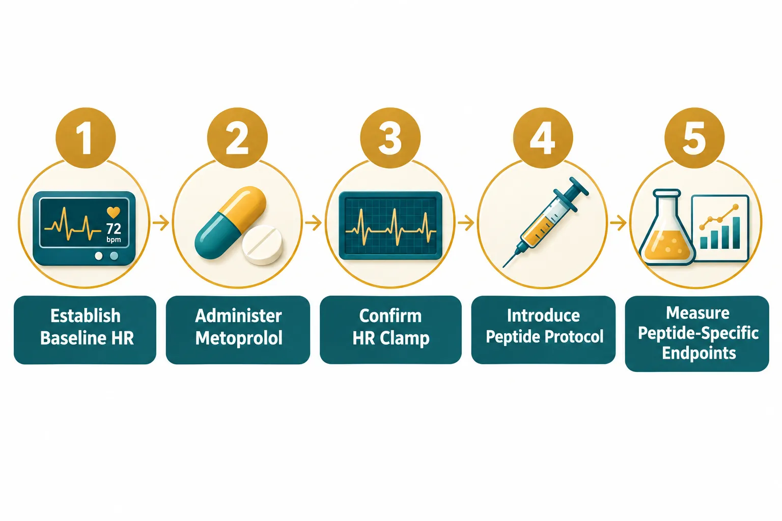
Applying metoprolol in incretin and growth-hormone studies: separating heart-rate effects from peptide responses effectively requires structured protocols. The following recommendations reflect current best practice for mechanistic research designs.

Before starting any intervention:

  • Record resting heart rate on at least three separate days to establish a stable baseline.
  • Document blood pressure, fasting glucose, and fasting insulin at each baseline visit.
  • Confirm that metoprolol has reached steady-state plasma levels (typically 4-5 half-lives after initiation or dose change) before introducing the peptide protocol.

During the study:

  • Use continuous or frequent intermittent heart-rate monitoring to confirm the "clamp" is maintained.
  • Measure peptide-specific endpoints (e.g., GLP-1 levels, GH pulse amplitude, insulin secretion) at standardized time points relative to peptide administration, not relative to drug administration.
  • In crossover designs, allow sufficient washout time for metoprolol; withdrawal data confirm that heart rate normalizes to the mid-60s bpm range after discontinuation, but full washout should be confirmed by repeated measurement rather than assumed from timing alone.

Repeated-measure considerations:

  • Collect hormone and metabolic samples at the same time of day across all study arms to control for circadian variation in GH pulsatility and insulin sensitivity.
  • Account for exercise tolerance changes: metoprolol blunts exercise-induced tachycardia, so any exercise challenge used to stimulate GH must be standardized by workload output, not by target heart rate.

Understanding peptide dosing protocols is equally important when designing repeated-measure studies, as dose timing relative to beta-blockade will affect the observed peptide response window.

For studies involving mitochondrial or cytoprotective peptides alongside cardiovascular variables, the SS-31 10mg research peptide considerations resource offers relevant parallel design guidance.


Conclusion

The value of metoprolol in incretin and growth-hormone studies lies in its predictability. A 5-7 bpm heart-rate reduction, reliably achieved and reversible, gives researchers a clean pharmacological lever to isolate peptide-specific effects from autonomic noise. The key practical steps are straightforward: establish a stable heart-rate baseline before intervention, confirm steady-state beta-blockade before introducing the peptide, use continuous monitoring to verify the clamp throughout the protocol, and build crossover or placebo-controlled arms to separate metoprolol's own endocrine effects from those of the peptide under study.

As incretin therapies expand in obesity and cardiometabolic medicine, mechanistic trials will increasingly need this kind of pharmacological scaffolding. Researchers and clinicians designing such studies should treat beta-blockade not as a background medication but as an active experimental variable that requires the same rigorous characterization as the peptide itself. Reviewing the broader landscape of peptides 101 for research-use only buyers is a practical next step for anyone building a comprehensive mechanistic protocol.

https://www.puretestedpeptides.com/wp-content/uploads/2026/10/metoprolol-in-incretin-and-growth-hormone-studies-separating-heart-rate-effects.webp 1024 1536 https://www.puretestedpeptides.com/wp-content/uploads/2026/01/buy-peptides-online.jpg 2026-10-04 13:04:572026-10-04 13:04:57Metoprolol in Incretin and Growth-Hormone Studies: Separating Heart-Rate Effects From Peptide Responses
Atorvastatin in Metabolic Peptide Experiments: Controlling Lipid-Lowering Effects in Retatrutide and Mitochondrial Models

Atorvastatin in Metabolic Peptide Experiments: Controlling Lipid-Lowering Effects in Retatrutide and Mitochondrial Models

October 4, 2026/0 Comments/in Uncategorized/by

Fewer than 10% of metabolic peptide experiments pre-specify how concurrent statin therapy will be handled, yet that omission can quietly invalidate a study's entire lipid endpoint. When the compound under investigation is a triple agonist as potent as retatrutide, and when mitochondrial probes such as MOTS-c or 5-Amino-1MQ are added to the design, the presence or absence of atorvastatin in the background becomes a variable that can dwarf the signal researchers are actually trying to measure. Understanding how to account for atorvastatin in metabolic peptide experiments, controlling lipid-lowering effects in retatrutide and mitochondrial models, is therefore not a minor procedural detail. It is a core element of experimental validity.

Key Takeaways

  • Atorvastatin's HMG-CoA reductase inhibition and pleiotropic effects can confound lipid endpoints in retatrutide and mitochondrial peptide studies if not systematically controlled.
  • Baseline lipid matching and pre-specified washout windows are the two most critical design decisions when atorvastatin is present in a study population.
  • Retatrutide's triple agonism (GLP-1, GIP, glucagon) independently lowers LDL, triglycerides, and hepatic lipids, making statin co-exposure a genuine confound rather than a neutral background variable.
  • Mitochondrial peptides such as MOTS-c and 5-Amino-1MQ interact with pathways that atorvastatin also touches, requiring careful arm construction to isolate each compound's contribution.
  • No completed published trial as of 2026 has formally used atorvastatin as a deliberate modulator of retatrutide's lipid effects; current guidance is therefore grounded in pharmacological principles and experimental design logic.

Why Atorvastatin Demands Attention in Metabolic Peptide Research

Why Atorvastatin Demands Attention in Metabolic Peptide Research

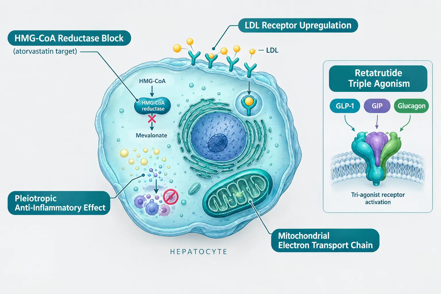
Atorvastatin is the world's most prescribed lipid-lowering drug. It blocks HMG-CoA reductase, the rate-limiting enzyme in hepatic cholesterol synthesis, and upregulates LDL receptors on liver cells. Beyond these primary actions, it exerts well-documented pleiotropic effects: reducing vascular inflammation, modulating endothelial nitric oxide, and influencing mitochondrial coenzyme Q10 levels. Each of these secondary actions intersects with pathways that metabolic peptides are designed to probe.

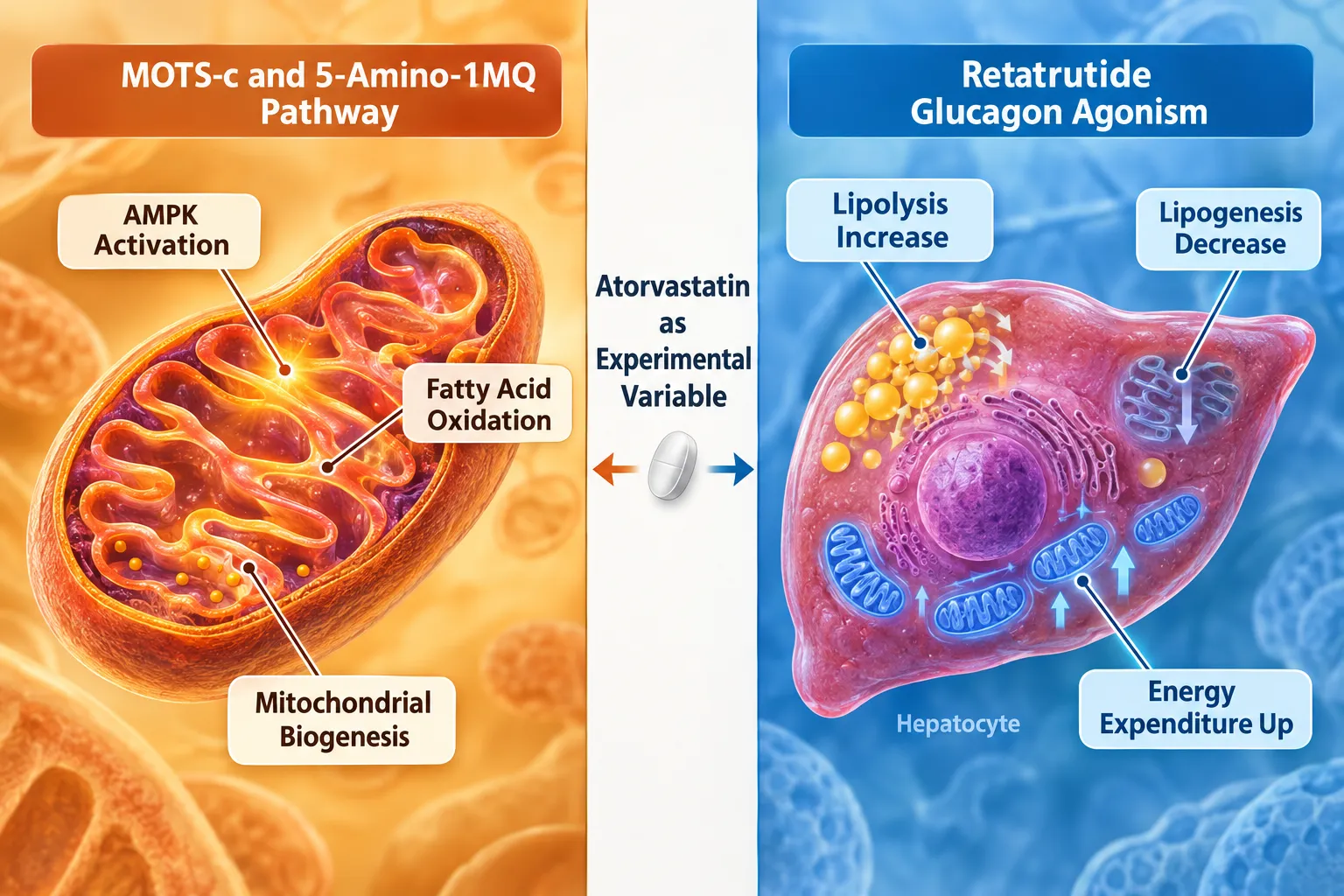
Retatrutide (LY3437943) activates GLP-1, GIP, and glucagon receptors simultaneously. Phase 2 trial data show mean weight reductions of approximately 22-24% over 48 weeks, accompanied by meaningful improvements in triglycerides, LDL cholesterol, and hepatic lipid content. Crucially, these lipid improvements arise from the peptide's own pharmacology, glucagon receptor agonism increases lipolysis and reduces lipogenesis, while GLP-1 and GIP components improve insulin sensitivity and reduce ectopic fat. When a participant is also taking atorvastatin, separating the statin's LDL effect from the peptide's LDL effect becomes statistically and mechanistically complex.

"The question is not whether atorvastatin matters, it clearly does. The question is whether the experimental design treats it as a known variable or an invisible confounder."

Researchers sourcing retatrutide peptide for preclinical work or reviewing GLP-3/retatrutide combination products should build statin status into their protocol from day one.


Experimental Design Principles for Atorvastatin in Metabolic Peptide Experiments

Experimental Design Principles for Atorvastatin in Metabolic Peptide Experiments

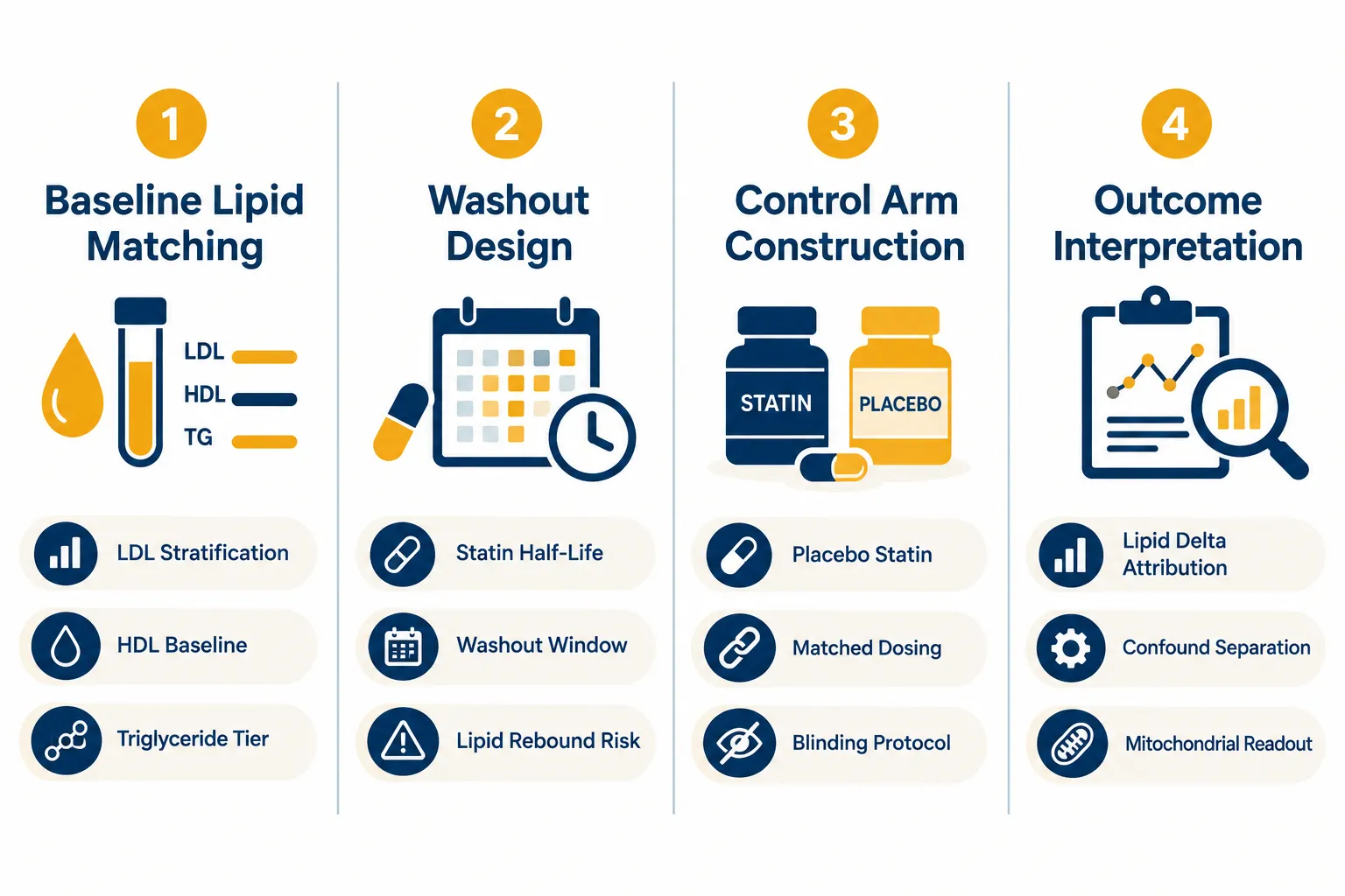
Rigorous control of atorvastatin's influence requires four interlocking design decisions.

Baseline Lipid Matching

Before randomization, participants or cell-line models should be stratified by:

  • LDL-C tier (optimal, borderline, elevated)
  • Triglyceride level (normal vs. elevated)
  • HDL-C baseline
  • Current statin dose (low, moderate, high intensity per ACC/AHA criteria)

Failure to match on these variables means that any observed lipid change could reflect pre-existing statin intensity differences rather than the peptide intervention. In retatrutide studies, where triglyceride reductions can exceed 30%, an unmatched high-intensity statin arm will produce misleading comparisons.

Washout Considerations

Atorvastatin has a plasma half-life of roughly 14 hours, but its pharmacodynamic effects on LDL receptor expression persist for several days after the last dose. A minimum two-week washout is generally recommended before lipid baseline measurements are taken in studies where statin use must be paused. However, washout carries its own risk: LDL rebound can occur within days, which may introduce a time-dependent confound if baseline measurements are taken at different points post-washout across participants.

Practical options:

Design Choice Advantage Risk
Full washout before baseline Clean lipid baseline LDL rebound, ethical concern in high-risk subjects
Stable dose maintained throughout Ethically safer Statin effect folded into all arms equally
Stratified by statin status (no washout) Preserves safety, enables subgroup analysis Requires larger sample size

Control Arm Construction

When atorvastatin cannot be washed out, as is often the case in cardiovascular-risk populations, the cleanest approach is to stratify randomization by statin use and ensure each treatment arm contains an equal proportion of statin users. This transforms atorvastatin from a confounder into a pre-specified effect modifier, allowing subgroup analyses to test whether the peptide's lipid effects differ by statin background.

Interpreting Outcomes Under Dual Therapy

In a dual-therapy scenario, the observed LDL reduction is a composite of atorvastatin's HMG-CoA blockade and retatrutide's glucagon-driven lipolysis and reduced hepatic lipogenesis. Statistical models should include statin dose as a covariate. Researchers interested in the GLP-3 peptide signaling pathway will recognize that receptor-level effects on lipid metabolism are distinct from enzyme-level inhibition, making additive versus synergistic modeling a meaningful analytical choice.


Mitochondrial Models: Where MOTS-c, 5-Amino-1MQ, and Atorvastatin Intersect

Mitochondrial Models: Where MOTS-c, 5-Amino-1MQ, and Atorvastatin Intersect

The complexity deepens considerably when mitochondrial peptides enter the experimental frame. MOTS-c is a mitochondria-derived peptide that activates AMPK, promotes fatty acid oxidation, and enhances mitochondrial biogenesis. 5-Amino-1MQ is a small-molecule inhibitor of NNMT (nicotinamide N-methyltransferase) that shifts cellular metabolism toward oxidative phosphorylation and reduces lipid accumulation. Both compounds operate on pathways that atorvastatin also touches, particularly through the coenzyme Q10 axis and mitochondrial electron transport chain dynamics.

Atorvastatin reduces endogenous coenzyme Q10 synthesis as a downstream consequence of HMG-CoA reductase inhibition. This can subtly impair mitochondrial respiratory chain efficiency, which is precisely the readout that MOTS-c and 5-Amino-1MQ experiments are designed to measure. An experiment that does not account for atorvastatin's CoQ10 effect may attribute impaired mitochondrial respiration to the peptide when the statin is the actual driver.

Key mitochondrial confounds introduced by atorvastatin:

  • Reduced CoQ10 availability in Complex I/III of the electron transport chain
  • Altered mitochondrial membrane potential in hepatocytes and myocytes
  • Possible upregulation of mitochondrial biogenesis as a compensatory response

For researchers working with SS-31 mitochondrial research models or reviewing SS-31 10mg research peptide considerations, the same logic applies: any statin in the background must be pre-specified, dose-recorded, and statistically controlled. Those exploring MOTS-c peptide for sale for metabolic research should document statin co-exposure in their experimental logs as a standard practice.

When retatrutide is layered on top of this mitochondrial model, the glucagon receptor agonism adds a third metabolic signal, increased hepatic fatty acid oxidation and energy expenditure, that further overlaps with both the peptide probes and the statin background. Isolating each compound's contribution requires:

  1. Pre-specified factorial or stratified designs
  2. Mitochondrial respiration assays (Seahorse XF or equivalent) run in statin-exposed versus statin-naive conditions
  3. CoQ10 plasma levels measured as a mechanistic covariate
  4. AMPK phosphorylation status as a shared downstream readout for MOTS-c, 5-Amino-1MQ, and glucagon agonism

Conclusion

Atorvastatin in metabolic peptide experiments, controlling lipid-lowering effects in retatrutide and mitochondrial models, is not a pharmacological afterthought. It is a design variable that, if ignored, can corrupt lipid endpoints, misattribute mitochondrial readouts, and produce irreproducible results. The actionable steps are clear:

  • Stratify by statin status at randomization, or pre-specify washout windows with lipid rebound monitoring.
  • Record atorvastatin dose intensity and include it as a covariate in all lipid and mitochondrial outcome models.
  • Measure CoQ10 levels when mitochondrial peptides such as MOTS-c, SS-31, or 5-Amino-1MQ are in the experimental design.
  • Use factorial subgroup analyses to test whether retatrutide's lipid effects differ by statin background, rather than assuming homogeneity.
  • Consult current phase 3 trial protocols as they become available; as of 2026, no completed study has formally used atorvastatin as a deliberate modulator of retatrutide's lipid effects, but the mechanistic rationale for doing so is strong.

Researchers who build these controls into their protocols from the outset will produce data that is interpretable, reproducible, and genuinely informative about the independent and combined contributions of each compound. Those who do not will spend far more time explaining away confounds than advancing the science.

https://www.puretestedpeptides.com/wp-content/uploads/2026/10/atorvastatin-in-metabolic-peptide-experiments-controlling-lipid-lowering-effects.webp 1024 1536 https://www.puretestedpeptides.com/wp-content/uploads/2026/01/buy-peptides-online.jpg 2026-10-04 13:04:572026-10-04 13:04:57Atorvastatin in Metabolic Peptide Experiments: Controlling Lipid-Lowering Effects in Retatrutide and Mitochondrial Models
Prednisone and Peptide Research Models: Separating Glucocorticoid Metabolic Effects From Tissue-Repair Signals

Prednisone and Peptide Research Models: Separating Glucocorticoid Metabolic Effects From Tissue-Repair Signals

October 4, 2026/0 Comments/in Uncategorized/by

Fewer than 15% of preclinical peptide studies formally account for background glucocorticoid exposure in their control arms, yet prednisone alone can shift glucose metabolism, body composition, and wound-healing readouts enough to invalidate a comparison. Understanding how to work with that interference is now a core competency in regenerative peptide research.

This article examines how prednisone and peptide research models interact, and provides a practical framework for deciding when steroid exposure should function as a control variable, an exclusion criterion, or a stratification factor.

Key Takeaways

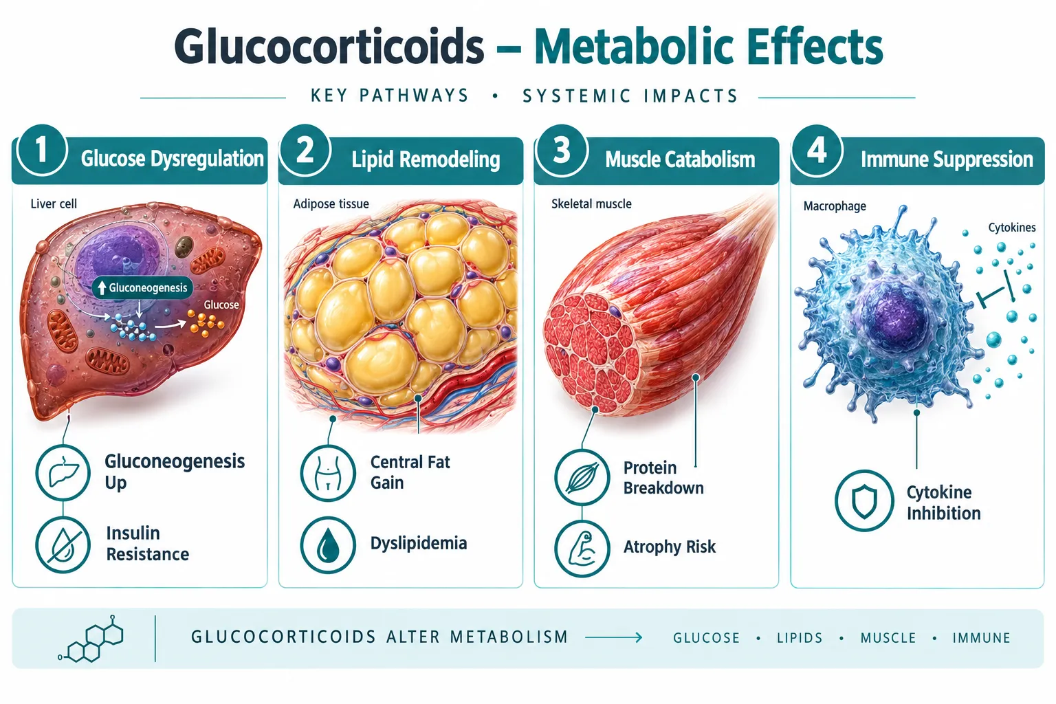
  • Prednisone exerts distinct metabolic effects (glucose dysregulation, lipid remodeling, muscle catabolism) that are mechanistically separate from its acute tissue-repair actions.
  • Peptide-based regenerative agents operate on slower, anabolic timelines, making direct head-to-head comparisons with glucocorticoids methodologically complex.
  • Pulsed or intermittent prednisone dosing can produce beneficial mitochondrial and sarcolemmal repair effects that overlap with peptide endpoints.
  • Researchers must classify steroid exposure as a control, exclusion criterion, or stratification variable before designing outcome readouts.
  • No Phase III trials currently pair peptides directly with long-term corticosteroid protocols, leaving a significant evidence gap.

How Prednisone Reshapes the Metabolic Landscape

Prednisone and peptide research models: separating glucocorticoid metabolic effects from tissue-repair signals begins with understanding what prednisone actually does to baseline physiology. Prednisone is a synthetic glucocorticoid that binds glucocorticoid receptors across virtually every tissue type. Its systemic metabolic profile is dominated by three well-documented disruptions.

How Prednisone Reshapes the Metabolic Landscape

1. Glucose and Insulin Metabolism
Prednisone stimulates hepatic gluconeogenesis and reduces peripheral insulin sensitivity. Even short-term use preferentially disrupts glucose metabolism before significant muscle protein changes appear. This matters enormously for any peptide study that uses fasting glucose, HOMA-IR, or glycemic variability as an outcome measure.

2. Lipid Remodeling and Body Composition
Prolonged prednisone use drives central adiposity and dyslipidemia. Importantly, research on intermittent weekly prednisone dosing shows sex-specific lipid remodeling in skeletal muscle, a finding that introduces a sex-as-biological-variable confound if not stratified properly.

3. Muscle Catabolism vs. Repair Duality
This is where the picture becomes nuanced. Chronic high-dose prednisone is catabolic to muscle. Yet pulsed glucocorticoid protocols in dystrophic muscle models have demonstrated enhanced physical performance through nutrient-utilization and repair pathways. Circadian timing of intermittent prednisone pulses has been shown to modulate mitochondrial remodeling, suggesting that dose, timing, and frequency determine whether the net effect is catabolic or restorative.

"The same compound that degrades muscle under chronic dosing can enhance sarcolemmal repair via annexin-mediated pathways under pulsed protocols, a distinction that most research designs fail to capture."

This duality means that labeling prednisone exposure simply as "harmful background noise" in a peptide study is an oversimplification that can distort results in either direction.


Tissue-Repair Signals: Where Peptides Operate Differently

To build a sound comparative framework, researchers must map where peptide mechanisms diverge from glucocorticoid mechanisms. For a broader foundation on peptide structure and receptor pharmacology, the Peptides 101 for Research-Use Only Buyers guide provides essential context.

Tissue-Repair Signals: Where Peptides Operate Differently

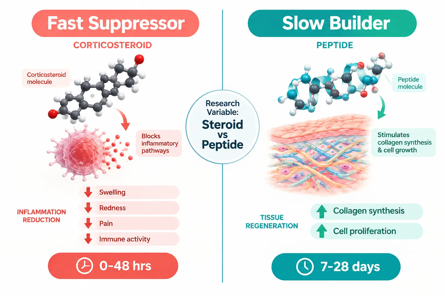
Corticosteroids act as fast suppressors: they reduce inflammatory cytokines within hours, providing rapid symptom relief. Clinical rheumatology data from 2026 confirm that prednisone reliably improves symptoms but impairs longer-term tissue healing when used continuously.

Peptides, by contrast, function as slow builders. They tend to stimulate collagen synthesis, cell proliferation, and mitochondrial biogenesis over days to weeks. Compounds such as GHK-Cu peptide have been studied for fibroblast activation and extracellular matrix remodeling, while SS-31 peptide research focuses on mitochondrial membrane stabilization, a mechanism directly relevant to steroid-induced mitochondrial disruption. For deeper reading on mitochondrial targets, SS-31 mitochondrial research offers a useful reference point.

Feature Prednisone (Pulsed) Peptide Agents
Onset of action Hours Days to weeks
Primary target Immune/inflammatory signaling Structural and metabolic repair
Mitochondrial effect Remodeling (timing-dependent) Biogenesis support
Glucose impact Dysregulation (dose-dependent) Neutral to beneficial
Evidence base Extensive Phase III data Preclinical/early Phase I-II

Peptide-based regenerative therapies remain insufficiently validated compared to glucocorticoids in head-to-head trials. No Phase III study currently pairs a peptide protocol directly with long-term corticosteroid therapy, which makes the need for rigorous study design even more critical.


A Framework for Handling Steroid Exposure in Peptide Research Models

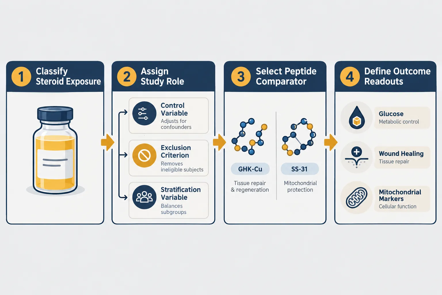
Applying the lens of prednisone and peptide research models: separating glucocorticoid metabolic effects from tissue-repair signals to study design requires a deliberate classification step before any outcome readout is chosen.

A Framework for Handling Steroid Exposure in Peptide Research Models

Researchers should ask three questions about any subject or model with prednisone exposure:

Step 1, Classify the exposure type
Is the steroid use chronic (daily, systemic), intermittent (weekly pulse), or acute (single-dose)? Each produces a different metabolic fingerprint. Intermittent once-weekly prednisone has been shown to improve ATP levels, NAD+ availability, and endurance in aged skeletal muscle, outcomes that directly overlap with peptide endpoints. Treating all steroid exposure as equivalent will produce noise, not signal.

Step 2, Assign a study role

  • Control variable: Include steroid-exposed and steroid-naive arms, measure the exposure quantitatively, and model it as a covariate. This is appropriate when excluding steroid users would make recruitment impossible.
  • Exclusion criterion: Remove subjects with any current or recent steroid use when the primary endpoint is glucose metabolism or wound healing, because prednisone's effects on those readouts are too large to model reliably.
  • Stratification variable: Randomize within steroid-exposed and steroid-naive strata when the research question specifically asks how a peptide performs in a glucocorticoid-rich environment.

Step 3, Select peptide comparators with mechanistic logic
Choose peptides whose mechanisms are least likely to be masked or amplified by glucocorticoid background. For metabolic endpoints, compounds such as MOTS-C peptide, which targets mitochondrial AMPK pathways, may show cleaner signal separation from prednisone's glucose effects than growth-hormone-releasing peptides. For tissue-repair endpoints, Thymosin Alpha 4 peptide research offers a comparator that operates through actin cytoskeletal and immune-modulation pathways distinct from glucocorticoid receptor signaling.

Dose-dependent and tissue-specific adverse metabolic effects from prednisone further underscore why a single classification rule cannot apply across all study types. A low-dose pulse in muscle tissue is not the same biological event as a high-dose chronic regimen in adipose tissue.


Conclusion

Prednisone and peptide research models: separating glucocorticoid metabolic effects from tissue-repair signals is not merely a methodological nicety, it is a prerequisite for generating reproducible data. The metabolic disruptions prednisone introduces (glucose dysregulation, lipid remodeling, context-dependent muscle effects) operate on different timescales and through different receptors than the anabolic, repair-oriented signals that most peptides are designed to amplify.

Actionable next steps for researchers in 2026:

  • Audit existing datasets for undocumented steroid co-exposure before pooling results.
  • Pre-register the steroid classification decision (control, exclusion, or stratification) in study protocols.
  • Prioritize mechanistic alignment when selecting peptide comparators, match the peptide's primary pathway to an endpoint that prednisone does not dominate.
  • Monitor both glucose and mitochondrial markers simultaneously to distinguish metabolic interference from genuine tissue-repair signal.
  • Follow emerging pulsed-dosing literature closely, as intermittent prednisone protocols may eventually serve as a validated adjunct rather than a pure confounder in select repair models.

The field will only move forward when steroid exposure is treated with the same rigor applied to any other pharmacological variable in the experimental design.

https://www.puretestedpeptides.com/wp-content/uploads/2026/10/prednisone-and-peptide-research-models-separating-glucocorticoid-metabolic-effec.webp 1024 1536 https://www.puretestedpeptides.com/wp-content/uploads/2026/01/buy-peptides-online.jpg 2026-10-04 13:04:262026-10-04 13:04:26Prednisone and Peptide Research Models: Separating Glucocorticoid Metabolic Effects From Tissue-Repair Signals
Omeprazole and Famotidine in GLP-2-T Studies: Separating Acid Suppression From Intestinal Barrier Effects

Omeprazole and Famotidine in GLP-2-T Studies: Separating Acid Suppression From Intestinal Barrier Effects

October 4, 2026/0 Comments/in Uncategorized/by

Fewer than one pH unit of difference in luminal acidity is enough to alter epithelial permeability, reshape the gut microbiome, and either mask or amplify a peptide's barrier-tightening signal. Yet in many intestinal research protocols, the choice between a proton-pump inhibitor (PPI) and an H2 receptor antagonist (H2RA) is treated as a background detail rather than a true experimental variable. In studies examining GLP-2-T, a modified form of glucagon-like peptide-2 studied for its mucosal repair and barrier-protective properties, this oversight can produce misleading readouts. Understanding how omeprazole and famotidine in GLP-2-T studies: separating acid suppression from intestinal barrier effects demands a precise comparison of two pharmacologically distinct drug classes, their divergent effects on pH, microbiota, and tight-junction biology, and a structured framework for controlling them in intestinal, epithelial, and organoid systems.

Key Takeaways

  • Omeprazole (PPI) and famotidine (H2RA) produce fundamentally different pH profiles, microbiome shifts, and off-target mucosal effects that can independently alter intestinal barrier readouts.
  • GLP-2 itself inhibits gastric acid secretion, meaning GLP-2-T studies carry an intrinsic acid-modulating signal that must be distinguished from pharmacologic acid suppression.
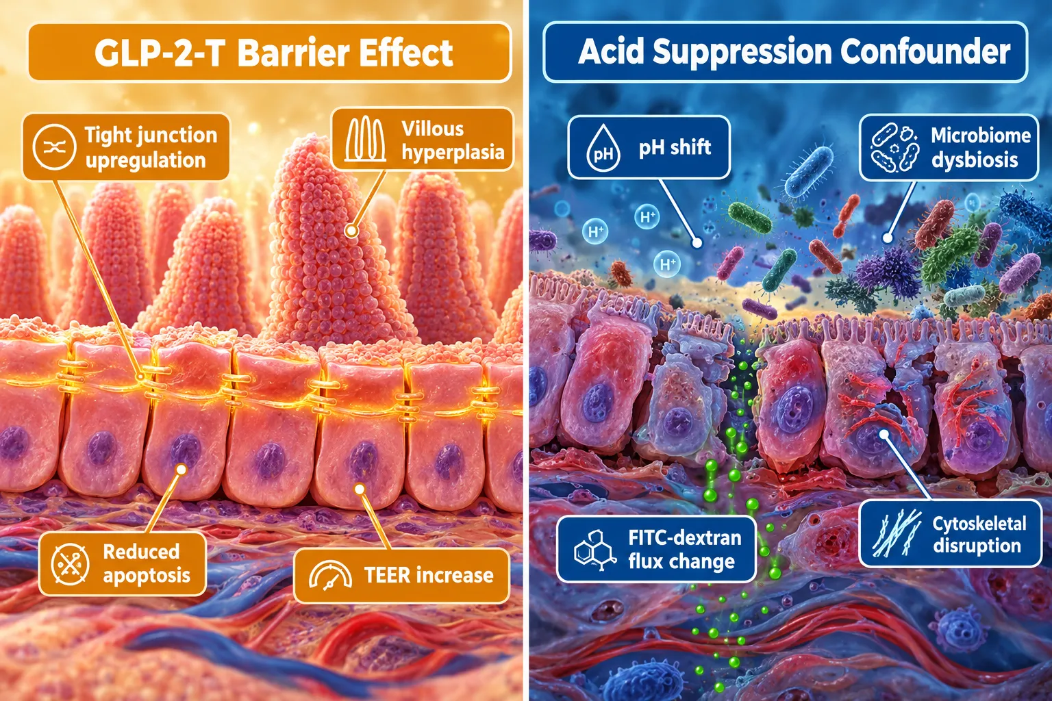
  • Transepithelial electrical resistance (TEER) and FITC-dextran flux, the two most common barrier endpoints, are pH-sensitive and require matched acid-control arms.
  • Famotidine is preferred for short-duration experiments where modest, reversible acid blunting is needed; omeprazole suits multi-day models where sustained pH clamping is the deliberate goal.
  • Treating the acid suppressor as a named experimental variable, not a background condition, is the single most impactful design change researchers can make in GLP-2-T barrier studies.

Why Acid Suppression Is Not a Neutral Background Condition

Why Acid Suppression Is Not a Neutral Background Condition

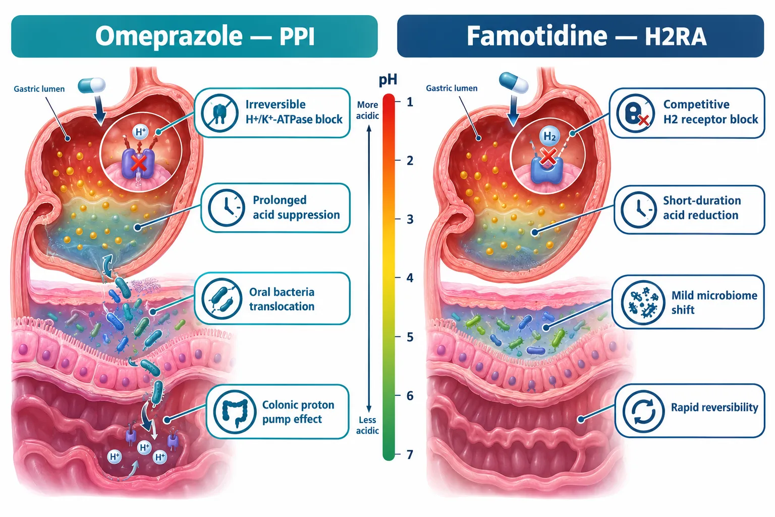
The assumption that acid-suppressive drugs simply "remove acid" and leave everything else unchanged is pharmacologically inaccurate. Omeprazole irreversibly inhibits the gastric H⁺/K⁺-ATPase, producing near-complete acid suppression for many hours even after the drug clears plasma. Famotidine competitively blocks H2 receptors, generating a shorter, milder, and fully reversible reduction in acid output.

These differences extend well beyond the stomach. Omeprazole also acts on colonic proton pumps, altering local electrolyte balance and immune tone. It induces smooth muscle relaxation and inhibits contractile activity, which can destabilize tight-junction regulation through the actin-myosin cytoskeleton, an effect entirely independent of gastric pH. In a longitudinal comparison (omeprazole 20 mg daily vs. famotidine 20 mg daily), the PPI produced significantly greater disruption of the gastric acid barrier and more pronounced translocation of oral bacteria into the lower gut, while famotidine caused milder, more localized changes. These microbiome shifts are not trivial: they alter the luminal environment in which GLP-2-T exerts its barrier effects.

Key pharmacodynamic differences:

Feature Omeprazole (PPI) Famotidine (H2RA)
Mechanism Irreversible H⁺/K⁺-ATPase inhibition Competitive H2 receptor blockade
Duration of acid suppression Prolonged (outlasts plasma half-life) Shorter, dose-dependent
Microbiome impact Greater oral-bacteria translocation Milder shifts
Off-target mucosal effects Colonic proton pump, smooth muscle Minimal beyond acid reduction
Preferred use in GLP-2-T models Multi-day sustained pH clamping Short experiments under 12 hours

For researchers sourcing compounds for controlled studies, reviewing GLP-3 RT peptide specifications alongside acid-suppressor selection is a practical starting point for protocol alignment.


GLP-2-T's Own Acid-Modulating Signal Complicates the Picture

GLP-2-T's Own Acid-Modulating Signal Complicates the Picture

A critical and often overlooked fact is that GLP-2 itself inhibits gastric acid secretion, independent of any co-administered PPI or H2RA. In controlled human infusion studies, native GLP-2 reduced pentagastrin-stimulated gastric acid secretion by roughly 15% and sham-feeding-stimulated secretion by approximately 65%. The dominant mechanism appears to be suppression of parasympathetic pathways rather than direct inhibition of parietal cell function.

This means that in any GLP-2-T experiment, the peptide is already modulating luminal acidity. Layering omeprazole or famotidine on top creates a compound acid-suppression signal whose individual components are difficult to disentangle unless the design explicitly accounts for them. When barrier endpoints such as TEER or FITC-dextran flux improve in a GLP-2-T arm that also received a PPI, the improvement could reflect:

  • Direct GLP-2-T effects on tight junctions and crypt cell proliferation
  • Reduced luminal acid damaging the epithelium (PPI effect)
  • Microbiome changes secondary to acid suppression
  • Smooth muscle relaxation altering cytoskeletal tension (omeprazole-specific)
  • Any combination of the above

"Treating the acid suppressor as a named experimental variable, not a background condition, is the single most impactful design change researchers can make in GLP-2-T barrier studies."

GLP-2's established biology reinforces this concern. The peptide stimulates crypt cell growth, promotes villous hyperplasia, reduces enterocyte apoptosis, suppresses epithelial permeability, increases intestinal blood flow, and improves nutrient absorption. These are robust, direct barrier-protective actions. But they occur alongside GLP-2-mediated reductions in gastric acid secretion and gastrointestinal motility, meaning the barrier signal is already multi-factorial before any drug is added.

For those exploring the broader landscape of GLP-related peptides, the GLP-3 peptide explained simply resource offers useful context on receptor biology. Researchers interested in sourcing reference compounds can also consult the best place to buy GLP guide for vendor evaluation criteria.


A Practical Framework for Omeprazole and Famotidine in GLP-2-T Studies: Separating Acid Suppression From Intestinal Barrier Effects

A Practical Framework for Omeprazole and Famotidine in GLP-2-T Studies: Separating Acid Suppression From Intestinal Barrier E

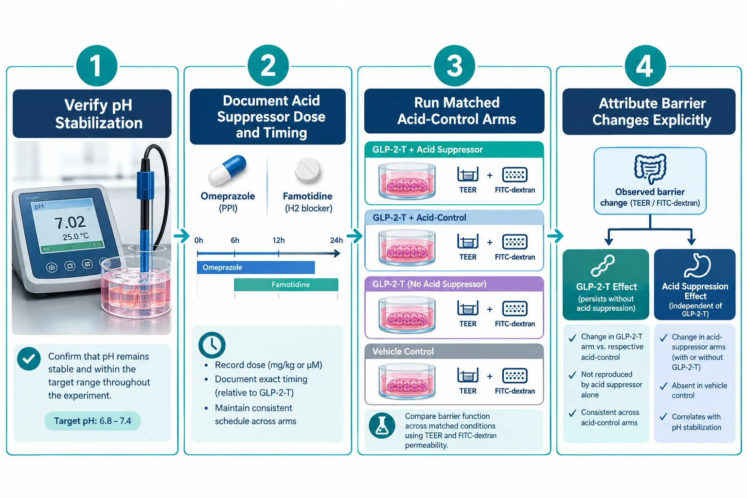
Current guidance on omeprazole and famotidine in GLP-2-T studies: separating acid suppression from intestinal barrier effects converges on four operational rules that apply equally to cell monolayer assays, organoid systems, and in vivo rodent models.

Step 1, Verify pH stabilization before introducing GLP-2-T.
Confirm that the chosen acid suppressor has reached its target pH range and that the system is stable. Introducing the peptide into a still-shifting pH environment conflates temporal acid changes with peptide-driven effects.

Step 2, Document the exact acid suppressor, dose, and time course.
This sounds obvious but is routinely omitted from methods sections. Stating "PPI was administered" without specifying omeprazole versus pantoprazole, dose, and dosing interval makes cross-study comparison impossible.

Step 3, Run matched acid-control arms.
Every GLP-2-T experiment should include:

  • A vehicle-only arm (no acid suppressor, no peptide)
  • An acid-suppressor-only arm (omeprazole or famotidine without GLP-2-T)
  • A GLP-2-T-only arm (no acid suppressor)
  • The combined arm (acid suppressor plus GLP-2-T)

This 2×2 factorial design allows direct attribution of TEER and FITC-dextran flux changes to acid suppression, GLP-2-T, or their interaction.

Step 4, Explicitly report the attribution.
Publication-ready methods must state whether observed barrier changes are attributable to acid suppression alone, GLP-2-T alone, or a synergistic interaction. In clinical management of short bowel syndrome, where both GLP-2 analogs and acid suppressors are used concurrently, this distinction is not merely academic; it informs dosing strategy.

Choosing between omeprazole and famotidine:

  • Use famotidine when the experiment runs under 12 hours, when rapid and reversible pH elevation is the goal, or when minimizing microbiome disruption is a priority.
  • Use omeprazole when the model deliberately incorporates strong, sustained acid suppression as a separate factor, for example, when studying mucosal repair in a high-acid damage model alongside GLP-2-T rescue.

This distinction also matters when comparing results against clinical data. PPIs outperform H2RAs in preventing gastroduodenal mucosal injury in high-risk patients, meaning omeprazole arms will produce an independent mucosal repair signal that must not be misread as GLP-2-T-specific barrier enhancement.

For researchers working with related peptide blends, the GLP-3R 30mg peptide GA9 and GLP3 RETA 20mg GA5 product pages provide formulation details relevant to multi-peptide experimental designs. Those examining visceral and metabolic endpoints alongside barrier function may also find the visceral fat research literature useful for contextualizing systemic readouts.


Conclusion

The core message for any laboratory working with GLP-2-T is straightforward: omeprazole and famotidine are not interchangeable background agents, they are active experimental variables with distinct pH profiles, microbiome consequences, and off-target mucosal effects that directly shape barrier readouts.

Actionable next steps for research teams:

  1. Audit existing protocols to identify whether the acid suppressor was treated as a variable or a background condition, and flag any published TEER or FITC-dextran data that lacked matched acid-control arms.
  2. Select the acid suppressor intentionally: famotidine for short, reversible, low-disruption experiments; omeprazole for sustained pH-clamping models where strong acid suppression is itself a study factor.
  3. Implement the 2×2 factorial design described above as a minimum standard for new GLP-2-T barrier studies.
  4. Account for GLP-2's intrinsic acid-suppressive effect in statistical models, it is a peptide-driven confounder that operates even in the absence of pharmacologic acid suppression.
  5. Report acid-suppressor identity, dose, and timing with the same rigor applied to the peptide itself.

Applying these principles will sharpen the signal-to-noise ratio in GLP-2-T research and produce barrier data that is genuinely attributable to the peptide rather than to its pharmacologic environment.

https://www.puretestedpeptides.com/wp-content/uploads/2026/10/omeprazole-and-famotidine-in-glp-2-t-studies-separating-acid-suppression-from-in.webp 1024 1536 https://www.puretestedpeptides.com/wp-content/uploads/2026/01/buy-peptides-online.jpg 2026-10-04 13:04:072026-10-04 13:04:07Omeprazole and Famotidine in GLP-2-T Studies: Separating Acid Suppression From Intestinal Barrier Effects
Retatrutide Phase 3 Results in 2026: How Researchers Should Interpret Weight Loss, A1C, and Cardiometabolic Endpoints

Retatrutide Phase 3 Results in 2026: How Researchers Should Interpret Weight Loss, A1C, and Cardiometabolic Endpoints

October 3, 2026/0 Comments/in Uncategorized/by

Fewer than five years after GLP-1 receptor agonists reshaped obesity medicine, a triple-receptor agonist is now producing weight loss figures that exceed nearly every benchmark set by its predecessors. Retatrutide Phase 3 Results in 2026, specifically from Eli Lilly's TRIUMPH program, have placed the research and clinical communities in the unusual position of needing new interpretive frameworks rather than simply updated numbers. Understanding what these results mean, how the trials were designed, and where the data should not be extrapolated is essential for any researcher working with this compound.

Key Takeaways

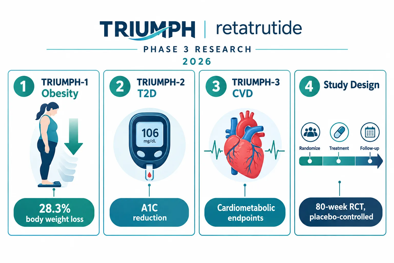
  • TRIUMPH-1 showed average body weight reductions of up to 28.3% at 80 weeks on the 12 mg dose, with roughly 45% of participants achieving at least 30% weight loss.
  • TRIUMPH-2 demonstrated meaningful A1C reductions alongside weight loss in adults with type 2 diabetes and obesity or overweight.
  • TRIUMPH-3 targeted adults with severe obesity and established cardiovascular disease, expanding the cardiometabolic endpoint dataset.
  • All three trials were 80-week, randomized, double-blind, placebo-controlled studies, setting a high methodological standard for interpretation.
  • Phase 3 clinical findings should not be directly extrapolated to research-use peptide materials without accounting for purity, formulation, and dosing differences.

Understanding the TRIUMPH Trial Architecture

The TRIUMPH program consists of three pivotal Phase 3 trials, each enrolling distinct populations to isolate different aspects of retatrutide's effects. TRIUMPH-1 enrolled adults with obesity or overweight without type 2 diabetes. TRIUMPH-2 enrolled adults who had both type 2 diabetes and obesity or overweight. TRIUMPH-3 focused on adults with severe obesity and established cardiovascular disease. Each trial ran for 80 weeks under a randomized, double-blind, placebo-controlled design, the gold standard for regulatory-grade evidence.

Understanding the TRIUMPH Trial Architecture

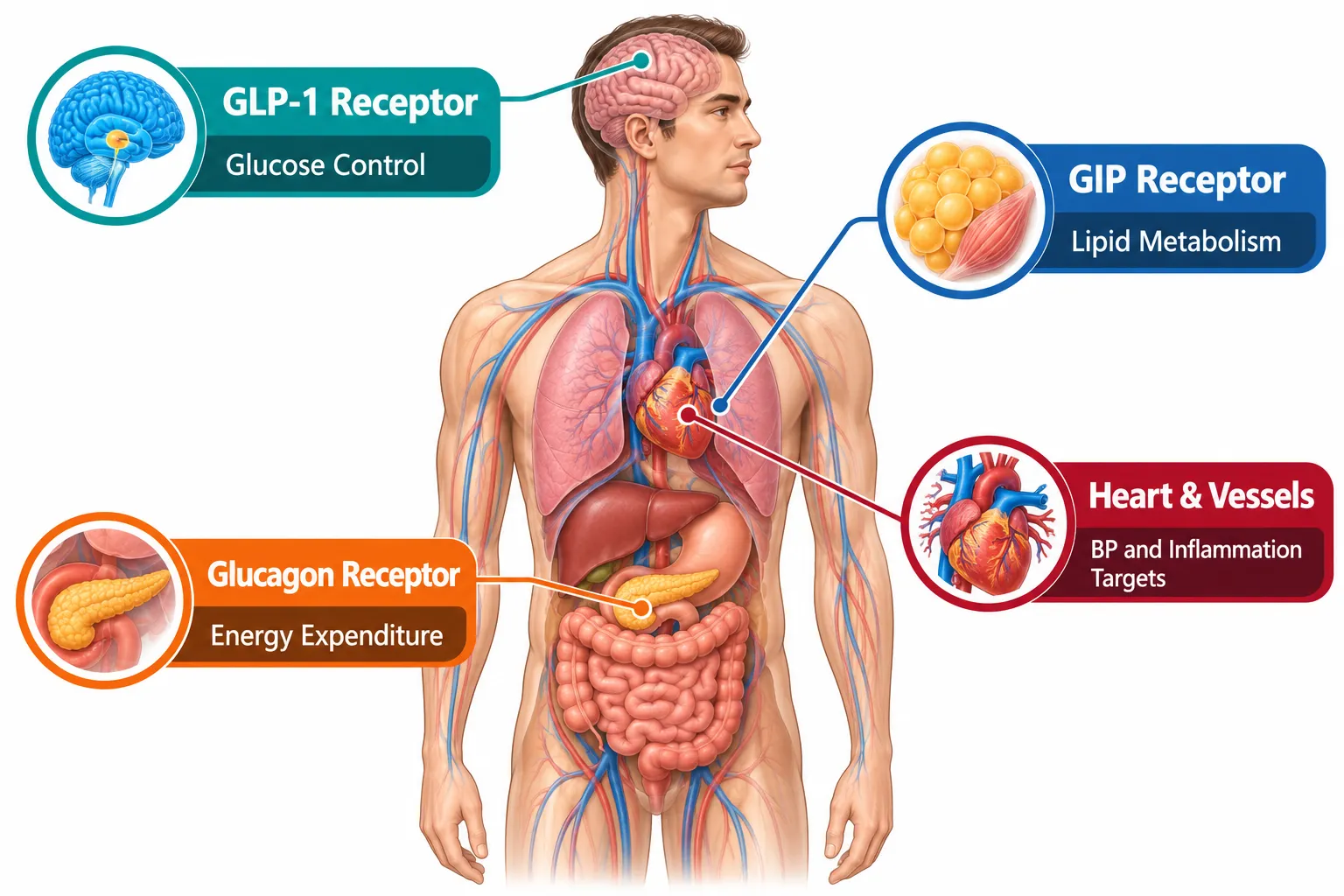
Retatrutide itself is a triple agonist acting simultaneously on GLP-1, GIP, and glucagon receptors. This mechanism distinguishes it from dual agonists like tirzepatide and single-receptor agents like semaglutide. Researchers reviewing the retatrutide clinical trial literature should note that the multi-receptor activity complicates direct comparisons with earlier compounds, since each receptor pathway contributes differently to energy expenditure, appetite suppression, and glucose metabolism.

Key dose arms tested across TRIUMPH trials:

Dose Trial Inclusion Primary Role
4 mg once weekly TRIUMPH-1, TRIUMPH-2 Lower-dose comparator
8 mg once weekly TRIUMPH-1, TRIUMPH-2 Mid-range efficacy arm
9 mg once weekly TRIUMPH-1 High-dose efficacy arm
12 mg once weekly TRIUMPH-1, TRIUMPH-2, TRIUMPH-3 Maximum dose efficacy arm

Interpreting Weight Loss Endpoints from Retatrutide Phase 3 Results in 2026

TRIUMPH-1 produced the most widely cited weight loss figures. Participants on the 9 mg dose lost an average of 64.4 lb, representing 25.9% of baseline body weight. Those on the 12 mg dose lost an average of 70.3 lb, or 28.3% of baseline weight. The placebo group lost approximately 2.2%. Approximately 45% of the roughly 2,500 participants achieved at least 30% weight loss, a threshold that had rarely appeared in prior obesity pharmacotherapy trials.

An extension to 104 weeks showed that participants with a BMI of 35 kg/m² or higher on the highest dose lost approximately 30.3% of body weight on average, suggesting the effect is durable rather than plateauing early.

"A 28% average weight reduction in a randomized controlled trial is not a ceiling, it is a new baseline expectation for next-generation obesity pharmacotherapy."

Researchers reviewing these figures alongside retatrutide endpoints data should apply several interpretive cautions:

  • Responder heterogeneity: Mean weight loss masks a wide distribution. Some participants lost far less; others exceeded 35%.
  • Baseline BMI matters: Higher baseline BMI was associated with greater absolute weight loss but not always greater percentage loss.
  • Titration schedules: The dose escalation protocol used in TRIUMPH trials is specific and structured. Rapid titration in research settings produces different tolerability profiles.
  • Placebo subtraction: The net treatment effect, not the absolute loss, is the relevant efficacy signal for mechanistic research.

A1C, Glycemic Control, and Cardiometabolic Endpoints: What the Data Show

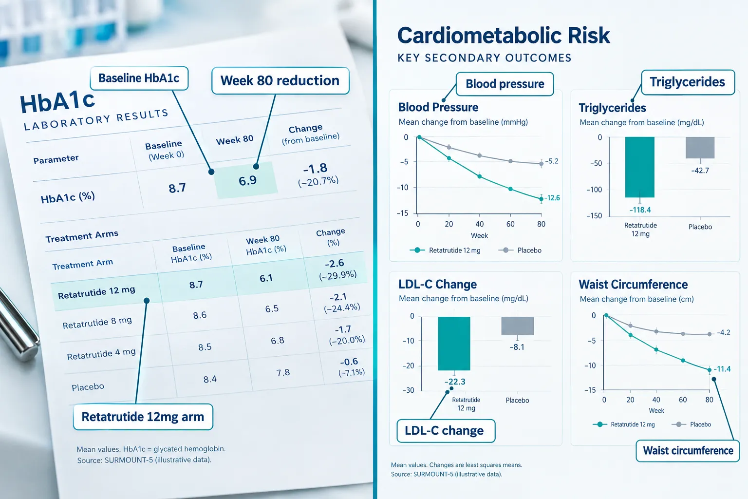
TRIUMPH-2 specifically addressed glycemic outcomes in adults with type 2 diabetes. Retatrutide produced substantial A1C reductions across all active dose arms, with the 12 mg arm achieving the largest decreases from baseline. These reductions were accompanied by meaningful weight loss, making it difficult to isolate the glycemic effect of the receptor agonism from the indirect benefit of fat mass reduction, a distinction that matters significantly for mechanistic researchers.

A1C, Glycemic Control, and Cardiometabolic Endpoints: What the Data Show

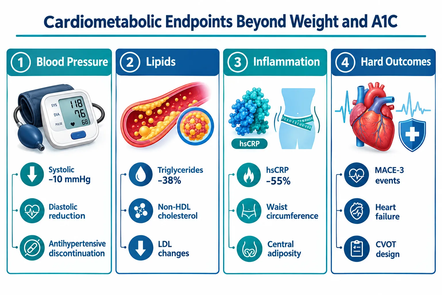
TRIUMPH-3 extended the dataset to cardiometabolic markers in a higher-risk population. Reported endpoints included changes in blood pressure, triglycerides, LDL-C, and waist circumference. While full cardiovascular outcomes data from a dedicated MACE trial are still pending as of 2026, the surrogate marker improvements observed in TRIUMPH-3 are consistent with the metabolic effects predicted by the triple-receptor mechanism.

Cardiometabolic endpoints researchers should track:

  • Systolic and diastolic blood pressure reduction
  • Fasting triglyceride change from baseline
  • HDL-C and LDL-C shifts
  • Waist circumference as a visceral adiposity proxy
  • Liver enzyme normalization in participants with metabolic-associated steatotic liver disease

How Researchers Should Avoid Misextrapolating Phase 3 Findings

Retatrutide Phase 3 Results in 2026 represent pharmaceutical-grade, GMP-manufactured material administered under controlled clinical conditions. Research-use peptide materials, such as those available as GLP-3 Reta 30mg or GLP-3 Reta 20mg for laboratory use, differ in formulation, excipient profile, and intended application. Researchers sourcing material should consult verified retatrutide peptide buy online resources that provide third-party purity documentation.

How Researchers Should Avoid Misextrapolating Phase 3 Findings

The following extrapolation errors are most common in the literature:

  1. Assuming equivalent bioavailability between pharmaceutical formulations and research-grade lyophilized peptides
  2. Applying TRIUMPH dose arms directly to non-human or in vitro research models without allometric scaling
  3. Conflating A1C reduction with direct receptor-mediated glycemic control when weight loss itself accounts for a significant portion of the glycemic benefit
  4. Ignoring the titration variable, TRIUMPH participants were dose-escalated over weeks, not started at maximum dose
  5. Overstating cardiovascular conclusions before dedicated MACE trial data are published

For researchers comparing retatrutide to other metabolic peptides, reviewing Tesamorelin dosage for fat loss frameworks offers a useful methodological contrast, since Tesamorelin's visceral fat reduction mechanism and endpoint structure differ substantially from triple-receptor agonism.


Conclusion

The Retatrutide Phase 3 Results in 2026 represent a landmark dataset for obesity, diabetes, and cardiometabolic research. The TRIUMPH program's three-trial architecture provides layered evidence across distinct populations, dose arms, and outcome categories. For researchers, the actionable steps are clear:

  • Treat the 80-week, placebo-controlled TRIUMPH data as the interpretive anchor for all mechanistic hypotheses involving retatrutide.
  • Distinguish between mean efficacy signals and responder distributions before drawing mechanistic conclusions.
  • Separate glycemic benefits attributable to weight loss from those attributable to direct receptor agonism when designing downstream studies.
  • Source research-grade material only from suppliers providing verified purity documentation, and never apply clinical dose arms directly to preclinical models without appropriate scaling.
  • Monitor the emerging MACE trial data before drawing cardiovascular outcome conclusions from surrogate markers alone.

The TRIUMPH results do not simply update a number, they reframe what metabolic pharmacotherapy can achieve and raise the methodological bar for how researchers must interpret, contextualize, and apply the data responsibly.

https://www.puretestedpeptides.com/wp-content/uploads/2026/10/retatrutide-phase-3-results-in-2026-how-researchers-should-interpret-weight-loss.webp 1024 1536 https://www.puretestedpeptides.com/wp-content/uploads/2026/01/buy-peptides-online.jpg 2026-10-03 13:08:402026-10-03 13:08:40Retatrutide Phase 3 Results in 2026: How Researchers Should Interpret Weight Loss, A1C, and Cardiometabolic Endpoints
Retatrutide Dose-Escalation Research: Comparing Exposure, Tolerability, and Endpoint Timing Across Study Designs

Retatrutide Dose-Escalation Research: Comparing Exposure, Tolerability, and Endpoint Timing Across Study Designs

October 3, 2026/0 Comments/in Uncategorized/by

Roughly 26% of adults worldwide meet clinical criteria for obesity, yet fewer than half of those enrolled in weight-loss drug trials complete the full escalation phase without a dose reduction or dropout. That gap between protocol intent and real-world completion sits at the center of retatrutide dose-escalation research: comparing exposure, tolerability, and endpoint timing across study designs, and it is now driving a new generation of deliberately structured trials.

Key Takeaways

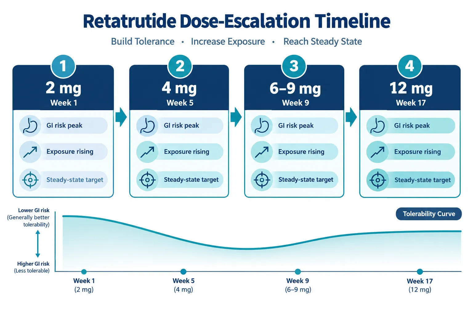
  • Retatrutide is titrated stepwise from 2 mg once weekly to target doses of 4, 9, or 12 mg, with escalation lasting approximately 16 weeks before a prolonged maintenance phase.
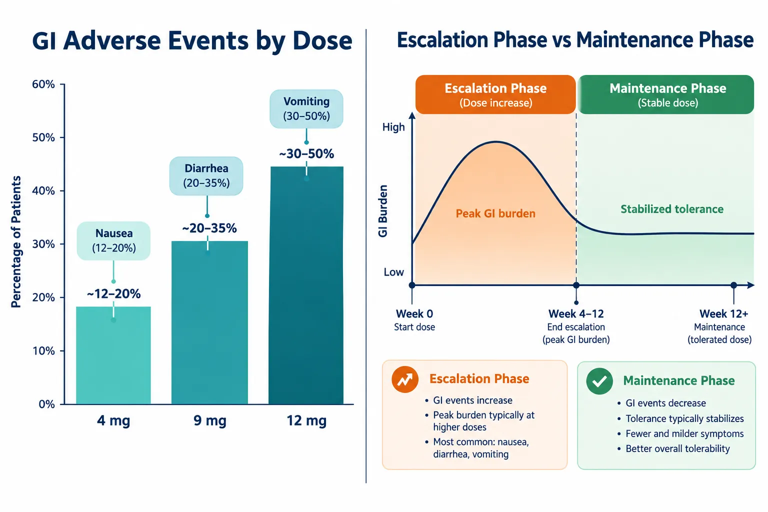
  • Gastrointestinal adverse events, nausea, diarrhea, and vomiting, are clearly dose-dependent and concentrate during the escalation phase rather than maintenance.
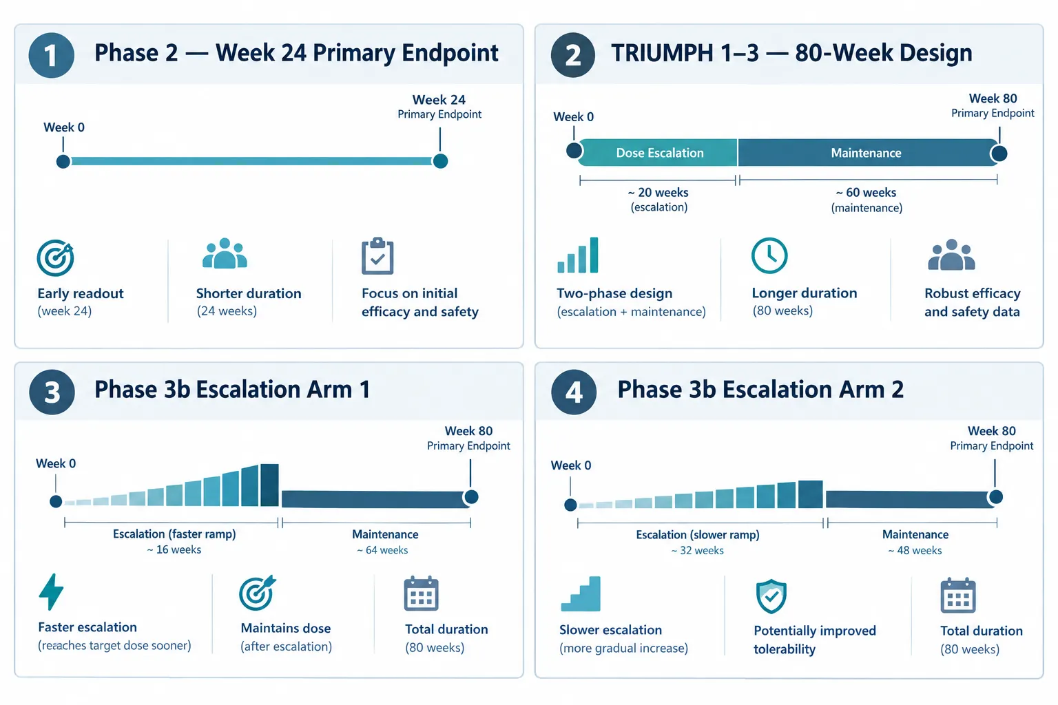
  • A dedicated phase 3b trial is directly comparing alternative escalation schedules to determine whether slower titration improves the efficacy-tolerability balance without sacrificing weight-loss outcomes.
  • Endpoint timing across studies is deliberately staggered: Week 24 captures early escalation impact, while 80-week designs assess long-term durability at steady-state exposure.
  • Emerging non-GI signals, including dysesthesia at the highest doses, are beginning to shape thinking about maximum recommended doses and future flexible titration labels.

How Escalation Schedules Shape Drug Exposure

The pharmacological foundation of any retatrutide dose-escalation study rests on a straightforward principle: the rate at which a participant reaches their target dose directly determines how quickly and how intensely the body is exposed to the drug's full activity. Retatrutide is a triple GIP, GLP-1, and glucagon receptor agonist, and its cardiometabolic mechanism differs meaningfully from classic small-molecule drugs.

How Escalation Schedules Shape Drug Exposure

In the TRIUMPH program trials, the standard titration schedule moves participants from 2 mg once weekly to their assigned maintenance dose in four-weekly steps. The 4 mg arm requires only one step up (2 mg to 4 mg). The 9 mg arm moves through 2, 4, 6, and 9 mg over approximately 12 weeks. The 12 mg arm adds one further step to reach the ceiling dose, with escalation spanning roughly 16 weeks in total before a 64-week maintenance period begins.

This design has an important implication for exposure measurement. Participants spend a relatively short time in the escalation phase and a much longer time at steady-state. Any pharmacokinetic or pharmacodynamic endpoint measured early in the study reflects a rapidly changing exposure environment, while endpoints measured later reflect stable, high-exposure conditions. Comparing results across studies that use different escalation speeds therefore requires careful attention to when in the titration curve each measurement was taken.

Key exposure variables across escalation designs:

Design Feature Faster Escalation Slower Escalation
Time to target dose Shorter Longer
Peak early exposure Higher Lower
GI adverse event timing Earlier, more intense Spread over more weeks
Steady-state onset Earlier Delayed
Week 24 endpoint context Near or at maintenance Still escalating

Tolerability Data Across Dose Levels and Titration Speeds

The tolerability profile of retatrutide is one of the most closely tracked variables in current dose-escalation research. Across TRIUMPH program data, nausea was reported in approximately 16%, 20%, and 27% of participants at 4, 9, and 12 mg respectively, compared with under 4% on placebo. Diarrhea followed a similar gradient, rising from roughly 19% at 4 mg to 23% at 12 mg. Vomiting appeared in 16% to 18% of actively treated participants across dose levels.

Tolerability Data Across Dose Levels and Titration Speeds

At the highest tested doses, pooled safety analyses report nausea in up to 43% of participants, diarrhea in approximately 33%, and vomiting in around 21%. These figures are substantial, but discontinuation rates remained relatively low, 2.2% at 4 mg, 4.5% at 9 mg, and 5.1% at 12 mg, suggesting that most participants tolerate the burden, particularly when escalation is gradual.

The phase 2 obesity trial published in the New England Journal of Medicine provided early evidence that starting at 2 mg or 4 mg and increasing every four weeks meaningfully reduced GI symptom severity compared with more aggressive approaches. This finding now underpins the entire TRIUMPH escalation framework.

"Gastrointestinal adverse events are not only dose-related but also mitigated by starting at a lower dose and escalating gradually, a principle that is now being tested head-to-head in dedicated escalation-comparison trials."

Beyond GI symptoms, emerging data from TRIUMPH-4 topline results identified dysesthesia, abnormal skin sensations, in approximately 21% of participants at the highest dose. While GI events remain the primary tolerability concern, this non-GI signal is beginning to inform discussions about maximum recommended doses and the design of future long-duration studies.


Endpoint Timing and Study Design Across Current Trials

The most direct test of retatrutide dose-escalation research: comparing exposure, tolerability, and endpoint timing across study designs is now underway in a phase 3b trial registered in June 2026. This randomized, double-blind, parallel-assignment study includes at least two experimental arms, labeled "Retatrutide Dose Escalation 1" and "Retatrutide Dose Escalation 2", specifically designed to determine whether alternative titration schedules can shift the efficacy-tolerability balance without reducing the final maintenance dose.

Endpoint Timing and Study Design Across Current Trials

A multicenter phase 2 study adds further granularity. Participants are randomized to maintenance doses of 1, 4, 8, and 12 mg, but the 4 mg and 8 mg arms each include alternative escalation schedules to isolate the effect of titration speed from the effect of final dose. The primary endpoint, percent body weight change at Week 24, is strategically placed at a point when some participants will have just reached maintenance while others, on slower schedules, may still be escalating. This deliberate misalignment is not a flaw; it is the research question.

Why endpoint timing matters for interpretation:

  • Week 24 endpoints capture the combined effect of escalation intensity and early maintenance exposure, making them sensitive to titration speed differences.
  • Week 48 assessments provide extended anthropometric and safety data that reflect more stable exposure conditions.
  • 80-week TRIUMPH designs (16 weeks escalation plus 64 weeks maintenance) are built to evaluate long-term durability, metabolic improvements, and safety at sustained high-dose exposure.

This staggered endpoint strategy allows researchers and regulators to distinguish between the short-term impact of how fast a patient escalates and the long-term consequences of sustained high-dose therapy, two questions that earlier single-arm trials could not cleanly separate.

For context on how other research peptides approach dosing and timing considerations, the literature on SS-31 ideal dosage and timing illustrates how titration decisions affect both tolerability and endpoint interpretation across different compound classes. Similarly, CJC-1295 pharmacokinetic comparison research demonstrates how half-life and exposure curves shape study design choices more broadly.

Researchers interested in the broader RETA 10mg research context or the GLP3-RETA 20mg formulation will find that these product-level considerations connect directly to the dose-level questions being tested in current escalation trials.


Conclusion

Retatrutide dose-escalation research: comparing exposure, tolerability, and endpoint timing across study designs has moved from a secondary consideration to the central question of the current trial generation. The evidence is clear that slower titration reduces early GI burden without eliminating it, that adverse events are dose-dependent and concentrated in the escalation phase, and that endpoint timing must be interpreted relative to where each participant sits on their titration curve.

Actionable next steps for researchers and clinicians following this space:

  1. Track the phase 3b escalation-comparison trial results, which will provide the first head-to-head data on alternative titration schedules at identical maintenance doses.
  2. When reviewing published trial data, note the escalation schedule used and the timing of primary endpoints relative to when participants reached maintenance dose.
  3. Anticipate that future regulatory labels for retatrutide at 9-12 mg may embed flexible titration options rather than a single fixed schedule, based on the tolerability evidence accumulating in 2026 trials.
  4. Consider non-GI signals, particularly dysesthesia at the highest doses, as part of the full exposure-tolerability profile when evaluating study designs.

The field is moving toward individualized titration, not as a clinical convenience, but as a scientifically validated strategy supported by rigorously designed comparative trials.

https://www.puretestedpeptides.com/wp-content/uploads/2026/10/retatrutide-dose-escalation-research-comparing-exposure-tolerability-and-endpoin.webp 1024 1536 https://www.puretestedpeptides.com/wp-content/uploads/2026/01/buy-peptides-online.jpg 2026-10-03 13:08:342026-10-03 13:08:34Retatrutide Dose-Escalation Research: Comparing Exposure, Tolerability, and Endpoint Timing Across Study Designs
Retatrutide and Cardiovascular Disease Research: Designing Endpoints Beyond Weight Loss and A1C

Retatrutide and Cardiovascular Disease Research: Designing Endpoints Beyond Weight Loss and A1C

October 3, 2026/0 Comments/in Uncategorized/by

"

Professional () hero image with 'Retatrutide and Cardiovascular Disease' in crisp white on a deep navy semi-transparent

Cardiovascular disease kills more people with obesity and type 2 diabetes than any other complication, yet most obesity drug trials still define success almost entirely by the number on a scale or a single blood sugar marker. Retatrutide and cardiovascular disease research is now forcing a rethink of that narrow framework, pushing investigators to design endpoints that capture blood pressure, lipid profiles, inflammatory biomarkers, and hard cardiovascular events alongside weight and A1C.

Key Takeaways

  • Retatrutide, a GLP-1/GIP/glucagon triple agonist, produces striking reductions in blood pressure, triglycerides, waist circumference, and high-sensitivity C-reactive protein (hsCRP), signals that go well beyond weight loss and glycemic control.
  • Phase 3 TRIUMPH-3 data showed roughly 22-25% weight reduction at 12 mg, with systolic blood pressure falling up to 10.8 mmHg and hsCRP dropping up to 58%, but cardiovascular event rates did not reach statistical significance.
  • A dedicated cardiovascular outcomes trial (CVOT) is underway, designed specifically to evaluate major adverse cardiovascular events (MACE) in high-risk populations.
  • Distinguishing exploratory cardiometabolic signals from validated cardiovascular-outcome evidence is essential for sound protocol planning and regulatory strategy.
  • Future endpoint frameworks will likely use integrated cardiometabolic composites, covering lipids, inflammation, central adiposity, and organ-specific outcomes, rather than relying on weight and A1C alone.

Why Triple-Agonist Biology Demands Broader Endpoint Thinking

Why Triple-Agonist Biology Demands Broader Endpoint Thinking

Retatrutide simultaneously activates GLP-1, GIP, and glucagon receptors, a pharmacological profile that creates metabolic effects across multiple organ systems at once. This broad reach is precisely why retatrutide clinical trials have moved quickly to incorporate cardiometabolic measures as prespecified exploratory endpoints rather than afterthoughts.

In the pivotal phase 2 obesity trial published in the New England Journal of Medicine, once-weekly retatrutide produced a mean weight reduction of 24.2% at 48 weeks at higher doses. Alongside that headline figure, investigators tracked systolic and diastolic blood pressure, waist circumference, fasting glucose, insulin, glycated hemoglobin, and most lipid parameters. That multi-factor design reflected an early recognition that glucagon receptor agonism affects hepatic lipid metabolism and energy expenditure in ways that GLP-1 alone does not.

A 2024 meta-analysis confirmed statistically significant reductions in systolic blood pressure (pooled weighted mean difference of approximately -4.70 mmHg) and diastolic blood pressure (-1.73 mmHg) across retatrutide studies. Complementary Lancet data showed systolic blood pressure falling roughly 5 mmHg with active doses versus about 1.5 mmHg on placebo over 40 weeks. These consistent signals across studies support blood pressure as a reliable intermediate endpoint, one that is biologically plausible, measurable, and clinically meaningful.

Protocol planning note: Blood pressure and lipid changes are reproducible across retatrutide studies, making them strong candidates for secondary or co-primary endpoints in future trials. They should not be treated as incidental observations.

Researchers designing studies around GLP-1 Reta compounds increasingly recognize that a single surrogate endpoint misrepresents the drug's full cardiometabolic footprint.


Cardiometabolic Endpoint Design: From Signals to Evidence

Cardiometabolic Endpoint Design: From Signals to Evidence

The TRIUMPH-3 phase 3 trial, which enrolled nearly 2,000 adults with severe obesity (BMI 35 or higher) and established cardiovascular disease, generated the most comprehensive cardiometabolic endpoint data to date. Results released in late September 2026 showed the following at the highest 12 mg dose:

Endpoint Approximate Change vs. Placebo
Systolic blood pressure -9.3 to -10.8 mmHg
Triglycerides -37 to -39.5%
Non-HDL cholesterol -16.5 to -19.6%
Waist circumference -16.9 to -19.0 cm
hsCRP -51 to -58%

These figures represent a multi-dimensional risk-modification profile that no single endpoint could capture. The hsCRP reduction in particular is notable: chronic low-grade inflammation is an independent cardiovascular risk factor, and a 51-58% drop in this marker suggests retatrutide may be modifying vascular biology beyond what weight loss alone would explain.

Equally important is the medication-burden signal. In the NEJM phase 2 trial, 41% of patients in the 8 mg group and 30% in the 12 mg group were able to discontinue at least one antihypertensive medication. This is a clinically relevant endpoint that regulatory agencies and payers increasingly value, it reflects real-world health impact, not just a surrogate number.

However, a critical distinction must be made. Despite these striking risk-factor improvements, TRIUMPH-3 did not show statistically significant reductions in major cardiovascular events. There were 44 MACE-5 events in the retatrutide group versus 52 in the placebo group, yielding a hazard ratio of 0.82 (95% CI 0.55-1.22). The MACE-3 hazard ratio was 1.12 (95% CI 0.64-1.96). The trial was not powered to detect event-level differences, and event counts were lower than projected. This is the essential gap between exploratory cardiometabolic signals and validated cardiovascular-outcome evidence.

For researchers working with related peptide frameworks such as SS-31 mitochondrial research or tesa dosage for fat loss protocols, this distinction between surrogate improvements and hard event outcomes is a recurring methodological challenge across the field.


Designing the Next Generation of Retatrutide Cardiovascular Endpoints

Designing the Next Generation of Retatrutide Cardiovascular Endpoints

A dedicated CVOT for retatrutide is now underway, targeting adults with overweight or obesity and established cardiovascular disease. This trial is explicitly designed to answer the question that TRIUMPH-3 could not: does retatrutide reduce major adverse cardiovascular events at a statistically powered level?

Recommended endpoint architecture for future retatrutide CVOTs:

  • Primary endpoint: MACE-3 (cardiovascular death, non-fatal myocardial infarction, non-fatal stroke) as the regulatory standard
  • Key secondary endpoints: Heart failure hospitalizations, kidney function trajectories, all-cause mortality
  • Exploratory biomarker endpoints: hsCRP, NT-proBNP, albuminuria, imaging-based myocardial structure measures
  • Safety hemodynamic monitoring: Pulse rate, arrhythmia surveillance, autonomic function, early diabetes studies showed increased pulse rate despite lower blood pressure, underscoring the need for comprehensive hemodynamic tracking

The inflammatory marker data from TRIUMPH-3 strengthens the case for including hsCRP and other inflammatory endpoints as prespecified secondary measures. A 51-58% reduction in hsCRP is not a minor signal; it warrants formal hypothesis testing in a powered trial.

Renal outcomes deserve explicit attention given the overlap between obesity, type 2 diabetes, and chronic kidney disease in high-risk populations. Albuminuria and estimated glomerular filtration rate trajectories should be incorporated as secondary endpoints, consistent with the approach taken in GLP-1 CVOT designs. For context on how related peptide research approaches multi-system endpoints, the GLP3 Reta compound research framework offers a useful parallel.

Lilly's March 2026 release on retatrutide in people with obesity and type 2 diabetes explicitly framed non-HDL cholesterol, triglyceride, and blood pressure improvements as key differentiating endpoints alongside glycemic control, signaling that the company's own endpoint strategy has already moved beyond weight and A1C.


Conclusion

Retatrutide and cardiovascular disease research is at a pivotal inflection point in 2026. The cardiometabolic signal is strong: blood pressure reductions of up to 10.8 mmHg, triglyceride drops approaching 40%, hsCRP reductions exceeding 50%, and clinically meaningful antihypertensive medication discontinuation rates all point to a drug with broad cardiovascular risk-modification potential.

What the evidence does not yet support is a validated claim of cardiovascular event reduction. TRIUMPH-3 was not powered for that conclusion, and the ongoing CVOT must deliver that answer before a formal cardiovascular risk-reduction indication can be pursued.

Actionable next steps for protocol designers and researchers:

  1. Build composite cardiometabolic endpoints into study designs from the start, do not treat blood pressure, lipids, and inflammation as secondary afterthoughts.
  2. Power dedicated CVOTs specifically for MACE, not just for weight loss, and include heart failure and renal outcomes as key secondary endpoints.
  3. Monitor hemodynamic safety comprehensively, including pulse rate and arrhythmia surveillance, given the mixed blood pressure and heart rate signals seen in early studies.
  4. Use inflammatory biomarkers such as hsCRP as prespecified exploratory endpoints to build the evidence base for future mechanistic claims.
  5. Follow retatrutide clinical trials progress closely, as CVOT results will determine whether the robust risk-factor improvements seen to date translate into the event-level cardiovascular benefit that patients and regulators require.

The science is compelling. The endpoint design must now match the ambition of the biology.

https://www.puretestedpeptides.com/wp-content/uploads/2026/10/retatrutide-and-cardiovascular-disease-research-designing-endpoints-beyond-weigh.webp 1024 1536 https://www.puretestedpeptides.com/wp-content/uploads/2026/01/buy-peptides-online.jpg 2026-10-03 13:04:482026-10-03 13:04:48Retatrutide and Cardiovascular Disease Research: Designing Endpoints Beyond Weight Loss and A1C
How to Read Hemoglobin, Hematocrit, and MCHC Results in Peptide Research Studies

How to Read Hemoglobin, Hematocrit, and MCHC Results in Peptide Research Studies

October 3, 2026/0 Comments/in Uncategorized/by

A single out-of-range CBC value can derail an entire peptide research protocol, yet most of those flags are artifacts, not true biological signals. Knowing how to read hemoglobin, hematocrit, and MCHC results in peptide research studies is therefore one of the most practical skills a researcher can develop. These three indices are tightly interconnected, and misreading one in isolation leads to faulty conclusions about safety and efficacy.

Key Takeaways

  • Hemoglobin (Hb), hematocrit (Hct), and MCHC are mathematically linked; changes in one almost always shift the others.
  • MCHC is primarily a quality-control flag, a value above 36 g/dL often signals a laboratory artifact rather than a true physiological change.
  • Hct thresholds (typically above 54% in males or 48% in females) serve as intervention triggers in peptide research monitoring protocols.
  • No single index should be interpreted in isolation; always cross-reference with MCV, RDW, and reticulocyte count.
  • Monitoring cadence matters: baseline, four-week, and twelve-week CBC panels are the current consensus practice for most peptide study designs.

Understanding the Three Core Red-Cell Indices

Understanding the Three Core Red-Cell Indices

Hemoglobin (Hb) is the iron-containing protein inside red blood cells that carries oxygen. It is measured in grams per deciliter (g/dL). Standard reference ranges are approximately 13.5-17.5 g/dL for adult males and 12.0-15.5 g/dL for adult females, though laboratory-specific ranges vary slightly.

Hematocrit (Hct) expresses the proportion of whole blood occupied by red blood cells, reported as a percentage. Normal values run roughly 41-53% for males and 36-46% for females. Because Hct is essentially three times the Hb value in healthy subjects, a large discrepancy between the two is itself a diagnostic clue.

MCHC (Mean Corpuscular Hemoglobin Concentration) measures the average concentration of hemoglobin within a given volume of packed red cells. It is calculated as:

MCHC (g/dL) = (Hb / Hct) x 100

Normal MCHC falls between 32 and 36 g/dL. Because hemoglobin physically cannot exceed its solubility limit inside a red cell, a result above 36-37 g/dL almost always indicates a pre-analytical error, lipemia, hemolysis, or a cold agglutinin, rather than a genuine finding.

Why These Three Indices Are Inseparable

The mathematical relationship among Hb, Hct, and MCHC means they must be read as a set. If Hb rises due to hemoconcentration during a peptide protocol, Hct will rise proportionally and MCHC will remain stable. If MCHC spikes while Hb and Hct stay flat, the sample is almost certainly compromised.


A Practical Workflow for Reading CBC Results in Peptide Studies

A Practical Workflow for Reading CBC Results in Peptide Studies

Researchers working with compounds such as SS-31 peptide or Ipamorelin peptide for sale formulations routinely collect complete blood counts (CBCs) to monitor hematological safety. The following six-step workflow reflects 2026 consensus practice.

Step 1, Establish a true baseline. Collect a CBC before any compound is introduced. A single pre-study value is insufficient; two baseline draws separated by at least one week reduce intra-individual variability noise.

Step 2, Evaluate Hb against sex-specific reference ranges. Flag values more than 1.5 g/dL above the upper reference limit. In peptide studies involving erythropoietic compounds, rising Hb is the earliest signal of red-cell mass expansion.

Step 3, Apply Hct thresholds as intervention triggers. Current guidance treats Hct above 54% (males) or 48% (females) as a threshold requiring protocol pause and clinical review. These numbers align with cardiovascular risk data from polycythemia research.

Step 4, Use MCHC as a quality-control gate, not a primary endpoint. Before interpreting any MCHC shift, confirm sample integrity. An MCHC above 36.5 g/dL should trigger a repeat draw before any protocol decision is made.

Step 5, Cross-reference MCV and RDW. Mean corpuscular volume (MCV) and red-cell distribution width (RDW) add context. A rising Hb with normal MCV and low RDW suggests true erythrocytosis. A rising Hb with low MCV and high RDW points toward iron-deficiency compensation, a very different finding.

Step 6, Contextualize within the peptide protocol timeline. Some compounds, including those explored in SS-31 mitochondrial research, affect cellular energy metabolism in ways that may indirectly influence reticulocyte production. Timing the CBC draw relative to dosing schedule matters.

Monitoring Cadence

Timepoint Recommended Panel
Baseline (pre-study) Full CBC with differential, iron studies
Week 4 CBC with differential
Week 12 Full CBC, iron studies, reticulocyte count
Any symptom event Immediate CBC plus MCHC quality check

Avoiding Overinterpretation: MCHC as a Quality-Control Flag

Avoiding Overinterpretation: MCHC as a Quality-Control Flag

The most common error in reading hemoglobin, hematocrit, and MCHC results in peptide research studies is treating every flagged value as a true biological event. MCHC is the index most vulnerable to this mistake.

Common pre-analytical artifacts that falsely elevate MCHC:

  • Lipemia (high triglycerides interfere with spectrophotometric Hb measurement)
  • In-vitro hemolysis from rough sample handling
  • Cold agglutinins causing red-cell clumping
  • Delayed sample processing beyond four hours

When sourcing compounds from a best peptide supplier for structured trials, researchers should also document sample collection procedures as rigorously as they document dosing. A poorly handled blood draw produces data that looks like a hematological crisis but is simply a laboratory error.

Low MCHC (hypochromia, below 32 g/dL) is more likely to be genuine. It points toward iron deficiency, thalassemia trait, or chronic disease anemia, all of which can confound peptide study endpoints and must be resolved before attributing changes to the compound under investigation.

Researchers exploring compounds such as those detailed in SS-31 mechanism and research should note that mitochondrial-targeted peptides may influence oxidative stress markers that interact with red-cell membrane integrity, making clean sample handling even more critical.

The Integration Principle

No single CBC index is a standalone decision-maker. Hb confirms oxygen-carrying capacity. Hct confirms red-cell mass. MCHC confirms data quality. Together, they form a three-point verification system. Researchers working with wholesale peptides across multi-subject trials should build this three-point check into their standard operating procedures.


Conclusion

Reading hemoglobin, hematocrit, and MCHC results in peptide research studies requires a systematic, index-by-index approach rather than a reflexive response to any single out-of-range flag. The practical next steps are clear:

  1. Set sex-specific baselines before any protocol begins, using two draws to reduce variability.
  2. Apply the Hct threshold rule (54%/48%) as the primary intervention trigger, not MCHC alone.
  3. Treat elevated MCHC as a quality-control alert first, repeat the draw before drawing any biological conclusion.
  4. Always cross-reference MCV, RDW, and reticulocyte count to distinguish true erythrocytosis from iron-deficiency compensation.
  5. Document sample handling procedures with the same rigor applied to dosing logs.

Researchers who build this workflow into their CBC review process will produce cleaner data, avoid false protocol pauses, and generate findings that hold up to scrutiny. As peptide research continues to mature in 2026, standardized hematological monitoring will increasingly separate rigorous studies from anecdotal reports.

https://www.puretestedpeptides.com/wp-content/uploads/2026/10/how-to-read-hemoglobin-hematocrit-and-mchc-results-in-peptide-research-studies.webp 1024 1536 https://www.puretestedpeptides.com/wp-content/uploads/2026/01/buy-peptides-online.jpg 2026-10-03 13:04:472026-10-03 13:04:47How to Read Hemoglobin, Hematocrit, and MCHC Results in Peptide Research Studies
LDL Cholesterol, Triglycerides, and Non-HDL Cholesterol: Building a Complete Metabolic Peptide Readout

LDL Cholesterol, Triglycerides, and Non-HDL Cholesterol: Building a Complete Metabolic Peptide Readout

October 3, 2026/0 Comments/in Uncategorized/by

Nearly half of adults who experience a first cardiovascular event have LDL cholesterol levels that fall within the "normal" range, a sobering fact that exposes the core weakness of relying on a single lipid marker. For researchers designing metabolic peptide experiments, that gap is not a minor inconvenience; it is a fundamental flaw in study design. Building a complete metabolic peptide readout around LDL cholesterol, triglycerides, and non-HDL cholesterol closes that gap and delivers a far more accurate picture of atherogenic particle burden.

Key Takeaways

  • LDL-C alone misses a significant portion of cardiovascular risk, especially in individuals with elevated triglycerides or metabolic syndrome.
  • Non-HDL cholesterol captures all atherogenic particles, LDL plus triglyceride-rich remnants, making it a superior global risk marker.
  • Current 2026 guidelines set paired goals: LDL-C and non-HDL-C must both be assessed and treated to target.
  • A complete metabolic lipid panel includes total cholesterol, LDL-C, HDL-C, triglycerides, and non-HDL-C, with optional apolipoprotein B and Lp(a) for high-risk or complex cases.
  • Lipid results gain the most interpretive power when paired with glucose and inflammatory endpoints in peptide research protocols.

Why One Lipid Marker Is Never Enough

The tradition of treating LDL-C as the sole lipid target made clinical sense when it was the best available tool. In 2026, that rationale no longer holds. Researchers now understand that triglyceride-rich lipoproteins, very-low-density lipoprotein (VLDL) and its remnants, drive atherosclerotic plaque independently of LDL-C. A patient or research subject can achieve an LDL-C below 100 mg/dL and still carry a dangerous burden of remnant cholesterol that a standard LDL reading will never detect.

Why One Lipid Marker Is Never Enough

This is where non-HDL cholesterol becomes indispensable. Calculated simply as total cholesterol minus HDL cholesterol, non-HDL-C captures every atherogenic particle in circulation, LDL, VLDL, IDL, and remnants. In populations with high triglycerides, obesity, type 2 diabetes, or metabolic syndrome, non-HDL-C consistently outperforms LDL-C as a predictor of cardiovascular outcomes. Research in patients with chronic kidney disease reinforces this point: non-HDL-C and remnant cholesterol show stronger, more consistent associations with adverse events than LDL-C alone.

For metabolic peptide experiments, this distinction matters enormously. Peptides that influence insulin signaling, energy metabolism, or inflammatory pathways, such as those explored in GLP-3 peptide research or Cagrilintide peptide studies, can shift triglyceride and remnant cholesterol levels without meaningfully moving LDL-C. A readout built on LDL alone would miss those effects entirely.

Key principle: Non-HDL-C should be no more than 30 mg/dL above the LDL-C goal. For a subject with an LDL target below 100 mg/dL, the non-HDL-C goal is below 130 mg/dL.


Assay Selection, Fasting Status, and Triglyceride Categories

A complete metabolic lipid readout in 2026 includes five core components:

Marker What It Measures Clinical Goal (High Risk)
Total Cholesterol All cholesterol fractions Context-dependent
LDL-C Primary atherogenic particle <70 mg/dL
HDL-C Protective lipoprotein >40 mg/dL (men), >50 mg/dL (women)
Triglycerides TG-rich lipoprotein burden <150 mg/dL
Non-HDL-C All atherogenic particles <100 mg/dL

Optional add-ons, apolipoprotein B (apoB) and lipoprotein(a) [Lp(a)], are now recommended at least once in a lifetime by major guidelines and are especially valuable in metabolically complex subjects.

Assay Selection, Fasting Status, and Triglyceride Categories

Fasting status is a practical decision point. Non-fasting samples are acceptable for most lipid panels and are now endorsed by major guidelines for routine screening. However, triglyceride measurements are most accurate in a fasted state (typically 9-12 hours), and when triglycerides are the primary endpoint in a peptide study, fasting samples are strongly preferred. Researchers should document fasting status consistently across all time points.

Triglyceride categories used clinically follow ATP III criteria that remain in active use:

  • Normal: below 150 mg/dL
  • Borderline-high: 150-199 mg/dL
  • High: 200-499 mg/dL
  • Very high: 500 mg/dL or above

Triglycerides at or above 200 mg/dL automatically trigger a non-HDL-C secondary goal. Levels at or above 500 mg/dL require urgent triglyceride-lowering intervention to prevent pancreatitis, a threshold relevant when studying peptides with metabolic or lipolytic activity, including those available through Mot-C peptide research lines.

"When triglycerides are elevated, non-HDL-C becomes the more reliable guide to atherogenic risk, not LDL-C."

For researchers sourcing study compounds, working with a verified best peptide supplier ensures purity standards that support reproducible lipid endpoint data.


Pairing Lipid Results With Glucose and Inflammatory Endpoints

Lipid data in isolation tells only part of the metabolic story. The 2026 ACC/AHA dyslipidemia guidelines explicitly incorporate the PREVENT-ASCVD equations, which account for cardiometabolic syndrome, type 2 diabetes, and chronic kidney disease, conditions where glucose dysregulation and systemic inflammation amplify lipid-driven risk. This is directly applicable to peptide research design.

Pairing Lipid Results With Glucose and Inflammatory Endpoints

When designing a complete metabolic peptide readout, researchers should pair lipid endpoints with:

  • Fasting glucose and HbA1c, to detect insulin resistance that elevates VLDL and triglycerides
  • High-sensitivity CRP (hsCRP), a marker of vascular inflammation that interacts with remnant cholesterol pathways
  • Fasting insulin or HOMA-IR, to quantify insulin resistance independent of glucose levels

This multi-marker approach is especially relevant when studying peptides with dual metabolic and anti-inflammatory mechanisms. For example, SS31 peptide research targeting mitochondrial oxidative stress may show effects on inflammatory markers alongside lipid shifts that would be invisible in a lipid-only readout. Similarly, IPA peptides with insulin-sensitizing properties may reduce triglycerides and non-HDL-C through glucose-mediated pathways that require concurrent glucose endpoint tracking to interpret correctly.

Practical checklist for a complete metabolic peptide readout:

  1. Collect fasting lipid panel: total cholesterol, LDL-C, HDL-C, triglycerides, non-HDL-C
  2. Add apoB and Lp(a) for high-risk or metabolically complex subjects
  3. Record fasting glucose, HbA1c, and fasting insulin
  4. Measure hsCRP as an inflammatory co-endpoint
  5. Calculate non-HDL-C and compare against the LDL-C goal plus 30 mg/dL
  6. Apply risk-stratified targets: LDL-C below 55 mg/dL and non-HDL-C below 85 mg/dL for very-high-risk profiles

When a subject reaches the LDL-C target but non-HDL-C remains elevated, current guidelines direct escalation of therapy, a principle that translates directly into peptide study design: if a compound lowers LDL-C without reducing non-HDL-C, residual atherogenic burden persists and must be addressed. Researchers seeking verified compounds for such protocols can explore options through a trusted best peptide website to ensure compound integrity across multi-endpoint studies.


Conclusion

Building a complete metabolic peptide readout is not optional for rigorous research, it is the standard. LDL cholesterol, triglycerides, and non-HDL cholesterol together capture the full spectrum of atherogenic particle burden that a single marker will always underestimate. The actionable steps are clear: use a five-marker fasting lipid panel as the baseline, add apoB and Lp(a) where risk complexity warrants it, document fasting status consistently, and pair every lipid endpoint with glucose and inflammatory markers. Apply the 2026 guideline targets, LDL-C below 70 mg/dL and non-HDL-C below 100 mg/dL for high-risk profiles, and more stringent thresholds for very-high-risk subjects, as interpretive anchors. Researchers who adopt this multi-marker framework will extract more meaningful, publishable, and clinically translatable data from every metabolic peptide experiment they run.

https://www.puretestedpeptides.com/wp-content/uploads/2026/10/ldl-cholesterol-triglycerides-and-non-hdl-cholesterol-building-a-complete-metabo.webp 1024 1536 https://www.puretestedpeptides.com/wp-content/uploads/2026/01/buy-peptides-online.jpg 2026-10-03 13:04:422026-10-03 13:04:42LDL Cholesterol, Triglycerides, and Non-HDL Cholesterol: Building a Complete Metabolic Peptide Readout
Page 7 of 81«‹56789›»
×

Helpful Links

  • My account
  • Cart
  • Checkout
  • Refund and Returns Policy
  • Privacy Policy
  • SMS Privacy Policy
  • Login
  • My Account
  • Logout

USA Made Lab Tested Peptides

All products are sold for research, laboratory, or analytical purposes only, and are not for human consumption


Pure Tested Peptides is a chemical supplier. Pure Tested Peptides is not a compounding / chemical compounding facility as defined under 503A of the Federal Food, Drug, and Cosmetic act. Pure Tested Peptides is not an outsourcing facility as defined under 503B of the Federal Food, Drug, and Cosmetic act.

The statements made within this website have not been evaluated by the US Food and Drug Administration. The products we offer are not intended to diagnose, treat, cure or prevent any disease.

Human/Animal Consumption Prohibited. Laboratory/In-Vitro Experimental Use Only

Scroll to top Scroll to top Scroll to top