Call or Text 727-513-9780
  • Shopping Cart Shopping Cart
    0Shopping Cart
Pure Tested Peptides | America's most trusted Peptides for sale online
  • Peptides for sale
    • Oral Peptides for sale
      • Peptide Capsules for sale
      • BPC 157 Capsules 1000mcg
      • SLU-PP-332 Capsules | 1000 mcg
      • 5-Amino-1MQ 50mg Capsules
      • Tesofensine 500mcg
    • All Peptides for sale
    • Peptide Sprays
      • BPC 157 Nasal Spray Kit
      • BPC-157 TB500 Nasal Spray Kit
      • Semax Nasal Spray 10mg
      • Selank – Nasal Spray Kit – 10mg
      • Epithalon 50MG Nasal Spray Kit
      • Ipamorelin 10mg Nasal Spray
      • Klow Nasal Spray (BPC-157 + TB-500 + GHK-Cu + KPV) | 80mg
      • Hulk Nasal Spray Tesa / Ipa Blend 6/3 MG
      • Klow Nasal Spray
      • NAD + 500 mg Nasal Spray
      • PT-141 Nasal Spray Kit
    • GHRH Peptides
      • Ipa Peptides
      • CJC-1295 Peptides
        • CJC-1295 with DAC 5 mg
        • CJC-1295 without DAC 5 mg
        • CJC-1295 Ipa 10mg
      • Tesa Peptides
        • Tesa Peptide
        • Tesa 20 mg
    • GHK-Cu Peptides
      • All GHK-Cu Peptides
      • GHK-Cu 100mg
      • KLOW Peptide Blend – Buy KLOW blend online
    • BPC Peptides
      • All BPC Peptides
      • BPC-157
      • BPC-157 TB-500
      • BPC 157 capsules 1000mcg
    • SLU-PP-332 Peptides
      • All SLU-PP-332 Peptides
      • SLU-PP-332 5mg
    • GLP3 Peptides
    • PT-141 Peptides
      • PT-141 Peptides for sale
      • PT-141 10mg
      • PT-141 Nasal Spray
    • CAG Peptides
      • Lipo-C Peptide Blend
      • CAG 5mg
      • CAG 10mg
    • MOTS-C Peptides
      • MOTS-C Peptides for sale
      • MOTS-c peptide
      • MOTS-c 10mg *6 pack*
    • 5 Amino 1MQ Peptides
      • 5 Amino 1MQ Peptides for sale
      • 5-Amino-1MQ 50mg Capsules
      • 5-Amino-1MQ 5mg
    • Epithalon Peptides
      • Epithalon Peptides for sale
      • Epithalon 10mg
      • Epithalon 50mg
  • Shop
    • GLPs
      • 5-Amino-1MQ 50mg Capsules
      • 5-Amino-1MQ 5mg
      • L-Carnitine 500mg/ml
      • Tesofensine 500mcg
      • SLU-PP-332 5mg
      • MOTS-c 10mg *6 pack*
    • Epithalon & BPC Peptides
      • Epithalon 10mg
      • Epithalon 50mg
      • BPC-157
      • BPC 157 capsules 1000mcg
      • BPC-157 TB-500
      • BPC-157 TB500 Nasal Spray Kit
      • BPC 157 Nasal Spray Kit
    • BPC TB-500 & NAD+ Peptides
      • NAD+ 500 mg
      • KLOW Peptide Blend – Buy KLOW blend online
      • GLOW Peptide Blend
      • TB 500 5mg
      • BPC 157 capsules 1000mcg – Supplement
      • BPC 157 Nasal Spray Kit
      • BPC-157
      • BPC-157 TB500 Nasal Spray Kit
      • BPC-157 TB-500
      • BPC 157 capsules 1000mcg
    • LL-37 Peptide
      • LL-37 10 mg
    • MOTS-C & Selank
      • MOTS-c peptide
      • Selank 10mg
    • GHK Peptides
      • GHK-Cu 100mg
      • GLOW Peptide Blend
      • KLOW Peptide Blend – Buy KLOW blend online
  • COAs
  • Wholesale
    • Wholesale Peptides for sale
  • PTP FAQ
  • Affiliates
    • Selank Peptide: Advanced Pharmacological Mechanisms Underlying Its Anxiolytic and Nootropic Effects in Research
      • 5-Amino-1MQ Peptide: Detailed Mechanisms of NNMT Inhibition and Its Impact on Cellular Metabolism Research
        • MOTS-C Peptide: Unraveling Its Role in Mitochondrial Dynamics and Energy Metabolism Research
          • Epithalon Peptide and Telomerase Regulation: Investigating Its Impact on Cellular Senescence and Lifespan Research Models
            • GLP2-T vs GLP2 Tirz Peptide: Understanding the Naming, Mechanistic Nuances, and Research Implications for Gut Health
      • Peptides and Polypeptides in Modern Pharmacology: What Research on Metoprolol, Prednisone, and Amlodipine Reveals
        • Polypeptide Peptides vs NSAIDs: What Naproxen and Diclofenac Teach Tissue-Repair Researchers About BPC‑157 and TB‑500
          • Peptides and Polypeptides in Endocrine Pharmacology: How GLP-1, GLP-2, and GLP-3 Retatrutide Differ From Classic Drugs Like Prednisone and Amlodipine
          • 5‑Amino‑1MQ Peptide and NNMT Inhibition: How It Compares With Statins Like Atorvastatin in Adipose and Lipid Metabolism Research
      • Enclomiphene, Estrogen Receptor Signaling, and Luteinizing Phase Biology: What Hormone Researchers Should Measure
        • Mesenchymal Stem Cells, BPC‑157, and GHK‑Cu: How Tissue Repair Peptides Compare With Classic NSAIDs Like Naproxen in Injury Models
          • Peptide Calculator Use Cases Beyond Growth Hormone: Working Through GLP‑3 Retatrutide, MOTS‑c, and BPC‑157 Research Dosing
        • Polypeptide Peptides in Endocrine and Metabolic Pharmacology: Lessons From Amlodipine, Prednisone, and Metoprolol
        • Peptides Calculator for Advanced Blends: Worked Examples for Tesamorelin, CJC‑1295, and Ipamorelin Stacks
          • Peptides and Polypeptides in Human Physiology: How Molecular Size Shapes Research Applications
            • Selank Peptide: Advanced Pharmacological Mechanisms Underlying Its Anxiolytic and Nootropic Effects in Research
          • 5-Amino-1MQ Peptide: Detailed Mechanisms of NNMT Inhibition and Its Impact on Cellular Metabolism Research
            • MOTS-C Peptide: Unraveling Its Role in Mitochondrial Dynamics and Energy Metabolism Research
              • Epithalon Peptide and Telomerase Regulation: Investigating Its Impact on Cellular Senescence and Lifespan Research Models
                • GLP2-T vs GLP2 Tirz Peptide: Understanding the Naming, Mechanistic Nuances, and Research Implications for Gut Health
                  • Peptides and Polypeptides in Endocrine Pharmacology: How GLP-1, GLP-2, and GLP-3 Retatrutide Differ From Classic Drugs Like Prednisone and Amlodipine
                    • Polypeptide Peptides vs NSAIDs: What Naproxen and Diclofenac Teach Tissue-Repair Researchers About BPC‑157 and TB‑500
                      • Peptides and Polypeptides in Modern Pharmacology: What Research on Metoprolol, Prednisone, and Amlodipine Reveals
                        • 5‑Amino‑1MQ Peptide and NNMT Inhibition: How It Compares With Statins Like Atorvastatin in Adipose and Lipid Metabolism Research
                        • Enclomiphene, Estrogen Receptor Signaling, and Luteinizing Phase Biology: What Hormone Researchers Should Measure
                        • Mesenchymal Stem Cells, BPC‑157, and GHK‑Cu: How Tissue Repair Peptides Compare With Classic NSAIDs Like Naproxen in Injury Models
                        • Peptide Calculator Use Cases Beyond Growth Hormone: Working Through GLP‑3 Retatrutide, MOTS‑c, and BPC‑157 Research Dosing
                        • Polypeptide Peptides in Endocrine and Metabolic Pharmacology: Lessons From Amlodipine, Prednisone, and Metoprolol
                        • Peptides Calculator for Advanced Blends: Worked Examples for Tesamorelin, CJC‑1295, and Ipamorelin Stacks
                        • Peptides and Polypeptides in Human Physiology: How Molecular Size Shapes Research Applications
                        • Selank Peptide: Advanced Pharmacological Mechanisms Underlying Its Anxiolytic and Nootropic Effects in Research
                        • MOTS-C Peptide: Unraveling Its Role in Mitochondrial Dynamics and Energy Metabolism Research
                        • Peptides and Polypeptides in Modern Pharmacology: What Research on Metoprolol, Prednisone, and Amlodipine Reveals
                        • Peptides and Polypeptides in Endocrine Pharmacology: How GLP-1, GLP-2, and GLP-3 Retatrutide Differ From Classic Drugs Like Prednisone and Amlodipine
                        • 5-Amino-1MQ Peptide: Detailed Mechanisms of NNMT Inhibition and Its Impact on Cellular Metabolism Research
                        • Mesenchymal Stem Cells, BPC‑157, and GHK‑Cu: How Tissue Repair Peptides Compare With Classic NSAIDs Like Naproxen in Injury Models
                        • Peptide Calculator Use Cases Beyond Growth Hormone: Working Through GLP‑3 Retatrutide, MOTS‑c, and BPC‑157 Research Dosing
                        • GLP2-T vs GLP2 Tirz Peptide: Understanding the Naming, Mechanistic Nuances, and Research Implications for Gut Health
                        • Polypeptide Peptides in Endocrine and Metabolic Pharmacology: Lessons From Amlodipine, Prednisone, and Metoprolol
                        • Polypeptide Peptides vs NSAIDs: What Naproxen and Diclofenac Teach Tissue-Repair Researchers About BPC‑157 and TB‑500
                        • Peptides and Polypeptides in Human Physiology: How Molecular Size Shapes Research Applications
                        • Peptides Calculator for Advanced Blends: Worked Examples for Tesamorelin, CJC‑1295, and Ipamorelin Stacks
                        • 5‑Amino‑1MQ Peptide and NNMT Inhibition: How It Compares With Statins Like Atorvastatin in Adipose and Lipid Metabolism Research
                        • Enclomiphene, Estrogen Receptor Signaling, and Luteinizing Phase Biology: What Hormone Researchers Should Measure
                        • Epithalon Peptide and Telomerase Regulation: Investigating Its Impact on Cellular Senescence and Lifespan Research Models
                        • Best research protocol Klow blend
                        • best time to take BPC-157
                        • best time to take DSIP (Delta Sleep Inducing Peptide)
                        • best time to take CJC-1295
                        • best time to take AOD-9604
                        • best time to take Follistatin 344
                        • best time to take Ipamorelin
                        • best time to take MK-677 (Ibutamoren)
                        • best time to take Ligandrol (LGD-4033) — research compound
                        • best time to take Ostarine (MK-2866) — research compound
                        • best time to take GHK-CU
                        • best time to take TB-500
                        • best time to take MOTS-c
                        • best time to take Semax
                        • best time to take RAD-140 (Testolone) — research compound
                        • best time to take Thymosin Alpha-1
                        • best time to take PEG-MGF
                        • Biolife Plasma, Octapharma Plasma, and Research Peptides: How Plasma Donation Labs Differ From Peptide Suppliers
                        • best time to take YK-11 — research compound
                        • best time to take PT-141 (Bremelanotide)
                        • Best research protocol Klow blend
                        • 5-Amino-1MQ and MOTS-C Synergy: Metabolic Signaling, Mitochondria, and Research Design
                        • BPC-157 and TB-500: Investigating Their Combined Effects on Angiogenesis and Cellular Migration in Tissue Repair Models
                        • BPC-157 Peptide: Gut Barrier Function, Inflammation, and Tissue-Recovery Research
                        • 5‑Amino‑1MQ and MOTS‑c Synergy in Metabolic Research: Designing NNMT and Mitochondrial Biogenesis Stacks
                        • CJC-1295 with DAC vs. Without DAC: Half-Life, Release Kinetics, and Research Implications
                        • CJC‑1295 with DAC vs. Without DAC: Expanding on Half‑Life Differences Using Tesamorelin and Ipamorelin Blend Case Studies
                        • Collagen Biology and Copper‑Binding Peptides: How GHK‑Cu, Glow Blend, and Klow Blend Interact with Skin and Connective Tissue
                        • Collagen Biology and Regenerative Peptides: How GHK‑Cu, Glow Blend, and Klow Blend Affect Extracellular Matrix Research
                    • DNA, Telomeres, and Longevity Peptides: Positioning Epithalon and MOTS‑c in Genetic Aging Research
                      • Enclomiphene Citrate: serm Mechanism, Testosterone Research, and Stack Compatibility
                        • Enclomiphene vs Enclomiphene Citrate: Formulation, Bioavailability, and Research Distinctions
                        • Epithalon Peptide Research: Telomerase Activation, Aging, and Pineal Gland Function
                        • Estrogen Receptor Signaling and Enclomiphene: How Selective Modulators Compare with Classic Polypeptide Hormones
                        • GHK-Cu Peptide: Advanced Mechanisms in Extracellular Matrix Remodeling and Wound Healing Research
                        • GHK-Cu Peptide: Collagen Synthesis, Wound Repair, and Skin-Barrier Research Models
                        • GLP-1 vs GLP-3 vs GLP-2: Peptide Classification and Research Applications
                        • GLP-2 Peptide Research Guide: Gut Barrier Function, Nutrient Absorption, and Intestinal Recovery Models
                        • GLP-3 Retatrutide vs. GLP-1 Drugs: What Triple-Agonist Biology Changes in Research Models
                        • Ipamorelin and Tesamorelin Combination: Synergistic GH Secretagogue Research and Dosing Protocols
                        • GLP2 Tirz Peptide: What It Is, Why the Name Exists, and How Researchers Should Interpret It
                        • Klow Blend Peptide Nasal Spray: What the Formulation Is Trying to Do in Cognitive Research
                        • Mitochondria, NNMT Inhibition, and Peptide Modulators: Where MOTS‑c and 5‑Amino‑1MQ Fit in Cellular Energy Research
                        • MOTS-c Peptide: Mitochondrial Function, Energy Metabolism, and What Researchers Measure
                        • MOTS-c vs. 5-Amino-1MQ: Which Metabolic Research Questions Each Compound Actually Answers
                        • Nasal Spray Peptides: Bioavailability, Administration, and Semax/Selank Research Applications
                        • PT-141 Peptide Research: Mechanism of Action and Melanocortin Receptor Signaling
                        • Retatrutide for Research: Mechanism, Structure, and GLP-1/GLP-3 Dual Action
                        • Retatrutide for Obesity and Type 2 Diabetes: What the Latest Trial Data Suggest
                        • Tesofensine Peptide Research: Mechanism, Appetite Suppression, and Neuropeptide Y Pathways
  • Contact
    • Contact Customer Service
    • Text Customer Support
  • About US
  • Shop all peptides
  • Affiliate Program
    • Affiliate Signup
  • Login / Register Login / Register Page Link Login / Register Page Link
  • Click to open the search input field Click to open the search input field Search
  • Menu Menu
Atorvastatin and Research Peptides: Designing Lipid and Liver-Control Groups for Metabolic Studies

Atorvastatin and Research Peptides: Designing Lipid and Liver-Control Groups for Metabolic Studies

October 6, 2026/0 Comments/in Uncategorized/by

Cardiovascular disease remains the leading cause of death worldwide, yet fewer than half of high-risk research subjects in preclinical metabolic models reach target lipid endpoints without a standardized control framework. Designing rigorous control groups is where most metabolic studies succeed or fail, and the intersection of Atorvastatin and Research Peptides: Designing Lipid and Liver-Control Groups for Metabolic Studies sits at the center of that challenge. As novel peptides such as retatrutide, MOTS-c, and 5-amino-1MQ enter metabolic research pipelines, pairing them against a well-characterized statin benchmark like atorvastatin is no longer optional, it is methodologically essential.

Key Takeaways

  • Atorvastatin reliably lowers LDL, triglycerides, and non-HDL cholesterol, making it the gold-standard lipid control comparator in metabolic studies.
  • Liver safety endpoints, particularly ALT, AST, and histological steatosis scores, must be tracked in every atorvastatin-containing control arm.
  • Peptide arms (retatrutide, MOTS-c, 5-amino-1MQ) require a statin-exposed control group to isolate peptide-specific metabolic contributions.
  • Western-type diet (WTD) models with and without atorvastatin remain the canonical structure for NAFLD and hyperlipidemia research designs.
  • Multi-arm study designs that combine diet, statin exposure, and peptide co-therapy now represent the evolving standard in 2026 metabolic research.

Why Atorvastatin Anchors Lipid and Liver-Control Group Design
Why Atorvastatin Anchors Lipid and Liver-Control Group Design

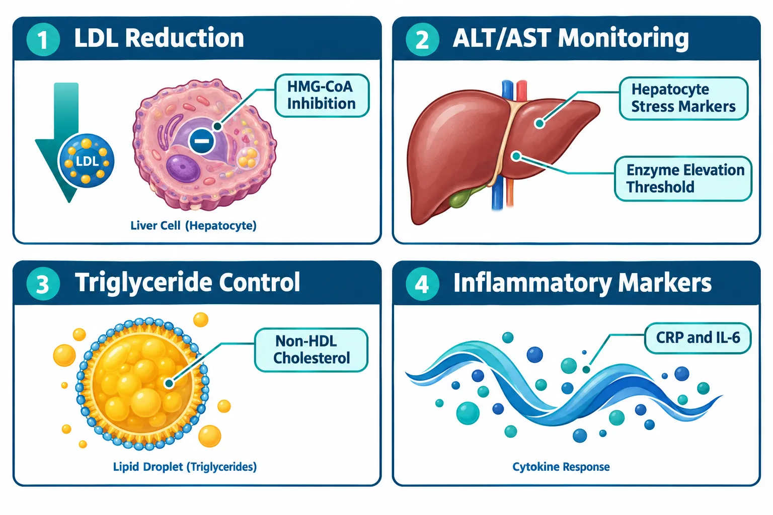
Atorvastatin is a high-intensity HMG-CoA reductase inhibitor with a well-mapped pharmacological profile. In metabolic research, that predictability is an asset. When a study introduces a novel peptide, researchers need a control arm whose lipid and liver effects are already understood, atorvastatin provides exactly that anchor.

Key lipid biomarkers affected by atorvastatin exposure include:

  • LDL cholesterol, typically reduced 40-55% at standard research doses
  • Triglycerides, modestly lowered, relevant in high-fat diet models
  • Non-HDL cholesterol, a composite marker that captures atherogenic particle burden
  • HDL cholesterol, marginally elevated, though not the primary endpoint

On the liver side, atorvastatin is metabolized extensively through CYP3A4 hepatic pathways. This makes ALT (alanine aminotransferase) and AST (aspartate aminotransferase) critical safety and efficacy readouts in every control arm. Current evidence from 2026 confirms that atorvastatin is safe even in subjects with advanced chronic liver disease, though transient enzyme elevations can occur and must be tracked systematically.

"A control group without liver enzyme monitoring is not a control group, it is an incomplete experiment."

For NAFLD and NASH models, atorvastatin also reduces hepatic steatosis scores and portal pressure markers. This means the "normal + atorvastatin" control arm carries meaningful liver biology, not just a lipid signal.


Structuring Multi-Arm Designs for Retatrutide, MOTS-c, and 5-Amino-1MQ Studies
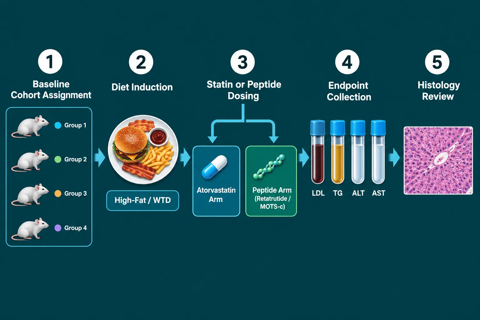
Structuring Multi-Arm Designs for Retatrutide, MOTS-c, and 5-Amino-1MQ Studies

When Atorvastatin and Research Peptides: Designing Lipid and Liver-Control Groups for Metabolic Studies are combined in a multi-arm preclinical model, the architecture of the study determines what conclusions are actually possible. A poorly structured design cannot separate statin effects from peptide effects, and that ambiguity destroys interpretive value.

Recommended Multi-Arm Framework

Arm Diet Statin Peptide Primary Readouts
Vehicle Control Standard chow None None Baseline LDL, ALT, AST
Lipid Control WTD Atorvastatin None LDL, TG, non-HDL, ALT
Peptide Arm A WTD None Retatrutide GLP receptor activity, lipids
Peptide Arm B WTD None MOTS-c Mitochondrial markers, AST
Combination Arm WTD Atorvastatin Retatrutide or MOTS-c All lipid + liver endpoints

Retatrutide targets GLP-1, GIP, and glucagon receptors simultaneously, producing meaningful reductions in body weight and triglycerides. For researchers exploring this compound, the Buy Retatrutide Peptide resource provides relevant formulation context. When placed against an atorvastatin lipid-control arm, the incremental triglyceride and non-HDL reduction attributable to retatrutide becomes statistically isolatable.

MOTS-c operates through mitochondrial AMPK activation, influencing glucose metabolism and fatty acid oxidation. Its liver effects, particularly on hepatic lipid accumulation, overlap with atorvastatin's steatosis-reducing properties. Researchers sourcing this compound can reference the Buy MOTS-c Peptide page for purity and dosing specifications. The MOTS-c Peptide Dosage Calculator is also a practical tool for designing consistent dosing protocols.

5-amino-1MQ inhibits NNMT (nicotinamide N-methyltransferase), shifting cellular metabolism toward fat oxidation. Its inflammatory marker profile, including CRP and IL-6, makes it particularly important to track alongside ALT and AST in the atorvastatin control arm, since both compounds affect hepatic inflammatory tone.

For broader context on how these peptides compare mechanistically to small-molecule drugs, the article on Polypeptide Peptides in Cardiometabolic Models provides a strong conceptual foundation.


Liver-Centric Endpoints and Inflammatory Marker Integration
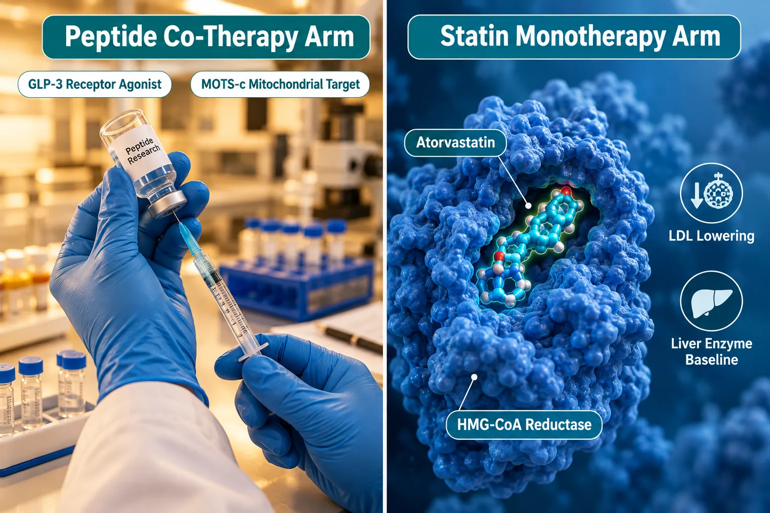
Liver-Centric Endpoints and Inflammatory Marker Integration

The most common failure in Atorvastatin and Research Peptides: Designing Lipid and Liver-Control Groups for Metabolic Studies is treating liver safety as secondary. In 2026, the evidence base has shifted firmly: liver endpoints are co-primary, not supplementary.

Liver-centric endpoints to include in every control arm:

  1. ALT and AST, tracked at baseline, mid-study, and endpoint; atorvastatin can cause transient elevations that must be distinguished from peptide-induced changes
  2. Hepatic steatosis score, histological grading from liver biopsy or MRI-based fat fraction
  3. Portal pressure markers, relevant in advanced NAFLD/NASH models
  4. Liver-to-body weight ratio, a rapid structural readout in rodent models

Inflammatory markers add a second interpretive layer. CRP, IL-6, and TNF-alpha are all modulated by atorvastatin through pleiotropic anti-inflammatory mechanisms. When a peptide like MOTS-c or GLP-3 peptide also reduces inflammatory load, the atorvastatin control arm provides the benchmark against which that reduction is measured.

Combination therapy designs, such as "phosphatidylcholine (PPC) alone" versus "PPC + atorvastatin", demonstrate how isolating statin-specific contributions requires a clean single-agent control. The same logic applies when adding SS-31 peptides to a metabolic model: the mitochondria-targeted antioxidant effects of SS-31 must be separated from atorvastatin's lipid-lowering signal through deliberate arm structure.

Monitoring schedule best practices:

  • Collect lipid panels at weeks 0, 4, 8, and 12 minimum
  • Run liver enzyme panels at the same intervals
  • Reserve histology for endpoint only (terminal in rodent models)
  • Include fasting insulin and HOMA-IR as metabolic co-endpoints

Conclusion

Designing rigorous control groups for metabolic peptide research is not a procedural formality, it is the scientific foundation on which every meaningful comparison rests. Atorvastatin's well-characterized effects on LDL, triglycerides, non-HDL cholesterol, ALT, and AST make it the most defensible lipid and liver-control comparator available for studies involving retatrutide, MOTS-c, or 5-amino-1MQ.

Actionable next steps for researchers in 2026:

  • Build a minimum five-arm design that separates vehicle, statin, peptide, and combination conditions
  • Pre-specify all liver enzyme thresholds as stopping rules and safety endpoints
  • Use Western-type diet induction as the metabolic stressor to activate both lipid and hepatic pathways
  • Align inflammatory marker panels (CRP, IL-6) with lipid and liver readouts for mechanistic depth
  • Consult formulation-specific resources for peptide purity verification before dosing begins

A well-structured control group does not slow research down, it makes the data worth publishing.

https://www.puretestedpeptides.com/wp-content/uploads/2026/10/atorvastatin-and-research-peptides-designing-lipid-and-liver-control-groups-for.webp 672 1008 https://www.puretestedpeptides.com/wp-content/uploads/2026/01/buy-peptides-online.jpg 2026-10-06 13:10:132026-10-06 13:10:13Atorvastatin and Research Peptides: Designing Lipid and Liver-Control Groups for Metabolic Studies
Metoprolol in Peptide Experiments: Controlling Heart Rate, Blood Pressure, and Adrenergic Confounding

Metoprolol in Peptide Experiments: Controlling Heart Rate, Blood Pressure, and Adrenergic Confounding

October 6, 2026/0 Comments/in Uncategorized/by

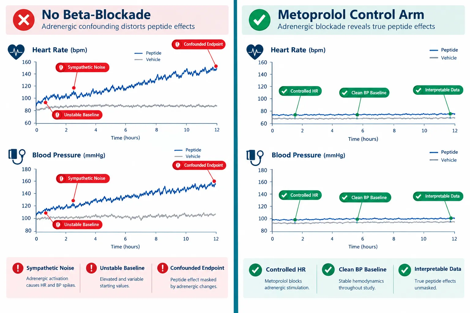
Cardiovascular endpoints fail silently when sympathetic noise is not controlled. Across retatrutide trials, growth-hormone-secretagogue models, and metabolic peptide studies, unmanaged adrenergic tone can shift heart rate by five to ten beats per minute and systolic blood pressure by several millimeters of mercury, differences large enough to obscure or mimic a genuine peptide effect. Understanding the role of metoprolol in peptide experiments for controlling heart rate, blood pressure, and adrenergic confounding is therefore not a minor methodological footnote; it is a foundational design decision.

Key Takeaways

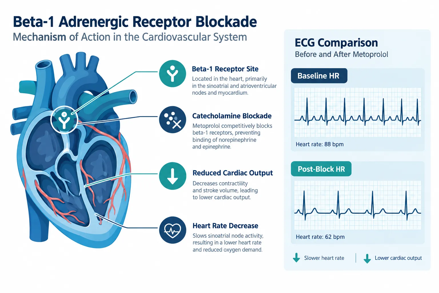
  • Metoprolol is a beta-1 selective blocker that reduces heart rate, systolic blood pressure, and cardiac output without raising peripheral resistance, making it a precise tool for quieting adrenergic noise.
  • Placebo-corrected reductions of 3-6 mmHg systolic and 5-7 beats per minute are typical, but inter-subject variability, partly driven by CYP2D6 metabolizer status, can be substantial.
  • Stable beta-blockade takes approximately one week of oral dosing; acute sympathetic blockade begins within 15 minutes of a single dose, so experimental timing must match the intended control strategy.
  • Metoprolol controls adrenergic tone but does not normalize all advanced structural or functional endpoints, meaning it cannot substitute for a peptide's mechanism of action.
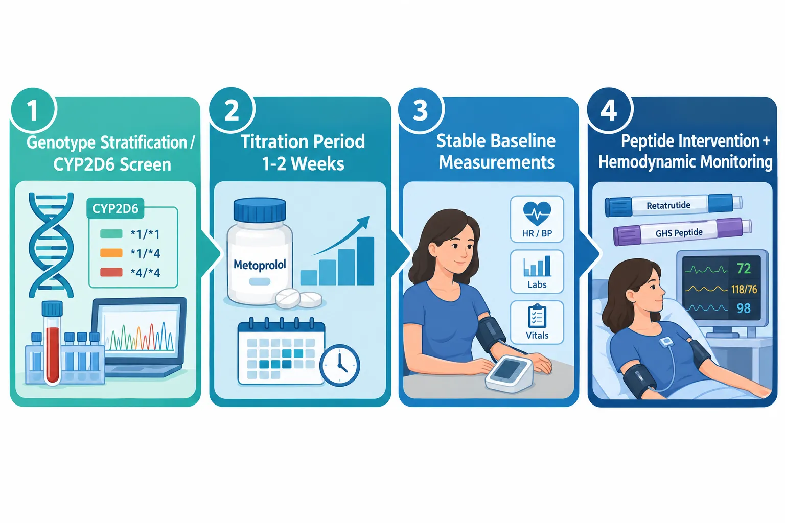
  • Proper use requires genotype stratification, defined titration periods, and both resting and exercise-state baseline measurements before any peptide intervention begins.

Why Adrenergic Tone Confounds Peptide Cardiovascular Readouts

Why Adrenergic Tone Confounds Peptide Cardiovascular Readouts

Many peptides under active investigation, including metabolic agents like retatrutide and GLP-3 class compounds, exert direct or indirect effects on the cardiovascular system. Growth-hormone secretagogues, vasoactive intestinal peptides such as VIP-5, and mitochondria-targeted peptides like SS-31 all operate in physiological environments where sympathetic tone fluctuates with stress, fasting state, exercise, and even the act of venipuncture.

Catecholamines, adrenaline and noradrenaline, bind beta-1 receptors on the myocardium and raise heart rate, contractility, and blood pressure. If a peptide also raises or lowers these parameters through a separate pathway, the two signals overlap. Without a pharmacological anchor, investigators cannot cleanly attribute observed hemodynamic changes to the peptide alone.

This is the core problem metoprolol solves. By selectively blocking beta-1 adrenergic receptors, it attenuates catecholamine-driven cardiovascular fluctuations, providing a more stable hemodynamic backdrop against which peptide-specific effects can be measured.

"Metoprolol does not eliminate cardiovascular physiology, it standardizes the sympathetic component of it, allowing other signals to emerge more clearly."

Key adrenergic phenomena that metoprolol suppresses include:

  • Resting tachycardia driven by anxiety or metabolic stimulation
  • Exercise-induced heart rate overshoot
  • Isoproterenol-provoked tachycardia used in challenge protocols
  • Reflex orthostatic tachycardia during postural changes

Each of these can appear as a false-positive or false-negative peptide effect if left uncontrolled.


Practical Application of Metoprolol in Peptide Experiments: Controlling Heart Rate, Blood Pressure, and Adrenergic Confounding

Practical Application of Metoprolol in Peptide Experiments: Controlling Heart Rate, Blood Pressure, and Adrenergic Confoundin

Dose Range and Titration Timing

Oral metoprolol doses in hypertension and heart-rate control protocols typically range from 100 to 450 mg per day, titrated at weekly intervals. A single 50-80 mg dose begins reducing exercise systolic blood pressure within approximately 15 minutes, but stable, chronic antihypertensive effect requires roughly one week of consistent dosing.

This distinction matters enormously for experimental design:

Control Strategy Onset Use Case
Acute single dose ~15 minutes Rapid sympathetic stabilization before a one-time challenge
Chronic titration (1 week+) ~7 days Stable baseline for multi-day peptide dosing protocols

For most mechanistic peptide trials, including those assessing cardiometabolic polypeptide models, chronic titration is preferred because it removes day-to-day adrenergic variability rather than just blunting a single measurement window.

Accounting for CYP2D6 Metabolizer Status

A critical and often overlooked source of confounding within the metoprolol control arm itself is pharmacogenomic variability. Poor CYP2D6 metabolizers accumulate significantly higher serum metoprolol concentrations, producing greater and more consistent reductions in heart rate, diastolic blood pressure, and mean arterial pressure compared to extensive metabolizers at the same dose.

If subjects are not stratified by CYP2D6 status, the beta-blockade "control" becomes a new source of variance. Practical steps include:

  1. Pre-screen subjects for CYP2D6 poor-metabolizer status
  2. Stratify randomization accordingly
  3. Monitor resting and exercise heart rate at baseline, after titration, and at each peptide dosing interval

Baseline Measurement Protocol

Baseline measurements must capture both resting and dynamic (exercise) cardiovascular state. Metoprolol reduces heart rate and systolic blood pressure at rest and during exercise, these are separate physiological states that peptides may affect differently. Recording only resting values misses exercise-induced adrenergic surges that some metabolic peptides, including growth-hormone secretagogues, actively modulate.


Interpretation Cautions: What Metoprolol Cannot Control

Interpretation Cautions: What Metoprolol Cannot Control

Metoprolol's selectivity is both its strength and its limitation. As a beta-1 selective agent, it does not block alpha-1 receptors or beta-2 receptors. Contrast this with carvedilol, a non-selective blocker with additional alpha-1 antagonism, which suppresses adrenergic activity through a broader mechanism. The choice between these agents changes the nature of residual adrenergic confounding, a distinction highlighted in comparative cardiovascular outcome protocols.

For researchers working with peptides in endocrine pharmacology contexts or GH-analogue peptides, additional considerations include:

  • Structural endpoints: Beta-blockade does not normalize advanced structural or functional cardiac endpoints. Recent data from myosin inhibitor trials confirm that metoprolol, despite controlling heart rate and blood pressure, did not replicate the specific functional improvements seen with next-generation myosin-targeted agents. Peptide trials must not assume that hemodynamic control equals functional equivalence.
  • Peripheral vascular effects: Metoprolol does not increase peripheral resistance, but it also does not reduce it. Peptides with vasodilatory mechanisms, such as VIP-class agents, will still show vascular effects that metoprolol leaves unmasked.
  • Metabolic interactions: Some metabolic peptides affect insulin sensitivity and glucose metabolism in ways that secondarily alter sympathetic tone. Metoprolol blunts the cardiovascular expression of this interaction but does not eliminate the underlying metabolic signal.

Conclusion

Metoprolol in peptide experiments for controlling heart rate, blood pressure, and adrenergic confounding represents one of the most practical pharmacological tools available to cardiovascular researchers in 2026. Its beta-1 selectivity, well-characterized dose-response profile, and rapid onset make it well suited for both acute challenge designs and chronic multi-week protocols.

Actionable next steps for research teams:

  • Determine whether acute or chronic beta-blockade better matches the experimental timeline before dosing begins
  • Screen and stratify subjects by CYP2D6 metabolizer status to prevent pharmacogenomic variance from contaminating the control arm
  • Collect resting and exercise-state hemodynamic baselines after titration, not before
  • Document which adrenergic phenomena remain unblocked (alpha-1, beta-2 pathways) and account for them in statistical models
  • Avoid interpreting stable hemodynamic readings as evidence that all cardiovascular endpoints are normalized, structural and functional parameters require independent assessment

Selecting high-purity, well-characterized peptides from a reliable peptide supplier is equally important; compound variability introduces its own confounding that no pharmacological control can fully correct.

https://www.puretestedpeptides.com/wp-content/uploads/2026/10/metoprolol-in-peptide-experiments-controlling-heart-rate-blood-pressure-and-adre.webp 672 1008 https://www.puretestedpeptides.com/wp-content/uploads/2026/01/buy-peptides-online.jpg 2026-10-06 13:10:002026-10-06 13:10:00Metoprolol in Peptide Experiments: Controlling Heart Rate, Blood Pressure, and Adrenergic Confounding
Prednisone and Research-Use Peptides: How Glucocorticoid Exposure Can Distort Metabolic, Immune, and Tissue-Repair Readouts

Prednisone and Research-Use Peptides: How Glucocorticoid Exposure Can Distort Metabolic, Immune, and Tissue-Repair Readouts

October 6, 2026/0 Comments/in Uncategorized/by

Roughly one in five research subjects enrolled in metabolic or immune-modulating studies carries an active or recent glucocorticoid prescription, yet fewer than half of published peptide protocols formally account for this exposure. The problem of Prednisone and Research-Use Peptides: How Glucocorticoid Exposure Can Distort Metabolic, Immune, and Tissue-Repair Readouts is not theoretical. When a subject is taking prednisone, the drug reshapes glucose handling, cytokine profiles, body composition, and wound biology simultaneously, making it nearly impossible to attribute changes in those endpoints to a peptide compound without deliberate experimental controls.

This guide is written for researchers, protocol designers, and informed buyers of research-use peptide compounds who need a clear, actionable map of where glucocorticoid confounding is most severe and how to design around it.


Key Takeaways

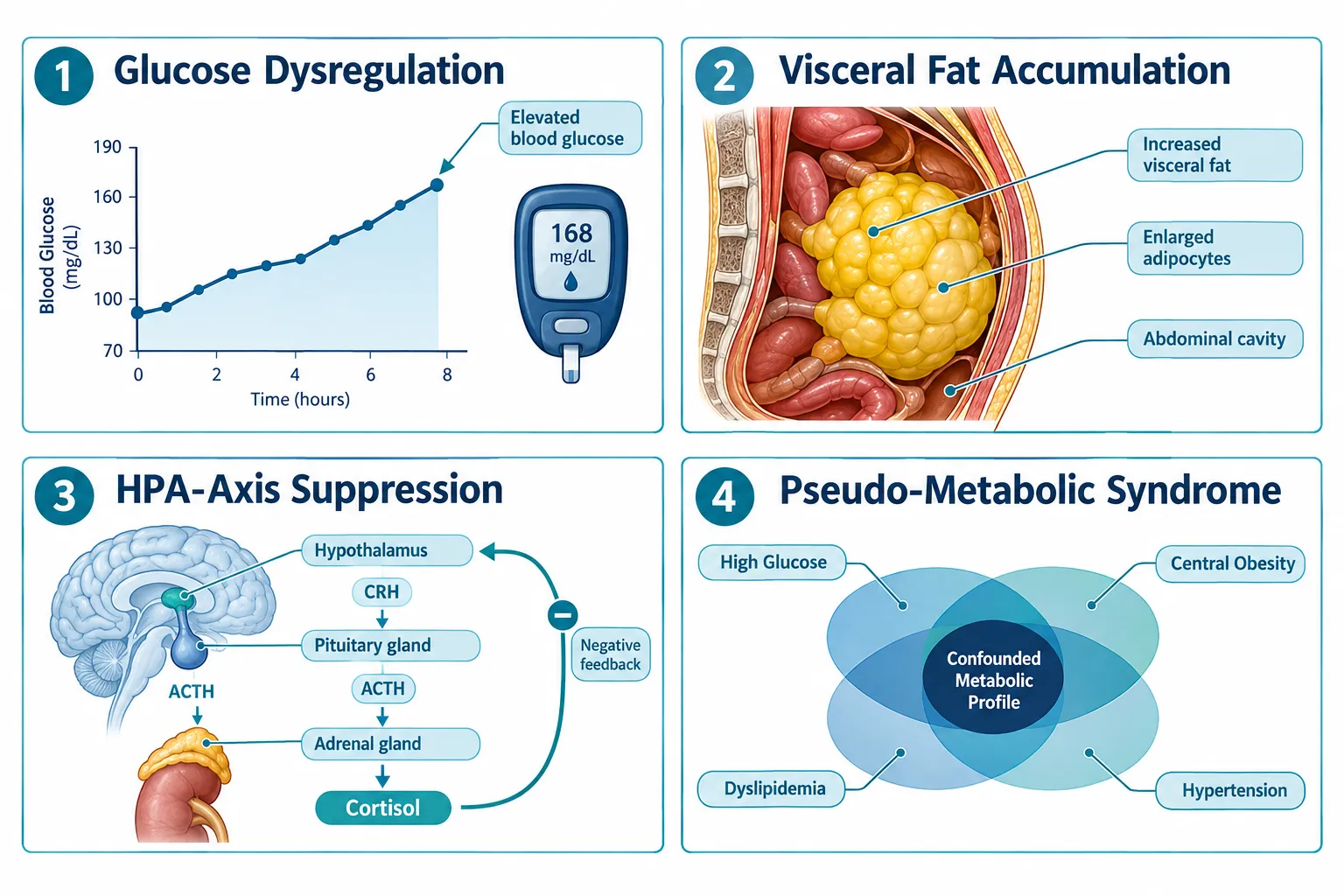
  • Prednisone induces a "pseudo-metabolic syndrome" that directly mimics or masks outcomes targeted by metabolic peptides.
  • Standard inflammatory biomarkers, CRP, leukocyte count, neutrophil ratio, are dose-dependently suppressed by glucocorticoids, distorting immune readouts.
  • Tissue-repair and wound-healing endpoints are particularly vulnerable because glucocorticoids both dampen acute inflammation and impair collagen remodeling.
  • Anabolic and sarcopenia-targeted peptides face the strongest confounding from glucocorticoid-driven muscle catabolism.
  • Glucocorticoid exposure should be treated as a core design variable, not background noise, in any peptide research protocol.

How Glucocorticoids Create a Pseudo-Metabolic Syndrome

How Glucocorticoids Create a Pseudo-Metabolic Syndrome

Prednisone does not simply suppress inflammation. At doses commonly used in clinical and research-adjacent settings, it triggers a cascade of metabolic changes that closely resemble metabolic syndrome: elevated fasting glucose, increased visceral adiposity, dyslipidemia, and insulin resistance. For researchers studying cardiometabolic peptide compounds, this is a direct collision.

The core metabolic confounders introduced by prednisone include:

Endpoint Glucocorticoid Effect Research Risk
Fasting glucose Elevated (hepatic gluconeogenesis) Masks GLP-class peptide efficacy
Visceral fat Increased via lipid redistribution Distorts body-composition endpoints
Insulin sensitivity Reduced Confounds metabolic peptide response
Appetite / caloric intake Increased Alters weight-loss peptide readouts
Cortisol / HPA axis Suppressed endogenous output Disrupts hormonal biomarker panels

The HPA-axis suppression point deserves particular attention. When exogenous glucocorticoids are present, endogenous cortisol production falls. This changes the hormonal baseline against which peptide pharmacodynamics are measured. For GH-analogue peptides and compounds that interact with the growth hormone axis, this hormonal shift can produce entirely misleading dose-response curves.

Researchers studying visceral fat redistribution should also note that glucocorticoid-driven adiposity preferentially accumulates centrally, the same compartment tracked in visceral fat research models. Without a glucocorticoid-naive control arm, it becomes impossible to separate peptide-driven changes from steroid-driven ones.


Immune Biomarker Distortion: What Prednisone Does to CRP, Cytokines, and Leukocytes

Immune Biomarker Distortion: What Prednisone Does to CRP, Cytokines, and Leukocytes

The immune-modulating effects of prednisone are both well-documented and deeply problematic for peptide research. The drug produces strong, dose-dependent suppression of nearly every standard inflammatory biomarker used in research protocols.

Biomarkers most severely distorted by glucocorticoid exposure:

  • C-reactive protein (CRP): Hepatic CRP synthesis is directly suppressed, producing artificially low readings that can mimic anti-inflammatory peptide effects.
  • Interleukin-6 (IL-6) and TNF-alpha: Both are blunted by glucocorticoid receptor activation, masking inflammatory resolution that a peptide may or may not be driving.
  • Leukocyte and neutrophil counts: Prednisone causes a characteristic leukocytosis with neutrophilia and lymphopenia, a pattern that can be mistaken for an immune-activation signal from a peptide compound.
  • Tissue-resident immune cells: Recent work confirms that glucocorticoids selectively modulate tissue-resident macrophages and mast cells in ways that differ from circulating immune cells. This matters enormously for regenerative peptides like BPC-157 and TB-500, where local tissue immune activity is the primary readout.

"When prednisone is present, the immune landscape a peptide encounters is fundamentally different from the one it was designed to modulate. Treating that as a minor covariate is a protocol error."

The practical consequence is that an immune-modulating peptide may appear highly effective, completely ineffective, or even counterproductive depending entirely on the glucocorticoid dose the subject carries. Without stratifying subjects by steroid exposure, or excluding active users entirely, the data cannot be interpreted with confidence.


Tissue-Repair and Anabolic Readouts: Where Glucocorticoid Confounding Is Most Severe

Tissue-Repair and Anabolic Readouts: Where Glucocorticoid Confounding Is Most Severe

Wound healing and tissue repair represent perhaps the most complex confounding scenario in Prednisone and Research-Use Peptides: How Glucocorticoid Exposure Can Distort Metabolic, Immune, and Tissue-Repair Readouts. Glucocorticoids produce a paradoxical two-phase effect on repair biology.

Phase 1 (acute): Inflammation is suppressed, which can make a wound-healing peptide appear to accelerate early healing simply because the steroid has already cleared the inflammatory field.

Phase 2 (remodeling): Glucocorticoids impair fibroblast proliferation, reduce collagen synthesis, and suppress angiogenesis. A peptide that promotes collagen remodeling may show blunted or absent efficacy, not because it failed, but because the steroid has suppressed the very cellular machinery the peptide targets.

For anabolic and sarcopenia-targeted peptides, the problem is equally stark. Glucocorticoids drive skeletal muscle catabolism through multiple mechanisms: they upregulate muscle-specific ubiquitin ligases, suppress IGF-1 signaling, and reduce protein synthesis rates. Any peptide designed to promote lean mass retention or muscle repair is working against a pharmacologically imposed catabolic state. The net readout will underestimate peptide efficacy, sometimes dramatically.

Mitochondrial function is also affected. Glucocorticoids impair mitochondrial biogenesis in muscle tissue, which is directly relevant for compounds studied in SS-31 mitochondrial research contexts.

Designing Around Glucocorticoid Confounding: Practical Protocol Controls

Researchers in 2026 have several validated strategies to reduce or detect glucocorticoid confounding:

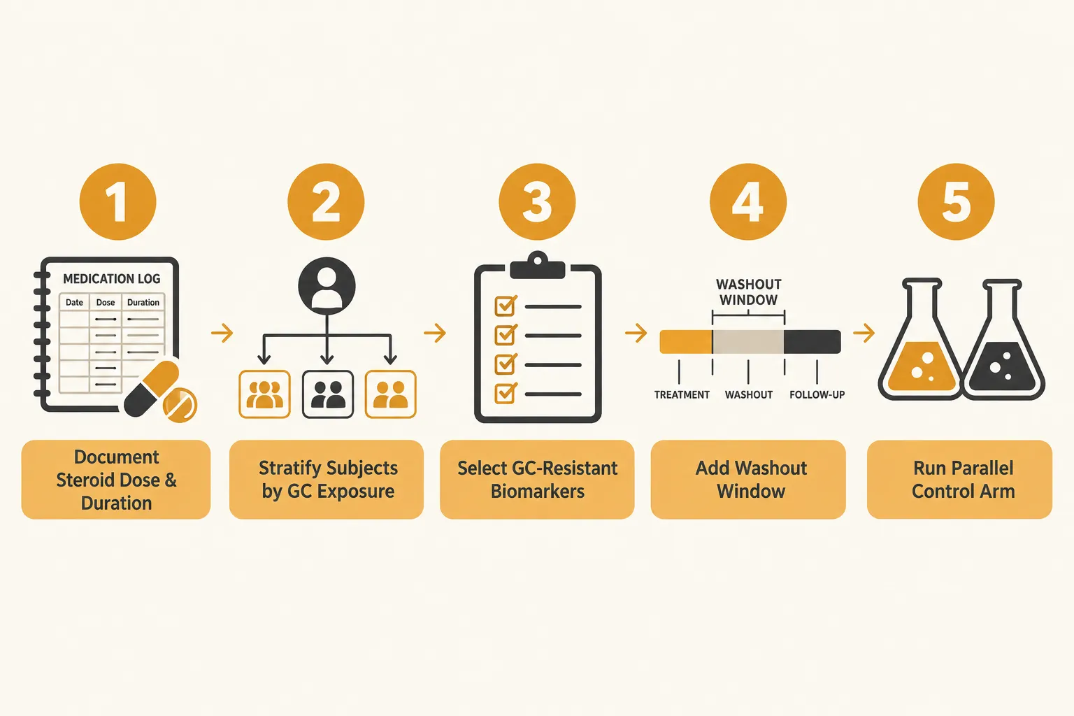
  1. Screen and stratify at enrollment. Collect steroid use history, current dose, and duration. Stratify rather than exclude where possible to preserve sample size.
  2. Use glucocorticoid-resistant biomarkers where available. Some cytokines and repair markers are less sensitive to steroid suppression and can serve as secondary endpoints.
  3. Build in washout windows. For subjects who can safely discontinue steroids, a documented washout period before peptide exposure begins reduces confounding substantially.
  4. Run parallel control arms. A glucocorticoid-exposed control arm receiving vehicle only allows direct quantification of steroid-driven endpoint changes.
  5. Report steroid exposure as a primary covariate. Statistical models should treat glucocorticoid dose as a continuous variable, not a binary yes/no flag.

Conclusion

The relationship between Prednisone and Research-Use Peptides: How Glucocorticoid Exposure Can Distort Metabolic, Immune, and Tissue-Repair Readouts is now well enough characterized that ignoring it constitutes a preventable design flaw. Across glucose metabolism, inflammatory cytokines, collagen remodeling, body composition, and immune-cell measurements, prednisone introduces quantifiable, directional biases that can make an effective peptide look inert, or an inert one look effective.

Actionable next steps for research protocol designers:

  • Add glucocorticoid screening to all enrollment checklists for metabolic, immune, or repair-focused peptide studies.
  • Pre-specify steroid dose as a covariate in the statistical analysis plan before data collection begins.
  • Consult updated 2026 biomarker guidance on glucocorticoid-resistant immune endpoints when designing immune-modulating peptide studies.
  • Review compound-specific confounding risk, peptides targeting GH axis, collagen synthesis, or mitochondrial function carry the highest vulnerability.

Treating glucocorticoid exposure as a core design variable rather than background noise is the single highest-leverage methodological improvement available to most peptide research teams today.

https://www.puretestedpeptides.com/wp-content/uploads/2026/10/prednisone-and-research-use-peptides-how-glucocorticoid-exposure-can-distort-met.webp 1024 1536 https://www.puretestedpeptides.com/wp-content/uploads/2026/01/buy-peptides-online.jpg 2026-10-06 13:06:132026-10-06 13:06:13Prednisone and Research-Use Peptides: How Glucocorticoid Exposure Can Distort Metabolic, Immune, and Tissue-Repair Readouts
Amlodipine in Peptide Research: Separating Calcium-Channel Effects From Blood-Pressure and Vascular Readouts

Amlodipine in Peptide Research: Separating Calcium-Channel Effects From Blood-Pressure and Vascular Readouts

October 6, 2026/0 Comments/in Uncategorized/by

Fewer than one in five preclinical peptide studies that co-administer a calcium-channel blocker formally account for the drug's independent vascular effects, a methodological gap that can quietly invalidate entire datasets. When amlodipine in peptide research enters the experimental picture, the challenge is not simply adjusting for lower blood pressure. It is disentangling at least three overlapping biological layers: direct L-type calcium-channel blockade, blood-pressure-dependent vascular changes, and blood-pressure-independent vascular remodeling. Getting these layers wrong means attributing amlodipine's known anti-proliferative and anti-atherosclerotic actions to the peptide under study, or, just as problematically, masking a real peptide signal behind the drug's dominant pharmacology.

Key Takeaways

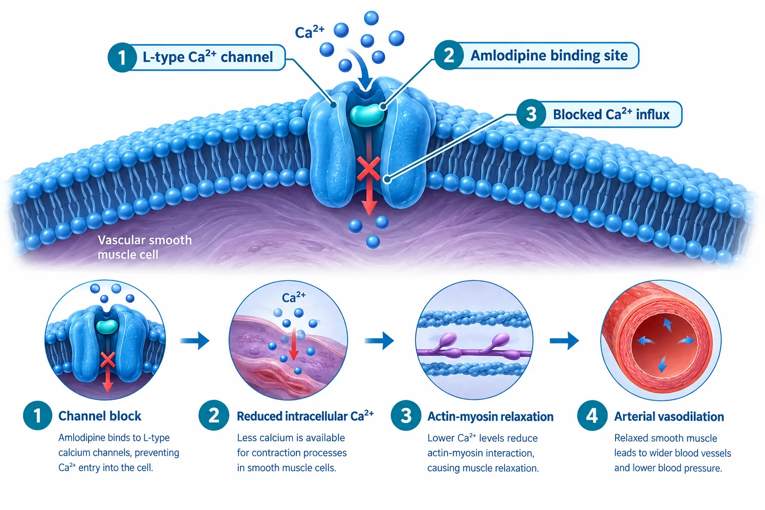
  • Amlodipine blocks L-type calcium channels in vascular smooth muscle, reducing intracellular Ca²⁺ and causing arterial relaxation independently of any peptide co-administered in a study.
  • The drug produces vascular benefits, including inhibited smooth-muscle cell proliferation and slowed atherosclerosis, that are separate from its blood-pressure-lowering effect.
  • At clinically relevant concentrations, amlodipine does not activate CRAC channels and does not interfere with fluorescence-based Ca²⁺ imaging dyes such as Cal-520, Fluo-4, or Fura-Red.
  • Researchers should pre-specify which readouts are calcium-channel-mediated, blood-pressure-dependent, or peptide-specific before data collection begins.
  • Orthogonal biomarkers, endothelial nitric oxide, arterial compliance, and vascular remodeling indices, can help isolate a peptide's contribution from amlodipine's background pharmacology.

How Amlodipine Works: The Calcium-Channel Mechanism Researchers Must Account For

How Amlodipine Works: The Calcium-Channel Mechanism Researchers Must Account For

Amlodipine is a dihydropyridine calcium antagonist. Its primary action is inhibiting the transmembrane influx of calcium ions through L-type voltage-gated channels, predominantly in vascular smooth muscle cells. The result is reduced intracellular Ca²⁺, diminished actin-myosin interaction, smooth-muscle relaxation, and arterial vasodilation. Its effect on vascular smooth muscle is substantially greater than its effect on cardiac muscle, a selectivity profile that matters when interpreting any cardiovascular or vascular readout in a peptide experiment.

This mechanism is not trivial background noise. It is a dominant pharmacological signal. Any peptide co-administered with amlodipine that also influences vascular tone, intracellular calcium, or smooth-muscle behavior will generate data that reflects both agents simultaneously unless the study design explicitly separates them.

What this means practically:

  • Vascular tone measurements (e.g., wire myography, pressure myography) will reflect amlodipine's baseline relaxation effect.
  • Intracellular Ca²⁺ flux assays will show a suppressed resting Ca²⁺ level before the peptide is even introduced.
  • Any peptide with vasodilatory properties may appear to have a blunted or absent effect simply because the vascular bed is already partially relaxed.

Researchers working with mitochondria-targeted peptides such as those explored in SS-31 10mg research peptide considerations should note that oxidative-stress and membrane-potential readouts can also be indirectly affected when intracellular calcium homeostasis is altered by a co-administered channel blocker.


Separating Calcium-Channel Effects From Blood-Pressure and Vascular Readouts: A Practical Framework

Separating Calcium-Channel Effects From Blood-Pressure and Vascular Readouts: A Practical Framework

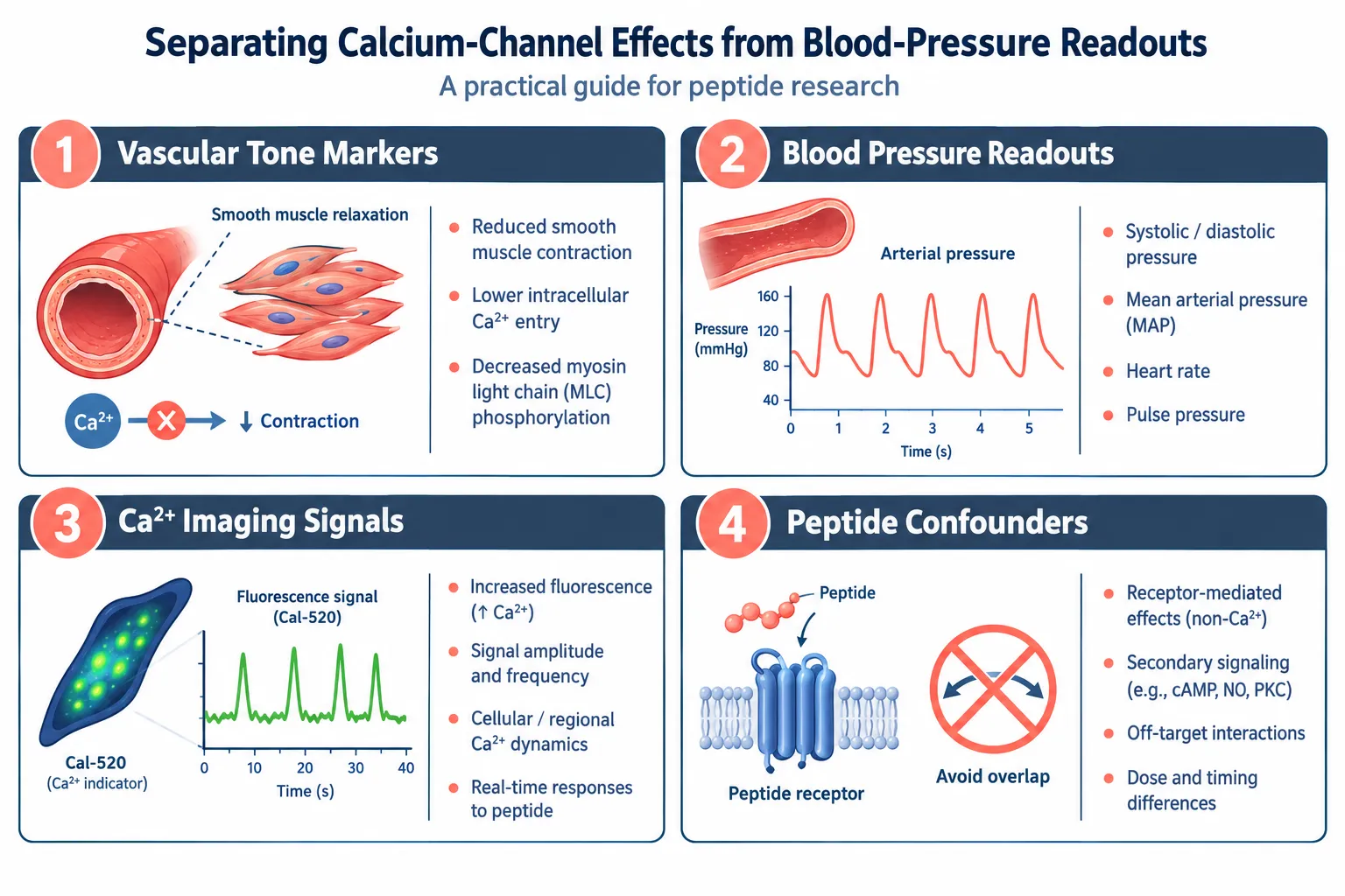
The central challenge in amlodipine in peptide research, separating calcium-channel effects from blood-pressure and vascular readouts, requires a tiered classification of every endpoint before data collection begins.

Tier 1: Blood-Pressure-Dependent Readouts

These are endpoints that change because amlodipine lowers arterial pressure. They include:

  • Brachial or carotid blood pressure values
  • Cardiac afterload indices
  • Glomerular filtration rate changes driven by reduced perfusion pressure

Practical step: Include a vehicle-treated, amlodipine-only control group at the same dose. Any change in these readouts in that group represents the amlodipine baseline, not a peptide effect.

Tier 2: Blood-Pressure-Independent Vascular Effects

Contemporary cardiovascular literature consistently identifies a second category of amlodipine actions that persist even when blood pressure is matched to comparator agents. These include:

Effect Mechanism Research Implication
Smooth-muscle cell anti-proliferation Direct Ca²⁺ signaling suppression May confound peptide anti-proliferative claims
Anti-atherosclerotic remodeling Reduced oxidative stress in vessel wall Arterial stiffness data needs amlodipine-only control
Improved arterial compliance Structural vascular changes Central pulse-wave velocity requires correction

For peptide studies examining vascular remodeling, such as those involving VIP peptide mechanisms or vasoactive signaling pathways, these BP-independent effects represent the most likely source of confounding.

Tier 3: Calcium-Channel-Specific Molecular Readouts

This tier covers direct molecular measurements of L-type channel activity and downstream Ca²⁺ signaling. A key methodological advantage here: at therapeutic plasma concentrations of approximately 0.5 µM, amlodipine does not activate calcium-release-activated calcium (CRAC) channels. This means CRAC-mediated signaling events attributed to a peptide are not being driven by amlodipine itself.

Additionally, fluorescence-based Ca²⁺ imaging using dyes such as Cal-520, Fluo-4, and Fura-Red is not confounded by amlodipine fluorescence at these concentrations. This is a meaningful green light for researchers using live-cell calcium imaging alongside peptide stimulation assays.

"Knowing which calcium compartment amlodipine does and does not touch is the single most useful piece of information a peptide researcher can carry into experimental design."

Researchers examining peptide signaling pathways, including those studying GLP-3 peptide signaling pathway interactions, benefit from this CRAC-channel clarity because receptor-operated calcium entry can then be attributed with greater confidence to the peptide's receptor engagement rather than to the channel blocker.


Designing the Experiment: Isolating Peptide-Specific Signals

Designing the Experiment: Isolating Peptide-Specific Signals

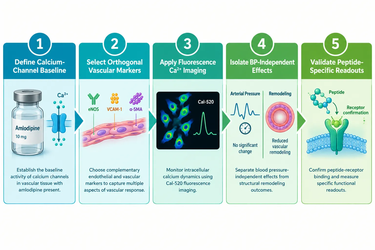
A rigorous experimental framework for amlodipine in peptide research, one that genuinely separates calcium-channel effects from blood-pressure and vascular readouts, involves five concrete design decisions.

Step 1, Define the calcium-channel baseline. Run amlodipine-only controls at the intended study concentration. Measure intracellular Ca²⁺, vascular tone, and any remodeling markers. This establishes the floor the peptide must be measured against.

Step 2, Select orthogonal vascular markers. Choose at least two endpoints that amlodipine does not directly regulate. Endothelial nitric oxide synthase (eNOS) activity, circulating endothelin-1 levels, and specific peptide receptor phosphorylation states are examples. These serve as peptide-specific signal channels.

Step 3, Apply fluorescence Ca²⁺ imaging with confidence. Given that amlodipine does not interfere with Cal-520, Fluo-4, or Fura-Red at relevant concentrations, live-cell imaging remains a valid tool. Pair it with a CRAC-channel inhibitor control to confirm that any observed Ca²⁺ flux is L-type-independent when the peptide is active.

Step 4, Separate central from peripheral blood pressure. Peripheral brachial pressure can be identical across treatment groups while central aortic pressure and pulse-wave velocity differ. Use central hemodynamic measurements to capture amlodipine's BP-independent arterial effects separately from peptide-driven changes.

Step 5, Validate with receptor-blocking controls. If the peptide of interest acts through a specific receptor, as is the case with many research peptides including those reviewed in LL-37 versus SS-31 benefits of each peptide comparisons, include a receptor-antagonist arm. Any vascular effect that disappears with receptor blockade but persists with amlodipine alone is attributable to the peptide.

Proper peptide dosing protocols also matter here: dose-response curves for the peptide should be generated in both the presence and absence of amlodipine to detect any shift in potency or efficacy caused by the background calcium-channel blockade.

For researchers sourcing compounds for controlled in vitro work, ensuring purity and verified composition is non-negotiable. Resources such as guidance on where to buy research-grade peptides evaluating purity underscore why compound quality directly affects the reliability of mechanistic conclusions.


Conclusion

Amlodipine is a well-characterized L-type calcium-channel blocker with a pharmacological footprint that extends well beyond simple blood-pressure reduction. In any peptide research context, failing to account for its anti-proliferative, anti-atherosclerotic, and Ca²⁺-suppressive actions risks misattributing drug effects to the peptide, or obscuring genuine peptide biology beneath a dominant pharmacological background.

Actionable next steps for researchers in 2026:

  1. Pre-classify every planned endpoint into one of the three tiers: blood-pressure-dependent, BP-independent vascular, or calcium-channel-specific molecular.
  2. Include an amlodipine-only control group at the study concentration in every experimental arm.
  3. Use fluorescence-based Ca²⁺ imaging with confidence at therapeutic concentrations, but pair it with CRAC-channel controls to confirm the signal source.
  4. Add orthogonal endothelial and receptor-specific biomarkers that amlodipine does not directly regulate.
  5. Run full dose-response curves for the peptide both with and without amlodipine present to detect potency shifts.

Rigorous separation of these layers does not complicate peptide research, it protects it. The result is data that can be defended, reproduced, and built upon.


https://www.puretestedpeptides.com/wp-content/uploads/2026/10/amlodipine-in-peptide-research-separating-calcium-channel-effects-from-blood-pre.webp 1024 1536 https://www.puretestedpeptides.com/wp-content/uploads/2026/01/buy-peptides-online.jpg 2026-10-06 13:05:532026-10-06 13:05:53Amlodipine in Peptide Research: Separating Calcium-Channel Effects From Blood-Pressure and Vascular Readouts
What Is a GLP-1 Pill? Comparing Oral Delivery Concepts With Research-Use GLP-3 Retatrutide Models

What Is a GLP-1 Pill? Comparing Oral Delivery Concepts With Research-Use GLP-3 Retatrutide Models

October 6, 2026/0 Comments/in Uncategorized/by

An oral tablet approved for weight loss sounds like a major leap forward, and in 2026, that leap has officially happened. But the question researchers, clinicians, and informed consumers are now asking is whether a GLP-1 pill and an injectable triple agonist like retatrutide are even playing the same game. Understanding what is a GLP-1 pill, comparing oral delivery concepts with research-use GLP-3 retatrutide models, reveals a surprisingly wide gap between two approaches that share a name but differ sharply in mechanism, scope, and research application.

Key Takeaways

  • GLP-1 pills are oral small-molecule drugs that activate the GLP-1 receptor; two were approved in the U.S. in 2026 for obesity.
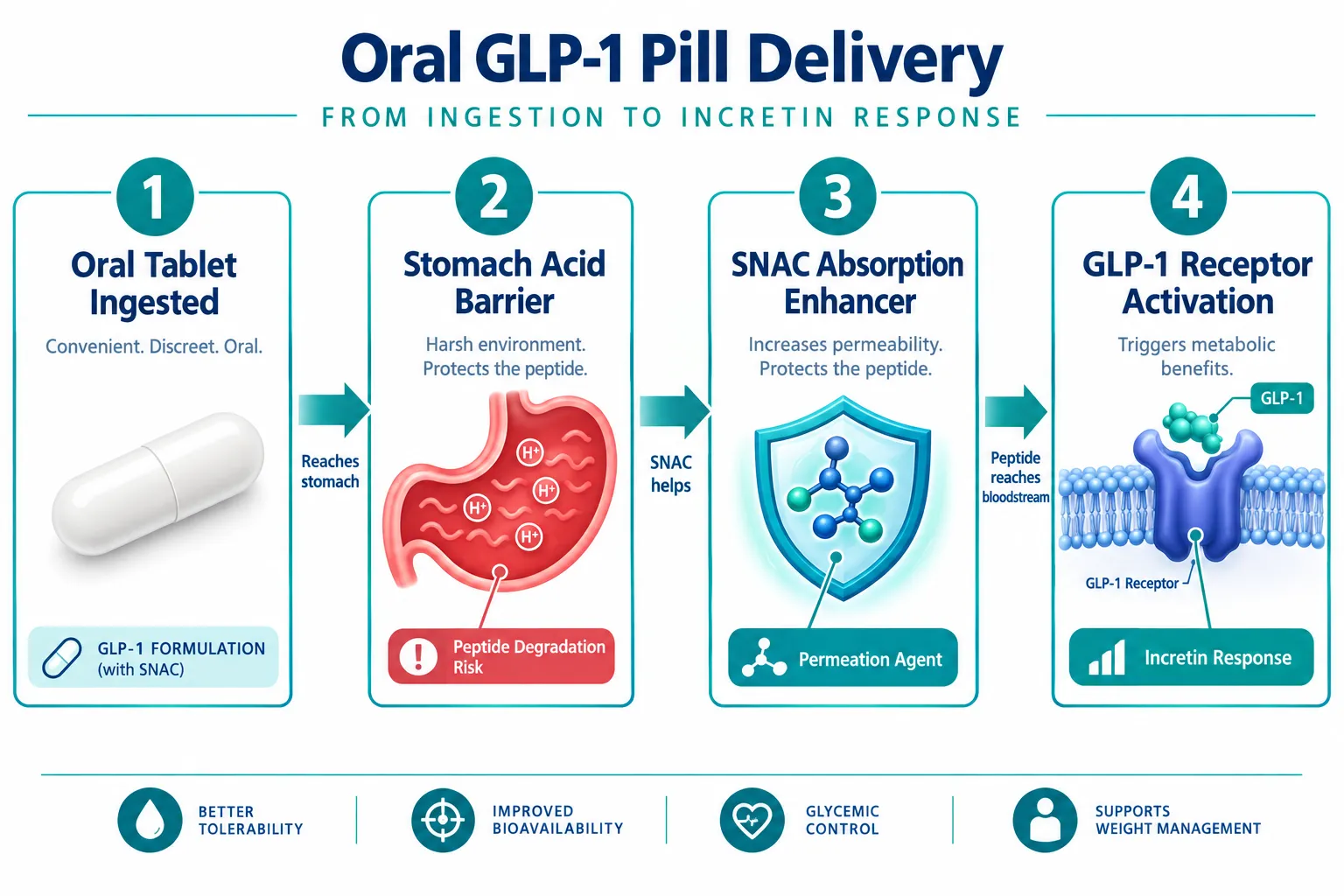
  • Oral delivery faces significant biochemical barriers, requiring absorption enhancers to survive the digestive tract.
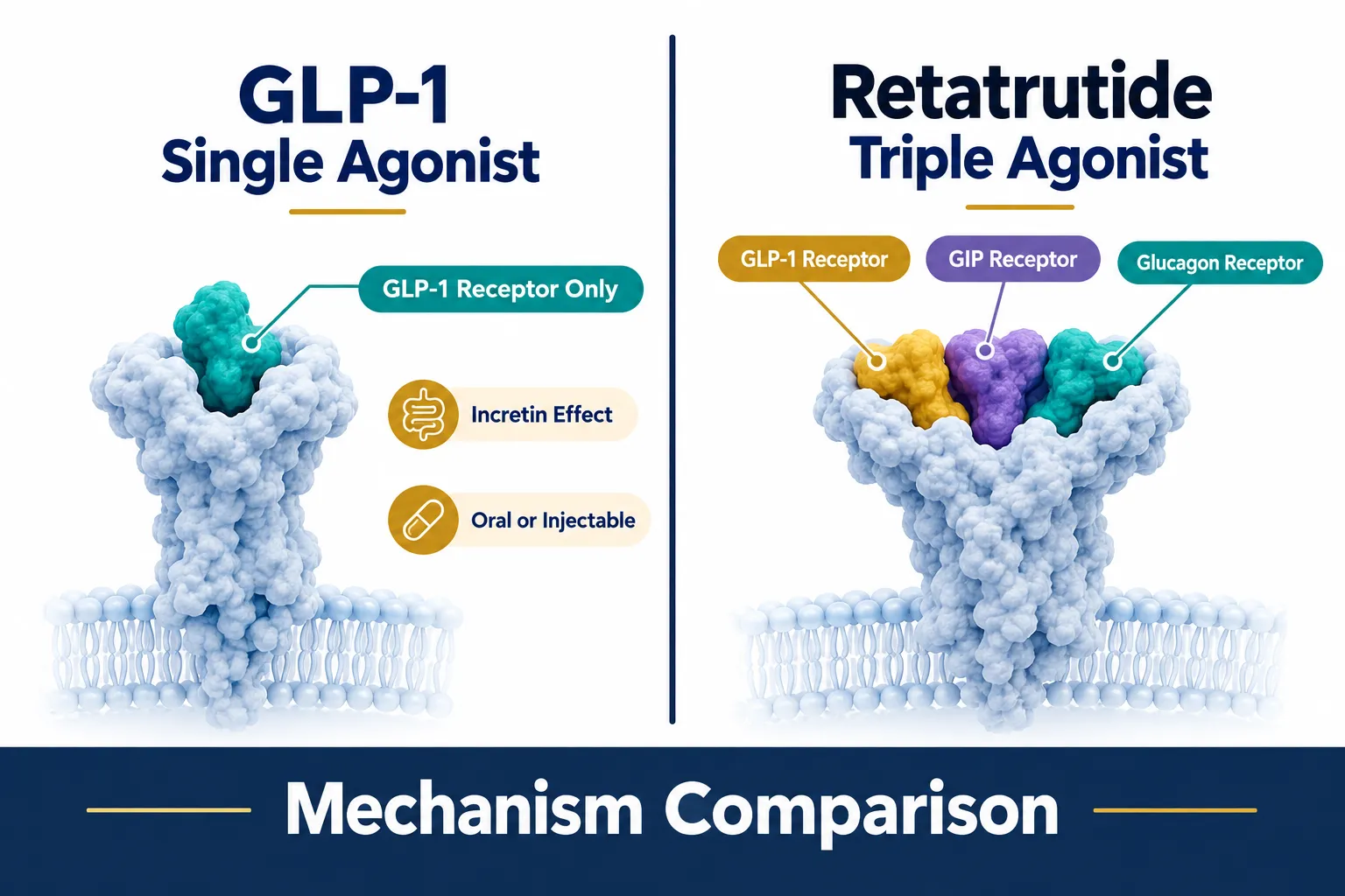
  • Retatrutide is a triple agonist targeting GLP-1, GIP, and glucagon receptors simultaneously, making it mechanistically distinct from any single-agonist pill.
  • The TRIUMPH Phase 3 program is testing retatrutide across obesity, type 2 diabetes, cardiovascular disease, and osteoarthritis populations.
  • An oral GLP-1 pill is not a substitute for a triple-agonist injectable in research or clinical contexts.

How a GLP-1 Pill Actually Works

The glucagon-like peptide-1 (GLP-1) receptor is a well-validated target for metabolic disease. When activated, it slows gastric emptying, reduces appetite, and stimulates insulin secretion in a glucose-dependent manner. Injectable GLP-1 receptor agonists have dominated this space for years, but 2026 marks a turning point for oral delivery.

How a GLP-1 Pill Actually Works

Two oral GLP-1 agents reached U.S. approval in 2026. Eli Lilly's orforglipron (brand name Foundayo) became the first approved oral-only GLP-1 pill specifically indicated for obesity. Novo Nordisk's semaglutide pill, already available for type 2 diabetes, also received approval for weight loss in adults in both the U.S. and EU markets. These are not peptide-based tablets, they are small-molecule non-peptide compounds or peptide formulations engineered to survive the hostile environment of the gastrointestinal tract.

The core challenge with oral peptide delivery is enzymatic degradation. Stomach acid and proteases break down peptide chains before they can reach systemic circulation. Semaglutide tablets overcome this using a co-formulation with sodium N-(8-[2-hydroxybenzoyl] amino) caprylate (SNAC), an absorption enhancer that temporarily raises local gastric pH and facilitates mucosal permeation. Orforglipron sidesteps this problem entirely by being a small-molecule compound that does not require an absorption enhancer.

Key oral delivery constraints include:

  • Low bioavailability compared to subcutaneous injection
  • Strict fasting requirements before dosing (for semaglutide tablets)
  • Narrower therapeutic window due to absorption variability
  • No flexibility in dose titration beyond what the formulation allows

For those exploring the broader landscape of peptides used in metabolic and weight-loss research, what is the strongest peptide for weight loss provides useful context on how different compound classes compare.


Retatrutide: The Triple-Agonist Model That Changes the Comparison

Calling retatrutide a "GLP-3" is a colloquial shorthand that has gained traction in research communities, but it is worth clarifying what that label actually means. Retatrutide is a single peptide molecule that simultaneously activates three receptors: GLP-1, glucose-dependent insulinotropic polypeptide (GIP), and glucagon. This triple agonism is why it is sometimes distinguished from standard GLP-1 agents with the informal "GLP-3" designation.

Retatrutide: The Triple-Agonist Model That Changes the Comparison

This mechanistic distinction is critical when comparing oral delivery concepts with research-use GLP-3 retatrutide models. A GLP-1 pill activates one receptor pathway. Retatrutide activates three, producing overlapping and synergistic metabolic effects that include:

  • GLP-1 activity: appetite suppression, insulin secretion, slowed gastric emptying
  • GIP activity: enhanced insulin response, potential adipose tissue effects
  • Glucagon activity: increased energy expenditure, hepatic fat mobilization

Phase 2 data for retatrutide showed weight reductions exceeding 24% of body weight at the highest dose over 48 weeks, a benchmark that no single GLP-1 agonist, oral or injectable, has matched in head-to-head comparisons at similar timepoints.

For more background on how retatrutide fits within the GLP-1 research landscape, see the GLP-1 retatrutide overview. Researchers seeking access to standardized research-grade material can also review GLP3 Reta 10mg and GLP-3 Reta 30mg for sale options for preclinical study use.


The TRIUMPH Phase 3 Program and What It Tells Researchers

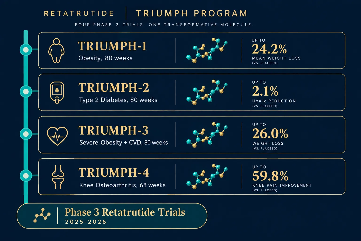
The most comprehensive data set currently being generated on retatrutide comes from the TRIUMPH Phase 3 program, a suite of large-scale trials running across 2025 and 2026. These trials are designed to test retatrutide across multiple disease populations, not just general obesity.

The TRIUMPH Phase 3 Program and What It Tells Researchers

Trial Population Duration Primary Focus
TRIUMPH-1 Obesity or overweight (no T2D) 80 weeks Weight loss efficacy and safety
TRIUMPH-2 Type 2 diabetes + obesity 80 weeks Weight loss and glycemic control
TRIUMPH-3 Severe obesity + CVD (BMI 35+) 80 weeks Cardiovascular and weight outcomes
TRIUMPH-4 Obesity + knee osteoarthritis 68 weeks Weight loss and knee pain reduction

Topline 2026 results from the TRIUMPH program and the parallel TRANSCEND-T2D-1 diabetes trial have shown meaningful reductions in A1C, body weight, and comorbidity burden. These outcomes extend well beyond what a single-receptor oral pill is designed to achieve.

For researchers tracking the evolution of this compound, the retatrutide clinical trials resource aggregates ongoing study data in one place.

"Retatrutide's triple-receptor mechanism produces metabolic effects that are qualitatively different from those of any approved oral GLP-1 agent, not just quantitatively larger."

It is also worth noting that retatrutide remains an injectable peptide in all clinical and research settings. There is no oral formulation in development at this stage, which means the oral vs. injectable distinction is not merely a delivery preference, it reflects a fundamental difference in what each compound can do.

For researchers interested in combinatorial peptide approaches in metabolic contexts, the article on combining Tesamorelin with CDC Ipamorelin offers relevant mechanistic parallels.


Conclusion

Understanding what is a GLP-1 pill, comparing oral delivery concepts with research-use GLP-3 retatrutide models, makes one conclusion unavoidable: these are fundamentally different tools. The 2026 approvals of orforglipron and oral semaglutide for obesity represent a genuine advance in patient convenience and access. However, their single-receptor mechanism, oral bioavailability constraints, and narrower metabolic scope place them in a separate category from retatrutide's triple-agonist profile.

Actionable next steps for researchers and informed readers:

  1. Review the TRIUMPH trial registry entries to follow Phase 3 retatrutide data as it becomes available.
  2. Distinguish between oral GLP-1 agents (small-molecule or SNAC-formulated peptide) and injectable triple agonists before drawing efficacy comparisons.
  3. If evaluating retatrutide for preclinical research, source only verified, tested-grade material and consult current retatrutide clinical trial literature before designing protocols.
  4. Do not assume that oral availability implies equivalent biological activity, receptor breadth, bioavailability, and dosing pharmacokinetics all differ substantially between compound classes.

The GLP-1 pill is a meaningful milestone. Retatrutide is a different kind of milestone entirely.

https://www.puretestedpeptides.com/wp-content/uploads/2026/10/what-is-a-glp-1-pill-comparing-oral-delivery-concepts-with-research-use-glp-3-re.webp 1024 1536 https://www.puretestedpeptides.com/wp-content/uploads/2026/01/buy-peptides-online.jpg 2026-10-06 13:05:492026-10-06 13:05:49What Is a GLP-1 Pill? Comparing Oral Delivery Concepts With Research-Use GLP-3 Retatrutide Models

Epithalon and Telomere Research: Designing Longitudinal Experiments That Separate Telomere Length From Senescence

October 5, 2026/0 Comments/in Uncategorized/by

Cover Image

Telomere length and cellular senescence are often treated as synonyms in aging research, but they are not the same thing, and conflating them produces flawed experiments. A 2025 study published in Biogerontology by Al-Dulaimi and colleagues demonstrated that the synthetic tetrapeptide epitalon (Ala-Glu-Asp-Gly) extends telomeres in four distinct human cell lines through two separate mechanisms, yet the study measured no downstream senescence endpoints. That gap is precisely where the field of Epithalon and Telomere Research: Designing Longitudinal Experiments That Separate Telomere Length From Senescence becomes both urgent and scientifically rich.

Key Takeaways

  • Epitalon extends telomeres in normal human cells primarily by upregulating hTERT and telomerase activity, and in certain cancer lines via the ALT pathway.
  • Telomere length and cellular senescence are related but distinct biological variables that require separate measurement in well-designed experiments.
  • Current evidence for epitalon is confined to in vitro and animal models; no well-controlled longitudinal human trials exist as of 2026.
  • Robust longitudinal study designs must track telomere metrics, telomerase activity, DNA-damage markers, and senescence-associated phenotypes in parallel arms over multiple passages.
  • Epitalon's greatest current value is as a probe compound for dissecting telomere biology in experimental systems, not as a clinically validated anti-aging therapy.

What Epitalon Does at the Molecular Level

Epithalon peptide telomere activation mechanism diagram

Epitalon is a four-amino-acid peptide originally derived from pineal gland research. Its molecular action is unusually well-characterized for a short peptide. In normal human fibroblasts and mammary epithelial cells, treatment at 0.2-1 μg/ml for four days produces dose-dependent telomere extension. The mechanism centers on hTERT, the catalytic subunit of telomerase. At 1 μg/ml, hTERT mRNA levels rose approximately 12-fold in one cell line and about 5-fold at 0.5 μg/ml in another, accompanied by measurable increases in telomerase enzyme activity.

In breast cancer cell lines (21NT and BT474), the picture shifts. Epitalon still extends telomeres, but the dominant mechanism is the Alternative Lengthening of Telomeres (ALT) pathway rather than canonical telomerase. This distinction matters enormously for experimental design: a peptide that engages two different telomere-maintenance mechanisms depending on cellular context cannot be treated as a single-mechanism probe without controlling for cell type.

For researchers exploring the broader landscape of peptide mechanisms, the Unlocking The Benefits Epithalon Peptide For Sale resource provides useful background on epitalon's molecular profile.

Key insight: Telomere extension via ALT in cancer lines raises legitimate safety questions about using telomere-activating agents in any setting where neoplastic cells may be present.


Why Telomere Length Is Not the Same as Senescence

Telomere length versus senescence marker split-screen comparison

The Hayflick limit describes how normal somatic cells stop dividing after a fixed number of replications. Short telomeres are one trigger for this arrest, but they are not the only one. Senescence can also be induced by oxidative stress, DNA double-strand breaks, oncogene activation, and mitochondrial dysfunction, none of which require telomere shortening.

This is why Epithalon and Telomere Research: Designing Longitudinal Experiments That Separate Telomere Length From Senescence demands a multi-marker framework. An experiment that measures only telomere length after epitalon treatment cannot determine whether:

  • Cells with longer telomeres are functionally younger
  • Senescence-associated secretory phenotype (SASP) factors have decreased
  • p16-INK4a expression has been suppressed
  • SA-beta-galactosidase (SA-β-gal) staining is reduced

The 2025 Biogerontology work confirmed telomere elongation but did not report these downstream senescence endpoints. A 2026 review on therapeutic peptides in gerontology reinforced this gap, noting that epitalon data remain confined to in vitro and animal models with no clinical evidence of systemic anti-senescence effects in humans.

For context on how mitochondrial dysfunction feeds into cellular aging independently of telomeres, the SS31 Mitochondrial Dynamics research overview is instructive.


Designing Longitudinal Experiments That Separate Telomere Length From Senescence

Longitudinal experiment design step-by-step flow for epitalon research

Rigorous longitudinal experiments in this space require parallel measurement arms and extended passage tracking. The table below outlines a practical framework:

Variable Category Specific Markers Measurement Method
Telomere Length Mean TRF length, telomere FISH Southern blot, q-FISH
Telomerase Activity hTERT mRNA, TRAP assay RT-qPCR, telomeric repeat amplification
DNA Damage gamma-H2AX foci, 53BP1 Immunofluorescence
Senescence Phenotype p16-INK4a, SA-β-gal Western blot, cytochemical staining
SASP Profile IL-6, IL-8, MMP-3 ELISA, multiplex cytokine panel
Functional Capacity Proliferation rate, differentiation BrdU incorporation, lineage assays

Three design principles stand out:

  1. Parallel arms with non-telomeric stressors. One arm receives epitalon or hTERT overexpression to extend telomeres. A second arm applies oxidative stress or mitochondrial inhibitors at matched passage numbers. Comparing outcomes isolates telomere-dependent from telomere-independent senescence pathways.

  2. Repeated sampling over extended passages. Short-term studies (3-4 days) capture telomere length changes but miss passage-level senescence dynamics. Sampling at passages 5, 10, 20, and 30 reveals whether early telomere gains translate into delayed senescence entry.

  3. Cell-type stratification. Because epitalon activates ALT in cancer lines and telomerase in normal cells, experiments must not pool these populations. Each cell type needs its own arm with matched controls.

The first independent replication of epitalon's telomere-extending effects outside the original Khavinson group, the Brunel University London work by Al-Dulaimi, Thomas, Matta, and Roberts, strengthens confidence in the core finding while also underscoring how narrow the current evidence base remains.

Researchers interested in how other peptides fit into aging-support research can explore the Aging Support category for comparative context. Those examining mitochondrial co-interventions may also find the SS-31 Kidney Health Research overview relevant, given the overlap between mitochondrial stress and senescence signaling.


The Translational Gap and What It Means for Researchers

As of 2026, there are no published, well-controlled longitudinal human trials measuring both telomere length and comprehensive senescence endpoints, frailty scores, organ function, senescent-cell biomarkers, over multi-year periods in epitalon-treated subjects. Multiple evidence summaries published in 2025-2026 agree on this point explicitly: while epitalon consistently increases telomerase activity and telomere length in cultured human cells, there is currently no strong clinical evidence that it lengthens telomeres in living humans or reverses biological aging.

A 2026 methodological commentary also warned that common anti-aging peptide protocols extrapolate epitalon doses from cell-culture experiments without appropriate scaling. The core evidence base comprises the 2025 four-cell-line study and earlier Khavinson-group work, a foundation that does not yet support clinical dosing recommendations.

For researchers comparing peptide mechanisms across different biological systems, the Polypeptide Peptides in Cardiometabolic Models article illustrates how preclinical-to-clinical translation challenges appear across peptide research broadly.


Conclusion

Epithalon and Telomere Research: Designing Longitudinal Experiments That Separate Telomere Length From Senescence is not merely a methodological concern, it is the central scientific question that will determine whether this peptide has genuine geroprotective value or remains a fascinating in vitro tool.

Actionable next steps for researchers:

  • Build multi-marker panels that capture telomere length, telomerase activity, DNA-damage response, and senescence phenotypes simultaneously in every experiment.
  • Include parallel arms with non-telomeric stressors to isolate telomere-dependent aging pathways.
  • Stratify by cell type to account for epitalon's context-dependent mechanism switching between telomerase and ALT.
  • Design passage-level longitudinal sampling rather than single-timepoint snapshots.
  • Resist translating in vitro dosing data to human protocols without appropriate pharmacokinetic scaling.

Epitalon's ability to extend telomeres without gene-therapy vectors makes it a uniquely tractable probe compound. Realizing that potential requires experiments built to answer the right question: not just whether telomeres get longer, but whether longer telomeres actually delay the senescence phenotype, and under what conditions.

https://www.puretestedpeptides.com/wp-content/uploads/2026/01/buy-peptides-online.jpg 0 0 https://www.puretestedpeptides.com/wp-content/uploads/2026/01/buy-peptides-online.jpg 2026-10-05 13:18:052026-10-05 13:18:05Epithalon and Telomere Research: Designing Longitudinal Experiments That Separate Telomere Length From Senescence
Semax Nasal Spray Reviews for Research Use: How to Evaluate Concentration, Excipients, and Batch Documentation

Semax Nasal Spray Reviews for Research Use: How to Evaluate Concentration, Excipients, and Batch Documentation

October 5, 2026/0 Comments/in Uncategorized/by

Fewer than one in ten research peptide products arrives with documentation detailed enough to support reproducible laboratory work, and Semax nasal spray is no exception. As demand for Semax nasal spray reviews for research use grows in 2026, the most useful evaluations focus not on subjective impressions but on measurable formulation attributes: verified concentration, disclosed excipients, and traceable batch records. This guide explains exactly what to look for and why it matters.

Key Takeaways

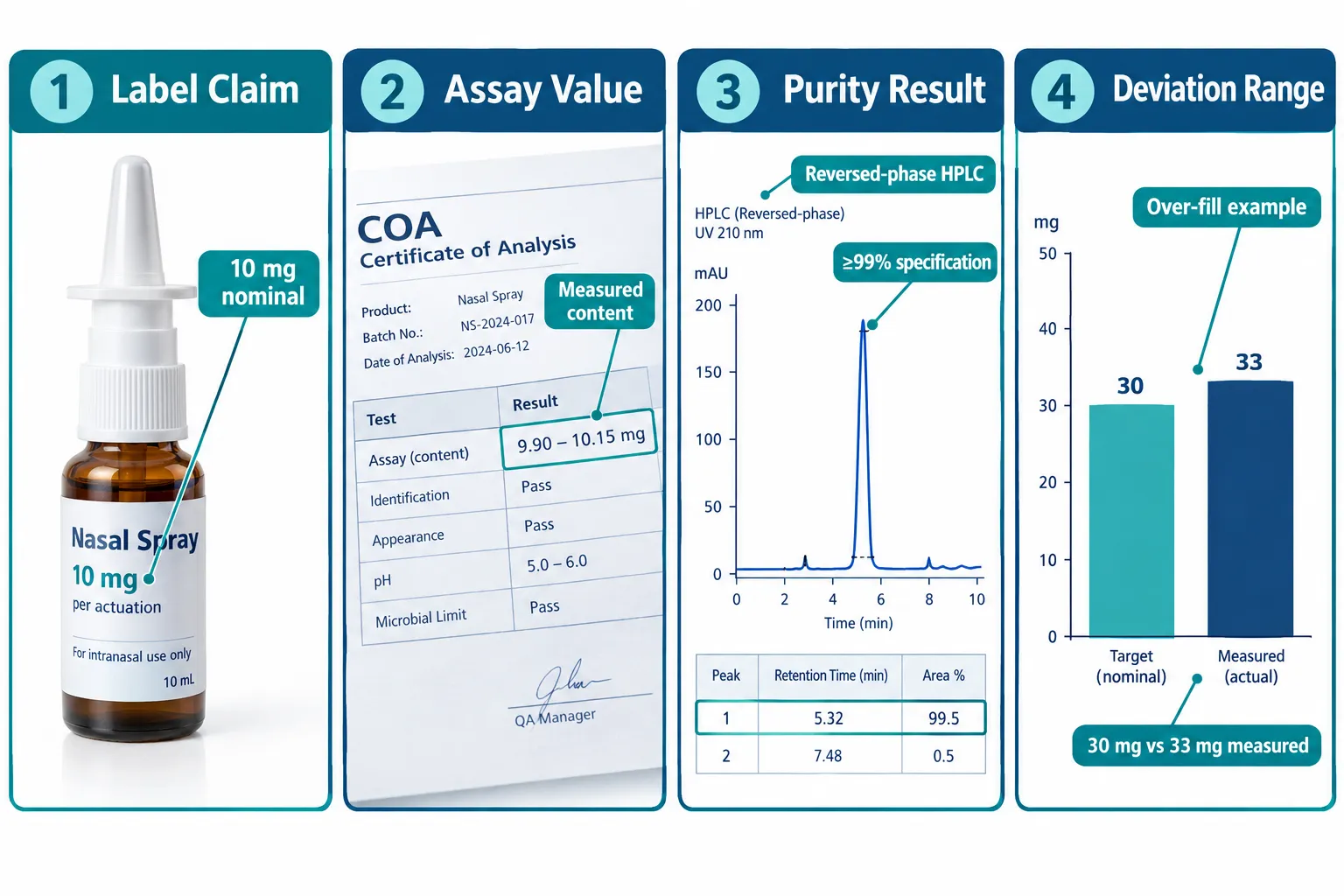
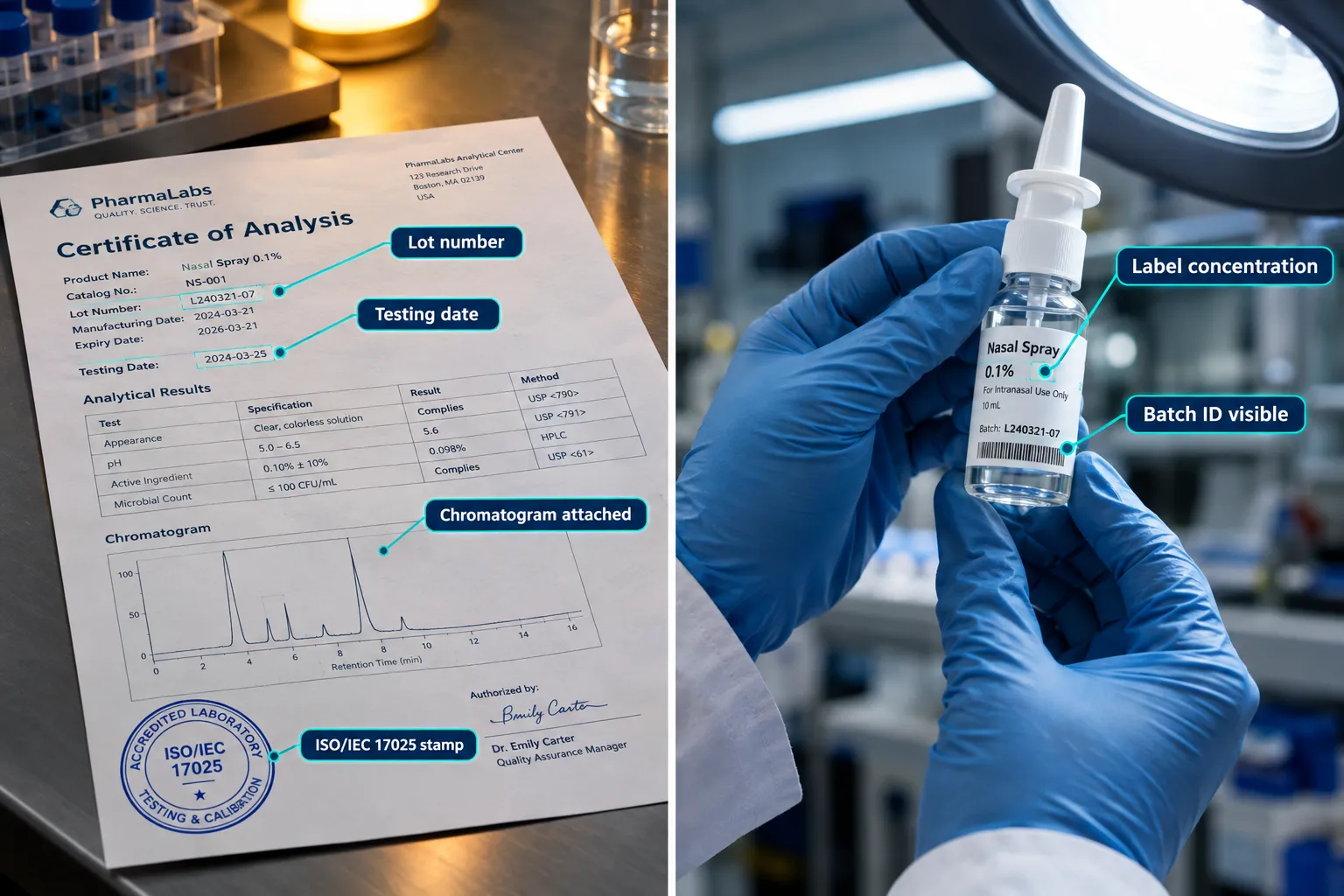
  • Assay values on a Certificate of Analysis (COA) often differ slightly from label claims, so always compare both before drawing conclusions about dose.
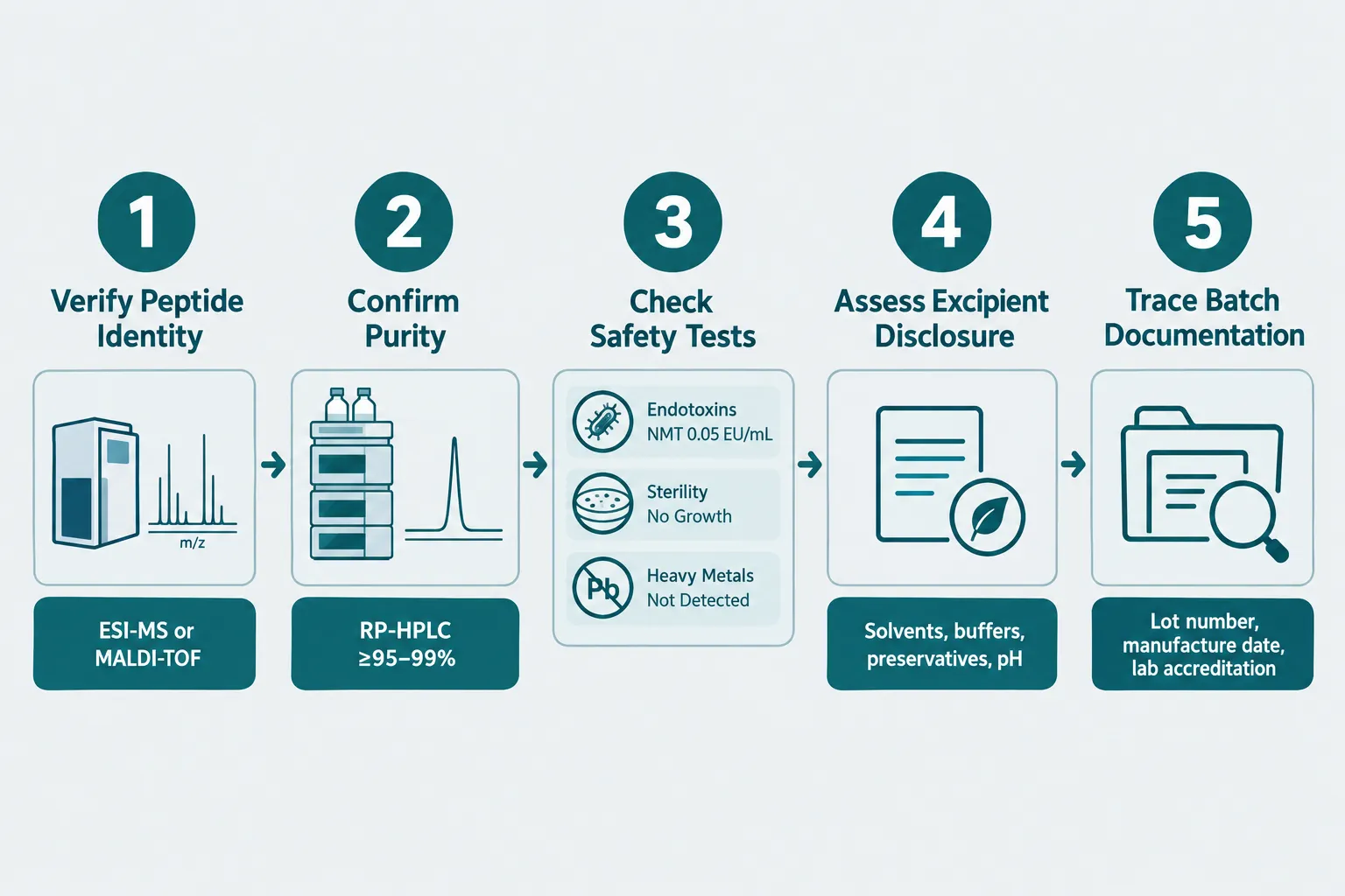
  • A research-grade COA should confirm peptide identity via mass spectrometry, demonstrate purity by HPLC, and report endotoxin, sterility, and heavy metal results.
  • Excipient disclosure, solvents, buffers, preservatives, pH, and osmolarity, is part of batch documentation, not a bonus detail.
  • Batch traceability requires lot numbers, manufacturing and testing dates, and ideally access to raw chromatograms or spectra.
  • Regulatory approval for Semax exists only in Russia; researchers in other jurisdictions should treat it strictly as an experimental compound and prioritize rigorous documentation over anecdotal claims.

Why Concentration Labeling Deserves Scrutiny

The nominal strength printed on a nasal spray label is a starting point, not a guarantee. When evaluating Semax nasal spray reviews for research use, the first task is to locate the assay value on the accompanying COA and compare it against the label claim.

Real-world data from 2026 batch reports illustrate the typical range of variation. A 10 mg labeled vial may test at 9.90-10.15 mg measured content. A 30 mg labeled nasal spray can come back at 33 mg confirmed by an independent laboratory. Neither result is necessarily alarming, but both change the effective per-spray dose calculation. Researchers who rely on the label alone will introduce systematic error into their work.

Why Concentration Labeling Deserves Scrutiny

Key questions to ask about concentration:

  • What is the labeled mg per container and the labeled volume (mL)?
  • What is the measured content (mg) on the COA, and what is the percentage deviation from label?
  • How many actuations does the container deliver, and what is the calculated dose per spray based on assay content, not nominal content?
  • Is the purity specification stated (typically 95-99% or higher), and does the assay value meet it?

Suppliers now advertising purity verified at 99% or above by reversed-phase HPLC and mass spectrometry are setting a useful benchmark. The expected average molecular mass for the Semax heptapeptide (C37H51N9O10S) is 813.93 Da; a COA that confirms this value via ESI-MS or MALDI-TOF provides identity assurance that a purity number alone cannot.

For broader context on how related nasal spray peptides are sourced and evaluated, see guidance on where to buy Selank nasal spray, a structurally related compound that faces similar documentation requirements.


A Practical Framework for Evaluating COA Quality

The most actionable Semax nasal spray reviews for research use are structured around four pillars: identity, purity, safety, and documentation. Researchers who apply this framework systematically can compare suppliers on objective grounds rather than marketing language.

A Practical Framework for Evaluating COA Quality

Step 1, Confirm Peptide Identity

Mass spectrometry is the only reliable tool for sequence confirmation. The COA should state the analytical method (ESI-MS or MALDI-TOF), the theoretical molecular mass, and the observed mass. A purity figure without identity confirmation is insufficient for research use.

Step 2, Verify Chromatographic Purity

Reversed-phase HPLC is the standard method. Look for the stated specification (e.g., ≥99%), the measured result, and ideally access to the chromatogram itself. Some vendors now offer COA libraries where researchers can download raw chromatograms and impurity profiles per lot.

Step 3, Review Safety-Related Metrics

This step is non-negotiable for any compound delivered via nasal mucosa. A complete safety panel includes:

Test Acceptable Result Example
Endotoxins NMT 0.05 EU/mL
Sterility No Growth
Heavy Metals Not Detected

Batch reports from leading suppliers in 2026 show that these metrics are now routinely included. If a COA omits them entirely, that absence is itself a red flag.

Step 4, Assess Excipient and Formulation Disclosure

Excipients affect nasal mucosal compatibility, peptide stability, and osmolarity. A research-grade product should disclose solvents, buffers, preservatives, pH, and osmolarity. When vendors treat excipient lists as proprietary, researchers lose the ability to assess formulation-related variables in their own protocols.

Step 5, Trace Batch Documentation

Full traceability means: lot number, manufacturing date, testing date, testing laboratory name, and ideally a statement of laboratory accreditation (ISO/IEC 17025 is the relevant standard for analytical testing labs). Some suppliers now offer searchable batch-report portals; others provide only a sparse index with no recorded measured values, requiring manual inspection of each PDF.

"Fewer accurate data points are more valuable than a dense document that omits the testing laboratory name."

This principle applies directly when comparing vendors. A COA that records the lot number, dates, accredited lab, chromatogram, and safety panel is categorically more useful than a glossy product page with a purity claim and no supporting data. Researchers interested in how documentation standards apply across the peptide category can explore the Semax peptide resource library for additional batch-level context.


Regulatory Context and Research-Only Use

Understanding the regulatory landscape is essential before procuring Semax nasal spray for any purpose. Semax has been approved as a nasal spray in Russia since 1994, primarily for cerebrovascular indications. It is not approved in the United States, the European Union, or most other jurisdictions.

Regulatory Context and Research-Only Use

A 2024 FDA scientific review, widely discussed in 2026 commentaries, found no human pharmacokinetic studies for Semax by any route of administration and judged the efficacy evidence insufficient for the nominated compounding uses. FDA staff recommended against placing Semax on the U.S. compounding list. An advisory committee vote in July 2026 reportedly favored inclusion by a narrow margin (8-5, one abstention), but no regulatory approval followed from that vote alone.

What this means for researchers:

  • Semax nasal spray must be treated as an experimental compound, not a validated therapeutic.
  • Trial data that does exist is largely from small, often unblinded studies concentrated in Russian research institutions.
  • The mismatch between Russian approval and Western regulatory standards makes COA scrutiny more important, not less, because researchers bear the burden of quality verification themselves.

For comparison, researchers working with other investigational peptides face similar documentation demands. The CJC-1295 mechanism and pharmacokinetic comparison resource illustrates how analytical rigor applies across the broader peptide research field. Similarly, those exploring metabolic research themes may find the SS-31 mitochondrial research themes overview useful for understanding how documentation standards translate across compound classes.


Conclusion

Evaluating Semax nasal spray reviews for research use in 2026 means moving past subjective reports and into the language of analytical chemistry. The most reliable suppliers distinguish themselves through measurable attributes: assay values that can be cross-checked against label claims, purity results backed by chromatograms, safety panels covering endotoxins and sterility, and batch documentation that includes lot numbers, testing dates, and accredited laboratory names.

Actionable next steps for researchers:

  1. Request or download the batch-specific COA before purchasing, not after.
  2. Calculate the per-spray dose using assay content, not the nominal label strength.
  3. Confirm that identity testing (MS) and purity testing (HPLC) are both present and method-described.
  4. Verify that endotoxin, sterility, and heavy metal results are explicitly stated.
  5. Check whether the testing laboratory is named and, ideally, ISO/IEC 17025 accredited.
  6. Demand explicit excipient disclosure to assess nasal mucosal compatibility and formulation stability.

Researchers sourcing other nasal spray peptides can apply the same framework. The where to buy Selank nasal spray guide and the GLP-3 RT 20mg peptide nasal spray product page both provide examples of how batch documentation can be presented at the point of purchase. Documentation quality is the single most controllable variable in research-grade peptide procurement, and it is the one most often overlooked.

https://www.puretestedpeptides.com/wp-content/uploads/2026/10/semax-nasal-spray-reviews-for-research-use-how-to-evaluate-concentration-excipie.webp 1254 1254 https://www.puretestedpeptides.com/wp-content/uploads/2026/01/buy-peptides-online.jpg 2026-10-05 13:09:502026-10-05 13:09:50Semax Nasal Spray Reviews for Research Use: How to Evaluate Concentration, Excipients, and Batch Documentation
CJC-1295 With Ipamorelin: How to Separate Additive GH-Pulse Effects From Blend and Timing Artifacts

CJC-1295 With Ipamorelin: How to Separate Additive GH-Pulse Effects From Blend and Timing Artifacts

October 5, 2026/0 Comments/in Uncategorized/by

Clinic-based reports consistently describe 3- to 5-fold increases in GH pulse amplitude when CJC-1295 is paired with ipamorelin, yet as of 2026, not a single published randomized controlled trial has tested the combination in humans. That gap between widespread clinical use and rigorous experimental evidence is not a minor footnote. It means that nearly every claim about "synergistic" or "additive" GH pulses from this blend is built on data that cannot cleanly distinguish true pharmacodynamic interaction from artifacts of dosing schedule, peptide form, meal timing, and background GH status.

Understanding CJC-1295 with ipamorelin: how to separate additive GH-pulse effects from blend and timing artifacts requires a disciplined study-design framework, one that treats synergy as a hypothesis to test, not a starting assumption.

Key Takeaways

  • No RCT has directly tested the CJC-1295 plus ipamorelin combination; all GH-pulse data are clinic-based or extrapolated from single-agent studies.
  • Using CJC-1295 with DAC creates a sustained GH elevation lasting days, which can masquerade as pulse synergy and must be avoided in pulse-focused research.
  • A 2×2 factorial design with vehicle controls is the minimum study architecture needed to isolate true additive GH-pulse effects from timing and exposure artifacts.
  • Fasted pre-sleep dosing and consistent circadian phase-locking reduce meal- and sleep-related variability that inflates apparent pulse amplitude.
  • Staggered dosing intervals (CJC-1295 first, ipamorelin 15-30 minutes later) versus simultaneous injection offer a practical within-subject comparison of pulse shape and kinetics.

Why the Peptide Form Changes Everything

Why the Peptide Form Changes Everything

The first design decision, and the most consequential, is which form of CJC-1295 is used. The DAC (Drug Affinity Complex) version binds albumin after injection, extending its half-life to several days. Single subcutaneous doses have produced 2- to 10-fold increases in mean plasma GH lasting six or more days, with IGF-1 elevations persisting 9-11 days. That pharmacokinetic profile is incompatible with pulsatile GH research. When baseline GH is elevated for nearly two weeks, any ipamorelin-triggered spike departs from an already-raised platform, making the combined peak appear larger than it actually is relative to a true resting state.

CJC-1295 no-DAC (mod-GRF 1-29) solves this problem. Its reported half-life is roughly 30 minutes. Ipamorelin's half-life is approximately 2 hours. Co-administering the two creates a short, overlapping signaling window that preserves a discrete pulsatile pattern. The GHRH receptor (targeted by CJC-1295) and the growth hormone secretagogue receptor (targeted by ipamorelin) are distinct but convergent pathways at the somatotroph cell. Activating both simultaneously amplifies GH release beyond what either agent achieves alone, but only the no-DAC form keeps that interaction measurable as a clean pulse rather than a tonic elevation.

Anyone exploring the Tesamorelin CJC-1295 Ipamorelin 12mg blend should understand this distinction before interpreting any GH data from a multi-peptide formulation.


A Factorial Study Design for CJC-1295 With Ipamorelin: How to Separate Additive GH-Pulse Effects From Blend and Timing Artifacts

A Factorial Study Design for CJC-1295 With Ipamorelin: How to Separate Additive GH-Pulse Effects From Blend and Timing Artifa

The minimum credible architecture for testing this combination is a 2×2 factorial design with four arms:

Arm CJC-1295 no-DAC Ipamorelin
1 Active Placebo
2 Placebo Active
3 Active Active
4 Placebo Placebo

This structure separates three distinct effects: the CJC-1295 main effect, the ipamorelin main effect, and the interaction term. If the combination arm (Arm 3) produces a GH pulse significantly larger than the sum of Arms 1 and 2 above the vehicle baseline (Arm 4), that constitutes evidence of true synergy. If it simply equals the sum, the effect is additive. If it falls short, there is antagonism or receptor saturation.

Pulse Sampling Protocol

Frequent blood sampling is non-negotiable. A practical schedule draws samples at 0, 15, 30, 45, 60, 90, and 120 minutes post-injection. This captures peak GH amplitude, time-to-peak, and pulse duration, three variables that can each shift independently depending on which agent is used.

IGF-1 timing adds a second layer. Because IGF-1 responds to cumulative GH exposure over 12-24 hours rather than to individual pulses, it should be measured at a fixed interval (typically 12 hours post-injection) across all arms. Comparing IGF-1 across arms that differ only in injection timing, without standardizing this window, is one of the most common sources of artifact in clinic-based data.

Vehicle Controls and Blinding

Vehicle controls (the diluent used for reconstitution, without active peptide) must match the injection volume and subcutaneous route of the active arms. For those reviewing reconstitution protocols for blend formulations, the vehicle composition matters: bacteriostatic water alone versus a buffered saline vehicle can affect subcutaneous absorption rate and therefore pulse timing.


Timing Artifacts and How to Control Them

Timing Artifacts and How to Control Them

Timing is the single largest source of non-pharmacodynamic artifact in GH-pulse studies with this combination. Three variables dominate:

1. Meal timing and insulin. Elevated insulin suppresses GH secretion. Guides updated through late 2026 consistently recommend injecting at least 2 hours after the last meal, particularly after carbohydrate intake. Pre-sleep fasted dosing is the standard recommendation because it aligns with the endogenous nocturnal GH surge and removes meal variability from the equation.

2. Circadian phase-locking. Dosing CJC-1295 no-DAC with ipamorelin in the early evening (roughly 18:00-20:00) can generate a GH spike near 01:00 that mirrors the body's natural GHRH-driven nocturnal surge. When all subjects receive the blend at the same circadian phase, observed differences in pulse amplitude between arms are more likely attributable to receptor co-activation than to random variation in sleep onset or meal timing.

3. Staggered versus simultaneous injection. Because GHRH receptor signaling and GHSR signaling have different kinetic profiles, injecting CJC-1295 no-DAC first and ipamorelin 15-30 minutes later can alter peak height and pulse width compared to a simultaneous injection. Analyzing these patterns within the same subject, a crossover element embedded in the factorial design, is one of the few practical methods currently available to distinguish true additive GH-pulse effects from artifacts of an arbitrary "both-at-once" schedule.

"Synergy claims for this blend rely on modeled receptor interactions and clinic experience rather than direct pulse-profiling RCTs, a distinction that matters enormously for interpreting amplitude data."

Practical dosing ranges reported in protocol literature run from 100 to 300 mcg of each peptide per injection, with titration every 1-2 weeks starting near 100 mcg. Cycles of 8-12 weeks followed by approximately 4 weeks off help preserve receptor sensitivity, a relevant consideration because receptor desensitization between cycles can alter baseline GH and create pseudo-synergy in later measurements. Researchers interested in ipamorelin sourcing can review available ipamorelin products and IPA 10mg formulations for reference-grade material.

For broader context on how ipamorelin compares to other growth hormone secretagogues, the Tesamorelin vs Ipamorelin comparison and Tesamorelin benefits overview provide useful mechanistic background. Those evaluating multi-peptide stacks can also reference Tesamorelin CJC-1295 blend dosage guidance for formulation-specific considerations.

Regulatory Context in 2026

Neither CJC-1295 nor ipamorelin is FDA-approved. Both peptides have been nominated and subsequently withdrawn from the 503A compounding list, meaning the combination is not permitted as a standard compounded product in the U.S. as of 2026. An FDA advisory committee specifically voted against allowing either peptide in routine compounding. This regulatory status reinforces that any GH-pulse data with the blend come from research or off-label clinic use, not from approved therapeutic programs, which further underscores the need for rigorous experimental controls when interpreting those data.


Conclusion

The central challenge with CJC-1295 with ipamorelin, how to separate additive GH-pulse effects from blend and timing artifacts, is fundamentally a study-design problem, not just a dosing question. The actionable steps are clear:

  • Use CJC-1295 no-DAC, not the DAC form, to preserve discrete pulsatile kinetics and prevent sustained baseline elevation from inflating apparent synergy.
  • Implement a 2×2 factorial design with vehicle controls so that main effects and the interaction term can be estimated independently.
  • Standardize timing by phase-locking injections to a consistent circadian window and enforcing a minimum 2-hour post-meal fast before every injection.
  • Sample frequently (at least seven time points over 120 minutes) and measure IGF-1 at a fixed 12-hour interval across all arms.
  • Test staggered versus simultaneous dosing within subjects to characterize how GHRH and GHSR kinetics interact across different injection intervals.

Until a registered trial applies this framework, the 3- to 5-fold pulse amplitude figures circulating in clinic literature should be treated as directional signals rather than confirmed pharmacodynamic facts. The combination may well be synergistic, but proving it requires separating the biology from the schedule.

https://www.puretestedpeptides.com/wp-content/uploads/2026/10/cjc-1295-with-ipamorelin-how-to-separate-additive-gh-pulse-effects-from-blend-an.webp 1024 1536 https://www.puretestedpeptides.com/wp-content/uploads/2026/01/buy-peptides-online.jpg 2026-10-05 13:04:402026-10-05 13:04:40CJC-1295 With Ipamorelin: How to Separate Additive GH-Pulse Effects From Blend and Timing Artifacts
GLP-3 Retatrutide vs GLP-2-T: Choosing Metabolic, Intestinal, and Organoid Readouts for Comparative Research

GLP-3 Retatrutide vs GLP-2-T: Choosing Metabolic, Intestinal, and Organoid Readouts for Comparative Research

October 5, 2026/0 Comments/in Uncategorized/by

Retatrutide produced mean weight reductions exceeding 24% over 48 weeks in phase 2 trials, a figure that no single-receptor incretin therapy had previously achieved. That single data point signals why researchers face a genuine decision problem when designing comparative studies: retatrutide operates through a fundamentally different biological model than GLP-2-type agents, and conflating their readout frameworks produces unreliable conclusions.

This article builds a practical decision framework for GLP-3 Retatrutide vs GLP-2-T: Choosing Metabolic, Intestinal, and Organoid Readouts for Comparative Research. The goal is to separate retatrutide's multi-receptor systemic model from GLP-2-T's intestinal-repair context, then map each to the endpoints, assay systems, and interpretation boundaries that fit them best.

Key Takeaways

  • Retatrutide simultaneously activates GLP-1R, GIPR, and GCGR, making systemic metabolic endpoints the primary readout domain.
  • GLP-2-type agents act principally on intestinal GLP-2 receptors; their readouts center on mucosal structure, barrier function, and organoid proliferation.
  • Mixing readout frameworks across these two agent classes introduces mechanistic confounds that weaken comparative data.
  • Receptor expression confirmation is a non-negotiable first step before running any parallel assay.
  • Organoid platforms can serve both arms only when receptor expression and endpoint domains are kept strictly separate.

Understanding the Mechanistic Divide

Understanding the Mechanistic Divide

The core challenge in GLP-3 Retatrutide vs GLP-2-T comparative research is that these two agent classes do not share a primary tissue target. Retatrutide (LY3437943) is the first GLP-1/GIP/glucagon triple agonist to complete phase 2 trials in both obesity and type 2 diabetes. It engages three distinct receptors, GLP-1R, GIPR, and GCGR, through a single peptide conjugated to a fatty diacid moiety. The downstream effects span appetite suppression, hepatic fat reduction, and systemic energy expenditure.

GLP-2-type agents (GLP-2-T), by contrast, bind the GLP-2 receptor expressed predominantly on intestinal subepithelial myofibroblasts and enteroendocrine cells. Their documented effects are gut-trophic: increased villus height, crypt cell proliferation, improved mucosal barrier integrity, and enhanced nutrient absorption. These are the readouts that define clinical use cases such as short bowel syndrome and intestinal failure.

The mechanistic divide is not a matter of degree, it is a matter of tissue domain. Retatrutide's phase 2 MASLD trial reinforced this by demonstrating meaningful reductions in liver fat measured by MRI-proton density fat fraction (MRI-PDFF), alongside improvements in hepatic enzymes and non-invasive fibrosis markers. None of those endpoints are relevant to GLP-2-T's primary mechanism.

"Choosing the wrong readout domain does not just weaken a study, it actively misrepresents the biology of both agents."

Researchers looking to source well-characterized material for this work can review options through the Buy Retatrutide Peptide page or explore the broader GLP-3 Retatrutide buy online USA resource for purity and specification details.


Building the Readout Decision Framework

Building the Readout Decision Framework

A structured five-step decision process reduces the risk of endpoint misalignment when designing GLP-3 Retatrutide vs GLP-2-T: Choosing Metabolic, Intestinal, and Organoid Readouts for Comparative Research studies.

Step 1: Define the Research Model

Choose whether the study is in vitro, ex vivo, or in vivo before selecting any endpoint. Retatrutide's receptor profile requires models that co-express GLP-1R, GIPR, and GCGR, hepatocyte lines, adipocyte cultures, pancreatic islet preparations, or diet-induced obesity rodent models. GLP-2-T studies require intestinal epithelial models: primary organoids derived from small intestinal crypts, Caco-2 monolayers, or murine intestinal preparations.

Step 2: Confirm Receptor Expression

This step is non-negotiable. Structural work published in 2024 clarified that retatrutide engages all three receptors with defined binding modes and biased signaling profiles. Before running any assay, confirm receptor expression using qPCR or immunofluorescence. A GLP-2-T study run in a model lacking adequate GLP-2R expression produces noise, not data.

Step 3: Select Primary Endpoints by Agent Class

Endpoint Category Retatrutide (GLP-3-like) GLP-2-T
Primary metabolic Body weight, HbA1c, fasting glucose, lipids Not applicable as primary
Hepatic MRI-PDFF, ALT/AST, fibrosis markers Minimal relevance
Intestinal structure Secondary / safety monitoring Villus height, crypt depth, cell proliferation
Barrier function GI tolerability (adverse event tracking) TEER, tight junction proteins (ZO-1, claudin)
Receptor signaling cAMP, beta-arrestin, downstream kinase activity cAMP via GLP-2R, PI3K/Akt pathway
Organoid readouts Hepatic organoid fat accumulation, glucose uptake Enterocyte and goblet cell differentiation, proliferation index

Step 4: Run Parallel Assays With Separated Domains

Phase 2 safety data for retatrutide identified dose-dependent gastrointestinal adverse events, nausea and diarrhea, alongside signals for lean mass changes and modest heart rate increases. These findings make body composition and heart rate monitoring essential safety-related readouts for the retatrutide arm. They are not shared endpoints with GLP-2-T, which does not produce systemic energy expenditure effects. Running parallel assays only works when each arm's endpoint set remains internally consistent.

Step 5: Interpret Within Defined Boundaries

No published 2025-2026 literature presents a formal GLP-3 vs GLP-2-T comparative schema. The framework described here is constructed from separate evidence streams. Researchers must therefore resist interpreting GLP-2-T intestinal organoid results as informative about systemic metabolic efficacy, and vice versa.

For additional context on related peptide research methodologies, the SLUPP332 oral versus injectable research resource offers useful parallel considerations on route-of-administration variables that affect readout selection.


Organoid Platforms: Where the Two Frameworks Can Intersect

Organoid Platforms: Where the Two Frameworks Can Intersect

Organoid technology is the one research context where GLP-3 Retatrutide vs GLP-2-T: Choosing Metabolic, Intestinal, and Organoid Readouts for Comparative Research can share a platform, but only with strict compartmentalization.

For retatrutide, hepatic organoids derived from primary hepatocytes or iPSC-derived liver cells provide a controlled system for measuring fat accumulation (lipid droplet quantification), glucose output, and receptor-level signaling (cAMP production, beta-arrestin recruitment). These models mirror the liver-fat and glycemic endpoints validated in the MASLD phase 2a trial.

For GLP-2-T, intestinal organoids derived from small intestinal crypts are the gold-standard platform. Key readouts include:

  • Organoid surface area expansion as a proxy for villus-like growth
  • Goblet cell and enterocyte differentiation ratios
  • Tight junction protein expression (ZO-1, occludin, claudin-3)
  • Transepithelial electrical resistance (TEER) as a barrier integrity measure
  • Nutrient transporter expression (SGLT1, GLUT2)

The intersection point is co-culture or multi-organ chip systems that link hepatic and intestinal compartments. In that setting, retatrutide's hepatic effects and GLP-2-T's intestinal effects can be observed in the same platform without conflating their primary readout domains. This remains a speculative but scientifically grounded direction for 2026 research programs.

Researchers sourcing material for organoid-based work should verify purity documentation carefully. The GLP3 RETA CAG 10mg for sale and GLP3 RETA CAG 10mg GA5 product pages provide specification details relevant to in vitro dosing decisions. For broader sourcing considerations, the GLP-3 Retatrutide buy online USA cheap resource addresses cost-quality tradeoffs that matter in multi-arm organoid studies.


Conclusion

The central lesson from aligning retatrutide's clinical trial evidence with GLP-2-T's intestinal biology is straightforward: these agents require separate readout frameworks, not a shared one. Retatrutide's primary endpoints are systemic, body weight, HbA1c, liver fat, lipids, and cardiometabolic markers. GLP-2-T's primary endpoints are structural and functional at the intestinal level, villus morphology, barrier integrity, and organoid proliferation.

Actionable next steps for research teams in 2026:

  1. Audit existing assay panels and remove any endpoints that do not align with the receptor biology of the agent being tested.
  2. Confirm GLP-1R, GIPR, and GCGR co-expression in every model used for retatrutide; confirm GLP-2R expression in every model used for GLP-2-T.
  3. Use MRI-PDFF, ALT/AST, and body composition as the metabolic arm's anchor endpoints.
  4. Use TEER, tight junction protein quantification, and organoid proliferation index as the intestinal arm's anchor endpoints.
  5. Consider multi-organ chip platforms only after single-domain validation is complete.

Maintaining this discipline protects the interpretive value of comparative data and positions research outputs for stronger translational relevance as triple-agonist therapies move through phase 3 development.

https://www.puretestedpeptides.com/wp-content/uploads/2026/10/glp-3-retatrutide-vs-glp-2-t-choosing-metabolic-intestinal-and-organoid-readouts.webp 1024 1536 https://www.puretestedpeptides.com/wp-content/uploads/2026/01/buy-peptides-online.jpg 2026-10-05 13:04:282026-10-05 13:04:28GLP-3 Retatrutide vs GLP-2-T: Choosing Metabolic, Intestinal, and Organoid Readouts for Comparative Research
Phosphate-Buffered Saline for Peptide Research: Osmolality, pH Drift, and Vehicle-Control Design

Phosphate-Buffered Saline for Peptide Research: Osmolality, pH Drift, and Vehicle-Control Design

October 5, 2026/0 Comments/in Uncategorized/by

Fewer than half of published peptide studies explicitly report the osmolality of their vehicle solution, yet that single omission can invalidate injection-site comparisons, confound receptor-binding data, and mask sequence-specific degradation. Getting phosphate-buffered saline for peptide research right, from osmolality and pH drift to vehicle-control design, is not a minor procedural detail. It is a foundational experimental decision.

Key Takeaways

  • Standard 1X PBS at pH 7.4 with an osmolality of 280-315 mOsm/kg is isotonic with plasma and appropriate for most short-term peptide assays.
  • pH drift during storage, not osmolality alone, is the primary driver of hydrolysis and deamidation in PBS-stored peptides.
  • PBS is not universally recommended as a primary reconstitution medium; marginally soluble or deamidation-prone peptides may need alternative buffers.
  • Vehicle controls must match the active peptide solution in both PBS formulation and osmolality to isolate pharmacologic effects.
  • Peptides reconstituted in PBS typically have a working shelf life of one to four weeks at 4 degrees C, depending on sequence sensitivity.

PBS Composition, Osmolality, and Why Isotonicity Matters

Standard 1X PBS used in peptide work contains approximately 137 mM NaCl, 2.7 mM KCl, and a 10 mM phosphate buffer. Together, these components yield an osmolality in the 280-315 mOsm/kg range, which closely mirrors physiological plasma osmolality. Modern commercial PBS products marketed specifically for peptide and cellular assays now explicitly specify osmolality around 290-310 mOsm/kg, framing this as "physiological osmotic conditions" rather than leaving it as an implied property.

PBS Composition, Osmolality, and Why Isotonicity Matters

Why does osmolality matter so much? Hypo-osmotic vehicles cause cells to swell; hyperosmotic vehicles cause shrinkage. Both alter membrane permeability and local pharmacokinetics at injection sites, introducing confounding variables that have nothing to do with the peptide being studied. A diluent-selection review from 2026 notes that vehicles outside the 280-300 mOsm/kg window are associated with increased injection-site irritation and altered local peptide uptake. For researchers sourcing peptides such as those reviewed in resources covering GH analogue peptides or SS-31 mitochondrial research themes, matching vehicle osmolality to the physiologic range is a prerequisite for clean data.

Practical osmolality checklist:

Parameter Target Value Risk if Outside Range
Osmolality 280-315 mOsm/kg Injection-site irritation, altered PK
pH 7.4 (± 0.1) Hydrolysis, deamidation, aggregation
NaCl concentration ~137 mM Precipitation for low-solubility peptides
Phosphate buffer ~10 mM Catalytic hydrolysis at higher concentrations

pH Drift in PBS: The Hidden Stability Risk

Osmolality sets the stage, but pH drift is the real antagonist in long-term peptide storage. PBS provides good initial pH control at 7.4, yet gradual drift during storage can accelerate hydrolysis, deamidation, and oxidation independently of osmolality. Even a shift of 0.2-0.3 pH units can materially shorten the working-stock stability of sensitive sequences.

pH Drift in PBS: The Hidden Stability Risk

The mechanism is specific. Phosphate buffers at physiologic pH promote general acid-base catalysis. Deamidation of asparagine-containing peptides, particularly those with Asn-Gly motifs, proceeds measurably faster in phosphate buffer than in unbuffered water at the same nominal pH. This means that even a well-prepared PBS stock does not offer indefinite protection.

"Peptides reconstituted in PBS typically have a working shelf life of about one to four weeks at 4 degrees C, depending on sequence and susceptibility to hydrolysis or deamidation."

Sequences at elevated risk in PBS:

  • Peptides containing Asn-Gly motifs (deamidation-prone)
  • Methionine-containing peptides (oxidation risk)
  • Peptides with labile ester or amide bonds (hydrolysis risk)
  • Marginally soluble peptides with high ionic-strength sensitivity

For researchers working with complex peptide formulations, such as those described in the polypeptide peptides in cardiometabolic models overview, understanding these sequence-specific risks before committing to PBS as a vehicle is essential.

When to consider alternatives to PBS:

  • Deamidation-prone sequences: histidine buffer (pH 5.5-7) or acetate buffer (pH 3.5-5.5)
  • Long-term storage beyond four weeks: reduced-phosphate or non-phosphate formulations
  • Marginally soluble peptides: HEPES-buffered saline or low-salt alternatives
  • Peptides with metal-binding motifs: avoid PBS due to phosphate-divalent cation interactions

Vehicle-Control Design Using Phosphate-Buffered Saline for Peptide Research

Designing a proper vehicle control is where phosphate-buffered saline for peptide research moves from chemistry into experimental rigor. The vehicle control must be identical to the active peptide solution in every parameter except the presence of the peptide itself. That means matching pH, osmolality, buffer concentration, and preparation method, not simply using "PBS" as a generic label.

Vehicle-Control Design Using Phosphate-Buffered Saline for Peptide Research

Core principles for matched vehicle-control design:

  1. Use the same PBS lot for both vehicle control and active peptide solution to eliminate batch-to-batch variability.
  2. Verify osmolality of both solutions with an osmometer before use; do not rely solely on calculated values.
  3. Monitor pH at each experimental timepoint, not just at preparation. Log any drift and flag deviations exceeding 0.1 pH units.
  4. Check solubility visually after adding PBS to the peptide. Cloudiness or particulate formation signals incompatibility; switch to an alternative buffer rather than proceeding.
  5. Store vehicle control under identical conditions, same temperature, same container type, same light exposure, as the peptide solution.

Researchers sourcing peptides from a best peptide supplier should request certificate-of-analysis data that includes solubility recommendations, as these directly inform whether PBS is an appropriate vehicle for a given sequence.

For studies involving receptor-binding assays, cell-based models, or preclinical in vivo work, including research with peptides such as those discussed in resources on where to buy research-grade Glow Blend peptide, a matched PBS vehicle control at pH 7.4 and 280-300 mOsm/kg remains the standard baseline. Deviating from this standard without documentation introduces uncontrolled variables that reviewers and regulatory bodies will flag.

Buffer alternatives at a glance:

Buffer pH Range Best Use Case
PBS (pH 7.4) 6.8-7.6 Cell-based assays, receptor binding, short-term in vivo
HEPES-buffered saline 7.0-7.6 High ionic-strength-sensitive peptides
Histidine buffer 5.5-7.0 Deamidation-prone sequences, formulation stability
Acetate buffer 3.5-5.5 Acidic-pH stability studies

Expert consensus in 2026 points toward conditional PBS use with tighter experimental controls, not abandonment. PBS remains the appropriate vehicle for physiologic, short-duration experiments where its osmolality and pH profile are genuine advantages. The movement is toward tailored buffer systems for long-term storage and sequence-specific challenges, with PBS retained as the default for well-characterized, water-soluble peptides in cell culture and receptor-binding contexts. Researchers exploring newer peptide classes, including those reviewed in the SLUPP332 oral versus injectable research discussion, will find that route of administration adds another layer of vehicle-selection complexity that PBS alone may not address.


Conclusion

Phosphate-buffered saline for peptide research remains a cornerstone vehicle, but it demands deliberate selection rather than default use. The practical steps are clear: verify osmolality falls within 280-315 mOsm/kg, confirm pH at 7.4 before and during storage, monitor for pH drift that accelerates deamidation and hydrolysis, and design vehicle controls that are compositionally identical to the active solution. For deamidation-prone, marginally soluble, or metal-binding peptides, pivot to histidine, acetate, or HEPES-based buffers before data collection begins rather than after anomalies appear. Document every parameter, use the same PBS lot across all arms of a study, and treat visible precipitation as a hard incompatibility signal. These steps will protect data integrity, reduce confounding, and produce vehicle-control comparisons that hold up to scrutiny.

https://www.puretestedpeptides.com/wp-content/uploads/2026/10/phosphate-buffered-saline-for-peptide-research-osmolality-ph-drift-and-vehicle-c.webp 1024 1536 https://www.puretestedpeptides.com/wp-content/uploads/2026/01/buy-peptides-online.jpg 2026-10-05 13:04:002026-10-05 13:04:00Phosphate-Buffered Saline for Peptide Research: Osmolality, pH Drift, and Vehicle-Control Design
Page 6 of 81«‹45678›»
×

Helpful Links

  • My account
  • Cart
  • Checkout
  • Refund and Returns Policy
  • Privacy Policy
  • SMS Privacy Policy
  • Login
  • My Account
  • Logout

USA Made Lab Tested Peptides

All products are sold for research, laboratory, or analytical purposes only, and are not for human consumption


Pure Tested Peptides is a chemical supplier. Pure Tested Peptides is not a compounding / chemical compounding facility as defined under 503A of the Federal Food, Drug, and Cosmetic act. Pure Tested Peptides is not an outsourcing facility as defined under 503B of the Federal Food, Drug, and Cosmetic act.

The statements made within this website have not been evaluated by the US Food and Drug Administration. The products we offer are not intended to diagnose, treat, cure or prevent any disease.

Human/Animal Consumption Prohibited. Laboratory/In-Vitro Experimental Use Only

Scroll to top Scroll to top Scroll to top