Call or Text 727-513-9780
  • Shopping Cart Shopping Cart
    0Shopping Cart
Pure Tested Peptides | America's most trusted Peptides for sale online
  • Peptides for sale
    • Oral Peptides for sale
      • Peptide Capsules for sale
      • BPC 157 Capsules 1000mcg
      • SLU-PP-332 Capsules | 1000 mcg
      • 5-Amino-1MQ 50mg Capsules
      • Tesofensine 500mcg
    • All Peptides for sale
    • Peptide Sprays
      • BPC 157 Nasal Spray Kit
      • BPC-157 TB500 Nasal Spray Kit
      • Semax Nasal Spray 10mg
      • Selank – Nasal Spray Kit – 10mg
      • Epithalon 50MG Nasal Spray Kit
      • Ipamorelin 10mg Nasal Spray
      • Klow Nasal Spray (BPC-157 + TB-500 + GHK-Cu + KPV) | 80mg
      • Hulk Nasal Spray Tesa / Ipa Blend 6/3 MG
      • Klow Nasal Spray
      • NAD + 500 mg Nasal Spray
      • PT-141 Nasal Spray Kit
    • GHRH Peptides
      • Ipa Peptides
      • CJC-1295 Peptides
        • CJC-1295 with DAC 5 mg
        • CJC-1295 without DAC 5 mg
        • CJC-1295 Ipa 10mg
      • Tesa Peptides
        • Tesa Peptide
        • Tesa 20 mg
    • GHK-Cu Peptides
      • All GHK-Cu Peptides
      • GHK-Cu 100mg
      • KLOW Peptide Blend – Buy KLOW blend online
    • BPC Peptides
      • All BPC Peptides
      • BPC-157
      • BPC-157 TB-500
      • BPC 157 capsules 1000mcg
    • SLU-PP-332 Peptides
      • All SLU-PP-332 Peptides
      • SLU-PP-332 5mg
    • GLP3 Peptides
    • PT-141 Peptides
      • PT-141 Peptides for sale
      • PT-141 10mg
      • PT-141 Nasal Spray
    • CAG Peptides
      • Lipo-C Peptide Blend
      • CAG 5mg
      • CAG 10mg
    • MOTS-C Peptides
      • MOTS-C Peptides for sale
      • MOTS-c peptide
      • MOTS-c 10mg *6 pack*
    • 5 Amino 1MQ Peptides
      • 5 Amino 1MQ Peptides for sale
      • 5-Amino-1MQ 50mg Capsules
      • 5-Amino-1MQ 5mg
    • Epithalon Peptides
      • Epithalon Peptides for sale
      • Epithalon 10mg
      • Epithalon 50mg
  • Shop
    • GLPs
      • 5-Amino-1MQ 50mg Capsules
      • 5-Amino-1MQ 5mg
      • L-Carnitine 500mg/ml
      • Tesofensine 500mcg
      • SLU-PP-332 5mg
      • MOTS-c 10mg *6 pack*
    • Epithalon & BPC Peptides
      • Epithalon 10mg
      • Epithalon 50mg
      • BPC-157
      • BPC 157 capsules 1000mcg
      • BPC-157 TB-500
      • BPC-157 TB500 Nasal Spray Kit
      • BPC 157 Nasal Spray Kit
    • BPC TB-500 & NAD+ Peptides
      • NAD+ 500 mg
      • KLOW Peptide Blend – Buy KLOW blend online
      • GLOW Peptide Blend
      • TB 500 5mg
      • BPC 157 capsules 1000mcg – Supplement
      • BPC 157 Nasal Spray Kit
      • BPC-157
      • BPC-157 TB500 Nasal Spray Kit
      • BPC-157 TB-500
      • BPC 157 capsules 1000mcg
    • LL-37 Peptide
      • LL-37 10 mg
    • MOTS-C & Selank
      • MOTS-c peptide
      • Selank 10mg
    • GHK Peptides
      • GHK-Cu 100mg
      • GLOW Peptide Blend
      • KLOW Peptide Blend – Buy KLOW blend online
  • COAs
  • Wholesale
    • Wholesale Peptides for sale
  • PTP FAQ
  • Affiliates
    • Selank Peptide: Advanced Pharmacological Mechanisms Underlying Its Anxiolytic and Nootropic Effects in Research
      • 5-Amino-1MQ Peptide: Detailed Mechanisms of NNMT Inhibition and Its Impact on Cellular Metabolism Research
        • MOTS-C Peptide: Unraveling Its Role in Mitochondrial Dynamics and Energy Metabolism Research
          • Epithalon Peptide and Telomerase Regulation: Investigating Its Impact on Cellular Senescence and Lifespan Research Models
            • GLP2-T vs GLP2 Tirz Peptide: Understanding the Naming, Mechanistic Nuances, and Research Implications for Gut Health
      • Peptides and Polypeptides in Modern Pharmacology: What Research on Metoprolol, Prednisone, and Amlodipine Reveals
        • Polypeptide Peptides vs NSAIDs: What Naproxen and Diclofenac Teach Tissue-Repair Researchers About BPC‑157 and TB‑500
          • Peptides and Polypeptides in Endocrine Pharmacology: How GLP-1, GLP-2, and GLP-3 Retatrutide Differ From Classic Drugs Like Prednisone and Amlodipine
          • 5‑Amino‑1MQ Peptide and NNMT Inhibition: How It Compares With Statins Like Atorvastatin in Adipose and Lipid Metabolism Research
      • Enclomiphene, Estrogen Receptor Signaling, and Luteinizing Phase Biology: What Hormone Researchers Should Measure
        • Mesenchymal Stem Cells, BPC‑157, and GHK‑Cu: How Tissue Repair Peptides Compare With Classic NSAIDs Like Naproxen in Injury Models
          • Peptide Calculator Use Cases Beyond Growth Hormone: Working Through GLP‑3 Retatrutide, MOTS‑c, and BPC‑157 Research Dosing
        • Polypeptide Peptides in Endocrine and Metabolic Pharmacology: Lessons From Amlodipine, Prednisone, and Metoprolol
        • Peptides Calculator for Advanced Blends: Worked Examples for Tesamorelin, CJC‑1295, and Ipamorelin Stacks
          • Peptides and Polypeptides in Human Physiology: How Molecular Size Shapes Research Applications
            • Selank Peptide: Advanced Pharmacological Mechanisms Underlying Its Anxiolytic and Nootropic Effects in Research
          • 5-Amino-1MQ Peptide: Detailed Mechanisms of NNMT Inhibition and Its Impact on Cellular Metabolism Research
            • MOTS-C Peptide: Unraveling Its Role in Mitochondrial Dynamics and Energy Metabolism Research
              • Epithalon Peptide and Telomerase Regulation: Investigating Its Impact on Cellular Senescence and Lifespan Research Models
                • GLP2-T vs GLP2 Tirz Peptide: Understanding the Naming, Mechanistic Nuances, and Research Implications for Gut Health
                  • Peptides and Polypeptides in Endocrine Pharmacology: How GLP-1, GLP-2, and GLP-3 Retatrutide Differ From Classic Drugs Like Prednisone and Amlodipine
                    • Polypeptide Peptides vs NSAIDs: What Naproxen and Diclofenac Teach Tissue-Repair Researchers About BPC‑157 and TB‑500
                      • Peptides and Polypeptides in Modern Pharmacology: What Research on Metoprolol, Prednisone, and Amlodipine Reveals
                        • 5‑Amino‑1MQ Peptide and NNMT Inhibition: How It Compares With Statins Like Atorvastatin in Adipose and Lipid Metabolism Research
                        • Enclomiphene, Estrogen Receptor Signaling, and Luteinizing Phase Biology: What Hormone Researchers Should Measure
                        • Mesenchymal Stem Cells, BPC‑157, and GHK‑Cu: How Tissue Repair Peptides Compare With Classic NSAIDs Like Naproxen in Injury Models
                        • Peptide Calculator Use Cases Beyond Growth Hormone: Working Through GLP‑3 Retatrutide, MOTS‑c, and BPC‑157 Research Dosing
                        • Polypeptide Peptides in Endocrine and Metabolic Pharmacology: Lessons From Amlodipine, Prednisone, and Metoprolol
                        • Peptides Calculator for Advanced Blends: Worked Examples for Tesamorelin, CJC‑1295, and Ipamorelin Stacks
                        • Peptides and Polypeptides in Human Physiology: How Molecular Size Shapes Research Applications
                        • Selank Peptide: Advanced Pharmacological Mechanisms Underlying Its Anxiolytic and Nootropic Effects in Research
                        • MOTS-C Peptide: Unraveling Its Role in Mitochondrial Dynamics and Energy Metabolism Research
                        • Peptides and Polypeptides in Modern Pharmacology: What Research on Metoprolol, Prednisone, and Amlodipine Reveals
                        • Peptides and Polypeptides in Endocrine Pharmacology: How GLP-1, GLP-2, and GLP-3 Retatrutide Differ From Classic Drugs Like Prednisone and Amlodipine
                        • 5-Amino-1MQ Peptide: Detailed Mechanisms of NNMT Inhibition and Its Impact on Cellular Metabolism Research
                        • Mesenchymal Stem Cells, BPC‑157, and GHK‑Cu: How Tissue Repair Peptides Compare With Classic NSAIDs Like Naproxen in Injury Models
                        • Peptide Calculator Use Cases Beyond Growth Hormone: Working Through GLP‑3 Retatrutide, MOTS‑c, and BPC‑157 Research Dosing
                        • GLP2-T vs GLP2 Tirz Peptide: Understanding the Naming, Mechanistic Nuances, and Research Implications for Gut Health
                        • Polypeptide Peptides in Endocrine and Metabolic Pharmacology: Lessons From Amlodipine, Prednisone, and Metoprolol
                        • Polypeptide Peptides vs NSAIDs: What Naproxen and Diclofenac Teach Tissue-Repair Researchers About BPC‑157 and TB‑500
                        • Peptides and Polypeptides in Human Physiology: How Molecular Size Shapes Research Applications
                        • Peptides Calculator for Advanced Blends: Worked Examples for Tesamorelin, CJC‑1295, and Ipamorelin Stacks
                        • 5‑Amino‑1MQ Peptide and NNMT Inhibition: How It Compares With Statins Like Atorvastatin in Adipose and Lipid Metabolism Research
                        • Enclomiphene, Estrogen Receptor Signaling, and Luteinizing Phase Biology: What Hormone Researchers Should Measure
                        • Epithalon Peptide and Telomerase Regulation: Investigating Its Impact on Cellular Senescence and Lifespan Research Models
                        • Best research protocol Klow blend
                        • best time to take BPC-157
                        • best time to take DSIP (Delta Sleep Inducing Peptide)
                        • best time to take CJC-1295
                        • best time to take AOD-9604
                        • best time to take Follistatin 344
                        • best time to take Ipamorelin
                        • best time to take MK-677 (Ibutamoren)
                        • best time to take Ligandrol (LGD-4033) — research compound
                        • best time to take Ostarine (MK-2866) — research compound
                        • best time to take GHK-CU
                        • best time to take TB-500
                        • best time to take MOTS-c
                        • best time to take Semax
                        • best time to take RAD-140 (Testolone) — research compound
                        • best time to take Thymosin Alpha-1
                        • best time to take PEG-MGF
                        • Biolife Plasma, Octapharma Plasma, and Research Peptides: How Plasma Donation Labs Differ From Peptide Suppliers
                        • best time to take YK-11 — research compound
                        • best time to take PT-141 (Bremelanotide)
                        • Best research protocol Klow blend
                        • 5-Amino-1MQ and MOTS-C Synergy: Metabolic Signaling, Mitochondria, and Research Design
                        • BPC-157 and TB-500: Investigating Their Combined Effects on Angiogenesis and Cellular Migration in Tissue Repair Models
                        • BPC-157 Peptide: Gut Barrier Function, Inflammation, and Tissue-Recovery Research
                        • 5‑Amino‑1MQ and MOTS‑c Synergy in Metabolic Research: Designing NNMT and Mitochondrial Biogenesis Stacks
                        • CJC-1295 with DAC vs. Without DAC: Half-Life, Release Kinetics, and Research Implications
                        • CJC‑1295 with DAC vs. Without DAC: Expanding on Half‑Life Differences Using Tesamorelin and Ipamorelin Blend Case Studies
                        • Collagen Biology and Copper‑Binding Peptides: How GHK‑Cu, Glow Blend, and Klow Blend Interact with Skin and Connective Tissue
                        • Collagen Biology and Regenerative Peptides: How GHK‑Cu, Glow Blend, and Klow Blend Affect Extracellular Matrix Research
                    • DNA, Telomeres, and Longevity Peptides: Positioning Epithalon and MOTS‑c in Genetic Aging Research
                      • Enclomiphene Citrate: serm Mechanism, Testosterone Research, and Stack Compatibility
                        • Enclomiphene vs Enclomiphene Citrate: Formulation, Bioavailability, and Research Distinctions
                        • Epithalon Peptide Research: Telomerase Activation, Aging, and Pineal Gland Function
                        • Estrogen Receptor Signaling and Enclomiphene: How Selective Modulators Compare with Classic Polypeptide Hormones
                        • GHK-Cu Peptide: Advanced Mechanisms in Extracellular Matrix Remodeling and Wound Healing Research
                        • GHK-Cu Peptide: Collagen Synthesis, Wound Repair, and Skin-Barrier Research Models
                        • GLP-1 vs GLP-3 vs GLP-2: Peptide Classification and Research Applications
                        • GLP-2 Peptide Research Guide: Gut Barrier Function, Nutrient Absorption, and Intestinal Recovery Models
                        • GLP-3 Retatrutide vs. GLP-1 Drugs: What Triple-Agonist Biology Changes in Research Models
                        • Ipamorelin and Tesamorelin Combination: Synergistic GH Secretagogue Research and Dosing Protocols
                        • GLP2 Tirz Peptide: What It Is, Why the Name Exists, and How Researchers Should Interpret It
                        • Klow Blend Peptide Nasal Spray: What the Formulation Is Trying to Do in Cognitive Research
                        • Mitochondria, NNMT Inhibition, and Peptide Modulators: Where MOTS‑c and 5‑Amino‑1MQ Fit in Cellular Energy Research
                        • MOTS-c Peptide: Mitochondrial Function, Energy Metabolism, and What Researchers Measure
                        • MOTS-c vs. 5-Amino-1MQ: Which Metabolic Research Questions Each Compound Actually Answers
                        • Nasal Spray Peptides: Bioavailability, Administration, and Semax/Selank Research Applications
                        • PT-141 Peptide Research: Mechanism of Action and Melanocortin Receptor Signaling
                        • Retatrutide for Research: Mechanism, Structure, and GLP-1/GLP-3 Dual Action
                        • Retatrutide for Obesity and Type 2 Diabetes: What the Latest Trial Data Suggest
                        • Tesofensine Peptide Research: Mechanism, Appetite Suppression, and Neuropeptide Y Pathways
  • Contact
    • Contact Customer Service
    • Text Customer Support
  • About US
  • Shop all peptides
  • Affiliate Program
    • Affiliate Signup
  • Login / Register Login / Register Page Link Login / Register Page Link
  • Click to open the search input field Click to open the search input field Search
  • Menu Menu
Vitamin D, MOTS-c, and 5-Amino-1MQ: Designing Nutrient-Controlled Cellular Energy Experiments

Vitamin D, MOTS-c, and 5-Amino-1MQ: Designing Nutrient-Controlled Cellular Energy Experiments

October 2, 2026/0 Comments/in Uncategorized/by

Researchers who publish conflicting results on metabolic peptides often share one overlooked flaw: they never standardized the vitamin D status of their cell cultures or animal models before the experiment began. That single omission can silently shift AMPK activity, mitochondrial respiration, and insulin signaling, making every downstream comparison unreliable. Understanding how to design nutrient-controlled cellular energy experiments around Vitamin D, MOTS-c, and 5-Amino-1MQ is therefore not a minor methodological footnote; it is the foundation of reproducible metabolic science.

Key Takeaways

  • Vitamin D, acting through the vitamin D receptor (VDR), directly modulates AMPK, mTOR, and mitochondrial oxidative capacity, making vitamin D status a critical baseline variable in any cellular energy experiment.
  • MOTS-c is a 16-amino-acid mitochondrial-derived peptide that activates AMPK by accumulating AICAR, linking mitochondrial stress signals to whole-cell energy regulation.
  • 5-Amino-1MQ inhibits nicotinamide N-methyltransferase (NNMT), redirecting nicotinamide toward NAD+ biosynthesis and reshaping cellular energy and lipid metabolism.
  • All three agents converge on the AMPK-SIRT1-PGC-1alpha axis, meaning their effects are not independent, experimental designs must account for this overlap.
  • Standardizing media composition, vitamin D concentration, and mitochondrial readout methods before adding peptide or small-molecule treatments is essential for valid, reproducible results.

Why Vitamin D Status Must Be Standardized First
Why Vitamin D Status Must Be Standardized First

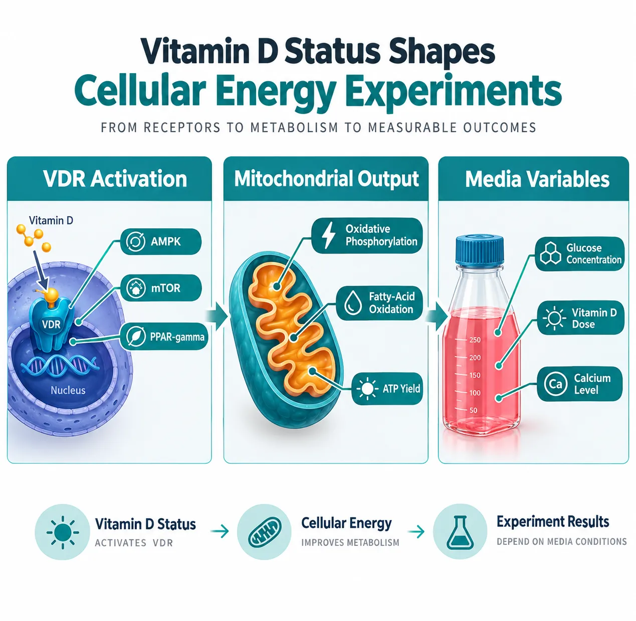
Most cell-culture media formulations contain little to no vitamin D, yet researchers rarely report the vitamin D content of their experimental system. This gap matters because the active form of vitamin D, calcitriol, enhances mitochondrial fatty-acid oxidation and oxidative phosphorylation in muscle cells. Human supplementation studies confirm that cholecalciferol increases oxidative phosphorylation capacity and improves muscle function, effects that translate directly to the readouts used in cellular energy assays.

The mechanism runs through the vitamin D receptor (VDR), which is expressed in adipocytes, hepatocytes, skeletal muscle, beta-cells, endothelial cells, and immune cells. VDR activation modulates several signaling nodes that are central to cellular energy regulation:

Signaling Node Role in Energy Metabolism
AMPK Cellular energy sensor; activates catabolic pathways
mTOR Anabolic signaling; suppressed during energy deficit
NF-kB Inflammatory regulator; linked to mitochondrial stress
PPAR-gamma Lipid metabolism and adipogenesis control

Because these pathways overlap with classic exercise and nutrient-stress responses, an experiment that ignores vitamin D status is effectively running with an uncontrolled variable sitting at the top of the signaling cascade. A 2026 mechanistic review on vitamin D and metabolic syndrome positions VDR-dependent signaling as a core modulator of insulin sensitivity, oxidative stress, and mitochondrial function, reinforcing that vitamin D belongs in the experimental design checklist, not the supplementary materials.

Practical standardization steps include:

  • Measuring baseline 25-hydroxyvitamin D in serum before animal studies
  • Supplementing cell culture media with a defined calcitriol concentration
  • Running a vitamin D-deficient control arm alongside replete conditions
  • Reporting media composition fully in the methods section

MOTS-c and 5-Amino-1MQ: Mechanisms Within a Nutrient-Controlled Framework
MOTS-c and 5-Amino-1MQ: Mechanisms Within a Nutrient-Controlled Framework

With a stable vitamin D baseline established, researchers can introduce the two experimental agents most relevant to nutrient-controlled cellular energy work: the mitochondrial peptide MOTS-c and the NNMT inhibitor 5-Amino-1MQ.

MOTS-c: A Mitochondrial Stress Peptide That Reads Nutrient State

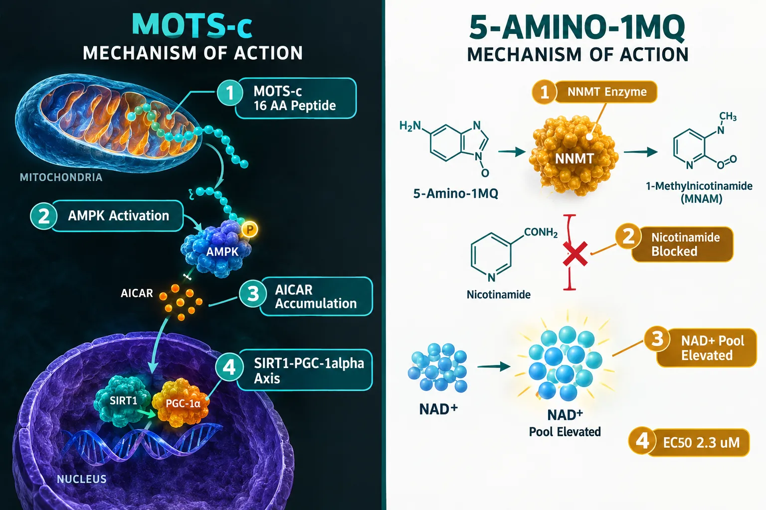
MOTS-c is a 16-amino-acid microprotein encoded within the 12S rRNA of mitochondrial DNA. Its primary mechanism involves inhibiting the folate cycle and de novo purine synthesis, which causes accumulation of AICAR, an endogenous AMPK agonist. This makes MOTS-c a direct link between mitochondrial stress and the cell's central energy "fuel gauge."

Under metabolic stress such as nutrient deprivation or mitochondrial dysfunction, MOTS-c translocates to the nucleus in an AMPK-dependent manner, where it regulates gene expression programs that optimize mitochondrial function and enhance antioxidant defenses. A 2026 translational review describes MOTS-c as part of a secreted mitochondrial microprotein network engaging the canonical AMPK → SIRT1 → PGC-1alpha axis, the same cascade driven by endurance exercise and caloric restriction.

Preclinical data from 2025 show that MOTS-c restores mitochondrial respiration in the type 2 diabetic heart, improving oxygen consumption and cardiac bioenergetics. Separate 2025 work reports that MOTS-c prevents metabolic derangements via regulation of both AMPK and mTOR pathways. Researchers exploring these effects can source MOTS-c peptide for in-vitro and preclinical work.

"MOTS-c does not act in isolation, its potency is shaped by the nutrient environment the cell is already experiencing when the peptide arrives."

5-Amino-1MQ: Redirecting NAD+ Through NNMT Inhibition

5-Amino-1MQ (5-amino-1-methylquinolinium) is a small-molecule inhibitor of nicotinamide N-methyltransferase (NNMT), the cytosolic enzyme that methylates nicotinamide, a NAD+ precursor, using SAM as the methyl donor. By occupying the nicotinamide-binding pocket of NNMT, 5-Amino-1MQ blocks this diversion and reshapes cellular NAD+ and methyl-donor pools.

In differentiated adipocyte models, 5-Amino-1MQ demonstrates concentration-dependent NNMT inhibition with a calculated EC50 of approximately 2.3 µM, providing a practical dose range for in-vitro studies. At effective concentrations, it reduces intracellular 1-methylnicotinamide, elevates NAD+ levels, and suppresses lipogenesis, demonstrating that NNMT inhibition can reroute nicotinamide toward energy metabolism rather than fat-storage pathways.

Preclinical obesity models show that 5-Amino-1MQ reverses diet-induced obesity, improves glucose tolerance, enhances insulin sensitivity, and attenuates fatty-liver changes. Research on 5-Amino-1MQ capsules and related 5-amino peptide compounds continues to expand as interest in NAD+ metabolism grows. Notably, NNMT inhibition also helps preserve nicotinamide and NMN as NAD+ precursors in skeletal muscle, connecting 5-Amino-1MQ to muscle bioenergetics research.


Designing the Three-Axis Experiment
Designing the Three-Axis Experiment

The convergence of all three agents on the AMPK-SIRT1-PGC-1alpha axis is both an opportunity and a methodological challenge. Vitamin D acts primarily via nuclear VDR-dependent transcription. MOTS-c operates as a mitochondria-encoded stress peptide converging on AMPK and SIRT1. 5-Amino-1MQ reshapes NAD+ availability, which directly feeds SIRT1 activity. These are not independent levers, pulling one affects the others.

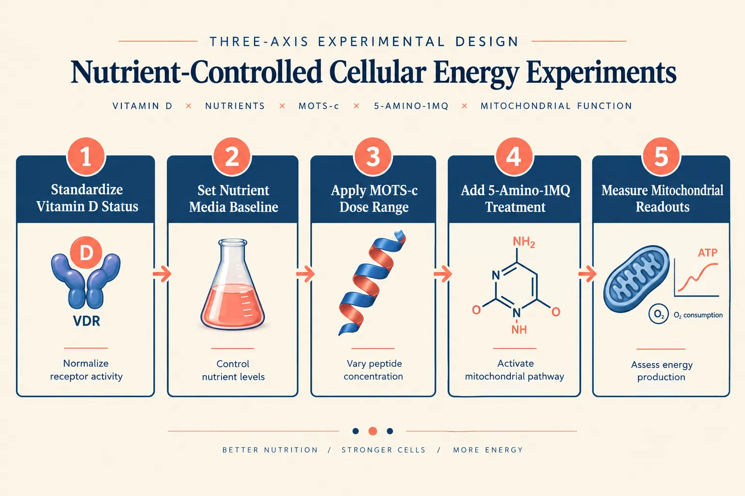
A rational three-axis experimental framework for Vitamin D, MOTS-c, and 5-Amino-1MQ: Designing Nutrient-Controlled Cellular Energy Experiments looks like this:

Axis 1, Vitamin D (VDR-dependent metabolic signaling)

  • Vary: deficient vs. replete calcitriol concentrations in media
  • Measure: VDR target gene expression, AMPK phosphorylation, mitochondrial membrane potential

Axis 2, MOTS-c (acute AMPK activation and stress response)

  • Vary: peptide concentration and timing relative to nutrient stress
  • Measure: oxygen consumption rate, AICAR levels, PGC-1alpha expression

Axis 3-5-Amino-1MQ (NAD+ salvage and NNMT inhibition)

  • Vary: concentration from sub-EC50 to supra-EC50 (reference: ~2.3 µM EC50)
  • Measure: intracellular NAD+/NADH ratio, 1-methylnicotinamide, lipogenesis markers

Combining these axes with controlled glucose, fatty-acid, and amino-acid availability allows researchers to interrogate how endocrine nutrients, mitochondrial microproteins, and NAD+ salvage pathways jointly determine ATP production, substrate choice, and resilience to metabolic stress. For researchers interested in related mitochondrial research tools, SS-31 mitochondrial research resources and NAD+ compounds offer complementary experimental angles. Additional context on how peptides fit into broader cardiometabolic models can be found in this overview of polypeptides in cardiometabolic models.

Critical readouts to standardize across all arms:

  • Oxygen consumption rate (OCR) and extracellular acidification rate (ECAR) via Seahorse or equivalent
  • ATP production rate separated by oxidative phosphorylation vs. glycolysis
  • Mitochondrial biogenesis markers (PGC-1alpha, TFAM, mtDNA copy number)
  • Oxidative stress indicators (mitochondrial ROS, glutathione ratio)

Conclusion

Designing nutrient-controlled cellular energy experiments with Vitamin D, MOTS-c, and 5-Amino-1MQ requires treating vitamin D not as background noise but as an active experimental variable. Before any peptide or small molecule is introduced, vitamin D status in the model system must be defined, controlled, and reported. From that stable foundation, MOTS-c can be used to probe acute AMPK-driven mitochondrial stress responses, while 5-Amino-1MQ reshapes the NAD+ landscape through NNMT inhibition, two complementary tools that, together with vitamin D, map the full AMPK-SIRT1-PGC-1alpha signaling network.

Actionable next steps for researchers in 2026:

  1. Audit current cell-culture and animal-model protocols for vitamin D content and add a deficient-versus-replete arm to ongoing studies.
  2. Select MOTS-c dose ranges based on published preclinical respiration data and confirm AICAR accumulation as a mechanistic marker.
  3. Use the ~2.3 µM EC50 reference for 5-Amino-1MQ to build a concentration-response curve before committing to a single treatment dose.
  4. Report all three axes, vitamin D status, peptide concentration, and NNMT inhibitor dose, alongside nutrient media composition in every methods section.
  5. Prioritize OCR and NAD+/NADH ratio as primary readouts to capture both mitochondrial and metabolic dimensions of the experiment.

All three agents remain experimental tools rather than approved therapeutics, but their mechanistic precision makes them among the most powerful instruments available for dissecting nutrient-sensing and energy-metabolism pathways in 2026.

https://www.puretestedpeptides.com/wp-content/uploads/2026/10/vitamin-d-mots-c-and-5-amino-1mq-designing-nutrient-controlled-cellular-energy-e.webp 1024 1536 https://www.puretestedpeptides.com/wp-content/uploads/2026/01/buy-peptides-online.jpg 2026-10-02 13:10:472026-10-02 13:10:47Vitamin D, MOTS-c, and 5-Amino-1MQ: Designing Nutrient-Controlled Cellular Energy Experiments
Enclomiphene Base vs Enclomiphene Citrate: Solubility, Counterion Effects, and Analytical Comparability

Enclomiphene Base vs Enclomiphene Citrate: Solubility, Counterion Effects, and Analytical Comparability

October 2, 2026/0 Comments/in Uncategorized/by

Salt form selection is not a footnote in pharmaceutical development, it determines whether two batches of the same active molecule will dissolve identically, measure identically, and dose identically. When researchers and formulators encounter enclomiphene base vs enclomiphene citrate: solubility, counterion effects, and analytical comparability, the differences between these two forms reach far beyond a simple name change. A mass discrepancy of roughly 47% between the freebase and salt forms means that a researcher who ignores salt-form identity risks systematic dosing errors, failed dissolution tests, and misinterpreted analytical results, all without any obvious red flag in the raw data.


Key Takeaways

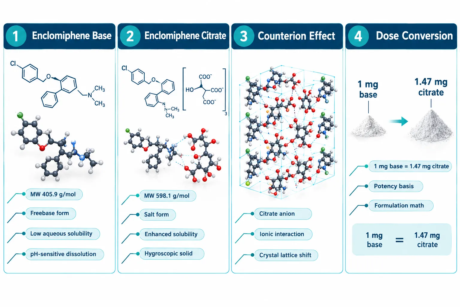
  • Enclomiphene base (MW ~405.9 g/mol) and enclomiphene citrate (MW ~598.1 g/mol) differ by approximately 47% in molecular weight, requiring a dose conversion factor for any potency-based calculation.
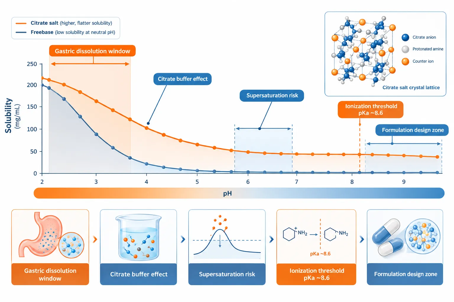
  • The citrate counterion substantially increases aqueous solubility compared to the freebase form, which is nearly insoluble at physiological pH.
  • pH-dependent solubility profiles differ sharply between the two forms, with critical implications for dissolution testing and oral formulation design.
  • Chromatographic and spectroscopic identity tests must be calibrated to the specific salt form in use; otherwise, retention times, response factors, and concentration calculations will be systematically offset.
  • Batch-to-batch comparability studies must confirm salt form identity before interpreting any apparent potency or purity discrepancy.

Chemical Identity and the Molecular Weight Gap

Chemical Identity and the Molecular Weight Gap

Enclomiphene is the trans-isomer of clomiphene, a selective estrogen receptor modulator (serm). In its freebase state, enclomiphene carries a molecular weight of approximately 405.9 g/mol. When combined with citric acid to form enclomiphene citrate, the resulting salt reaches approximately 598.1 g/mol, an increase of roughly 192 g/mol attributable entirely to the citrate counterion.

This gap has immediate practical consequences. A 25 mg dose expressed as the base equivalent requires approximately 36.8 mg of the citrate salt to deliver the same molar quantity of active compound. The conversion factor (citrate MW divided by base MW, approximately 1.47) must be applied consistently across:

  • Label claims on research-grade materials
  • Stock solution preparation for in vitro assays
  • Dissolution specifications in quality control testing
  • Comparative potency calculations across studies using different salt forms

Failure to apply this factor is one of the most common sources of apparent "potency discrepancy" between batches that are, in chemical terms, identical. Researchers sourcing a serm 10mg product, for example, should always confirm whether the stated weight reflects the base or the salt form before preparing working concentrations.


Solubility Profiles and the Role of the Citrate Counterion

Solubility Profiles and the Role of the Citrate Counterion

The phrase enclomiphene base vs enclomiphene citrate: solubility, counterion effects, and analytical comparability captures a genuine formulation challenge. Enclomiphene freebase is classified as practically insoluble in water at neutral to basic pH. Its ionization pKa sits near 8.6, meaning that at physiological pH (approximately 7.4), the molecule exists predominantly in its unionized, lipophilic form, a state that strongly limits aqueous dissolution.

The citrate salt changes this picture in two ways:

  1. Direct counterion solubilization: The citrate anion creates an ionic lattice that disrupts the hydrophobic crystal packing of the freebase, allowing water molecules to penetrate more readily.
  2. Microenvironmental pH buffering: Citric acid is a weak triprotic acid. As the salt dissolves, the local microenvironment becomes mildly acidic, which promotes ionization of the enclomiphene amine and further drives dissolution.

"The counterion is not an inert passenger, it actively shapes the thermodynamic and kinetic solubility of the active molecule."

pH-dependent solubility comparison:

pH Enclomiphene Base Enclomiphene Citrate
1.2 (gastric) Moderate High
4.5 (intestinal) Low Moderate-High
6.8 (intestinal) Very Low Moderate
7.4 (physiological) Practically Insoluble Low-Moderate

This profile explains why oral formulations almost universally use the citrate salt. The freebase form would require solubilization strategies, co-solvents, surfactants, or amorphous dispersion, to achieve comparable bioavailability. Hygroscopicity is a related concern: enclomiphene citrate absorbs atmospheric moisture more readily than the freebase, which affects storage conditions, weighing accuracy, and long-term stability. Proper desiccant storage and tightly sealed containers are non-negotiable for the citrate form.


Analytical Comparability: Where Salt Form Errors Become Data Errors

Understanding enclomiphene base vs enclomiphene citrate: solubility, counterion effects, and analytical comparability in an analytical context requires examining three distinct testing domains.

Analytical Comparability: Where Salt Form Errors Become Data Errors

Chromatographic Methods (HPLC/UPLC)

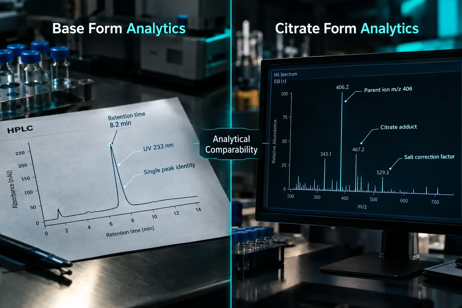
Both forms share the same enclomiphene chromophore and will produce UV absorbance at approximately 233 nm. However, the response factor, the relationship between peak area and concentration, must be calculated against a reference standard of matching salt form. Using a base-form reference standard to quantify a citrate-form sample introduces a systematic ~32% underestimation of true enclomiphene content (since the citrate sample contains proportionally less active molecule per unit mass).

Retention time is not affected by salt form under standard reversed-phase conditions, because the citrate counterion dissociates in aqueous mobile phases. This can mislead analysts into assuming the methods are fully interchangeable when they are not.

Mass Spectrometry

Electrospray ionization (ESI-MS) detects the enclomiphene molecular ion at m/z 406 regardless of salt form, since ionization strips the counterion. However, citrate adducts ([M + citrate]) can appear in negative-ion mode and may confuse automated library matching if the spectral database entry was built from the freebase.

Identity and Purity Testing

Infrared (IR) spectroscopy and X-ray powder diffraction (XRPD) will produce distinctly different spectra for the base and citrate forms. These are not interchangeable reference standards. A batch submitted with a base-form certificate of analysis but tested against a citrate-form IR reference will fail identity, not because the material is impure, but because the wrong reference was applied.

This is why well-designed analytical methods, much like the purity evaluation frameworks discussed in research-grade peptide purity evaluation, must explicitly state the salt form in every specification and reference standard description.

Stability and Forced Degradation

Enclomiphene citrate generates citric acid as a degradation-related impurity under hydrolytic stress conditions. This impurity is absent from freebase degradation profiles. Any forced degradation study or shelf-life specification must account for this difference, or citrate-derived impurities will be incorrectly flagged as enclomiphene-related degradants.


Practical Guidance for Batch Comparability

When two batches of enclomiphene appear to differ in potency or purity, the first diagnostic step is confirming salt form identity, not re-running the assay. A structured comparability checklist should include:

  • Confirm salt form via IR or XRPD before any quantitative comparison
  • Apply the correct MW conversion factor (base to citrate: multiply by 1.47; citrate to base: multiply by 0.679)
  • Match reference standards to the salt form of the test article
  • Adjust dissolution specifications to reflect the expected solubility profile of the specific form
  • Document hygroscopicity controls (desiccant storage, equilibration time before weighing)

Researchers working with peptide-based compounds face analogous salt-form and counterion considerations. Resources such as SS-31 10mg research peptide considerations and what is ideal dosage of SS31 illustrate how molecular weight and salt form affect concentration calculations across compound classes. Similarly, dosing frameworks for growth hormone secretagogues, such as those covered in tesa dosage for fat loss and tesa vs sermorelin, demonstrate how salt-form awareness is a universal analytical discipline, not specific to any single molecule.


Conclusion

The comparison of enclomiphene base vs enclomiphene citrate: solubility, counterion effects, and analytical comparability resolves into a single actionable principle: salt form is a primary analytical variable, not a secondary label detail. The ~47% molecular weight difference demands a consistent conversion factor in every concentration calculation. The citrate counterion's buffering and solubilization effects must be reflected in dissolution specifications and formulation design. And every analytical method, from HPLC quantitation to IR identity testing, must be explicitly validated against the salt form actually in use.

Actionable next steps for researchers and formulators in 2026:

  1. Audit all existing enclomiphene reference standards and certificates of analysis to confirm explicit salt form declaration.
  2. Recalculate any historical concentration data using the correct MW conversion factor for the form used.
  3. Establish separate XRPD or IR identity specifications for base and citrate forms and never cross-apply them.
  4. Implement moisture-control procedures specific to the hygroscopic citrate salt.
  5. Flag any batch comparability discrepancy for salt-form verification before concluding a purity or potency failure.

Treating these two forms as analytically equivalent is the fastest route to systematic, invisible error. Treating them as distinct chemical entities, which they are, is the foundation of reliable enclomiphene research.

https://www.puretestedpeptides.com/wp-content/uploads/2026/10/enclomiphene-base-vs-enclomiphene-citrate-solubility-counterion-effects-and-anal.webp 1024 1536 https://www.puretestedpeptides.com/wp-content/uploads/2026/01/buy-peptides-online.jpg 2026-10-02 13:04:102026-10-02 13:04:10Enclomiphene Base vs Enclomiphene Citrate: Solubility, Counterion Effects, and Analytical Comparability
Omeprazole, Famotidine, and GLP-2-T Research: Controlling Gastric-Acid Variables in Intestinal Models

Omeprazole, Famotidine, and GLP-2-T Research: Controlling Gastric-Acid Variables in Intestinal Models

October 2, 2026/0 Comments/in Uncategorized/by

Gastric acid suppression is one of the most frequently overlooked confounders in intestinal peptide research, yet pH shifts as small as one unit can alter epithelial permeability, reshape the luminal microbiome, and mask or amplify the very signals a study is designed to measure. When the research involves a novel compound like GLP-2-T alongside established acid modulators, the stakes for rigorous variable control become even higher. This article examines how omeprazole, famotidine, and GLP-2-T research intersects in the context of controlling gastric-acid variables in intestinal models, and what investigators must document to produce reproducible, interpretable data.

Key Takeaways

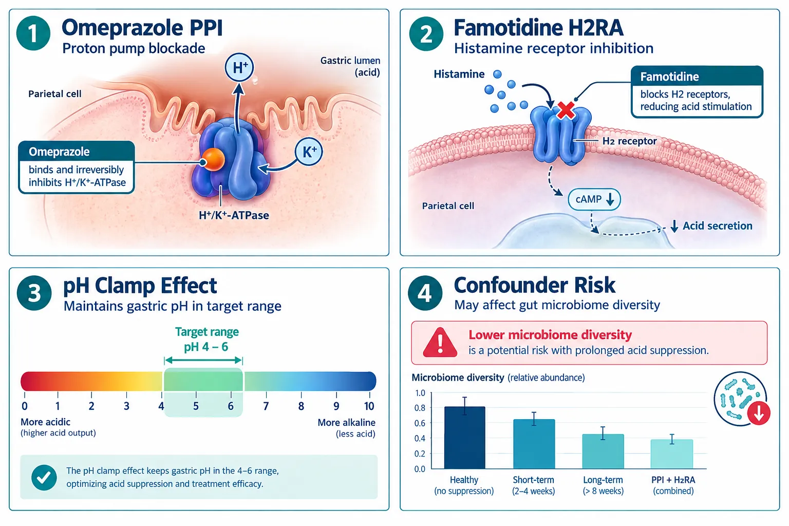
  • Omeprazole (PPI) and famotidine (H2RA) suppress gastric acid through distinct mechanisms, producing different pH profiles, microbiome effects, and experimental confounders.
  • Acid suppression alone can accelerate mucosal repair, meaning it must be treated as an active variable, not a neutral background condition, in intestinal models.
  • GLP-2-T is positioned as a downstream effector of mucosal physiology; its readouts are directly influenced by the acid environment established before or during exposure.
  • Combination "pH clamp" regimens using both drug classes can stabilize luminal pH rapidly, but they introduce additive microbiome disruption that requires dedicated endpoint tracking.
  • Endpoint selection, particularly tight-junction integrity, transepithelial resistance, and microbial diversity indices, must be pre-specified with acid exposure conditions fully recorded.

Why Acid Suppression Becomes a Study Confounder

Why Acid Suppression Becomes a Study Confounder

Omeprazole is a proton pump inhibitor (PPI). It irreversibly blocks the H+/K+-ATPase enzyme on gastric parietal cells, producing sustained acid suppression that persists well beyond the compound's plasma half-life. Famotidine is an H2 receptor antagonist (H2RA). It competes with histamine at the H2 receptor, producing faster but more transient pH elevation. These two mechanisms matter enormously for experimental design because they create different temporal pH profiles inside any intestinal model system.

Key pharmacodynamic differences:

Feature Omeprazole (PPI) Famotidine (H2RA)
Onset of action 1-4 hours 30-60 minutes
Duration of effect 24-72 hours 6-12 hours
pH ceiling Higher, more sustained Moderate, variable
Microbiome impact Greater oral-to-gut bacterial shift Lower, more localized
Reversibility Irreversible (enzyme turnover needed) Reversible, competitive

When either agent is present in a model without being explicitly recorded as an independent variable, it can independently accelerate gastric wound repair, alter tight-junction protein expression, and change luminal bacterial composition. Research using stress-ulcer and mucosal-injury models has confirmed that acid suppression is a dominant variable, not a passive background condition.

"Acid suppression is not a neutral scaffold. It is an active intervention that reshapes the biological terrain on which every other compound must act."

For investigators exploring GLP peptides for research purposes, this means the acid environment must be treated with the same rigor as the peptide dose itself.


GLP-2-T in Intestinal Models: Where Acid Control Intersects

GLP-2-T in Intestinal Models: Where Acid Control Intersects

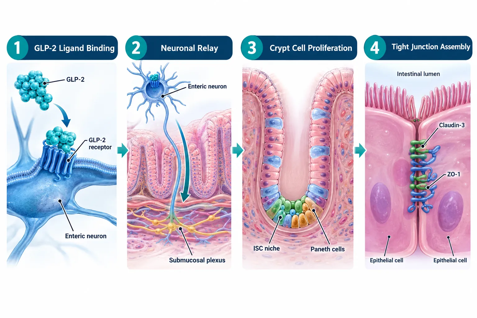
GLP-2 (glucagon-like peptide-2) is a 33-amino-acid hormone secreted by intestinal L-cells. Its primary roles include promoting mucosal growth, reducing intestinal permeability, and modulating gastric acid secretion itself, making it uniquely entangled with the acid variables described above. Clinical GLP-2 analogs such as teduglutide have established this peptide class as a benchmark for intestinal trophic research.

GLP-2-T refers to a research-stage analog designed to engage GLP-2 receptors with modified pharmacokinetics or receptor selectivity. Because GLP-2-T is predicted to act downstream of the acid environment, the luminal pH at the time of peptide exposure directly conditions the model's response. Specifically:

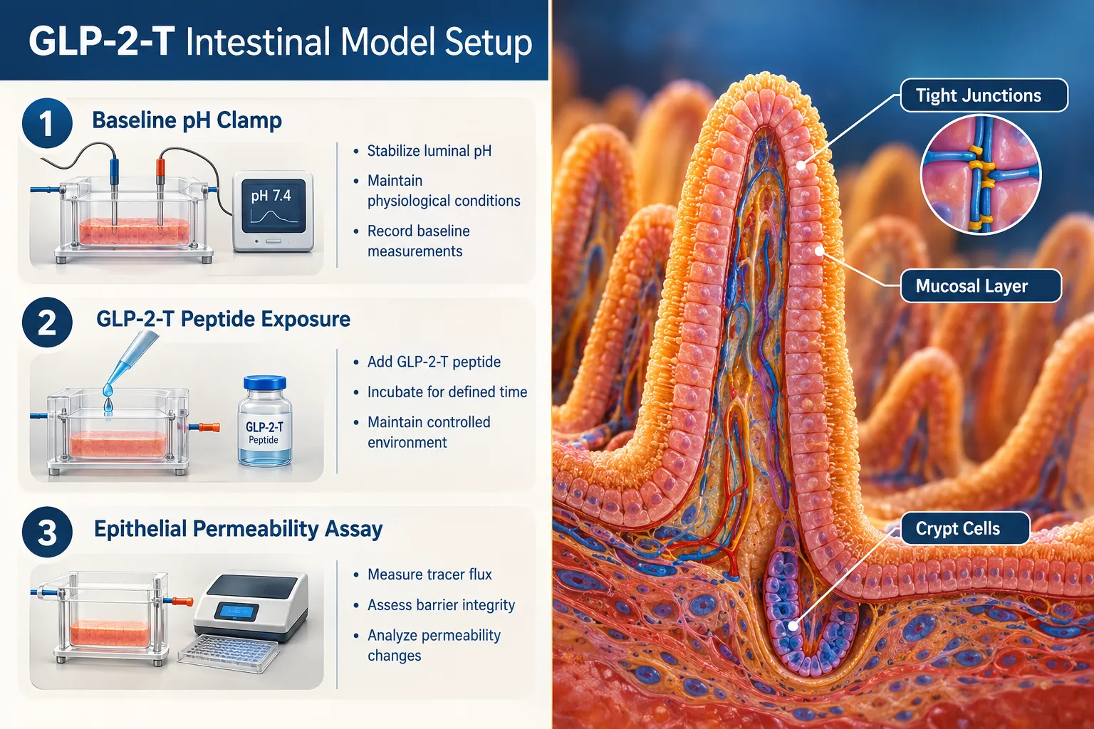
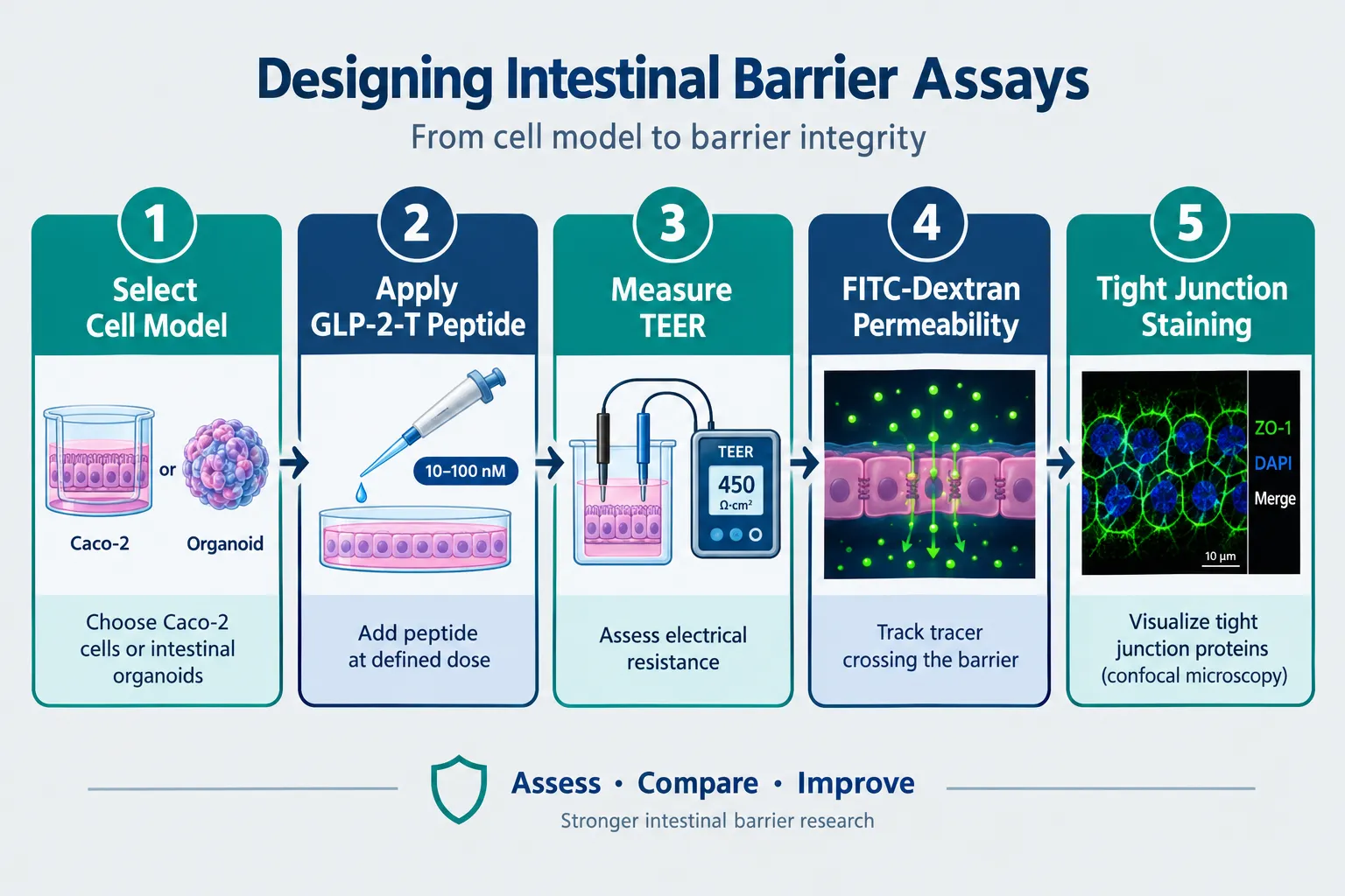
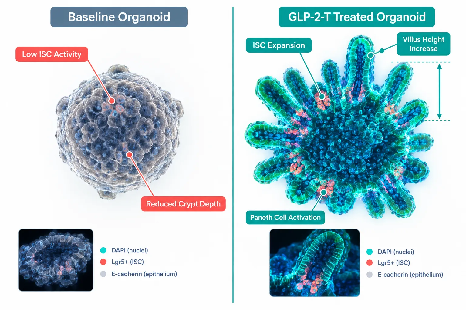
  • Epithelial permeability endpoints (transepithelial electrical resistance, FITC-dextran flux) are sensitive to baseline pH; an uncontrolled acid shift can mimic or cancel GLP-2-T's barrier-tightening effects.
  • Mucosal proliferation markers (crypt cell mitosis, villus height) respond to both acid suppression and GLP-2R activation, creating overlapping signal if both variables are active simultaneously.
  • Microbiome diversity indices are altered by PPIs more profoundly than H2RAs; since GLP-2-T research often includes microbiome endpoints, the choice between omeprazole and famotidine is not interchangeable.

For context on related peptide mechanisms, the comparison of LL-37 versus SS-31 peptide benefits illustrates how overlapping biological targets demand careful endpoint separation, a principle that applies directly here.

Researchers exploring GLP-3 Reta compounds will recognize that the same acid-variable logic governs any GLP-family peptide study conducted in an intestinal model system.


Experimental Workflow: Recording and Controlling Acid Variables

Experimental Workflow: Recording and Controlling Acid Variables

Controlling gastric-acid variables in intestinal models requires a structured approach to both pre-treatment conditions and endpoint documentation. The following workflow reflects current preclinical best practice when coupling acid suppression with GLP-2 or GLP-2-T exposure.

Step-by-step acid control protocol:

  1. Define the pH target range before any peptide is introduced. Most intestinal epithelial models require a luminal pH between 4.5 and 6.5 for physiologically relevant responses.
  2. Select the acid suppressor based on study duration. Use famotidine for short-window experiments (under 12 hours) where reversibility is needed. Use omeprazole for sustained pH clamping over multi-day models.
  3. Consider combination regimens cautiously. Omeprazole-plus-famotidine combinations can achieve rapid, stable pH clamping, useful for acute injury models, but they amplify microbiome disruption and must be documented as a compound intervention, not a single variable.
  4. Record acid exposure as an independent variable in all statistical models. Do not treat it as a preprocessing step.
  5. Pre-specify microbiome endpoints separately from epithelial endpoints. PPI-driven oral-to-gut bacterial translocation can confound 16S rRNA diversity data if not controlled with a matched acid-suppression arm.
  6. Introduce GLP-2-T only after pH stabilization is confirmed. Introducing the peptide during an acid transient will produce unreliable permeability and proliferation data.

Endpoints requiring dedicated acid controls:

  • Tight-junction protein expression (claudin-1, occludin, ZO-1)
  • Transepithelial electrical resistance (TEER)
  • Villus-to-crypt ratio
  • Mucosal cytokine profiles (IL-6, TNF-alpha)
  • Microbial alpha- and beta-diversity indices

For researchers interested in how mitochondrial peptides interact with similar tissue-integrity endpoints, the work on SS-31 mitochondrial research and wound healing models provides useful methodological parallels for endpoint separation strategies.

Choosing Between PPIs and H2RAs for GLP-2-T Studies

The choice is not merely pharmacological, it is experimental design. PPIs offer superior pH stability for multi-day intestinal organoid or ex-vivo tissue models, but their microbiome effects introduce a confounder that is difficult to isolate. H2RAs preserve a more physiological luminal bacterial environment at the cost of pH variability. Neither is universally superior; the correct choice depends on which endpoints are primary.

For GLP-1 Reta research contexts, where metabolic and mucosal endpoints often overlap, this trade-off is especially consequential and should be addressed explicitly in the methods section of any publication or report.


Conclusion

Omeprazole, famotidine, and GLP-2-T research demands that controlling gastric-acid variables in intestinal models be treated as a first-order experimental priority, not an afterthought. The core actionable steps are clear: select the acid suppressor that matches study duration and microbiome sensitivity requirements, document pH exposure as an independent variable in every statistical model, stabilize luminal pH before introducing GLP-2-T, and pre-specify epithelial permeability and microbiome endpoints with matched acid-control arms.

Investigators should also cross-reference their endpoint selection against established peptide research frameworks, including VEGF upregulation studies and wound healing compound models, where overlapping biological targets have required similar variable-isolation strategies. As GLP-2-T research matures in 2026, the quality of acid-variable documentation will likely become a primary criterion for peer review and reproducibility assessment.

https://www.puretestedpeptides.com/wp-content/uploads/2026/10/omeprazole-famotidine-and-glp-2-t-research-controlling-gastric-acid-variables-in.webp 1024 1536 https://www.puretestedpeptides.com/wp-content/uploads/2026/01/buy-peptides-online.jpg 2026-10-02 13:04:042026-10-02 13:04:04Omeprazole, Famotidine, and GLP-2-T Research: Controlling Gastric-Acid Variables in Intestinal Models
Glow Blend vs Klow Blend in Fibroblast Studies: Comparing Matrix, Barrier, and Formulation-Dependent Outcomes

Glow Blend vs Klow Blend in Fibroblast Studies: Comparing Matrix, Barrier, and Formulation-Dependent Outcomes

October 2, 2026/0 Comments/in Uncategorized/by

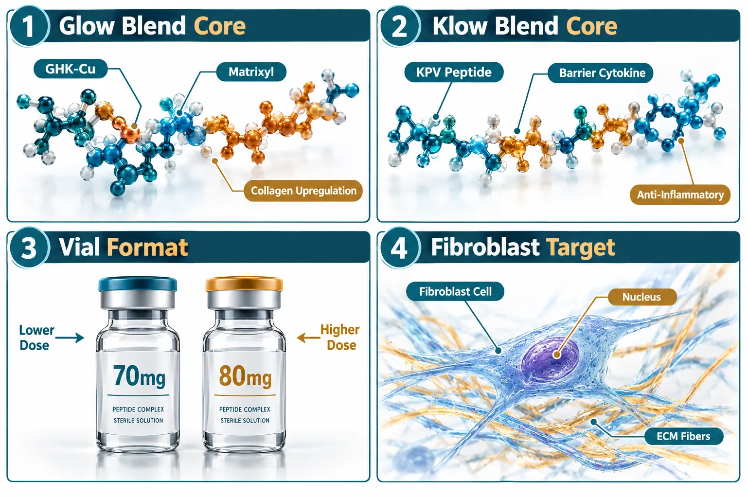
Fewer than 15% of multi-peptide blends sold through research networks in 2026 carry full ingredient disclosure at the component level, yet formulation identity is the single most important variable when interpreting fibroblast study data. The debate around Glow Blend vs Klow Blend in fibroblast studies: comparing matrix, barrier, and formulation-dependent outcomes sits at the center of this problem. Both blends are marketed as skin-matrix research tools, both arrive in 70-80 mg vials, and both show measurable fibroblast activity in cell culture models. What separates them is the composition logic behind each formula and the biological endpoints each one is best positioned to influence.

Key Takeaways

  • Glow Blend is a tri-peptide formula primarily studied for extracellular matrix remodeling and FAK-paxillin fibroblast signaling.
  • Klow Blend is a quad-peptide formula that adds KPV as a differentiating component, shifting focus toward barrier integrity and cytokine modulation.
  • Vial formats differ (70 mg vs 80 mg), and batch-to-batch reproducibility is a documented concern for both products.
  • No peer-reviewed, head-to-head fibroblast trial comparing these two blends directly existed as of late 2026.
  • Formulation transparency, knowing exactly what is in each vial, is the foundational requirement for any valid comparison.

Blend Identity and Ingredient Disclosure: The Starting Point

Blend Identity and Ingredient Disclosure: The Starting Point

Before any fibroblast data can be interpreted, the composition of each blend must be confirmed. Glow Blend is formulated as a tri-peptide combination. Its core components target collagen synthesis pathways and receptor-level signaling in dermal fibroblasts. Klow Blend is a quad-peptide formula, meaning it carries one additional active component, KPV (Lys-Pro-Val), a tripeptide fragment of alpha-MSH with well-characterized anti-inflammatory properties.

This structural difference matters more than vial size. The 70 mg (Glow) versus 80 mg (Klow) distinction reflects the additional mass contributed by the fourth peptide, not a simple dosing escalation. Researchers sourcing either product should verify certificate-of-analysis documentation before use. For those exploring verified multi-peptide options, resources like BPC-157 and TB-500 blend research pages offer a useful reference point for how combination peptide transparency is handled in practice.

"Formulation identity is not a background variable, it is the independent variable. Without it, fibroblast data from blend studies is uninterpretable."


Fibroblast Matrix Outcomes: Where Glow Blend Shows Stronger Signals

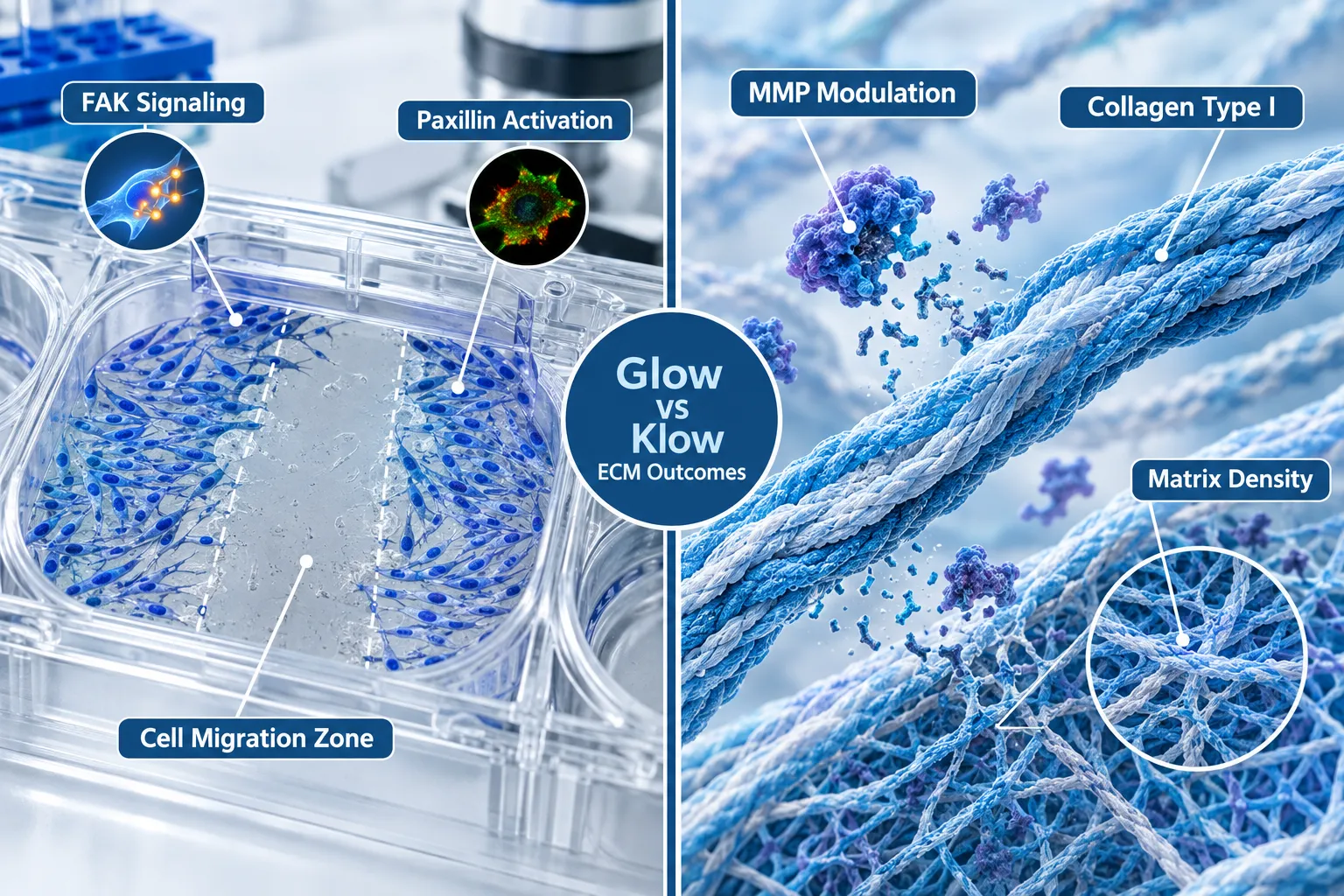
The extracellular matrix (ECM) remodeling narrative is where Glow Blend generates its most discussed research signals. In cell culture models using human dermal fibroblasts, the tri-peptide combination in Glow Blend has been associated with:

  • Upregulation of collagen Type I and Type III synthesis markers
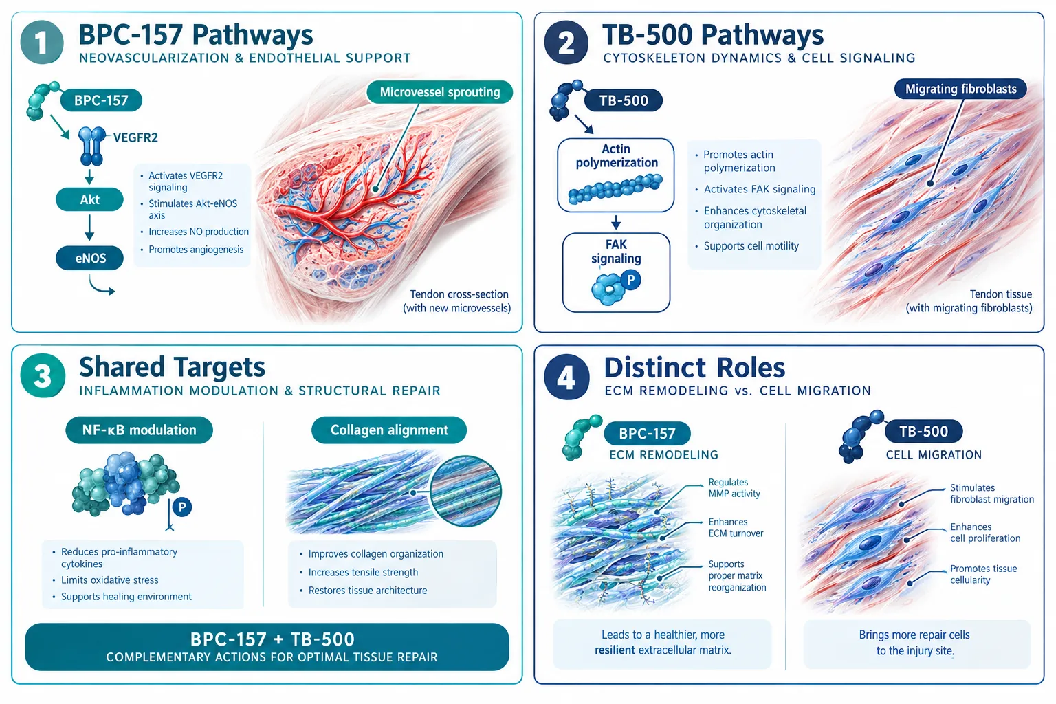
  • Activation of FAK (focal adhesion kinase) and paxillin signaling cascades, which govern cell migration and matrix attachment
  • Increased fibroblast proliferation rates at physiologically relevant concentrations

The FAK-paxillin pathway is particularly significant. When fibroblasts migrate into a wound matrix or a collagen scaffold, FAK phosphorylation drives the cytoskeletal reorganization that allows cells to pull and remodel surrounding fibers. Glow Blend's tri-peptide profile appears to amplify this process more consistently than Klow Blend in matrix-focused assays.

For researchers interested in how growth hormone-related peptide blends interact with fibroblast activity at the systemic level, a Tesamorelin comparison provides useful context on how different peptide classes influence tissue remodeling through distinct receptor pathways.


Barrier and Inflammatory Endpoints: Where Klow Blend Differentiates

Barrier and Inflammatory Endpoints: Where Klow Blend Differentiates

The addition of KPV in Klow Blend shifts the research profile toward barrier function and cytokine regulation. KPV has been studied for its ability to reduce pro-inflammatory cytokine expression, specifically IL-6 and TNF-alpha, in keratinocyte and fibroblast co-culture models. This makes Klow Blend more relevant when the research question involves:

  • Skin barrier integrity under inflammatory challenge conditions
  • Cytokine-mediated fibroblast activation (as opposed to direct mechanical signaling)
  • Dermal-epidermal junction support in models simulating compromised barrier states

The distinction is not that Klow Blend is "better", it is that it addresses a different biological question. Matrix remodeling and barrier repair are related but mechanistically separate processes. A study designed to measure collagen deposition will likely see stronger results with Glow Blend. A study designed to measure inflammatory resolution in a damaged skin model may see more relevant signals with Klow Blend.

Endpoint Glow Blend Signal Klow Blend Signal
Collagen Type I synthesis Strong Moderate
FAK-paxillin activation Strong Moderate
IL-6 cytokine reduction Weak Strong
Barrier integrity markers Moderate Strong
Fibroblast migration rate Strong Moderate

Formulation-Dependent Outcomes and Batch Reproducibility

The phrase "formulation-dependent outcomes" in the context of Glow Blend vs Klow Blend in fibroblast studies: comparing matrix, barrier, and formulation-dependent outcomes carries a specific warning. Both blends have shown batch-to-batch variability in independent quality assessments conducted through multi-vendor networks in 2026. This variability affects:
  • Peptide purity percentages (ranging from 92% to 98%+ depending on supplier)
  • Solubility profiles under standard reconstitution protocols
  • Stability during shipping and storage at non-ideal temperatures

Reproducibility is the largest unresolved challenge in this research space. A fibroblast study run with a high-purity Glow Blend batch may produce collagen synthesis data that cannot be replicated with a lower-purity batch from a different vendor. Researchers should treat batch documentation as mandatory, not optional.

For those evaluating sourcing standards, reviewing what constitutes a best peptide supplier in the current market is a practical first step. Similarly, understanding wholesale peptides for sale quality tiers helps contextualize why two ostensibly identical blend products can produce divergent fibroblast data.

Formulation-Dependent Outcomes and Batch Reproducibility

The lack of a peer-reviewed, head-to-head fibroblast trial comparing Glow and Klow Blends directly remains the field's most significant gap. Current comparative data comes from monograph releases, vendor-sponsored summaries, and informal multi-lab networks, none of which meet the evidentiary standard required for clinical extrapolation. Researchers using either blend should design studies with matched controls, verified batch documentation, and endpoint-specific assay selection.

For researchers exploring related peptide combination protocols, the IPA Sermorelin stack research page and SS-31 mechanism and research resource offer parallel examples of how multi-component peptide interactions are documented in preclinical settings.


Conclusion

The comparison of Glow Blend vs Klow Blend in fibroblast studies: comparing matrix, barrier, and formulation-dependent outcomes is not a question with a single winner. Glow Blend's tri-peptide composition makes it the stronger candidate for ECM remodeling and FAK-paxillin-driven fibroblast migration studies. Klow Blend's quad-peptide formula, anchored by KPV, positions it as the more relevant tool for barrier integrity and cytokine-modulated fibroblast research.

Actionable next steps for researchers in 2026:

  1. Confirm full ingredient disclosure and certificate of analysis before any fibroblast experiment.
  2. Select the blend based on the specific endpoint, matrix remodeling or barrier/inflammatory outcomes, not on vial size or price.
  3. Document batch numbers and purity percentages as part of the study methodology.
  4. Design matched-control experiments to isolate blend-specific effects from vehicle or solubility artifacts.
  5. Treat all current comparative data as preliminary until peer-reviewed head-to-head trials are published.

The field will benefit most from standardized protocols and transparent sourcing, not from vendor-driven narratives that obscure the formulation differences that actually drive the outcomes.

https://www.puretestedpeptides.com/wp-content/uploads/2026/10/glow-blend-vs-klow-blend-in-fibroblast-studies-comparing-matrix-barrier-and-form.webp 1024 1536 https://www.puretestedpeptides.com/wp-content/uploads/2026/01/buy-peptides-online.jpg 2026-10-02 13:03:512026-10-02 13:03:51Glow Blend vs Klow Blend in Fibroblast Studies: Comparing Matrix, Barrier, and Formulation-Dependent Outcomes
5-Amino-1MQ and MOTS-c Co-Treatment: A Matrix for Dose-Response, Additivity, and Interaction Testing

5-Amino-1MQ and MOTS-c Co-Treatment: A Matrix for Dose-Response, Additivity, and Interaction Testing

October 2, 2026/0 Comments/in Uncategorized/by

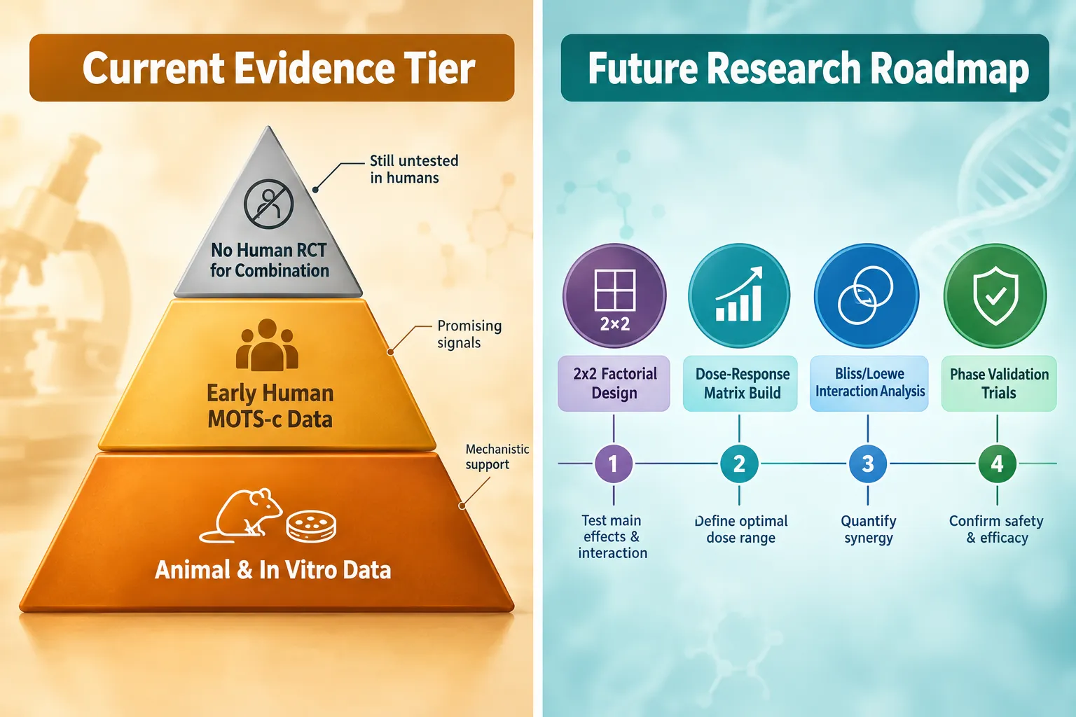
Preclinical data show that combining NNMT inhibition with mitochondrial peptide signaling can increase fat oxidation by roughly 47% above baseline, yet no published, peer-reviewed study has formally tested 5-Amino-1MQ and MOTS-c together in any model, human or animal. That gap between mechanistic promise and experimental evidence is precisely why a rigorous combination-study framework matters. Designing 5-Amino-1MQ and MOTS-c co-treatment: a matrix for dose-response, additivity, and interaction testing is not a dosing guide, it is a blueprint for generating credible data where none currently exists.

Key Takeaways

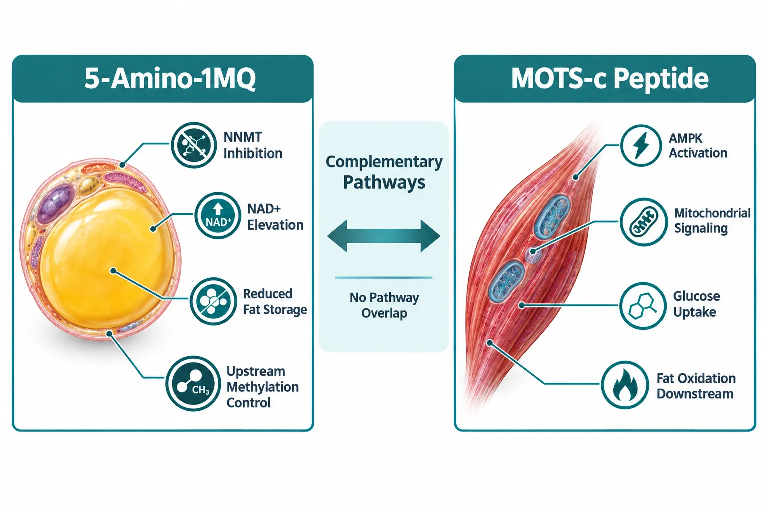
  • 5-Amino-1MQ inhibits NNMT and raises NAD+ in adipose tissue; MOTS-c activates AMPK in skeletal muscle, the pathways are complementary, not overlapping.
  • No published study has tested the two compounds together; any combination matrix is currently hypothetical experimental design, not completed research.
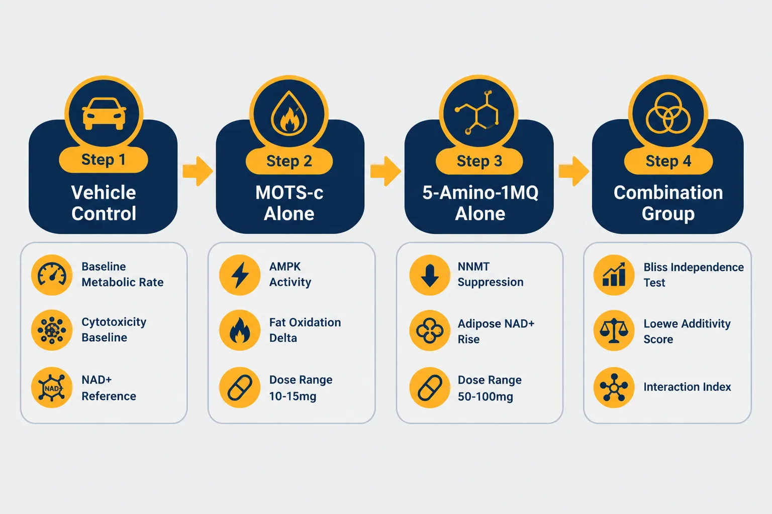
  • A 2×2 factorial layout (vehicle, each agent alone, combination) is the minimum structure needed to detect true interaction effects.
  • Bliss independence and Loewe additivity models are the standard analytical tools for quantifying whether a combination is additive, synergistic, or antagonistic.
  • Both compounds are sold for research use only; 5-Amino-1MQ has no registered human trials, while MOTS-c has limited early human mechanistic data.

Why These Two Compounds Belong in the Same Experimental Frame

Why These Two Compounds Belong in the Same Experimental Frame

Understanding the rationale for 5-Amino-1MQ and MOTS-c co-treatment begins with their distinct, non-overlapping mechanisms.

5-Amino-1MQ is a small-molecule inhibitor of nicotinamide N-methyltransferase (NNMT), an enzyme that consumes NAD+ precursors in white adipose tissue. By suppressing NNMT, the compound elevates intracellular NAD+ and alters methylation-dependent regulation of fat storage. In obese mouse models, it reduced body weight without changing food intake or lean mass, a profile that suggests a direct effect on adipose metabolism rather than appetite suppression. Researchers can explore 5-Amino-1MQ capsules as a reference-grade research compound.

MOTS-c is a 16-amino-acid peptide encoded in mitochondrial DNA. It activates AMPK and related metabolic stress pathways, primarily in skeletal muscle, enhancing glucose uptake and fat oxidation. Unlike 5-Amino-1MQ, MOTS-c has early human mechanistic data in metabolic and exercise contexts. Researchers can review MOTS-c and Elamipretide for context on mitochondrial-targeted peptide combinations.

The conceptual logic for pairing them is straightforward: 5-Amino-1MQ acts upstream on NNMT and NAD+ in white fat; MOTS-c acts downstream on mitochondrial signaling and AMPK in muscle. There is no obvious pathway competition, which creates a plausible, though unproven, basis for additivity or synergy.

"Mechanistic complementarity is a hypothesis, not a result. It justifies building a dose-response matrix; it does not replace one."


Building the Experimental Matrix: Factorial Design and Dose Selection

Building the Experimental Matrix: Factorial Design and Dose Selection

The foundation of 5-Amino-1MQ and MOTS-c co-treatment: a matrix for dose-response, additivity, and interaction testing is a properly structured factorial experiment. A 2×2 factorial layout, vehicle control, MOTS-c alone, 5-Amino-1MQ alone, and the combination, is the minimum design capable of detecting interaction effects. Anything less cannot distinguish true synergy from simple additive responses.

Dose Range Selection

Research-oriented protocols commonly reference the following ranges, extrapolated from animal and mechanistic data rather than large randomized human trials:

Compound Typical Research Range Route
5-Amino-1MQ 50-100 mg once daily Oral
MOTS-c 10-15 mg once daily Subcutaneous

For a proper dose-response matrix, each compound should be tested at a minimum of three concentration levels (low, mid, high) within these ranges. This produces a 3×3 grid of nine treatment conditions plus vehicle control, sufficient to fit a response surface and identify the concentration pair that maximizes effect while minimizing off-target risk.

Endpoints to Measure

A credible matrix requires mechanistic endpoints, not just body-weight readouts:

  • NAD+ and SAM pool quantification, both pathways influence NAD+ availability by different mechanisms, and their interaction on methyl donor pools is currently unknown
  • AMPK phosphorylation, primary downstream marker of MOTS-c activity
  • NNMT enzyme activity, direct measure of 5-Amino-1MQ target engagement
  • Mitochondrial oxygen consumption rate (OCR), Seahorse or equivalent assay
  • Cytotoxicity controls, cell viability at every dose level to separate metabolic effects from toxicity artifacts

Omitting cytotoxicity controls is a common methodological error in preclinical peptide research. Any apparent "synergy" in a metabolic endpoint is uninterpretable if cell viability is simultaneously compromised.


Interaction Analysis: Bliss, Loewe, and What the Data Actually Need to Show

Interaction Analysis: Bliss, Loewe, and What the Data Actually Need to Show

Once dose-response data are collected, the analytical framework determines whether the combination qualifies as additive, synergistic, or antagonistic. Two models dominate combination pharmacology:

Bliss Independence assumes that each agent acts independently. The expected combined effect is calculated as: E(A+B) = E(A) + E(B) − E(A)×E(B). If the observed effect exceeds this prediction, synergy is indicated; if it falls short, antagonism is present.

Loewe Additivity treats the two drugs as if they were dilutions of each other. It calculates a combination index (CI): CI < 1 indicates synergy, CI = 1 indicates additivity, CI > 1 indicates antagonism. Loewe is generally preferred when the two compounds share a common downstream endpoint, in this case, fat oxidation or AMPK activity.

For 5-Amino-1MQ and MOTS-c co-treatment: a matrix for dose-response, additivity, and interaction testing, researchers should apply both models and report where they agree and diverge. Discordance between Bliss and Loewe often signals that the mechanistic assumption of independence or dose-equivalence needs revision, itself a valuable finding.

A Critical Caution on NAD+ Interaction

Both compounds influence NAD+ availability, though by different mechanisms. 5-Amino-1MQ raises NAD+ by blocking its consumption via NNMT; MOTS-c influences NAD+-dependent AMPK signaling from the mitochondrial side. Whether co-administration amplifies NAD+ effects, creates redundancy, or produces unexpected depletion of shared methyl donor (SAM) pools is unknown. This interaction should be treated as an explicit hypothesis within the matrix, not an assumed benefit.

Multi-compound stacks that combine these agents with NAD+ precursors, some commercial vials contain approximately 10 mg 5-Amino-1MQ, 10 mg MOTS-c, and 100 mg NAD+, add a third variable that further complicates interaction modeling. Researchers interested in related mitochondrial peptide combinations can also review SS-31 and MOTS-c stacks for comparative mechanistic context.

When sourcing compounds for any in-vitro or animal study, purity verification is non-negotiable. Lab-tested peptides with documented certificate-of-analysis data are the baseline standard for credible preclinical work.


Conclusion

The scientific case for exploring 5-Amino-1MQ and MOTS-c co-treatment: a matrix for dose-response, additivity, and interaction testing rests on mechanistic complementarity, upstream NNMT inhibition in adipose tissue paired with downstream AMPK activation in skeletal muscle. That complementarity is conceptually sound, but it remains a hypothesis. No peer-reviewed study has yet tested this combination formally.

Actionable next steps for researchers:

  1. Design a 3×3 dose-response matrix with vehicle control and measure at least four mechanistic endpoints, including cytotoxicity at every concentration.
  2. Apply both Bliss independence and Loewe additivity models to the combination data; report the combination index and flag any discordance between models.
  3. Explicitly test NAD+ and SAM pool effects as a primary endpoint, not a secondary observation, this is the most likely site of unexpected interaction.
  4. Use only purity-verified, lab-tested compounds and document batch-level CoA data in any publication.
  5. Frame all findings as hypothesis-generating until Phase-grade human trial data exist; 5-Amino-1MQ remains entirely preclinical, and no human dose-response validation for either compound in combination has been registered.

The matrix described here does not yet exist in the literature. Building it rigorously is the work that needs to happen before any clinical extrapolation is warranted.

https://www.puretestedpeptides.com/wp-content/uploads/2026/10/5-amino-1mq-and-mots-c-co-treatment-a-matrix-for-dose-response-additivity-and-in.webp 1024 1536 https://www.puretestedpeptides.com/wp-content/uploads/2026/01/buy-peptides-online.jpg 2026-10-02 13:03:482026-10-02 13:03:485-Amino-1MQ and MOTS-c Co-Treatment: A Matrix for Dose-Response, Additivity, and Interaction Testing
Retatrutide, GLP-1 Pills, and Small-Molecule Weight-Loss Options: What Researchers Should Compare Before Selecting a Model

Retatrutide, GLP-1 Pills, and Small-Molecule Weight-Loss Options: What Researchers Should Compare Before Selecting a Model

October 1, 2026/0 Comments/in Uncategorized/by

Fewer than five years ago, the only oral option for metabolic research was a single approved peptide formulation with modest weight-loss signals. By mid-2026, the landscape includes a triple-receptor injectable peptide, a first-in-class non-peptide oral GLP-1 agonist, and a reformulated oral semaglutide, each with a distinct mechanism, tolerability profile, and regulatory standing. Choosing the wrong model for a given research question wastes resources and produces data that cannot be compared across studies. Understanding the key variables in Retatrutide, GLP-1 Pills, and Small-Molecule Weight-Loss Options is the first step toward designing a rigorous protocol.

Key Takeaways

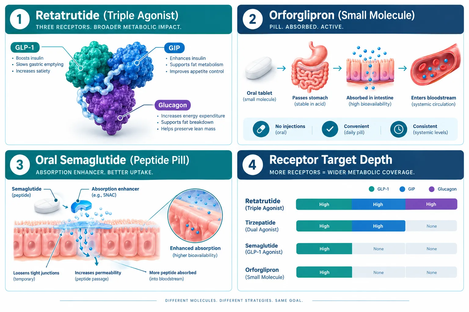
  • Retatrutide is a triple agonist (GLP-1, GIP, glucagon receptors) with phase 3 data showing substantially higher weight-loss magnitude than single-agonist comparators.
  • Orforglipron (Foundayo) is a non-peptide small molecule that achieves oral bioavailability without an absorption enhancer, making it mechanistically distinct from oral semaglutide.
  • Oral semaglutide (Wegovy pill) is a peptide formulation requiring a specific absorption agent; it is broadly available in the US but shows more modest weight-loss numbers in smaller patient cohorts.
  • Regulatory status differs sharply: some agents are approved, others remain in late-stage trials or are available only for research purposes.
  • Researchers must explicitly encode receptor target depth, delivery route, glycemic endpoints, and tolerability variables before selecting any model.

Understanding the Compound Classes in Play

Understanding the Compound Classes in Play

Not all weight-loss agents work the same way, and conflating them in a research design leads to flawed conclusions. The current field breaks into three meaningful categories.

Retatrutide is a single-molecule triple agonist that activates GLP-1, GIP, and glucagon receptors simultaneously. Phase 3 data from the TRIUMPH-1 trial in obesity showed weight reductions that outpaced earlier dual-agonist benchmarks. In a separate type 2 diabetes cohort, weight loss was more modest and gastrointestinal adverse events were notably more frequent, a signal researchers must account for when selecting a population model. The broader TRIUMPH program also covers comorbidity-focused indications including obstructive sleep apnea, knee osteoarthritis, and cardiovascular disease, making it one of the most expansive obesity drug programs currently running. The planned TRIUMPH-5 head-to-head against tirzepatide will eventually serve as a key benchmark, but that data is not yet available. Researchers interested in the evidence base can review Retatrutide clinical trial summaries and Retatrutide clinical trials data as the program evolves.

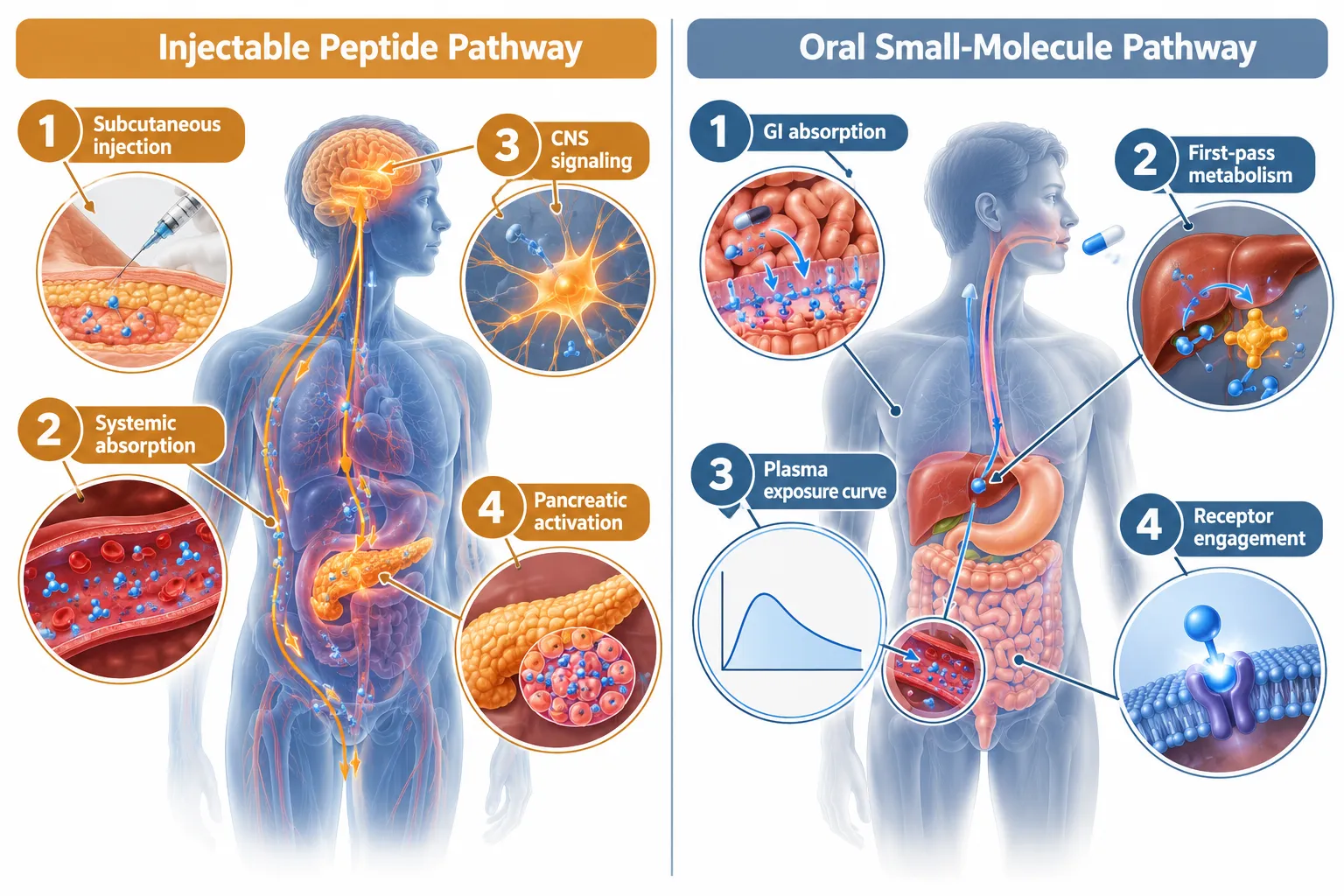
Orforglipron (Foundayo) is a non-peptide small molecule, meaning it is not built from amino acids and does not require an absorption enhancer to survive the GI tract. ATTAIN-1 phase 3 data for obesity showed meaningful weight loss with an all-oral dosing schedule. Because it engages only the GLP-1 receptor, its appetite and glycemic effects are narrower than retatrutide's triple-agonist profile.

Oral semaglutide (Wegovy pill) is a peptide reformulated with the absorption enhancer SNAC. It is broadly available in the US market as of 2026 but carries more modest weight-loss numbers in published cohorts compared to injectable semaglutide or the newer agents. Its primary research value lies in head-to-head oral comparisons.

For a broader orientation to peptide classes and how GLP-3 compounds fit alongside these agents, the Peptides 101 for research-use only buyers resource provides useful structural context.


Core Variables Researchers Must Encode When Comparing Retatrutide, GLP-1 Pills, and Small-Molecule Weight-Loss Options

Core Variables Researchers Must Encode When Comparing Retatrutide, GLP-1 Pills, and Small-Molecule Weight-Loss Options

Selecting a model without explicitly defining the following variables produces data that cannot be interpreted cleanly.

Receptor Target Depth

"A single receptor target and a triple receptor target are not interchangeable models, even when the primary endpoint is body weight."

The table below summarizes receptor engagement across the three compound classes:

Compound GLP-1 GIP Glucagon Delivery
Retatrutide Yes Yes Yes Injectable
Orforglipron Yes No No Oral (non-peptide)
Oral Semaglutide Yes No No Oral (peptide + SNAC)

Glucagon receptor activation drives additional energy expenditure beyond appetite suppression. Researchers modeling thermogenic pathways must account for this difference. Researchers sourcing retatrutide for in-vitro or preclinical work can reference GLP-3 Reta 10mg 99% pure third-party tested or GLP-3 Reta 30mg for sale for research-grade supply options.

Oral Exposure and Bioavailability

Oral bioavailability varies dramatically between a non-peptide small molecule and a peptide-based oral agent. Orforglipron achieves systemic exposure without an absorption enhancer, producing a cleaner pharmacokinetic curve. Oral semaglutide depends on SNAC to open tight junctions in the gastric mucosa, which introduces variability based on food timing and gastric pH. Researchers designing oral exposure models must encode this difference explicitly in their protocols.

Glycemic vs. Weight Endpoints

Retatrutide's type 2 diabetes cohort showed that glycemic and weight outcomes do not move in parallel. Models built around HbA1c reduction require different dose-escalation assumptions than models built around percent body weight change. Researchers should not assume that a compound's obesity-trial weight-loss magnitude will replicate in a glycemic-focused design.

Tolerability Variables

Gastrointestinal adverse events, nausea, vomiting, diarrhea, appear across all three agents but with different frequency profiles. Retatrutide's diabetes cohort showed notably higher GI event rates than its obesity cohort, likely reflecting dose-escalation speed and baseline metabolic status. Any comparative model must pre-specify tolerability endpoints rather than treating them as secondary observations.


Regulatory Status and Research-Use Considerations in 2026

Regulatory Status and Research-Use Considerations in 2026

As of 2026, orforglipron (Foundayo) has received regulatory approval for obesity, making it the first non-peptide oral GLP-1 agonist to reach that status. Oral semaglutide for obesity is also approved and commercially available in the US. Retatrutide remains in phase 3 trials and has not yet received approval; it is available only as a research compound through verified research-grade suppliers.

This distinction matters for study design. Approved agents can be studied in clinical settings with standard IRB frameworks. Research compounds require additional documentation, sourcing from qualified suppliers, and strict adherence to research-use-only protocols.

Researchers building comparative models should also consider how retatrutide's comorbidity-focused TRIUMPH sub-studies (OSA, knee OA, CVD) may eventually generate approval pathways beyond obesity, expanding the range of valid research questions.

For researchers exploring related peptide mechanisms, resources on Tesamorelin dosage for fat loss and Tesamorelin before and after offer useful comparisons for growth-hormone-mediated fat metabolism models alongside GLP-1-axis compounds.


Conclusion

The question of which model to select from Retatrutide, GLP-1 Pills, and Small-Molecule Weight-Loss Options is not answered by weight-loss magnitude alone. Receptor target depth, oral bioavailability mechanism, glycemic versus weight endpoints, tolerability profiles, and current regulatory status all shape what a given compound can and cannot tell a researcher.

Actionable next steps for researchers:

  1. Define the primary endpoint first, weight loss, glycemic control, or comorbidity reduction, before selecting a compound.
  2. Map receptor targets explicitly; do not treat GLP-1-only agents as equivalent to triple agonists in mechanistic models.
  3. Account for oral exposure variability when designing pharmacokinetic comparisons between peptide-based and non-peptide oral agents.
  4. Pre-specify tolerability endpoints, particularly in diabetic population models where GI event rates differ from obesity cohorts.
  5. Verify regulatory status before sourcing; approved agents and research-stage compounds require different procurement and documentation protocols.

The TRIUMPH-5 head-to-head data, when available, will provide the most direct comparative benchmark in this field. Until then, rigorous model selection depends on encoding the variables above with precision.

https://www.puretestedpeptides.com/wp-content/uploads/2026/10/retatrutide-glp-1-pills-and-small-molecule-weight-loss-options-what-researchers.webp 1024 1536 https://www.puretestedpeptides.com/wp-content/uploads/2026/01/buy-peptides-online.jpg 2026-10-01 13:05:002026-10-01 13:05:00Retatrutide, GLP-1 Pills, and Small-Molecule Weight-Loss Options: What Researchers Should Compare Before Selecting a Model
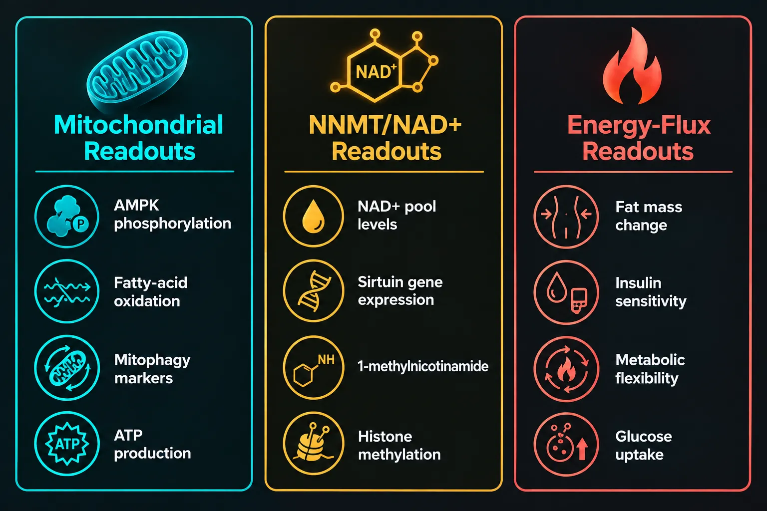
MOTS-c and 5-Amino-1MQ: A Practical Guide to Separating Mitochondrial, NNMT, and Energy-Flux Readouts

MOTS-c and 5-Amino-1MQ: A Practical Guide to Separating Mitochondrial, NNMT, and Energy-Flux Readouts

October 1, 2026/0 Comments/in Uncategorized/by

Attributing an observed metabolic effect to the wrong mechanism is one of the most common errors in multi-compound research protocols. When two agents act on overlapping but distinct pathways, as MOTS-c and 5-Amino-1MQ do, a poorly designed assay panel can make a mitochondrial signal look like an NAD+ effect, or vice versa. This practical guide to MOTS-c and 5-Amino-1MQ covers how to separate mitochondrial, NNMT, and energy-flux readouts so that each intervention gets credited for what it actually does.

Key Takeaways

  • MOTS-c is a mitochondrial-derived peptide that activates AMPK primarily in skeletal muscle, driving glucose uptake and fatty-acid oxidation.
  • 5-Amino-1MQ inhibits NNMT, preserving the intracellular NAD+ pool and activating sirtuin-linked pathways mainly in white adipose tissue and liver.
  • Both compounds can influence NAD+ levels, but through mechanistically different routes that require distinct assays to distinguish.
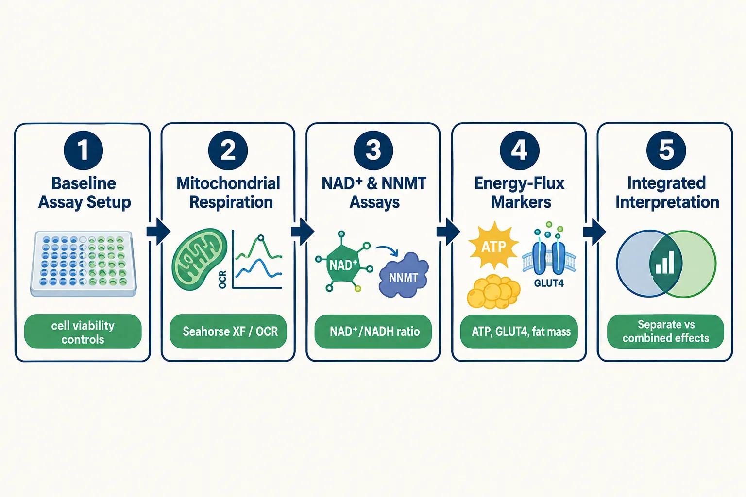
  • A well-designed study pairs mitochondrial respiration assays, NAD-related measurements, NNMT-linked markers, ATP quantification, and cell-viability controls in the same protocol.
  • The evidence base for both agents remains preclinical; neither is an approved therapeutic.

Understanding the Two Pathways Before Designing Any Assay

Understanding the Two Pathways Before Designing Any Assay

Any practical guide to MOTS-c and 5-Amino-1MQ must begin with a clear picture of what each compound does independently. Confusing their mechanisms at the design stage leads to assay panels that cannot answer the questions being asked.

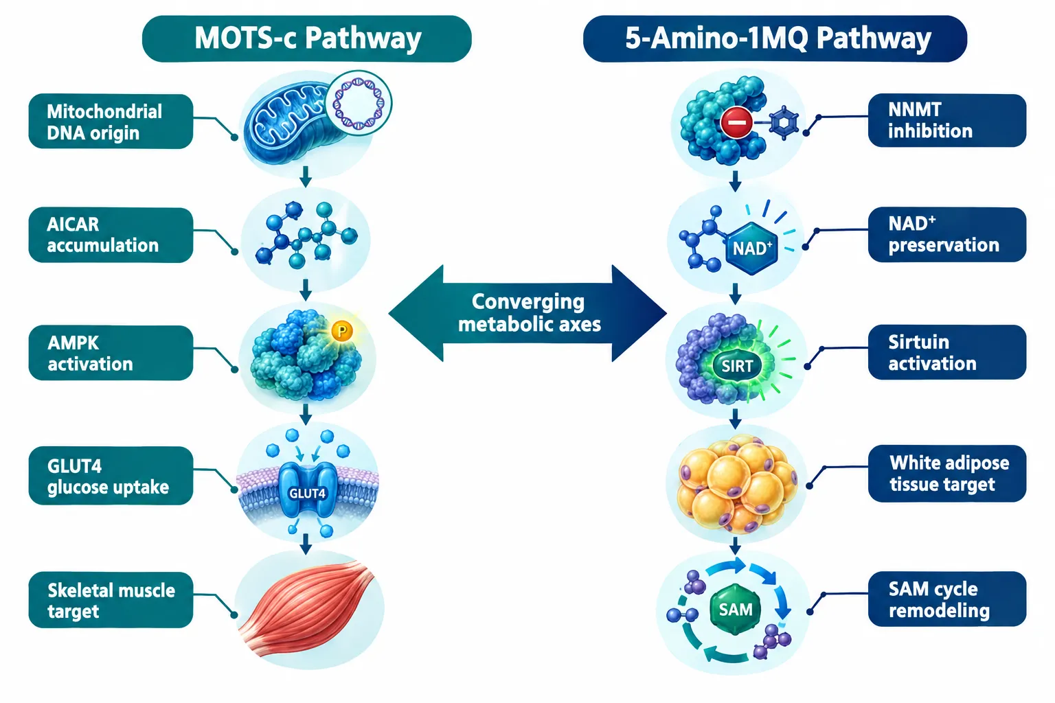
MOTS-c is a 16-amino-acid peptide encoded within mitochondrial DNA, first characterized by a University of Southern California research group in 2015. It interferes with the folate-methionine one-carbon cycle and de novo purine biosynthesis inside cells, causing AICAR to accumulate. AICAR is a well-established indirect activator of AMP-activated protein kinase (AMPK). The downstream result is increased fatty-acid oxidation, enhanced GLUT4-mediated glucose uptake, and improved insulin sensitivity, effects observed most strongly in skeletal muscle in animal models. Animal studies have shown prevention of diet-induced obesity and age-related insulin resistance.

Researchers interested in MOTS-c peptide research materials should note that its primary readout territory is mitochondrial bioenergetics and substrate oxidation, not direct NAD+ salvage.

5-Amino-1MQ (5-amino-1-methylquinolinium) is a small, membrane-permeable molecule that directly inhibits nicotinamide N-methyltransferase (NNMT). NNMT converts nicotinamide to 1-methylnicotinamide, consuming S-adenosylmethionine (SAM) in the process. By blocking this enzyme, 5-Amino-1MQ reduces SAM consumption, preserves the intracellular NAD+ pool, and activates NAD+-dependent pathways including sirtuins and mitochondrial biogenesis. Preclinical mouse data have shown reductions in fat mass without reduced food intake. Its primary action zone is white adipose tissue and liver, where NNMT is upregulated in obesity.

"Both compounds can nudge NAD+ levels upward, but they pull different levers to get there, and conflating those levers produces uninterpretable data."

This mechanistic divergence is exactly why a guide to separating mitochondrial, NNMT, and energy-flux readouts is necessary before any combined protocol begins.


How to Separate Mitochondrial, NNMT, and Energy-Flux Readouts in Practice

How to Separate Mitochondrial, NNMT, and Energy-Flux Readouts in Practice

The core design principle is simple: assign each readout category its own assay layer, and run all three layers in the same experimental unit so that confounding is controlled rather than ignored.

Mitochondrial Readouts

These assays capture AMPK-centric activity attributable to MOTS-c.

Readout Recommended Assay What It Captures
AMPK activation Western blot for p-AMPK/AMPK ratio Direct MOTS-c downstream signal
Oxygen consumption rate (OCR) Seahorse XF Analyzer Mitochondrial respiration flux
Fatty-acid oxidation Radiolabeled palmitate oxidation Substrate shift toward lipids
Mitophagy/biogenesis LC3-II, PGC-1alpha expression Mitochondrial quality control

NNMT and NAD+ Readouts

These assays isolate the 5-Amino-1MQ contribution.

  • NAD+/NADH ratio: A direct readout of NAD+ pool preservation. If 5-Amino-1MQ is working, this ratio rises without a proportional change in NAMPT activity.
  • 1-methylnicotinamide (MNA) levels: MNA is the product of NNMT activity. Reduced MNA confirms NNMT inhibition, not just a generic NAD+ boost.
  • SAM/SAH ratio: Reflects the methylation economy downstream of NNMT blockade.
  • Sirtuin-linked gene expression: SIRT1 and SIRT3 targets confirm NAD+-dependent pathway activation.

Critically, if MOTS-c is present in the same well and AMPK is active, NAD+ consumption will increase through energy-demanding pathways. That can mask 5-Amino-1MQ's preserving effect. Measuring MNA and SAM/SAH alongside NAD+/NADH disambiguates the source.

Energy-Flux Readouts

These whole-system markers capture the integrated output of both interventions.

  • Fat mass change (in animal models): Reflects combined lipolytic and oxidative effects.
  • Insulin sensitivity: Glucose tolerance tests and HOMA-IR scores capture GLUT4-mediated improvements driven by MOTS-c.
  • Metabolic flexibility: The ability to switch between glucose and fat oxidation, measurable via respiratory exchange ratio (RER).
  • ATP quantification: Total cellular ATP reflects the net energy-production outcome and serves as a cross-check on both mitochondrial respiration and NAD+ pathway activity.

For context on how other mitochondrial-targeting peptides are studied, the SS-31 mitochondrial research themes resource illustrates how cardiolipin-targeted agents use similar OCR-based designs, offering useful methodological parallels. Additional background on SS-31 elamipretide research approaches can inform how to structure mitochondrial membrane integrity controls.


Building an Attribution-Proof Protocol: Step-by-Step

Building an Attribution-Proof Protocol: Step-by-Step

The following five-step framework minimizes attribution error when running MOTS-c and 5-Amino-1MQ together.

Step 1, Establish baseline cell viability controls. Run MTT or CellTiter-Glo assays at every dose tested. If viability drops, any observed change in OCR or NAD+ is a toxicity artifact, not a mechanism signal.

Step 2, Run single-agent arms first. Before combining, characterize each compound alone. This produces a mechanistic fingerprint: MOTS-c alone should shift OCR and raise p-AMPK; 5-Amino-1MQ alone should lower MNA and raise the NAD+/NADH ratio.

Step 3, Apply the full assay panel to the combination arm. All readouts from Steps 1 and 2 must be measured simultaneously. Partial panels in the combination arm are the primary source of attribution errors.

Step 4, Use pathway inhibitors as mechanistic controls. An AMPK inhibitor (such as Compound C) can silence the MOTS-c arm pharmacologically, allowing the NNMT signal to be read in isolation within the same combined-treatment context. An NNMT-overexpression control does the reverse.

Step 5, Interpret energy-flux markers last. Fat mass, insulin sensitivity, and ATP changes are downstream integrations of both pathways. They should be interpreted only after the upstream mitochondrial and NNMT readouts have been attributed.

For researchers also exploring growth-hormone-mediated metabolic comparisons, reviewing how peptides in cardiometabolic models differ from small-molecule drugs provides useful framing for why AMPK agonists, NNMT inhibitors, and GH-driven agents require separate readout categories. Similarly, understanding visceral fat research methodology helps contextualize fat-mass endpoints.


Conclusion

A practical guide to MOTS-c and 5-Amino-1MQ separating mitochondrial, NNMT, and energy-flux readouts comes down to one discipline: never let overlapping downstream effects collapse distinct upstream mechanisms into a single unattributed signal.

Actionable next steps for researchers:

  1. Map each compound to its primary assay layer before designing the protocol.
  2. Always include MNA quantification alongside NAD+/NADH to distinguish NNMT inhibition from AMPK-driven NAD+ consumption.
  3. Run single-agent arms before the combination arm to establish mechanistic fingerprints.
  4. Include cell-viability controls at every dose and time point.
  5. Reserve energy-flux endpoints (fat mass, insulin sensitivity, ATP) for last-stage interpretation, not primary attribution.

Both MOTS-c and 5-Amino-1MQ remain research-stage compounds with a predominantly preclinical evidence base. No large, controlled human trials have confirmed the animal findings, and neither agent is an approved drug. All protocols should be conducted within appropriate research frameworks and regulatory guidelines.

https://www.puretestedpeptides.com/wp-content/uploads/2026/10/mots-c-and-5-amino-1mq-a-practical-guide-to-separating-mitochondrial-nnmt-and-en.webp 1024 1536 https://www.puretestedpeptides.com/wp-content/uploads/2026/01/buy-peptides-online.jpg 2026-10-01 13:04:082026-10-01 13:04:08MOTS-c and 5-Amino-1MQ: A Practical Guide to Separating Mitochondrial, NNMT, and Energy-Flux Readouts
GLP-2-T and GLP2 Tirz Peptide Research: How to Design Intestinal Barrier, Organoid, and Epithelial-Repair Assays

GLP-2-T and GLP2 Tirz Peptide Research: How to Design Intestinal Barrier, Organoid, and Epithelial-Repair Assays

October 1, 2026/0 Comments/in Uncategorized/by

Roughly 70% of the body's immune activity is concentrated in the gut lining, yet most peptide research programs treat intestinal barrier function as an afterthought. GLP-2-T and GLP2 Tirz Peptide Research: How to Design Intestinal Barrier, Organoid, and Epithelial-Repair Assays has emerged as a rigorous discipline precisely because the gut epithelium is both a therapeutic target and a sensitive readout system. Designing assays that capture permeability shifts, tight-junction remodeling, and organoid growth requires a structured approach and a clear understanding of what these peptide names actually represent.

Key Takeaways

  • GLP-2 drives intestinal growth indirectly through enteric neurons, not directly through epithelial receptors, assay design must account for this.
  • "GLP-2-T" and "GLP2 Tirz" are working nomenclature terms, not validated compound names; confirm identity through mass spectrometry and receptor-binding assays before interpreting data.
  • Core barrier readouts include transepithelial electrical resistance (TEER), FITC-dextran permeability, and tight-junction protein immunostaining.
  • Organoid models using intestinal stem cells (ISCs) and Paneth cells provide the most physiologically relevant epithelial-repair endpoints.
  • Purity and sequence verification of research peptides are non-negotiable prerequisites for reproducible results.

Understanding GLP-2 Biology Before Building Assays

Understanding GLP-2 Biology Before Building Assays

GLP-2 (glucagon-like peptide-2) is a 33-amino-acid hormone secreted by enteroendocrine L-cells in response to luminal nutrients. Its primary role is to stimulate intestinal growth, increase mucosal surface area, and reinforce epithelial barrier integrity. A critical detail that shapes every assay design: the GLP-2 receptor is expressed on enteric neurons and subepithelial myofibroblasts, not on enterocytes themselves. This means GLP-2 acts indirectly, triggering a neuronal relay that ultimately drives crypt cell proliferation and tight-junction assembly.

This neuroepithelial circuit has direct implications for assay selection. Simple monolayer cell cultures lacking a neuronal component will not fully recapitulate GLP-2 signaling. Researchers working on GLP-2-T and GLP2 Tirz concepts must either use co-culture systems or organoid platforms that preserve the cellular architecture of the crypt-villus axis.

Nomenclature clarity matters. The terms "GLP-2-T" and "GLP2 Tirz" circulate in research communities as shorthand for modified GLP-2 analogs or multi-agonist concepts that may incorporate tirzepatide-like dual-receptor targeting. Neither term corresponds to a single, fully characterized compound as of 2026. Before any assay begins, researchers should confirm peptide identity via mass spectrometry, HPLC purity analysis, and receptor-binding confirmation. Explore available GLP peptide research options to understand the range of characterized analogs on the market.


Designing Intestinal Barrier Assays for GLP-2-T and GLP2 Tirz Peptide Research

Designing Intestinal Barrier Assays for GLP-2-T and GLP2 Tirz Peptide Research

Barrier function assays form the quantitative backbone of GLP-2-T and GLP2 Tirz Peptide Research: How to Design Intestinal Barrier, Organoid, and Epithelial-Repair Assays programs. Two readouts dominate the literature:

Transepithelial Electrical Resistance (TEER)

TEER measures the ionic resistance across a cell monolayer grown on a permeable Transwell insert. Higher resistance values indicate a tighter, more intact barrier. For GLP-2 analog research:

  • Seed Caco-2 or T84 cells on 0.4-micron Transwell membranes
  • Allow 21 days for full differentiation and tight-junction maturation
  • Apply peptide at physiologically relevant concentrations (typically 1-100 nM)
  • Record TEER at 24, 48, and 72 hours post-treatment

Key point: A meaningful TEER increase after GLP-2-T treatment suggests tight-junction reinforcement, but must be paired with protein-level confirmation.

FITC-Dextran Permeability

This assay quantifies paracellular leakage by measuring how much fluorescently labeled dextran crosses the monolayer from apical to basolateral compartments. A reduction in FITC-dextran flux after peptide treatment confirms barrier-sealing activity.

Tight-Junction Protein Immunostaining

Immunofluorescence staining for ZO-1, claudin-3, and occludin provides mechanistic evidence. Disrupted tight junctions appear as fragmented or discontinuous staining patterns. Successful GLP-2 analog treatment should restore linear, continuous junction morphology.

Endpoint summary table:

Assay Readout Positive Result
TEER Ohms x cm2 Increased resistance
FITC-Dextran Fluorescence (ng/mL) Decreased flux
ZO-1 Immunostaining Confocal imaging Continuous junctions
NLRP3 Inflammasome Western blot / ELISA Reduced activation
LPS Translocation ELISA (basolateral) Reduced LPS signal

For researchers exploring related multi-agonist frameworks, reviewing GLP-3 peptide research compounds can help contextualize receptor-specificity differences when designing controls.


Organoid and Epithelial-Repair Assays in GLP-2-T and GLP2 Tirz Peptide Research

Organoid and Epithelial-Repair Assays in GLP-2-T and GLP2 Tirz Peptide Research

Organoid models represent the most physiologically faithful platform for GLP-2-T and GLP2 Tirz Peptide Research: How to Design Intestinal Barrier, Organoid, and Epithelial-Repair Assays. Intestinal organoids derived from crypts maintain ISC populations, Paneth cells, goblet cells, and enterocytes in a self-organizing three-dimensional structure.

Setting Up the Organoid Assay

  1. Isolate crypts from murine small intestine or human biopsy tissue using EDTA chelation
  2. Embed in Matrigel at 37°C and overlay with ENR medium (EGF, Noggin, R-spondin)
  3. Introduce peptide at day 3 post-seeding, once basal organoids are established
  4. Quantify growth by measuring organoid surface area, budding frequency, and villus-like domain length at 24-hour intervals

Key Epithelial-Repair Endpoints

  • ISC marker expression: Lgr5+ cell frequency by immunofluorescence or flow cytometry
  • Paneth cell density: Lysozyme staining as a proxy for niche-support capacity
  • Villus height and crypt depth: Morphometric analysis from confocal z-stacks
  • Proliferation index: Ki-67 staining in crypt-base columnar cells

GLP-2 analogs consistently increase organoid budding efficiency and villus domain length in preclinical models. Modified analogs with extended half-lives, relevant to the GLP-2-T concept, show amplified effects on crypt proliferation compared to native GLP-2, making organoid size and budding frequency sensitive discriminators between compound variants.

Researchers interested in complementary barrier-repair peptide tools may find value in reviewing SS-31 peptide research for mitochondrial-protection endpoints that can run alongside epithelial-repair assays. Similarly, cagrilinitide peptide research offers a relevant comparator for multi-agonist gut-axis studies.

Disease Model Applications

  • IBD models: LPS-challenged organoids or DSS-treated murine intestine provide a relevant inflammatory context for testing GLP-2-T barrier-rescue activity
  • Short bowel syndrome (SBS) surrogates: Remnant intestine organoids from partial resection models show blunted adaptation that GLP-2 analogs can rescue, a validated translational endpoint
  • GVHD models: Graft-versus-host disease involves severe epithelial damage; GLP-2 receptor agonism has shown protective effects in these systems, making crypt survival the primary endpoint

For those sourcing characterized analogs for these models, GLP-3R peptide options and Reta peptide research compounds represent structurally related tools worth including as comparators.


Conclusion

Rigorous GLP-2-T and GLP2 Tirz Peptide Research: How to Design Intestinal Barrier, Organoid, and Epithelial-Repair Assays programs rest on three pillars: verified peptide identity, mechanistically appropriate cell models, and multi-endpoint readout strategies. The following steps will strengthen any research program:

  1. Verify compound identity with mass spectrometry and HPLC before any biological assay
  2. Pair TEER with FITC-dextran and tight-junction immunostaining for a complete barrier profile
  3. Use organoid models when ISC proliferation and villus morphology are primary endpoints
  4. Include inflammatory challenge conditions (LPS, DSS) to stress-test barrier-repair claims
  5. Source research-grade peptides from suppliers with documented purity data, see guidance on evaluating purity for research-grade peptides for a practical framework

As multi-agonist concepts combining GLP-2 activity with tirzepatide-like GIP/GLP-1 receptor engagement continue to develop, the assay toolkit described here will serve as the foundation for evaluating any new analog claiming intestinal barrier or epithelial-repair activity.

https://www.puretestedpeptides.com/wp-content/uploads/2026/10/glp-2-t-and-glp2-tirz-peptide-research-how-to-design-intestinal-barrier-organoid.webp 1024 1536 https://www.puretestedpeptides.com/wp-content/uploads/2026/01/buy-peptides-online.jpg 2026-10-01 13:03:562026-10-01 13:03:56GLP-2-T and GLP2 Tirz Peptide Research: How to Design Intestinal Barrier, Organoid, and Epithelial-Repair Assays
BPC-157 and TB-500 in Tendon and Muscle Models: Separating Angiogenesis, Matrix Remodeling, and Inflammation Readouts

BPC-157 and TB-500 in Tendon and Muscle Models: Separating Angiogenesis, Matrix Remodeling, and Inflammation Readouts

October 1, 2026/0 Comments/in Uncategorized/by

Roughly 35 animal and in vitro studies have examined BPC-157 and TB-500 in tendon and muscle repair, yet only a single human clinical study exists across the entire combined literature. That ratio alone explains why researchers in 2026 are pushing for a more disciplined, endpoint-specific framework rather than broad claims about "healing peptides." Understanding BPC-157 and TB-500 in tendon and muscle models: separating angiogenesis, matrix remodeling, and inflammation readouts requires moving beyond stacked-peptide narratives and into the granular language of scratch assays, collagen histology, vascular markers, and mechanical testing.

Key Takeaways

  • BPC-157 operates primarily through VEGFR2, Akt-eNOS, and TGF-beta pathways, making it the stronger candidate for angiogenesis and matrix remodeling endpoints in tendon models.
  • TB-500 drives actin polymerization and cell migration via MAPK and FAK signaling, positioning it as a cytoskeletal and motogenic agent rather than a direct ECM organizer.
  • Inflammation readouts differ: BPC-157 has detailed cytokine and NF-kB profiling in musculoskeletal models, while TB-500's anti-inflammatory data are less systematically mapped in tendon tissue.
  • Combining both peptides shows early synergistic signals in rodent models, but no completed Phase 2 or Phase 3 human trials exist for either compound in tendon or muscle repair.
  • Both compounds remain experimental and are banned or under regulatory scrutiny in organized sport and clinical compounding as of 2026.

Mechanistic Profiles: How Each Peptide Acts at the Tissue Level

Mechanistic Profiles: How Each Peptide Acts at the Tissue Level

Understanding the mechanistic separation between BPC-157 and TB-500 is the foundation for interpreting any experimental readout.

BPC-157 is a synthetic pentadecapeptide derived from a gastric protein. In tendon and muscle models, it activates VEGFR2 and the Akt-eNOS nitric oxide axis, stimulates fibroblast proliferation, and engages focal adhesion kinase (FAK)-paxillin signaling. These pathways converge on three measurable outcomes: new blood vessel formation, extracellular matrix (ECM) reorganization, and inflammatory resolution. In rat Achilles tendon transection models, aggregated data report 30-40% shortening of healing time alongside improved collagen fiber organization and ultimate tensile strength versus untreated controls.

TB-500, a synthetic derivative of thymosin beta-4 (Tβ4), works through a different axis. Its core action is promoting actin polymerization and cytoskeletal stabilization, enabling rapid cell migration into injury zones. Signaling involves MAPK, NF-kB modulation, and FAK, but the tendon-specific mechanistic map is less detailed than BPC-157's. In muscle models, TB-500 has demonstrated satellite cell (Pax7+) mobilization, enhanced fibrillar regeneration, and reduced fibrosis in murine muscular dystrophy models.

Feature BPC-157 TB-500
Primary signaling VEGFR2, Akt-eNOS, TGF-beta MAPK, FAK, actin dynamics
Main tissue action Angiogenesis + ECM remodeling Cell migration + cytoskeletal repair
Inflammation data depth Detailed (cytokine, NF-kB, COX-2) Moderate (early-phase modulation)
Tendon-specific mapping Extensive preclinical Limited
Human trial data Single study (non-musculoskeletal) None in tendon/muscle

Researchers interested in broader peptide mechanisms may also find the discussion of wound healing compounds useful for contextualizing these tissue-repair pathways.


Separating the Three Readouts: Angiogenesis, Matrix Remodeling, and Inflammation

Separating the Three Readouts: Angiogenesis, Matrix Remodeling, and Inflammation

When evaluating BPC-157 and TB-500 in tendon and muscle models: separating angiogenesis, matrix remodeling, and inflammation readouts, each endpoint demands its own assay logic and interpretive standard.

Angiogenesis

BPC-157 reliably increases microvessel density and restores blood flow in ischemic hind-limb models, reflecting VEGF-driven angiogenic activity and nitric-oxide-mediated vasoregulation. Measurable outputs include CD31-positive vessel counts, laser Doppler perfusion imaging, and microvessel density scoring on histological sections.

TB-500/Tβ4 is also described as pro-angiogenic, but its angiogenesis data in orthopaedic models are summarized more broadly across wound, cardiac, and dermal contexts rather than tendon-specific dose-response curves. This makes direct comparison at the tissue level difficult without purpose-designed studies.

Matrix Remodeling

BPC-157 engages TGF-beta-linked ECM pathways, improves collagen alignment, and reduces scar formation across tendon, ligament, and muscle injuries. Polarized-light microscopy and second-harmonic generation imaging of collagen fiber organization, combined with mechanical tensile testing, are the standard readouts. Partial restoration of normal tendon collagen architecture and biomechanical strength has been reported in multiple rodent studies.

TB-500's contribution to matrix remodeling is characterized mainly as accelerating migratory infiltration of repair cells into damaged ECM rather than directly orchestrating collagen synthesis or fiber alignment. This is a meaningful distinction: TB-500 may prepare the matrix environment for remodeling without driving the structural reorganization itself.

Inflammation

BPC-157 modulates the nitric oxide system, downregulates pro-inflammatory cytokines, interacts with COX-2, and inhibits NF-kB activation. These actions produce faster resolution of the inflammatory phase in musculoskeletal injury models, with measurable reductions in IL-6, TNF-alpha, and prostaglandin markers.

TB-500 modulates the early inflammatory phase and facilitates mesenchymal stem-cell migration to injury zones, but detailed cytokine profiling in tendon and muscle models remains limited. Researchers working on wound healing endpoints should note this gap when designing comparative studies.

"BPC-157 has the widest preclinical footprint across musculoskeletal, vascular, and neurological models, while TB-500 has a more focused literature in musculoskeletal and wound healing, a distinction that matters enormously when selecting assay panels."


Combining BPC-157 and TB-500: Synergy Evidence and Research Gaps

Combining BPC-157 and TB-500: Synergy Evidence and Research Gaps

The rationale for combining BPC-157 and TB-500, sometimes called the "Wolverine stack" in research circles, rests on the idea of orthogonal mechanisms. BPC-157 provides localized angiogenesis, ECM remodeling, and inflammation control; TB-500 drives broad cell migration and cytoskeletal repair. One rat model using combined BPC-157 plus Tβ4 reported earlier restoration of contractile function than either peptide alone, supporting a synergistic interpretation.

However, these data are entirely preclinical. As of 2026:

  • No completed Phase 2 or Phase 3 trials exist for injectable BPC-157 or TB-500 in tendon or muscle injuries.
  • No human randomized controlled trials confirm musculoskeletal healing benefits.
  • TB-500/Tβ4 remains a banned substance in organized sport.
  • Regulatory discussions around BPC-157 include FDA proposals not to add it to the traditional compounding bulks list.

For researchers studying related peptide stacks, the IPA Sermorelin stack research page offers a useful parallel framework for thinking about combinatorial peptide effects in preclinical models.

Expert analyses from 2026 predict that future work will focus on mapping dose-dependent effects on discrete endpoints, vessel density, collagen architecture, tensile strength, and cytokine signatures, within standardized injury models. Any clinical adoption in orthopaedics or sports medicine will require long-term human trials demonstrating that mechanistic benefits translate into faster, safer recovery without promoting maladaptive angiogenesis or fibrosis.

Researchers exploring mitochondrial and cellular repair mechanisms alongside these peptides may find the SS-31 mitochondrial research literature relevant, as oxidative stress pathways intersect with ECM remodeling in chronic tendon injury. Similarly, those examining broader tissue repair contexts may reference LL-37 versus SS-31 benefits of each peptide for comparative mechanistic framing.


Conclusion

Properly evaluating BPC-157 and TB-500 in tendon and muscle models: separating angiogenesis, matrix remodeling, and inflammation readouts demands endpoint-specific thinking rather than generalized healing claims. BPC-157 is the stronger candidate for angiogenesis and ECM remodeling assays, with detailed VEGFR2 and TGF-beta mechanistic data and measurable improvements in collagen organization and tensile strength. TB-500 is better framed as a migration and cytoskeletal repair agent, with its most robust data in satellite cell mobilization and early-phase inflammatory modulation.

Actionable next steps for researchers and clinicians:

  1. Design studies with discrete endpoint panels, vessel density counts, collagen histology scoring, cytokine multiplex assays, and mechanical tensile testing, rather than composite "healing" outcomes.
  2. Treat combination data as hypothesis-generating only; no human trial has validated synergistic claims.
  3. Monitor regulatory developments closely, as both compounds face evolving restrictions in compounding and sport.
  4. Prioritize standardized rodent injury models (Achilles transection, muscle crush) to enable cross-study comparison before any translational step.

Both compounds remain promising but investigational. The next phase of research depends on disciplined assay design, not further stacking claims.

https://www.puretestedpeptides.com/wp-content/uploads/2026/10/bpc-157-and-tb-500-in-tendon-and-muscle-models-separating-angiogenesis-matrix-re.webp 1024 1536 https://www.puretestedpeptides.com/wp-content/uploads/2026/01/buy-peptides-online.jpg 2026-10-01 13:03:562026-10-01 13:03:56BPC-157 and TB-500 in Tendon and Muscle Models: Separating Angiogenesis, Matrix Remodeling, and Inflammation Readouts
Hemoglobin, LDL Cholesterol, and Blood Pressure in Peptide Studies: Building a Cardiometabolic Monitoring Panel

Hemoglobin, LDL Cholesterol, and Blood Pressure in Peptide Studies: Building a Cardiometabolic Monitoring Panel

October 1, 2026/0 Comments/in Uncategorized/by

Across incretin analogue trials, systolic blood pressure drops of 2-5 mmHg and glycated hemoglobin reductions of 0.7-1.5 percentage points have been recorded consistently, yet many researchers still track these endpoints in isolation rather than as a coordinated panel. Building a structured approach to hemoglobin, LDL cholesterol, and blood pressure in peptide studies is not just good laboratory hygiene; it is the difference between detecting a meaningful cardiometabolic signal and missing it entirely.

Key Takeaways

  • HbA1c, LDL-C, and systolic blood pressure form the core triad for monitoring cardiometabolic effects in GLP-class and metabolic-peptide research.
  • Effect sizes are modest but reproducible: roughly -0.25% HbA1c, -0.17 mmol/L LDL-C, and -4 mmHg systolic BP per meta-analytic estimates.
  • Timing matters, baseline, week 4, week 12, and week 26 assessments capture the full trajectory of peptide-driven changes.
  • Composite indices (e.g., Hemoglobin-Myeloid Index) outperform single markers for cardiometabolic risk sensitivity.
  • Confounders such as diet, hydration status, and concurrent medications must be controlled before attributing changes to the peptide under study.

Why These Three Markers Define Cardiometabolic Risk in Peptide Research

Why These Three Markers Define Cardiometabolic Risk in Peptide Research

Researchers studying GLP-1 receptor agonists, GH secretagogues, and other metabolic peptides need a consistent framework for interpreting laboratory data. The three markers, glycated hemoglobin (HbA1c), low-density lipoprotein cholesterol (LDL-C), and blood pressure (BP), were not chosen arbitrarily. Each reflects a distinct but interconnected pathway that peptide agents are known to modulate.

HbA1c captures average glycemic exposure over roughly 90 days, making it ideal for tracking slow-moving metabolic shifts. LDL-C reflects atherogenic lipid burden, which GLP-1 receptor agonists reduce modestly but reproducibly across overweight, type 2 diabetes, and chronic kidney disease populations. Systolic blood pressure responds to natriuretic and vasodilatory effects that several peptide classes trigger, with reductions averaging approximately 4 mmHg in well-controlled trials.

Together, these three endpoints mirror what commercial cardiometabolic panels, including those combining apolipoprotein B, small dense LDL-C, and insulin resistance scores, already treat as mandatory. Researchers exploring GLP-3 peptide formulations or GLP-1 peptides will find that aligning their monitoring to this triad makes cross-study comparisons far more tractable.

"A monitoring panel is only as strong as the consistency of its collection windows. Sporadic lab draws produce noise, not signal."

The Role of Composite Indices

Single-marker tracking has a well-documented sensitivity ceiling. The Hemoglobin-Myeloid Index (HMI), a composite of hemoglobin and leukocyte counts, has shown greater discriminatory power for cardiometabolic risk than hemoglobin alone. This finding supports building multi-parameter panels rather than relying on any one biomarker, a principle directly applicable to peptide study design.


Structuring a Monitoring Panel: Timing, Confounders, and Collection Windows

Structuring a Monitoring Panel: Timing, Confounders, and Collection Windows

A well-designed cardiometabolic monitoring panel for peptide studies requires more than choosing the right markers. Timing and confounder control are equally critical.

Recommended Assessment Windows

Timepoint Primary Markers Secondary Markers
Baseline (Day 0) HbA1c, LDL-C, Systolic BP HDL-C, triglycerides, fasting glucose
Week 4 Systolic BP, fasting glucose Body weight, heart rate
Week 12 HbA1c, LDL-C, Systolic BP Triglycerides, HDL-C
Week 26+ Full panel + apolipoprotein B hs-CRP, lipoprotein(a)

HbA1c requires at least 8-12 weeks of exposure before a meaningful change is detectable, making early draws useful only for baseline anchoring. Blood pressure, by contrast, can shift within weeks, as demonstrated in orforglipron trials where a mean systolic reduction of approximately 4.3 mmHg was observed alongside HbA1c reductions approaching 1.36%.

Key Confounders to Control

Failing to account for confounders is the most common reason peptide-related cardiometabolic signals are misinterpreted.

  • Dietary changes: Caloric restriction independently lowers LDL-C and BP; weight loss attributable to the peptide must be separated from intentional dieting.
  • Hydration status: Dehydration elevates hemoglobin concentration artificially, inflating apparent HbA1c values.
  • Concurrent medications: Statins, antihypertensives, and metformin each affect one or more panel markers directly.
  • Assay standardization: Inconsistent laboratory methods between draws introduce measurement noise. Reviewing reference standards for peptide benchmarks helps researchers apply the same rigor to biomarker assays.
  • Time of day for BP: Circadian variation in systolic BP can exceed 10 mmHg; standardized morning measurements are essential.

For GH secretagogue research, including studies involving MOTS-c peptide, insulin sensitivity and triglyceride levels should be added to the secondary panel, as GH-axis activation has distinct lipid and glucose effects compared to GLP-class agents.


Applying the Panel Across GLP-Class, GH-Secretagogue, and Metabolic-Peptide Experiments

Applying the Panel Across GLP-Class, GH-Secretagogue, and Metabolic-Peptide Experiments

The core triad of hemoglobin, LDL cholesterol, and blood pressure in peptide studies applies broadly, but the expected effect profile varies by peptide class.

GLP-Class Peptides

Network meta-analyses in type 2 diabetes with chronic kidney disease place semaglutide and liraglutide at the top of probability rankings for LDL-C reduction (SUCRA 100%) and HbA1c improvement. The long-term SELECT trial confirmed that semaglutide 2.4 mg reduces total cholesterol, LDL-C, and triglycerides while raising HDL-C over roughly 40 months, even in patients without type 2 diabetes. Researchers using GLP-3R peptide formulations should establish whether their compound shares this lipid-modulating profile by anchoring measurements to the same triad.

GH-Secretagogue and Mitochondria-Targeted Peptides

Peptides that act on the GH axis or mitochondrial pathways, such as those reviewed in SS-31 research peptide considerations, have distinct cardiovascular signatures. SS-31 targets mitochondrial cardiolipin and may influence cardiac efficiency without directly lowering LDL-C. For these agents, blood pressure and hemoglobin remain relevant endpoints, but the lipid panel should be expanded to include triglycerides and HDL-C to capture the full metabolic picture.

Natriuretic and Cardiac Peptides

BNP and NT-proBNP are increasingly used alongside HbA1c, LDL-C, and BP in comprehensive panels for patients on cardiometabolic peptide therapies. This reflects a broader trend: researchers and clinicians are converging on integrated panels that treat metabolic and cardiac biomarkers as complementary rather than separate. Understanding how peptides interface with endocrine pharmacology is essential context for interpreting these expanded panels correctly.

For a foundational overview of peptide classes and their mechanisms, the Peptides 101 research-use guide provides useful structural context before designing a monitoring protocol.


Conclusion

Building a cardiometabolic monitoring panel around hemoglobin, LDL cholesterol, and blood pressure in peptide studies is not optional for rigorous research, it is the baseline standard. The actionable steps are straightforward:

  1. Set a baseline before any peptide exposure using HbA1c, fasting LDL-C, and standardized morning systolic BP.
  2. Schedule follow-up draws at weeks 4, 12, and 26, matching the biological half-life of each marker to the collection window.
  3. Document and control confounders, diet, medications, hydration, and assay methods, before attributing any change to the peptide.
  4. Expand the panel with apolipoprotein B, hs-CRP, and triglycerides when the peptide class warrants it.
  5. Use composite indices such as HMI where single-marker sensitivity is insufficient.

Researchers who apply this framework consistently will produce data that is both internally valid and comparable across studies, the foundation of any meaningful contribution to the cardiometabolic peptide literature in 2026.

https://www.puretestedpeptides.com/wp-content/uploads/2026/10/hemoglobin-ldl-cholesterol-and-blood-pressure-in-peptide-studies-building-a-card.webp 1024 1536 https://www.puretestedpeptides.com/wp-content/uploads/2026/01/buy-peptides-online.jpg 2026-10-01 13:03:512026-10-01 13:03:51Hemoglobin, LDL Cholesterol, and Blood Pressure in Peptide Studies: Building a Cardiometabolic Monitoring Panel
Page 8 of 81«‹678910›»
×

Helpful Links

  • My account
  • Cart
  • Checkout
  • Refund and Returns Policy
  • Privacy Policy
  • SMS Privacy Policy
  • Login
  • My Account
  • Logout

USA Made Lab Tested Peptides

All products are sold for research, laboratory, or analytical purposes only, and are not for human consumption


Pure Tested Peptides is a chemical supplier. Pure Tested Peptides is not a compounding / chemical compounding facility as defined under 503A of the Federal Food, Drug, and Cosmetic act. Pure Tested Peptides is not an outsourcing facility as defined under 503B of the Federal Food, Drug, and Cosmetic act.

The statements made within this website have not been evaluated by the US Food and Drug Administration. The products we offer are not intended to diagnose, treat, cure or prevent any disease.

Human/Animal Consumption Prohibited. Laboratory/In-Vitro Experimental Use Only

Scroll to top Scroll to top Scroll to top