Call or Text 727-513-9780
  • Shopping Cart Shopping Cart
    0Shopping Cart
Pure Tested Peptides | America's most trusted Peptides for sale online
  • Peptides for sale
    • Oral Peptides for sale
      • Peptide Capsules for sale
      • BPC 157 Capsules 1000mcg
      • SLU-PP-332 Capsules | 1000 mcg
      • 5-Amino-1MQ 50mg Capsules
      • Tesofensine 500mcg
    • All Peptides for sale
    • Peptide Sprays
      • BPC 157 Nasal Spray Kit
      • BPC-157 TB500 Nasal Spray Kit
      • Semax Nasal Spray 10mg
      • Selank – Nasal Spray Kit – 10mg
      • Epithalon 50MG Nasal Spray Kit
      • Ipamorelin 10mg Nasal Spray
      • Klow Nasal Spray (BPC-157 + TB-500 + GHK-Cu + KPV) | 80mg
      • Hulk Nasal Spray Tesa / Ipa Blend 6/3 MG
      • Klow Nasal Spray
      • NAD + 500 mg Nasal Spray
      • PT-141 Nasal Spray Kit
    • GHRH Peptides
      • Ipa Peptides
      • CJC-1295 Peptides
        • CJC-1295 with DAC 5 mg
        • CJC-1295 without DAC 5 mg
        • CJC-1295 Ipa 10mg
      • Tesa Peptides
        • Tesa Peptide
        • Tesa 20 mg
    • GHK-Cu Peptides
      • All GHK-Cu Peptides
      • GHK-Cu 100mg
      • KLOW Peptide Blend – Buy KLOW blend online
    • BPC Peptides
      • All BPC Peptides
      • BPC-157
      • BPC-157 TB-500
      • BPC 157 capsules 1000mcg
    • SLU-PP-332 Peptides
      • All SLU-PP-332 Peptides
      • SLU-PP-332 5mg
    • GLP3 Peptides
    • PT-141 Peptides
      • PT-141 Peptides for sale
      • PT-141 10mg
      • PT-141 Nasal Spray
    • CAG Peptides
      • Lipo-C Peptide Blend
      • CAG 5mg
      • CAG 10mg
    • MOTS-C Peptides
      • MOTS-C Peptides for sale
      • MOTS-c peptide
      • MOTS-c 10mg *6 pack*
    • 5 Amino 1MQ Peptides
      • 5 Amino 1MQ Peptides for sale
      • 5-Amino-1MQ 50mg Capsules
      • 5-Amino-1MQ 5mg
    • Epithalon Peptides
      • Epithalon Peptides for sale
      • Epithalon 10mg
      • Epithalon 50mg
  • Shop
    • GLPs
      • 5-Amino-1MQ 50mg Capsules
      • 5-Amino-1MQ 5mg
      • L-Carnitine 500mg/ml
      • Tesofensine 500mcg
      • SLU-PP-332 5mg
      • MOTS-c 10mg *6 pack*
    • Epithalon & BPC Peptides
      • Epithalon 10mg
      • Epithalon 50mg
      • BPC-157
      • BPC 157 capsules 1000mcg
      • BPC-157 TB-500
      • BPC-157 TB500 Nasal Spray Kit
      • BPC 157 Nasal Spray Kit
    • BPC TB-500 & NAD+ Peptides
      • NAD+ 500 mg
      • KLOW Peptide Blend – Buy KLOW blend online
      • GLOW Peptide Blend
      • TB 500 5mg
      • BPC 157 capsules 1000mcg – Supplement
      • BPC 157 Nasal Spray Kit
      • BPC-157
      • BPC-157 TB500 Nasal Spray Kit
      • BPC-157 TB-500
      • BPC 157 capsules 1000mcg
    • LL-37 Peptide
      • LL-37 10 mg
    • MOTS-C & Selank
      • MOTS-c peptide
      • Selank 10mg
    • GHK Peptides
      • GHK-Cu 100mg
      • GLOW Peptide Blend
      • KLOW Peptide Blend – Buy KLOW blend online
  • COAs
  • Wholesale
    • Wholesale Peptides for sale
  • PTP FAQ
  • Affiliates
    • Selank Peptide: Advanced Pharmacological Mechanisms Underlying Its Anxiolytic and Nootropic Effects in Research
      • 5-Amino-1MQ Peptide: Detailed Mechanisms of NNMT Inhibition and Its Impact on Cellular Metabolism Research
        • MOTS-C Peptide: Unraveling Its Role in Mitochondrial Dynamics and Energy Metabolism Research
          • Epithalon Peptide and Telomerase Regulation: Investigating Its Impact on Cellular Senescence and Lifespan Research Models
            • GLP2-T vs GLP2 Tirz Peptide: Understanding the Naming, Mechanistic Nuances, and Research Implications for Gut Health
      • Peptides and Polypeptides in Modern Pharmacology: What Research on Metoprolol, Prednisone, and Amlodipine Reveals
        • Polypeptide Peptides vs NSAIDs: What Naproxen and Diclofenac Teach Tissue-Repair Researchers About BPC‑157 and TB‑500
          • Peptides and Polypeptides in Endocrine Pharmacology: How GLP-1, GLP-2, and GLP-3 Retatrutide Differ From Classic Drugs Like Prednisone and Amlodipine
          • 5‑Amino‑1MQ Peptide and NNMT Inhibition: How It Compares With Statins Like Atorvastatin in Adipose and Lipid Metabolism Research
      • Enclomiphene, Estrogen Receptor Signaling, and Luteinizing Phase Biology: What Hormone Researchers Should Measure
        • Mesenchymal Stem Cells, BPC‑157, and GHK‑Cu: How Tissue Repair Peptides Compare With Classic NSAIDs Like Naproxen in Injury Models
          • Peptide Calculator Use Cases Beyond Growth Hormone: Working Through GLP‑3 Retatrutide, MOTS‑c, and BPC‑157 Research Dosing
        • Polypeptide Peptides in Endocrine and Metabolic Pharmacology: Lessons From Amlodipine, Prednisone, and Metoprolol
        • Peptides Calculator for Advanced Blends: Worked Examples for Tesamorelin, CJC‑1295, and Ipamorelin Stacks
          • Peptides and Polypeptides in Human Physiology: How Molecular Size Shapes Research Applications
            • Selank Peptide: Advanced Pharmacological Mechanisms Underlying Its Anxiolytic and Nootropic Effects in Research
          • 5-Amino-1MQ Peptide: Detailed Mechanisms of NNMT Inhibition and Its Impact on Cellular Metabolism Research
            • MOTS-C Peptide: Unraveling Its Role in Mitochondrial Dynamics and Energy Metabolism Research
              • Epithalon Peptide and Telomerase Regulation: Investigating Its Impact on Cellular Senescence and Lifespan Research Models
                • GLP2-T vs GLP2 Tirz Peptide: Understanding the Naming, Mechanistic Nuances, and Research Implications for Gut Health
                  • Peptides and Polypeptides in Endocrine Pharmacology: How GLP-1, GLP-2, and GLP-3 Retatrutide Differ From Classic Drugs Like Prednisone and Amlodipine
                    • Polypeptide Peptides vs NSAIDs: What Naproxen and Diclofenac Teach Tissue-Repair Researchers About BPC‑157 and TB‑500
                      • Peptides and Polypeptides in Modern Pharmacology: What Research on Metoprolol, Prednisone, and Amlodipine Reveals
                        • 5‑Amino‑1MQ Peptide and NNMT Inhibition: How It Compares With Statins Like Atorvastatin in Adipose and Lipid Metabolism Research
                        • Enclomiphene, Estrogen Receptor Signaling, and Luteinizing Phase Biology: What Hormone Researchers Should Measure
                        • Mesenchymal Stem Cells, BPC‑157, and GHK‑Cu: How Tissue Repair Peptides Compare With Classic NSAIDs Like Naproxen in Injury Models
                        • Peptide Calculator Use Cases Beyond Growth Hormone: Working Through GLP‑3 Retatrutide, MOTS‑c, and BPC‑157 Research Dosing
                        • Polypeptide Peptides in Endocrine and Metabolic Pharmacology: Lessons From Amlodipine, Prednisone, and Metoprolol
                        • Peptides Calculator for Advanced Blends: Worked Examples for Tesamorelin, CJC‑1295, and Ipamorelin Stacks
                        • Peptides and Polypeptides in Human Physiology: How Molecular Size Shapes Research Applications
                        • Selank Peptide: Advanced Pharmacological Mechanisms Underlying Its Anxiolytic and Nootropic Effects in Research
                        • MOTS-C Peptide: Unraveling Its Role in Mitochondrial Dynamics and Energy Metabolism Research
                        • Peptides and Polypeptides in Modern Pharmacology: What Research on Metoprolol, Prednisone, and Amlodipine Reveals
                        • Peptides and Polypeptides in Endocrine Pharmacology: How GLP-1, GLP-2, and GLP-3 Retatrutide Differ From Classic Drugs Like Prednisone and Amlodipine
                        • 5-Amino-1MQ Peptide: Detailed Mechanisms of NNMT Inhibition and Its Impact on Cellular Metabolism Research
                        • Mesenchymal Stem Cells, BPC‑157, and GHK‑Cu: How Tissue Repair Peptides Compare With Classic NSAIDs Like Naproxen in Injury Models
                        • Peptide Calculator Use Cases Beyond Growth Hormone: Working Through GLP‑3 Retatrutide, MOTS‑c, and BPC‑157 Research Dosing
                        • GLP2-T vs GLP2 Tirz Peptide: Understanding the Naming, Mechanistic Nuances, and Research Implications for Gut Health
                        • Polypeptide Peptides in Endocrine and Metabolic Pharmacology: Lessons From Amlodipine, Prednisone, and Metoprolol
                        • Polypeptide Peptides vs NSAIDs: What Naproxen and Diclofenac Teach Tissue-Repair Researchers About BPC‑157 and TB‑500
                        • Peptides and Polypeptides in Human Physiology: How Molecular Size Shapes Research Applications
                        • Peptides Calculator for Advanced Blends: Worked Examples for Tesamorelin, CJC‑1295, and Ipamorelin Stacks
                        • 5‑Amino‑1MQ Peptide and NNMT Inhibition: How It Compares With Statins Like Atorvastatin in Adipose and Lipid Metabolism Research
                        • Enclomiphene, Estrogen Receptor Signaling, and Luteinizing Phase Biology: What Hormone Researchers Should Measure
                        • Epithalon Peptide and Telomerase Regulation: Investigating Its Impact on Cellular Senescence and Lifespan Research Models
                        • Best research protocol Klow blend
                        • best time to take BPC-157
                        • best time to take DSIP (Delta Sleep Inducing Peptide)
                        • best time to take CJC-1295
                        • best time to take AOD-9604
                        • best time to take Follistatin 344
                        • best time to take Ipamorelin
                        • best time to take MK-677 (Ibutamoren)
                        • best time to take Ligandrol (LGD-4033) — research compound
                        • best time to take Ostarine (MK-2866) — research compound
                        • best time to take GHK-CU
                        • best time to take TB-500
                        • best time to take MOTS-c
                        • best time to take Semax
                        • best time to take RAD-140 (Testolone) — research compound
                        • best time to take Thymosin Alpha-1
                        • best time to take PEG-MGF
                        • Biolife Plasma, Octapharma Plasma, and Research Peptides: How Plasma Donation Labs Differ From Peptide Suppliers
                        • best time to take YK-11 — research compound
                        • best time to take PT-141 (Bremelanotide)
                        • Best research protocol Klow blend
                        • 5-Amino-1MQ and MOTS-C Synergy: Metabolic Signaling, Mitochondria, and Research Design
                        • BPC-157 and TB-500: Investigating Their Combined Effects on Angiogenesis and Cellular Migration in Tissue Repair Models
                        • BPC-157 Peptide: Gut Barrier Function, Inflammation, and Tissue-Recovery Research
                        • 5‑Amino‑1MQ and MOTS‑c Synergy in Metabolic Research: Designing NNMT and Mitochondrial Biogenesis Stacks
                        • CJC-1295 with DAC vs. Without DAC: Half-Life, Release Kinetics, and Research Implications
                        • CJC‑1295 with DAC vs. Without DAC: Expanding on Half‑Life Differences Using Tesamorelin and Ipamorelin Blend Case Studies
                        • Collagen Biology and Copper‑Binding Peptides: How GHK‑Cu, Glow Blend, and Klow Blend Interact with Skin and Connective Tissue
                        • Collagen Biology and Regenerative Peptides: How GHK‑Cu, Glow Blend, and Klow Blend Affect Extracellular Matrix Research
                    • DNA, Telomeres, and Longevity Peptides: Positioning Epithalon and MOTS‑c in Genetic Aging Research
                      • Enclomiphene Citrate: serm Mechanism, Testosterone Research, and Stack Compatibility
                        • Enclomiphene vs Enclomiphene Citrate: Formulation, Bioavailability, and Research Distinctions
                        • Epithalon Peptide Research: Telomerase Activation, Aging, and Pineal Gland Function
                        • Estrogen Receptor Signaling and Enclomiphene: How Selective Modulators Compare with Classic Polypeptide Hormones
                        • GHK-Cu Peptide: Advanced Mechanisms in Extracellular Matrix Remodeling and Wound Healing Research
                        • GHK-Cu Peptide: Collagen Synthesis, Wound Repair, and Skin-Barrier Research Models
                        • GLP-1 vs GLP-3 vs GLP-2: Peptide Classification and Research Applications
                        • GLP-2 Peptide Research Guide: Gut Barrier Function, Nutrient Absorption, and Intestinal Recovery Models
                        • GLP-3 Retatrutide vs. GLP-1 Drugs: What Triple-Agonist Biology Changes in Research Models
                        • Ipamorelin and Tesamorelin Combination: Synergistic GH Secretagogue Research and Dosing Protocols
                        • GLP2 Tirz Peptide: What It Is, Why the Name Exists, and How Researchers Should Interpret It
                        • Klow Blend Peptide Nasal Spray: What the Formulation Is Trying to Do in Cognitive Research
                        • Mitochondria, NNMT Inhibition, and Peptide Modulators: Where MOTS‑c and 5‑Amino‑1MQ Fit in Cellular Energy Research
                        • MOTS-c Peptide: Mitochondrial Function, Energy Metabolism, and What Researchers Measure
                        • MOTS-c vs. 5-Amino-1MQ: Which Metabolic Research Questions Each Compound Actually Answers
                        • Nasal Spray Peptides: Bioavailability, Administration, and Semax/Selank Research Applications
                        • PT-141 Peptide Research: Mechanism of Action and Melanocortin Receptor Signaling
                        • Retatrutide for Research: Mechanism, Structure, and GLP-1/GLP-3 Dual Action
                        • Retatrutide for Obesity and Type 2 Diabetes: What the Latest Trial Data Suggest
                        • Tesofensine Peptide Research: Mechanism, Appetite Suppression, and Neuropeptide Y Pathways
  • Contact
    • Contact Customer Service
    • Text Customer Support
  • About US
  • Shop all peptides
  • Affiliate Program
    • Affiliate Signup
  • Login / Register Login / Register Page Link Login / Register Page Link
  • Click to open the search input field Click to open the search input field Search
  • Menu Menu
Retatrutide and Cardiovascular Disease Research: Interpreting Weight, Glycemic, and Safety Endpoints Together

Retatrutide and Cardiovascular Disease Research: Interpreting Weight, Glycemic, and Safety Endpoints Together

September 26, 2026/0 Comments/in Uncategorized/by

Fewer than 40% of adults with severe obesity and established cardiovascular disease achieve meaningful, sustained weight loss through lifestyle intervention alone, yet researchers increasingly face a more complex challenge: determining whether the dramatic weight reductions produced by newer agents like retatrutide actually translate into reduced cardiovascular events, or whether they simply improve the numbers on a lab report.

Retatrutide and cardiovascular disease research: interpreting weight, glycemic, and safety endpoints together has become one of the most consequential methodological questions in metabolic medicine as of 2026. The drug's triple agonist mechanism, targeting GIP, GLP-1, and glucagon receptors simultaneously, produces weight loss that surpasses nearly every prior pharmacological benchmark. But the population most likely to receive this therapy in clinical practice is not the uncomplicated obesity cohort studied in pivotal trials. It is the patient with prior myocardial infarction, heart failure, or advanced atherosclerosis, where surrogate endpoints and actual cardiovascular outcomes can diverge sharply.

Key Takeaways

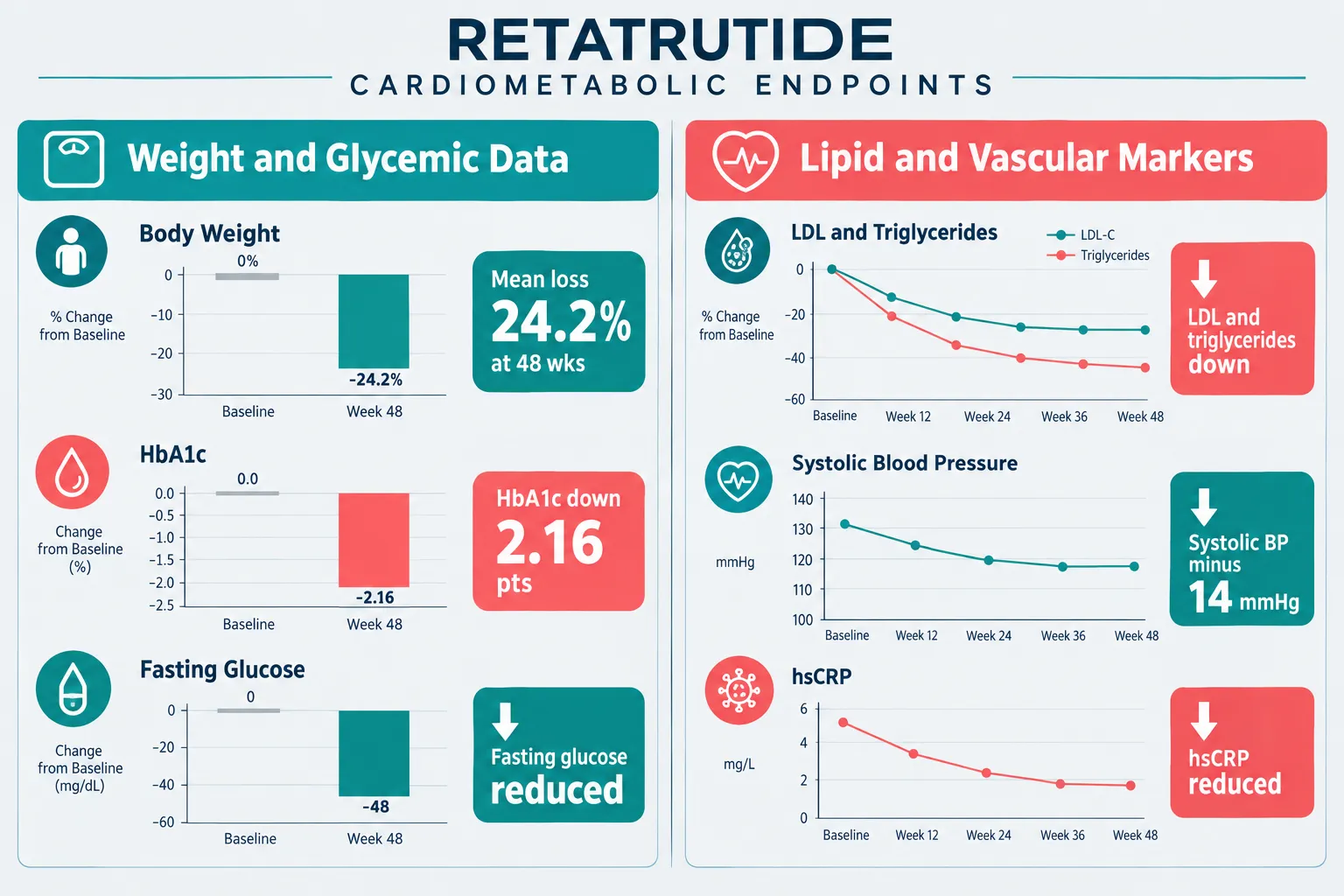
  • Retatrutide produced mean body weight reductions of up to 24.2% at 48 weeks in phase 2 trials, exceeding prior pharmacological benchmarks.
  • Glycemic improvements, including HbA1c reductions of approximately 2.16 percentage points, occurred alongside weight loss in both diabetic and non-diabetic populations.
  • Lipid panels, blood pressure, and inflammatory markers all shifted in a cardiovascular-favorable direction, but these remain surrogate endpoints.
  • No completed cardiovascular outcomes trials have yet confirmed reductions in major adverse cardiovascular events (MACE) for retatrutide.
  • Researchers must resist treating weight loss as a direct proxy for cardiovascular benefit, particularly in high-risk populations with established disease.

Why the Severe-Obesity CVD Population Demands a Different Analytical Frame
Why the Severe-Obesity CVD Population Demands a Different Analytical Frame

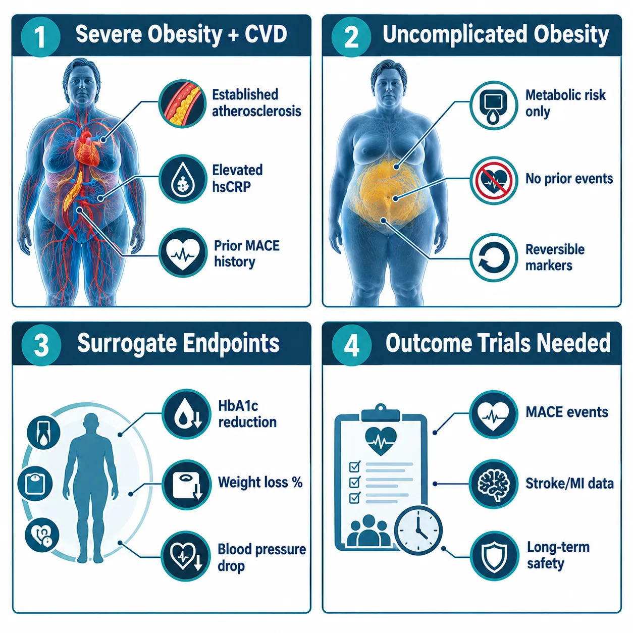
The distinction between uncomplicated obesity and obesity complicated by established cardiovascular disease is not semantic, it is mechanistically and statistically critical. In uncomplicated obesity cohorts, cardiovascular risk is largely driven by modifiable metabolic factors: insulin resistance, dyslipidemia, and hypertension. Meaningful weight loss reliably improves these factors, and the pathway from surrogate improvement to event reduction is relatively well-supported by prior evidence from bariatric surgery and GLP-1 receptor agonist outcomes trials.

In the severe-obesity CVD population, however, the risk architecture is different. Atherosclerotic plaques are already established. Inflammatory burden is higher. Cardiac remodeling may be present. The relationship between short-term biomarker improvement and long-term event reduction is less predictable, and the potential for adverse interactions, including changes in heart rate, volume shifts from rapid weight loss, or arrhythmia risk, requires independent evaluation rather than inference from surrogate data.

This is the central interpretive challenge for retatrutide and cardiovascular disease research: interpreting weight, glycemic, and safety endpoints together requires researchers to resist collapsing a multi-dimensional risk picture into a single headline number.

Researchers reviewing retatrutide endpoints data should note that the phase 2 obesity trial enrolled adults without diabetes and without prior cardiovascular events. Extrapolating those results to a post-MI population with preserved or reduced ejection fraction introduces assumptions that the data do not yet support.


What the Phase 2 Data Actually Show: Weight, Glycemic, and Lipid Endpoints
What the Phase 2 Data Actually Show: Weight, Glycemic, and Lipid Endpoints

The phase 2 evidence base for retatrutide is genuinely impressive across multiple dimensions. In the pivotal obesity trial involving 338 adults, once-weekly retatrutide at 12 mg produced a mean body weight reduction of 24.2% at 48 weeks. The 8 mg dose achieved 22.8% mean loss. Placebo-adjusted weight reduction at 24 weeks was approximately 16%, with a continued downward trajectory through the trial period. Every participant receiving 8 or 12 mg weekly achieved at least 5% weight loss, a threshold widely cited in regulatory and guideline contexts as clinically meaningful for cardiovascular risk.

Glycemic endpoints reinforced the cardiometabolic signal. In a separate 36-week phase 2 study in adults with type 2 diabetes, the highest dose produced an average HbA1c decrease of approximately 2.16 percentage points alongside up to 16.9% weight reduction. Fasting glucose and fasting insulin also declined in parallel with weight loss in the obesity trial.

Key insight: A 2.16 percentage-point HbA1c reduction in a population with established type 2 diabetes is clinically substantial, but it is a surrogate marker for cardiovascular risk, not a direct measure of event prevention.

Lipid and inflammatory markers moved in favorable directions. Triglycerides, LDL cholesterol, non-HDL cholesterol, and total cholesterol all declined. HDL modestly improved. Systolic blood pressure fell by roughly 4-8 mmHg across effective dose arms in the obesity trial, and by approximately 14 mmHg at the highest dose in a manufacturer-reported analysis using an efficacy estimand. High-sensitivity C-reactive protein (hsCRP), a marker of systemic inflammation strongly associated with cardiovascular events, was also reduced.

Waist circumference decreased by approximately 10-15 cm across effective dose arms, a change relevant to visceral adiposity and its downstream effects on insulin sensitivity and hepatic lipid metabolism.

For researchers seeking GLP-3 Reta 30mg formulations for preclinical or translational research purposes, these multi-endpoint findings provide a rich framework for designing studies that go beyond single-biomarker outcomes.


Interpreting Safety Endpoints Alongside Efficacy: What Researchers Must Not Overlook
Interpreting Safety Endpoints Alongside Efficacy: What Researchers Must Not Overlook

Safety data from phase 2 trials show a profile broadly consistent with potent incretin-based therapies. Adverse events were predominantly gastrointestinal, nausea, vomiting, and diarrhea, and were more frequent at higher doses, consistent with dose-dependent receptor engagement. Serious adverse events were infrequent, and the overall safety profile was considered acceptable for continued development.

Critically, available phase 2 summaries do not signal major cardiovascular safety concerns over 36-48 weeks. But this observation carries an important caveat: 36-48 weeks is insufficient to characterize arrhythmia risk, heart failure decompensation risk, or ischemic event rates in a population with established cardiovascular disease.

Researchers must treat the absence of a safety signal as a starting point for investigation, not a conclusion.

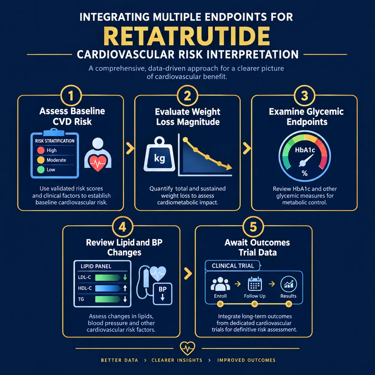
The integrated interpretation framework for retatrutide and cardiovascular disease research, interpreting weight, glycemic, and safety endpoints together, should follow a structured logic:

  1. Stratify baseline cardiovascular risk before interpreting any endpoint change.
  2. Evaluate weight loss magnitude in the context of rate and composition, not just percentage.
  3. Examine glycemic endpoints as independent contributors to risk, not simply as weight-loss correlates.
  4. Review lipid and blood pressure changes with attention to clinical significance thresholds.
  5. Identify gaps: no completed MACE outcomes trial exists for retatrutide as of 2026.

Researchers exploring adjacent cardiometabolic peptide research, including SS-31 kidney health research, will recognize this multi-endpoint interpretive discipline as standard practice in high-risk populations where organ-level interactions are complex.

Those sourcing compounds for translational research can review available Reta 20mg for sale options or GLP-3 Reta CAG 10mg for sale formulations through verified research-grade suppliers.


Conclusion

The surrogate endpoint data for retatrutide are among the most compelling seen in pharmacological obesity research. Weight loss of 24% at 48 weeks, combined with substantial HbA1c reduction, improved lipid panels, lower blood pressure, and reduced hsCRP, constructs a plausible and mechanistically coherent case for cardiovascular benefit.

But plausibility is not proof, particularly in the severe-obesity cardiovascular disease population where the stakes are highest and the biology is most complex.

Actionable next steps for researchers and clinicians:

  • Design studies that explicitly enroll patients with established cardiovascular disease, not just metabolic risk factors.
  • Pre-specify cardiovascular safety endpoints, including arrhythmia monitoring and heart failure hospitalization, as primary or co-primary outcomes, not secondary analyses.
  • Avoid reporting weight loss as a cardiovascular outcome; report it as one component of a multi-endpoint risk profile.
  • Track cardiovascular outcomes trial timelines for retatrutide and resist extrapolating event-level benefit from surrogate data until those trials report.
  • When reviewing buy Reta online options for preclinical research, ensure purity and dosing specifications align with the phase 2 protocols to maintain translational relevance.

The science surrounding retatrutide and cardiovascular disease research: interpreting weight, glycemic, and safety endpoints together is advancing rapidly. The discipline required to interpret that science rigorously, especially in high-risk populations, must advance at the same pace.

https://www.puretestedpeptides.com/wp-content/uploads/2026/09/retatrutide-and-cardiovascular-disease-research-interpreting-weight-glycemic-and.webp 1024 1536 https://www.puretestedpeptides.com/wp-content/uploads/2026/01/buy-peptides-online.jpg 2026-09-26 13:11:232026-09-26 13:11:23Retatrutide and Cardiovascular Disease Research: Interpreting Weight, Glycemic, and Safety Endpoints Together
Retatrutide Phase 3 Results: How Weight Loss, A1C, and Cardiovascular Findings Change Research Interpretation

Retatrutide Phase 3 Results: How Weight Loss, A1C, and Cardiovascular Findings Change Research Interpretation

September 26, 2026/0 Comments/in Uncategorized/by

Fewer than one in ten obesity drugs in history have produced mean weight loss exceeding 20 percent in a phase 3 trial. Retatrutide, a triple receptor agonist targeting GIP, GLP-1, and glucagon pathways simultaneously, has now cleared that threshold by a wide margin, and the implications reach far beyond a single efficacy number.

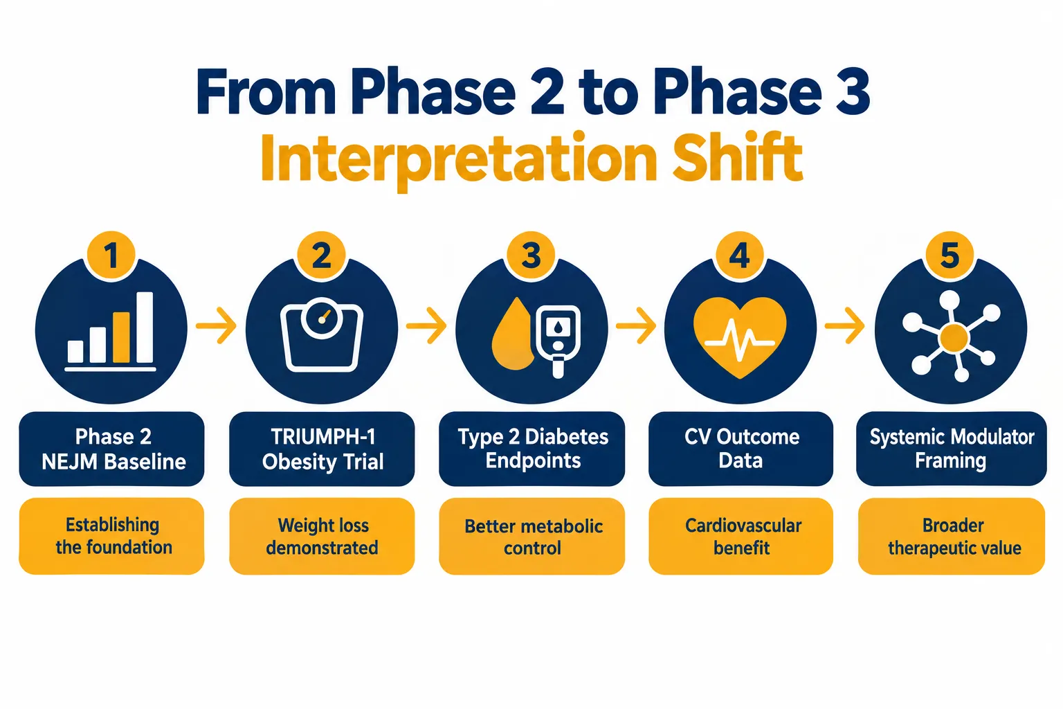
The full scope of Retatrutide Phase 3 Results: How Weight Loss, A1C, and Cardiovascular Findings Change Research Interpretation is still being absorbed by the research community. Topline data from the TRIUMPH master protocol and its obesity sub-programs show outcomes that reframe earlier phase 2 benchmarks, raise new questions about cardiovascular labeling, and complicate how scientists define a "weight-loss drug" in the first place.

Key Takeaways

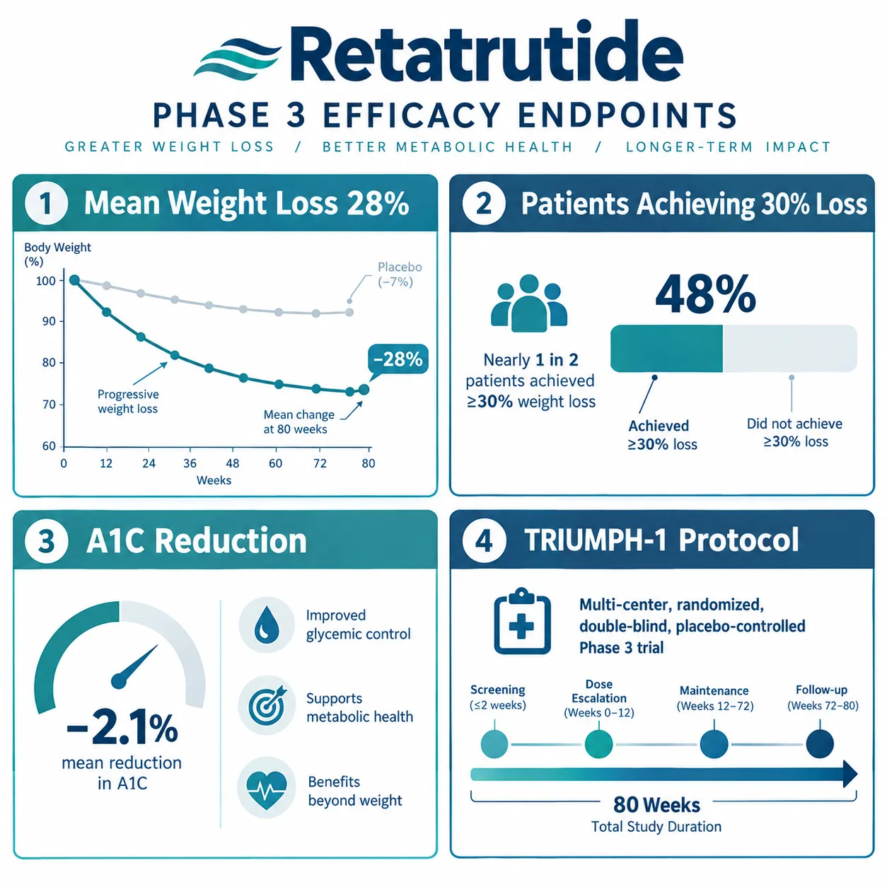
  • Phase 3 obesity data show approximately 28 percent mean weight loss at 80 weeks, with nearly 45 percent of participants achieving 30 percent or greater body weight reduction.
  • A1C improvements in the type 2 diabetes arm reinforce retatrutide's glycemic potential beyond weight loss alone.
  • Cardiovascular outcome data from the obesity-plus-established-CVD trial show MACE hazard ratios that are neutral to modestly favorable, but do not yet confirm a cardiovascular label claim.
  • Phase 2 NEJM results are now viewed as a conservative baseline rather than a peak effect.
  • Retatrutide remains investigational; no regulatory approval has been granted as of 2026.

What the Obesity Efficacy Numbers Actually Mean

What the Obesity Efficacy Numbers Actually Mean

The headline figure from the pivotal obesity phase 3 program is approximately 28 percent mean body weight reduction at 80 weeks. That number demands context. Semaglutide 2.4 mg produced roughly 15 percent in its landmark trial; tirzepatide reached approximately 22 percent at its highest dose. Retatrutide's figure, if confirmed in peer-reviewed publication, would represent the largest mean weight loss ever reported for a pharmacological agent in a phase 3 obesity study.

Equally striking is the distribution of response. Roughly 45 percent of participants achieved 30 percent or greater weight loss, a threshold that, in earlier eras, was associated only with bariatric surgery. That figure matters because it shifts the clinical conversation from average outcomes to what a subset of patients might realistically expect.

"The ceiling for pharmacological weight loss may not have been reached yet, but retatrutide has moved it substantially higher."

Researchers tracking Retatrutide Clinical Trials note that discontinuation rates and tolerability data are critical to interpreting these numbers fairly. Gastrointestinal adverse events, nausea, vomiting, diarrhea, remain the most common side effects, consistent with the incretin class. Dropout rates tied to adverse events have not been negligible, which means the 28 percent figure reflects a population that tolerated the drug well enough to complete the protocol.


Glycemic and Cardiovascular Findings: Separating Signal from Claim

Glycemic and Cardiovascular Findings: Separating Signal from Claim

The type 2 diabetes arm of the phase 3 program adds a second layer of significance to the overall data picture. A1C reductions were clinically meaningful, and the breadth of cardiometabolic risk-factor improvements, including blood pressure, triglycerides, and waist circumference, reinforces the argument that retatrutide acts as a systemic cardiometabolic modulator rather than a simple appetite suppressant.

This distinction matters for research interpretation. When a compound simultaneously reduces body weight, lowers blood glucose, improves lipid panels, and reduces blood pressure, the mechanism of benefit becomes difficult to attribute to any single pathway. That complexity is scientifically valuable but also complicates regulatory discussions about labeling.

The cardiovascular outcome trial enrolled participants with obesity and established cardiovascular disease. MACE (major adverse cardiovascular events) hazard ratios came in neutral to modestly favorable, a result that is encouraging but not yet sufficient to support a dedicated cardiovascular risk-reduction label claim. Researchers reviewing Retatrutide Clinical Trial data consistently note that risk-factor improvement and hard cardiovascular event reduction are not interchangeable endpoints.

Key distinctions from the cardiovascular data:

  • Improved risk factors (blood pressure, lipids, weight) are confirmed across trials
  • MACE hazard ratios are neutral to modestly favorable, not definitively protective
  • A cardiovascular label claim would require additional event-driven data
  • Monitoring for cardiovascular and gastrointestinal signals remains active in ongoing maintenance studies

How Phase 3 Data Reshape Research Interpretation Going Forward

How Phase 3 Data Reshape Research Interpretation Going Forward

Understanding Retatrutide Phase 3 Results: How Weight Loss, A1C, and Cardiovascular Findings Change Research Interpretation requires stepping back from individual endpoints and examining what the data collectively signal about the incretin class ceiling.

Phase 2 results published in the New England Journal of Medicine were widely cited as evidence of exceptional efficacy. Phase 3 data now reframe those results as a conservative baseline, not a peak effect. That inversion is unusual in drug development, where phase 3 trials frequently show more modest outcomes than earlier studies due to broader, more heterogeneous populations.

Several interpretive questions now dominate expert commentary:

Research Question Why It Matters
Duration of effect Does weight loss plateau, reverse, or persist beyond 80 weeks?
Head-to-head trials How does retatrutide compare directly to tirzepatide or semaglutide?
Class generalizability Do triple agonism benefits extend to other compounds in development?
Real-world CV impact Will risk-factor gains translate to event reduction outside trial conditions?
Cost and access High efficacy is only meaningful if patients can sustain treatment

The TRIUMPH maintenance study is still active, and its results will be central to answering the durability question. Industry framing has leaned heavily on "next-generation" language, but researchers caution that approval timelines and cardiovascular label expectations remain separate conversations from efficacy data.

For those following the science of triple-receptor agonism alongside related peptide research, including work on IPA Sermorelin Stack Research and metabolic modulation, the retatrutide data set offers a rare opportunity to observe how mechanism-of-action complexity translates into clinical outcomes at scale.


Conclusion

Retatrutide Phase 3 Results represent a genuine inflection point in obesity and cardiometabolic research, not because any single number is record-breaking in isolation, but because the combination of weight loss magnitude, glycemic improvement, and cardiovascular signal breadth forces a reclassification of what this compound is and what it does.

Actionable next steps for researchers and clinicians following this space:

  1. Wait for peer-reviewed publication of full TRIUMPH-1 data before drawing conclusions from topline press releases.
  2. Distinguish between risk-factor improvements and hard cardiovascular event data when evaluating cardiovascular claims.
  3. Track the maintenance study for durability evidence beyond 80 weeks.
  4. Monitor head-to-head trial announcements, which will provide the most clinically relevant comparative data.
  5. Evaluate discontinuation and tolerability rates alongside efficacy figures to form a complete picture of real-world applicability.

Retatrutide remains investigational as of 2026. The data are compelling, the mechanistic rationale is strong, and the research interpretation is still evolving. That combination makes this one of the most closely watched drug development programs in metabolic medicine today.

https://www.puretestedpeptides.com/wp-content/uploads/2026/09/retatrutide-phase-3-results-how-weight-loss-a1c-and-cardiovascular-findings-chan.webp 1024 1536 https://www.puretestedpeptides.com/wp-content/uploads/2026/01/buy-peptides-online.jpg 2026-09-26 13:10:202026-09-26 13:10:20Retatrutide Phase 3 Results: How Weight Loss, A1C, and Cardiovascular Findings Change Research Interpretation
Hemoglobin and Hematocrit in Peptide Research: How to Interpret Oxygen-Carrying Capacity and Safety Signals

Hemoglobin and Hematocrit in Peptide Research: How to Interpret Oxygen-Carrying Capacity and Safety Signals

September 26, 2026/0 Comments/in Uncategorized/by

A hematocrit reading of 54% in a research subject is not just a number, it is a stop signal. Yet many researchers encounter elevated red-cell indices in their bloodwork panels and lack a clear framework for deciding what those values mean, whether they reflect a true physiological change, and what action, if any, is warranted. Understanding hemoglobin and hematocrit in peptide research: how to interpret oxygen-carrying capacity and safety signals is one of the most practical skills any serious investigator can develop.

Key Takeaways

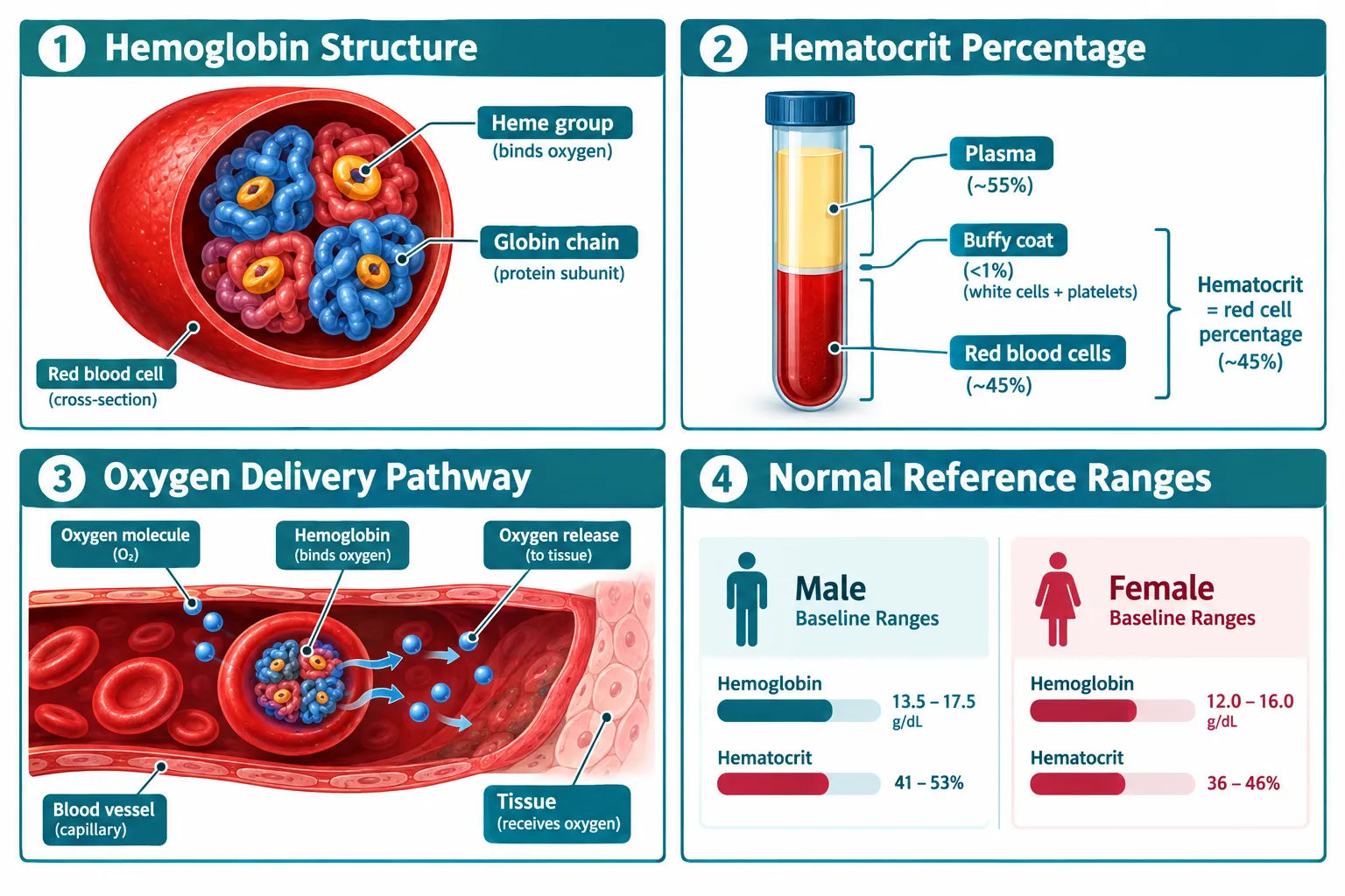
  • Hemoglobin measures the protein that carries oxygen; hematocrit measures the percentage of blood volume occupied by red cells, both are essential safety markers in peptide research.
  • Baseline values must be established before any research protocol begins, because pre-existing elevations change how subsequent readings are interpreted.
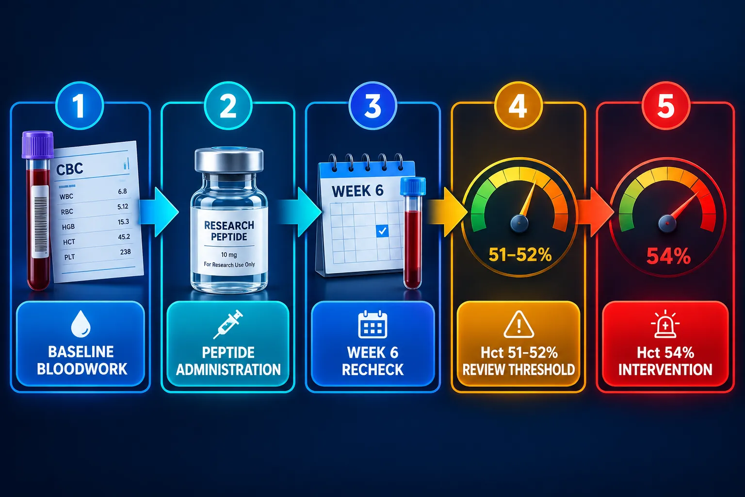
  • A hematocrit of 51-52% warrants review; 54% or above typically triggers intervention or protocol cessation.
  • Dehydration and red-cell indices like MCV can confound hematocrit readings and must be ruled out before attributing a rise to a peptide compound.
  • Monitoring frequency, action thresholds, and management strategies follow a guideline-style algorithm that researchers should apply consistently.

What Hemoglobin and Hematocrit Actually Measure

What Hemoglobin and Hematocrit Actually Measure

Hemoglobin (Hb) is the iron-containing protein inside red blood cells. Its job is to bind oxygen in the lungs and release it to tissues. It is measured in grams per deciliter (g/dL). Normal ranges sit roughly at 13.5-17.5 g/dL for adult males and 12.0-15.5 g/dL for adult females, though reference intervals vary slightly by laboratory.

Hematocrit (Hct) is a different but related measurement. It expresses the proportion of whole blood that is made up of red blood cells, reported as a percentage. Because hematocrit is a ratio of cells to total blood volume, it can be influenced by both the number of red cells and the volume of plasma. This distinction matters enormously when interpreting results in a research context.

Red-cell indices, including mean corpuscular volume (MCV), mean corpuscular hemoglobin (MCH), and red cell distribution width (RDW), add important context. A high hematocrit paired with a normal MCV and normal RDW looks very different from one paired with microcytic, hypochromic cells, which would suggest iron deficiency rather than true erythrocytosis.

The Difference Between True Erythrocytosis and a Spurious Rise

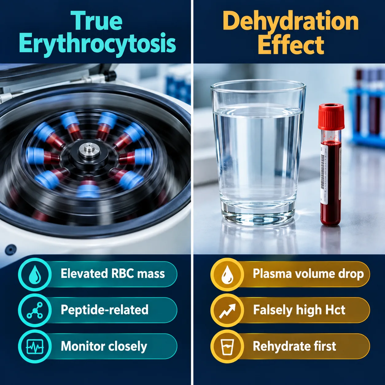
True erythrocytosis means the body is producing more red blood cells, increasing the absolute red cell mass. A spurious rise in hematocrit, by contrast, happens when plasma volume decreases, most commonly through dehydration, without any actual increase in red cell production. A subject who is even mildly dehydrated at the time of blood draw can show a hematocrit several points higher than their true baseline.

Researchers should always confirm adequate hydration before attributing a hematocrit elevation to a peptide compound. When in doubt, repeating the draw after proper rehydration is the most straightforward corrective step.


Monitoring Protocols: Hemoglobin and Hematocrit in Peptide Research

Monitoring Protocols: Hemoglobin and Hematocrit in Peptide Research

Establishing a monitoring schedule is not optional, it is the foundation of responsible research. The general framework follows a logical sequence:

  1. Baseline bloodwork before the protocol begins, capturing Hb, Hct, and a full red-cell panel.
  2. Recheck at 6-8 weeks, which is typically when androgenic or peptide-adjacent compounds begin to exert measurable effects on erythropoiesis.
  3. Ongoing monitoring every 3-6 months once values are stable.

This schedule mirrors the approach used in testosterone replacement and gender-affirming hormone research, where hematocrit elevation is documented to be several-fold more common than in placebo groups. The same logic applies when investigating compounds that interact with growth hormone axes or metabolic pathways. Researchers exploring options like Tesamorelin peptide benefits or mitochondria-targeted agents such as SS-31 peptides for sale should incorporate hematological monitoring from the outset.

Action Thresholds Every Researcher Should Know

Hematocrit Level Recommended Action
Below 50% Continue protocol; monitor per schedule
51-52% Flag for review; assess hydration and confounders
53% Increase monitoring frequency; evaluate risk factors
54% or above Pause protocol; consider intervention or cessation

These thresholds are not arbitrary. At hematocrit levels above 54%, blood viscosity increases meaningfully, raising the theoretical risk of thromboembolic events. Even in research settings, that risk profile demands a conservative response.


Confounders, Red Flags, and Interpreting Safety Signals

Confounders, Red Flags, and Interpreting Safety Signals

Several factors can confound hematocrit readings and lead to misinterpretation. Researchers using lab tested peptides should be aware of the following:

  • Altitude: Living or training at elevation stimulates erythropoietin production naturally, raising baseline Hct independent of any compound.
  • Sleep apnea: Chronic intermittent hypoxia drives compensatory erythrocytosis and can be mistaken for a compound-induced effect.
  • Polycythemia vera: A rare myeloproliferative condition that elevates red cell mass; it must be ruled out when hematocrit rises without a clear cause.
  • Androgens and peptides with androgenic overlap: Compounds like MT-2 research peptides operate through melanocortin receptors, but any compound that indirectly influences testosterone or IGF-1 pathways warrants careful hematological oversight.

When a true elevation is confirmed, management options include dose reduction, increased hydration, therapeutic phlebotomy in severe cases, and in some protocols, temporary cessation. Researchers investigating compounds with systemic metabolic effects, including those in the GLP-3 peptide class or IPA peptides, should document any hematological changes as part of their safety reporting.

It is also worth noting that severe erythrocytosis remains relatively rare when protocols are well-managed. Evidence from gender-affirming testosterone care shows that diligent monitoring catches elevations early, before they reach clinically dangerous levels. The same principle applies to peptide research: early detection through consistent bloodwork is far more effective than reactive management.

Researchers sourcing compounds from a best peptide manufacturer should also confirm purity, since contaminants or misidentified compounds can introduce unexpected physiological effects, including on erythropoiesis.


Conclusion

Interpreting hemoglobin and hematocrit in peptide research: how to interpret oxygen-carrying capacity and safety signals requires more than reading a number off a lab report. It demands understanding what the values represent, what can distort them, and what action each threshold calls for.

Actionable next steps for researchers:

  • Establish a full baseline CBC before starting any peptide protocol.
  • Rule out dehydration and altitude effects before attributing any Hct rise to the compound under study.
  • Apply the 51-52% review threshold and the 54% intervention threshold consistently across all protocols.
  • Document red-cell indices alongside Hb and Hct to distinguish true erythrocytosis from nutritional or plasma-volume artifacts.
  • Recheck values at 6-8 weeks and every 3-6 months thereafter, adjusting frequency if values trend upward.

Rigorous hematological monitoring is not a bureaucratic formality. It is the mechanism by which researchers protect subject safety and generate data that is actually interpretable. Build it into every protocol from day one.

https://www.puretestedpeptides.com/wp-content/uploads/2026/09/hemoglobin-and-hematocrit-in-peptide-research-how-to-interpret-oxygen-carrying-c.webp 1024 1536 https://www.puretestedpeptides.com/wp-content/uploads/2026/01/buy-peptides-online.jpg 2026-09-26 13:10:032026-09-26 13:10:03Hemoglobin and Hematocrit in Peptide Research: How to Interpret Oxygen-Carrying Capacity and Safety Signals
Peptide Research Samples and Common Prescription Drugs: How to Map Metoprolol, Lisinopril, Omeprazole, and Statin Confounders

Peptide Research Samples and Common Prescription Drugs: How to Map Metoprolol, Lisinopril, Omeprazole, and Statin Confounders

September 26, 2026/0 Comments/in Uncategorized/by

More than 60 percent of adults enrolled in cardiometabolic research studies take at least one of four drug classes, beta-blockers, ACE inhibitors, proton pump inhibitors, or statins, yet fewer than half of published peptide study protocols include a formal confounder mapping strategy for these agents. That gap creates reproducibility problems that surface long after data collection ends.

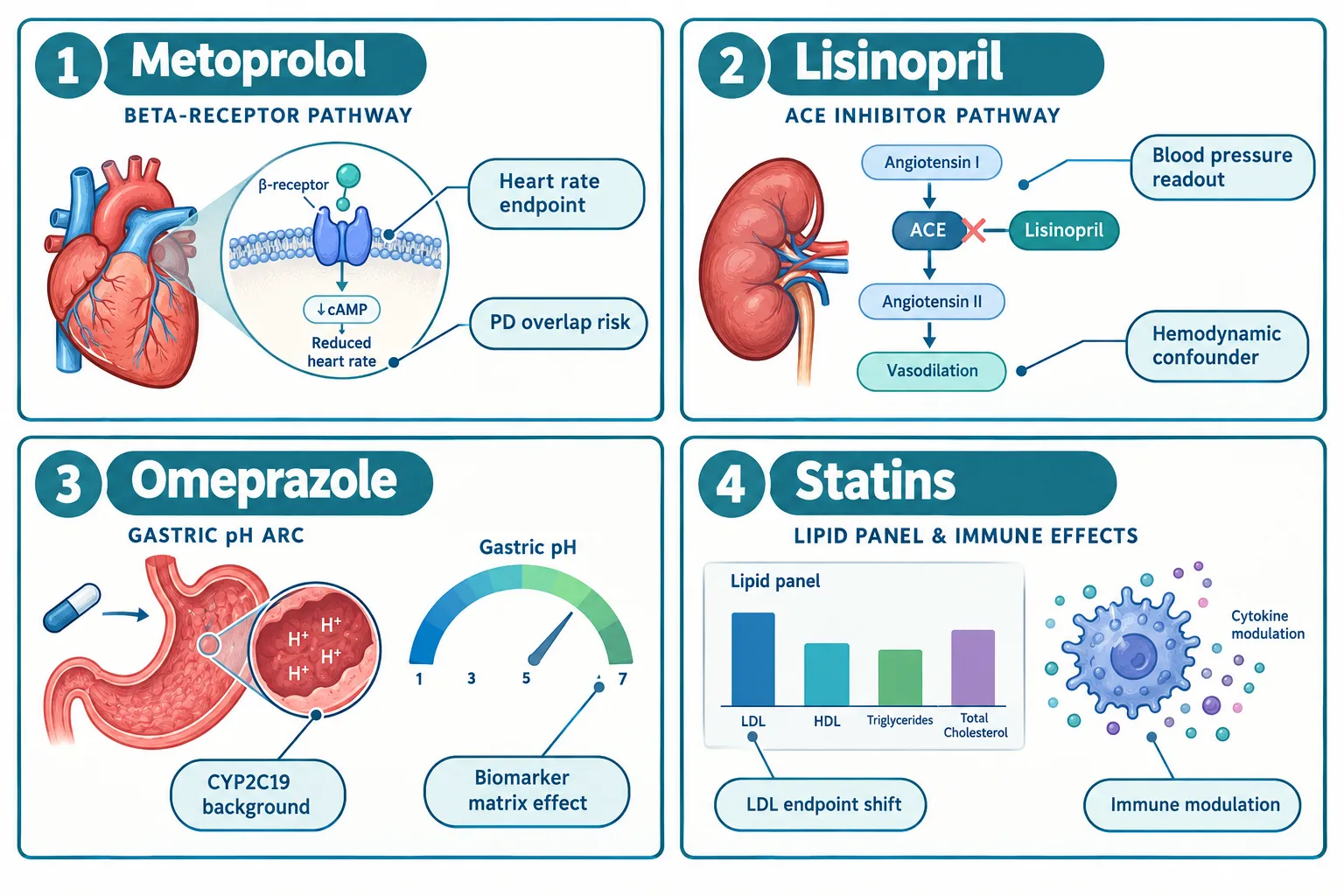
Mapping peptide research samples and common prescription drugs, specifically metoprolol, lisinopril, omeprazole, and statin confounders, is not a clinical pharmacology exercise reserved for late-stage trials. It is a foundational study-design task that applies equally to cell-based models, ex vivo tissue preparations, and preclinical animal work. The sections below build a practical medication-confounder matrix organized around exposure timing, physiological readouts, assay controls, and documentation.

Key Takeaways

  • Peptide compounds largely bypass CYP-mediated metabolism, so pharmacokinetic drug-drug interactions with metoprolol, lisinopril, omeprazole, and statins are minimal, but pharmacodynamic and assay-level interference is substantial.
  • A confounder matrix should address four dimensions: exposure timing, physiological endpoint overlap, chromatographic interference, and immunogenicity background.
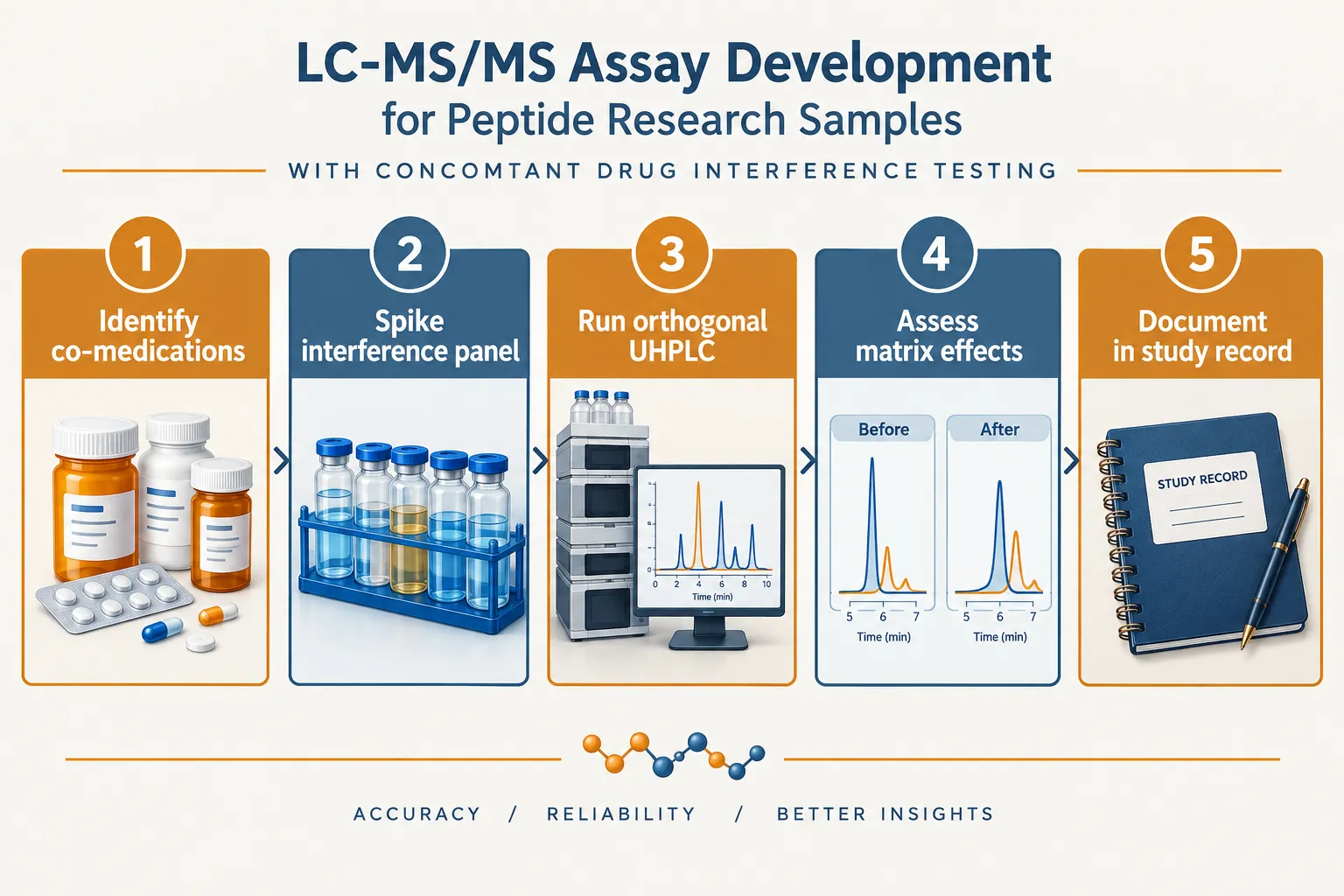
  • LC-MS/MS assays for peptide biomarkers must include spiked interference panels containing the major metabolites of each concomitant drug class.
  • Stratifying animal cohorts or ex vivo samples by background drug exposure before analysis prevents mis-attribution of endpoint changes to the peptide under study.
  • Formal documentation of confounder mapping, including inclusion/exclusion logic and assay selectivity data, is expected by regulatory agencies reviewing INDs, NDAs, and BLAs for peptide products.

Understanding Why These Four Drug Classes Matter in Peptide Research

Understanding Why These Four Drug Classes Matter in Peptide Research

Regulatory guidance from the FDA on clinical pharmacology for peptide drug products makes a clear distinction: because peptides are typically degraded by proteolytic enzymes rather than CYP enzymes, classical pharmacokinetic drug-drug interactions with small molecules are uncommon. That conclusion, however, does not mean background medications are irrelevant. It means the risk shifts from PK to PD and to bioanalytical interference.

Metoprolol is a beta-1 selective adrenergic blocker. In peptide research involving cardiovascular or autonomic endpoints, heart rate, cardiac output, blood pressure variability, metoprolol directly modulates the same physiological parameters a peptide may be expected to influence. Any cohort or animal group receiving metoprolol will show attenuated heart rate responses, which can mask or amplify a peptide's apparent hemodynamic activity.

Lisinopril, an ACE inhibitor, suppresses angiotensin II generation and elevates bradykinin levels. In ex vivo vascular models or animal studies examining endothelial function, inflammatory cytokine profiles, or renal perfusion, lisinopril creates a shifted physiological baseline. Peptide-mediated vasodilation or anti-inflammatory signals measured against that background will be systematically different from peptide signals measured in drug-naive tissue.

Omeprazole is a proton pump inhibitor and a known CYP2C19 inhibitor. While the peptide itself may not be a CYP substrate, co-administered small-molecule probes or metabolic tracers used alongside a peptide study may be affected. More critically, omeprazole alters gastric and intestinal pH, which can affect oral bioavailability of companion agents and shift matrix composition in gastrointestinal tissue samples.

Statins present a dual challenge. First, they directly alter lipid panel endpoints, LDL, HDL, triglycerides, that are commonly used as efficacy biomarkers in cardiometabolic peptide research. A peptide with lipid-modulating properties cannot be fairly evaluated in a statin-treated cohort without stratification. Second, certain statins have documented immunomodulatory effects, meaning they can influence immune biomarker readouts relevant to peptide immunogenicity assessments.

For researchers exploring mitochondrial or cardiometabolic angles, resources such as the SS-31 mitochondrial research themes overview and the discussion of peptides in cardiometabolic models provide useful context on how peptide mechanisms intersect with the same pathways these drugs target.


Building the Confounder Matrix: Exposure Timing, Readouts, and Assay Controls

Building the Confounder Matrix: Exposure Timing, Readouts, and Assay Controls

A confounder matrix is a structured reference document, not a statistical model, that maps each background drug to the specific dimensions where it can distort peptide research data. The matrix should be built before sample collection begins.

Dimension 1: Exposure Timing

The timing of background drug exposure relative to peptide administration or sample collection determines whether interference is acute or chronic. The table below summarizes key timing considerations.

Drug Relevant Exposure Window Primary Timing Risk
Metoprolol Acute (1-4 hrs) and chronic Blunted heart rate response during peptide challenge
Lisinopril Chronic (days to weeks) Shifted vascular baseline at time of tissue harvest
Omeprazole Chronic (steady-state CYP2C19 inhibition) Altered matrix pH; companion probe metabolism affected
Statins Chronic (lipid and immune effects) Lipid endpoint suppression; immune biomarker shift

Dimension 2: Physiological Readout Overlap

For each peptide under study, list the primary and secondary efficacy endpoints. Then cross-reference each drug class against those endpoints. If metoprolol and the peptide both affect resting heart rate, that endpoint requires either exclusion of metoprolol-treated subjects from the primary analysis or formal stratification with sufficient statistical power in each stratum.

Key principle: An endpoint that is also a pharmacological target of a background drug cannot serve as an unconfounded primary readout without stratification or covariate adjustment.

Dimension 3: Assay Controls and Chromatographic Interference

FDA bioanalytical method validation guidance for biomarkers explicitly requires sponsors to demonstrate assay selectivity and assess matrix effects across relevant patient subgroups and concomitant therapies. For LC-MS/MS workflows used to quantify peptide concentrations or peptide-related biomarkers, this means building an interference panel.

Interference panel construction steps:

  1. Obtain authenticated reference standards for the parent compound and major metabolites of metoprolol, lisinopril, omeprazole, and the relevant statin.
  2. Spike each compound individually into the biological matrix at clinically relevant concentrations.
  3. Run the spiked samples using the validated LC-MS/MS method and record retention times, peak areas, and any co-elution with peptide analyte peaks.
  4. Apply orthogonal chromatographic separation (e.g., UHPLC-HRMS/MS) to resolve any co-eluting species.
  5. Document selectivity data in the bioanalytical method validation report.

Published LC-MS assay work on cardiovascular medications has demonstrated that lisinopril and statins such as lovastatin exhibit peak tailing and matrix-dependent variability that can compromise quantification accuracy. These findings reinforce the need to include such compounds in interference testing rather than assuming they are chromatographically inert.

For researchers working with antioxidant or mitochondrially targeted peptides, the LL-37 versus SS-31 benefits comparison illustrates how different peptide classes engage distinct biological pathways, a factor that determines which background drugs are most likely to confound a given readout.

Dimension 4: Immunogenicity Background

Statins and long-term PPI use can influence immune biomarkers. FDA draft guidance on immunogenicity risk for synthetic peptides notes that impurities and background immunomodulatory agents can affect the summative immune response. When designing immunogenicity assays for a peptide program, the confounder matrix should flag statin and omeprazole exposure as variables requiring documentation in the sample metadata.


Documentation, Stratification, and Regulatory Alignment

Documentation, Stratification, and Regulatory Alignment

Mapping confounders is only useful if the map is formally embedded in study documentation. Regulatory science frameworks, including FDA product-specific guidances for generic peptide products updated in 2026, tighten expectations around impurity analysis and biological comparability, making it more important than ever to distinguish product-related signals from background medication effects in real-world samples.

Recommended documentation elements:

  • Inclusion/exclusion criteria log: Record which drug classes are permitted, restricted, or required to be stable-dose before enrollment or sample collection begins.
  • Stratification plan: Pre-specify strata for metoprolol use (yes/no), statin type and dose, and omeprazole use. Assign samples to strata before unblinding any endpoint data.
  • Assay selectivity appendix: Attach interference panel results to the bioanalytical method validation report. Include retention time tables and representative chromatograms for each spiked drug.
  • Biomarker qualification notes: For each lipid or cardiovascular endpoint used as a peptide efficacy biomarker, note whether statin or beta-blocker use was a pre-specified covariate in the analysis plan.

Regulatory guidance consistently positions concomitant medication mapping as part of clinical pharmacology planning rather than an afterthought. Practical recommendations from FDA workshop materials include prospectively capturing beta-blocker, ACE inhibitor, PPI, and statin use, stratifying PK/PD and biomarker analyses by these medications, and pre-screening bioanalytical methods for interference from their major metabolites.

Researchers sourcing verified peptide compounds for these studies can explore options through resources such as the best peptide website directory and the best place to buy peptide listings to ensure purity standards align with the stringent analytical requirements described above. For studies involving endocrine or receptor-level interactions, the discussion of peptides in endocrine pharmacology offers additional context on how background drug classes can shift receptor biology.


Conclusion

Mapping peptide research samples and common prescription drugs, metoprolol, lisinopril, omeprazole, and statin confounders, is a concrete, executable task that protects data integrity from the earliest stages of study design. The confounder matrix framework presented here addresses four actionable dimensions: exposure timing, physiological readout overlap, LC-MS/MS assay interference, and immunogenicity background documentation.

Actionable next steps for research teams:

  1. Before finalizing a study protocol, audit every primary and secondary endpoint against the pharmacological targets of the four drug classes covered here.
  2. Build an interference panel for all LC-MS/MS or ligand-binding assays using authenticated reference standards for each background drug and its major metabolites.
  3. Pre-specify stratification variables in the statistical analysis plan so that background drug exposure is a documented covariate, not a post-hoc explanation.
  4. Attach selectivity and matrix-effect data to the bioanalytical validation report as a dedicated confounder appendix.
  5. Review current FDA product-specific guidances and bioanalytical method validation guidance for biomarkers to ensure documentation meets the latest regulatory expectations.

Treating confounder mapping as a first-class study-design deliverable, not a footnote, is what separates reproducible peptide research from data that cannot be interpreted with confidence.

https://www.puretestedpeptides.com/wp-content/uploads/2026/09/peptide-research-samples-and-common-prescription-drugs-how-to-map-metoprolol-lis.webp 1024 1536 https://www.puretestedpeptides.com/wp-content/uploads/2026/01/buy-peptides-online.jpg 2026-09-26 13:06:382026-09-26 13:06:38Peptide Research Samples and Common Prescription Drugs: How to Map Metoprolol, Lisinopril, Omeprazole, and Statin Confounders
Retatrutide 2026 Trial Data vs Earlier Obesity Models: What Researchers Should Compare Before Drawing Conclusions

Retatrutide 2026 Trial Data vs Earlier Obesity Models: What Researchers Should Compare Before Drawing Conclusions

September 26, 2026/0 Comments/in Uncategorized/by

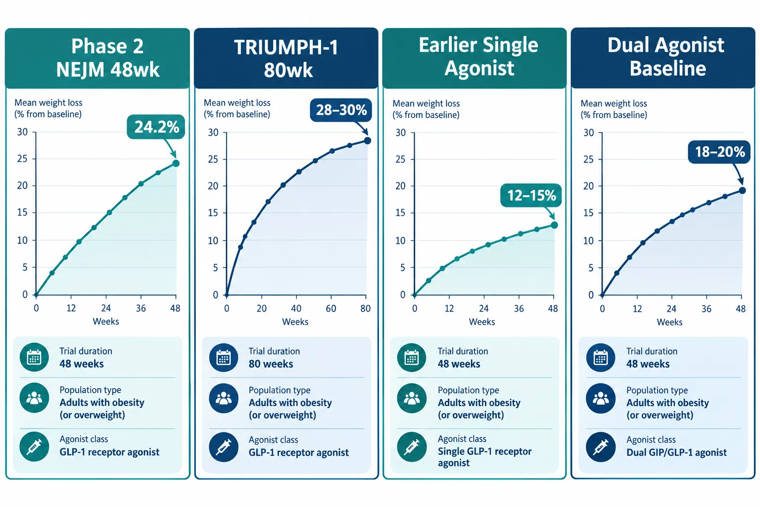
A single Phase 2 result, 24.2% mean body weight loss at 48 weeks, repositioned retatrutide as a potential turning point in obesity pharmacotherapy. Now, with the TRIUMPH program delivering Phase 3 data in 2026 and an FDA submission planned for early 2027, the research community faces a more demanding challenge: comparing these new numbers against earlier obesity models without letting headline figures obscure methodological differences that matter enormously.

Retatrutide 2026 trial data vs earlier obesity models: what researchers should compare before drawing conclusions is not simply a question of which drug produces a bigger number. It is a question of whether the numbers are even measuring the same thing across studies.

Key Takeaways

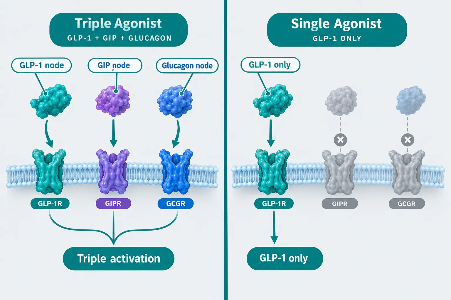
  • Retatrutide is a triple agonist (GLP-1, GIP, glucagon receptors), making direct comparisons to earlier single- or dual-agonist trials structurally complex.
  • TRIUMPH-1 reports up to 28-30% weight loss over 80 weeks, but population composition, baseline BMI, and trial duration differ from earlier benchmarks.
  • Estimand frameworks, dose escalation schedules, and completeness of adverse-event reporting vary significantly across generations of obesity trials.
  • Glycemic endpoints (A1C reduction) in TRIUMPH-2 require careful alignment with earlier diabetic-population studies before conclusions are drawn.
  • A transparent comparison framework covering at least seven variables is essential before any cross-trial efficacy claim holds scientific weight.

Why the Comparison Problem Is Harder Than It Looks

Why the Comparison Problem Is Harder Than It Looks

The excitement around retatrutide is understandable. The TRIUMPH-1 trial, targeting non-diabetic adults with obesity or overweight, reported weight reductions approaching 28-30% over 80 weeks, figures that have drawn frequent comparisons to bariatric surgery outcomes. TRIUMPH-2 extended the analysis to people with type 2 diabetes, and TRIUMPH-3 enrolled participants with severe obesity and established cardiovascular disease. A global registration trial completed enrollment in late 2025, broadening the population further.

Earlier obesity pharmacotherapy trials, including those for semaglutide and tirzepatide, ran for 68 weeks in many pivotal designs, enrolled populations with somewhat different baseline BMI distributions, and used dose escalation schedules that do not map cleanly onto retatrutide's protocol. Researchers exploring retatrutide clinical trial data will quickly notice that the surface-level weight-loss percentages are not the only variable worth examining.

Seven variables that demand alignment before cross-trial comparison:

Variable Why It Matters
Population baseline BMI Higher baseline BMI inflates absolute kg lost
Trial duration (weeks) Longer trials favor plateau-resistant compounds
Estimand framework Defines how dropouts and discontinuations are handled
Dose escalation schedule Steeper escalation can accelerate early weight loss
Completeness of AE reporting Affects tolerability profile comparisons
Glycemic endpoints (A1C) Critical for diabetic-population sub-analyses
Completers vs ITT analysis Changes the denominator and the headline figure

"A 30% weight loss figure means very little in isolation. The population, the protocol, and the statistical framework are the context that gives the number meaning."


Mechanism Differences That Complicate Direct Comparisons

Mechanism Differences That Complicate Direct Comparisons

Retatrutide is a triple agonist, activating GLP-1, GIP, and glucagon receptors simultaneously. Earlier approved agents in this space are single agonists (GLP-1 only) or dual agonists (GLP-1 and GIP). This mechanistic distinction is not merely academic, it directly affects which endpoints are most informative and which adverse-event profiles are expected.

Glucagon receptor activation drives additional energy expenditure and lipolysis pathways that are absent in earlier models. This means that comparing retatrutide's weight-loss magnitude against a GLP-1 monotherapy trial without accounting for the additional metabolic burden on participants is methodologically incomplete. Researchers interested in the GLP-3 Reta product tag context will recognize that the triple-agonist classification is central to understanding why the efficacy signal appears larger.

Key mechanistic comparison points:

  • GLP-1 component: Shared with earlier agents; allows partial endpoint alignment on nausea, satiety, and gastric motility outcomes.
  • GIP component: Shared with tirzepatide; permits limited dual-agonist comparison, but retatrutide's GIP binding affinity differs.
  • Glucagon component: Unique to retatrutide in this class; drives hepatic fat reduction and thermogenesis outcomes not tracked in earlier trial designs.

For researchers also examining peptides with distinct metabolic profiles, the Tesamorelin science and sourcing overview offers a useful reference point for understanding how growth-hormone-releasing peptides address visceral fat through a separate mechanism, underscoring why mechanism-first framing matters in any comparative analysis.


Building a Transparent Framework for Cross-Trial Analysis

Building a Transparent Framework for Cross-Trial Analysis

Retatrutide 2026 trial data vs earlier obesity models: what researchers should compare before drawing conclusions comes down to a structured, reproducible framework rather than selective headline matching. The following steps reflect best practice for cross-generational obesity trial comparison.

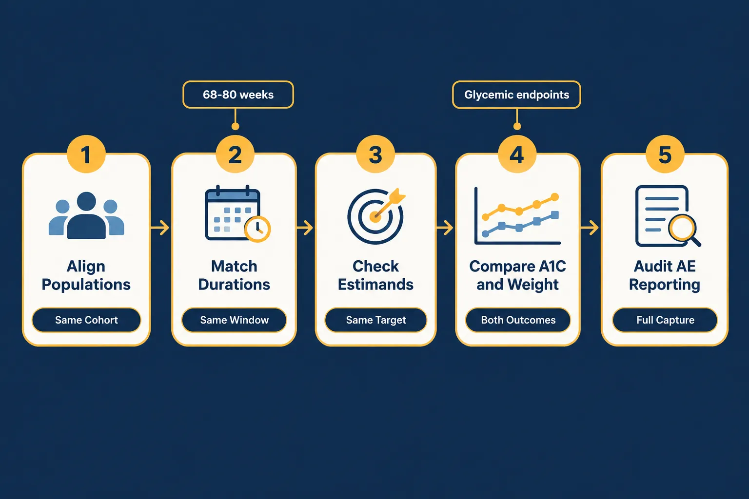
Step 1, Align populations. Confirm that baseline BMI, diabetes status, cardiovascular comorbidity burden, and geographic distribution are comparable. TRIUMPH-3's cardiovascular-disease population is not equivalent to a general overweight cohort.

Step 2, Match or adjust for duration. TRIUMPH-1's 80-week window exceeds the 68-week standard used in several earlier pivotal trials. Annualized weight-loss rates provide a more honest cross-trial metric than raw endpoint figures.

Step 3, Examine estimands explicitly. The treatment policy estimand (which includes all participants regardless of discontinuation) and the hypothetical estimand (which models outcomes assuming full adherence) produce materially different results. Earlier trials varied in which they pre-specified as primary.

Step 4, Compare A1C and glycemic outcomes in matched populations. TRIUMPH-2 data on weight and glycemic outcomes in type 2 diabetes must be benchmarked only against earlier trials that enrolled comparable diabetic populations with similar baseline A1C ranges.

Step 5, Audit adverse-event completeness. Gastrointestinal adverse events are consistently reported across generations, but cardiovascular safety signals, injection-site reactions, and lean mass loss data have not been uniformly collected. Retatrutide's glucagon component introduces a hepatic safety dimension that earlier single-agonist trial protocols did not systematically monitor.

Researchers building stacks or combination protocols may also find value in reviewing the IPA Sermorelin stack research resource for context on how multi-peptide frameworks are evaluated in research settings, and the SS-31 mitochondrial research tag for parallel work on metabolic and mitochondrial endpoints that may become relevant as retatrutide's full metabolic profile is characterized.

For researchers seeking the compound itself for preclinical work, the Reta 20mg product page provides sourcing details relevant to laboratory use.


Conclusion

The TRIUMPH program represents a genuine advance in obesity pharmacotherapy research, and the 2026 data deserve serious scientific attention. But the value of that data depends entirely on how carefully it is contextualized against earlier obesity models.

Actionable next steps for researchers:

  1. Build a comparison table that explicitly lists population baseline characteristics, trial duration, estimand type, and primary endpoint definition for each study being compared.
  2. Separate efficacy comparisons by population subgroup, non-diabetic, type 2 diabetes, and high-cardiovascular-risk cohorts require distinct reference studies.
  3. Request or locate full adverse-event appendices, not just summary tables, before drawing tolerability conclusions.
  4. Apply annualized weight-loss rates as a normalized cross-trial metric rather than relying on raw endpoint percentages.
  5. Track the FDA submission timeline (currently planned for Q1 2027) and review any agency briefing documents for additional population and estimand disclosures.

Retatrutide 2026 trial data vs earlier obesity models: what researchers should compare before drawing conclusions is ultimately a call for methodological discipline. The numbers are impressive, but the framework used to evaluate them will determine whether the conclusions hold.

https://www.puretestedpeptides.com/wp-content/uploads/2026/09/retatrutide-2026-trial-data-vs-earlier-obesity-models-what-researchers-should-co.webp 1024 1536 https://www.puretestedpeptides.com/wp-content/uploads/2026/01/buy-peptides-online.jpg 2026-09-26 13:05:022026-09-26 13:05:02Retatrutide 2026 Trial Data vs Earlier Obesity Models: What Researchers Should Compare Before Drawing Conclusions
Semax Nasal Spray Research Products Compared: Formulation Transparency, Assay Documentation, and Use-Case Fit

Semax Nasal Spray Research Products Compared: Formulation Transparency, Assay Documentation, and Use-Case Fit

September 25, 2026/0 Comments/in Uncategorized/by

Fewer than half of Semax nasal spray products currently available to researchers disclose the complete formulation data needed to run reproducible experiments. That gap matters because a 375 mcg-per-spray bottle and a 100 mcg-per-spray bottle are not interchangeable in a CNS research protocol, yet both may carry the same "research-grade" label. This article applies a structured buyer framework to the key variables that separate credible Semax nasal spray research products from those that fall short on formulation transparency, assay documentation, and use-case fit.

Key Takeaways

  • Semax nasal sprays vary widely in per-spray dose (100-375 mcg) and total peptide load (10-30 mg), making concentration disclosure a non-negotiable first filter.
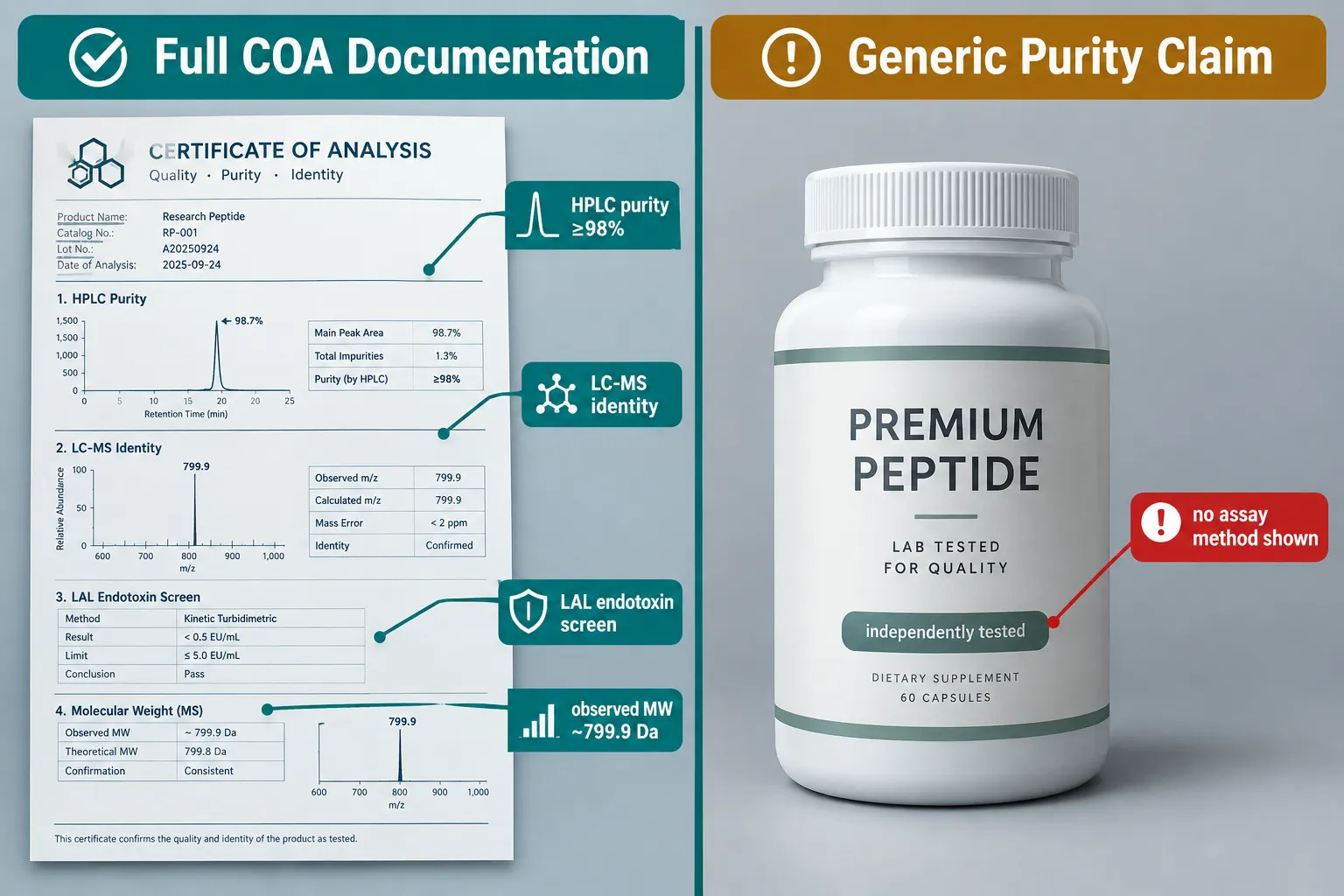
  • Batch-level COA documentation should include HPLC purity, LC-MS identity confirmation, and LAL endotoxin screening, not just a generic "≥99% purity" claim.
  • Intranasal delivery is pharmacokinetically justified for CNS research, offering roughly tenfold greater relative blood-brain barrier penetration versus intravenous injection.
  • Combination Semax/Selank sprays are a growing niche in 2026 but require dual-peptide assay documentation to be scientifically valid.
  • Researchers should request full COA files and explicit assay methods before committing to any supplier for critical experiments.

Why Formulation Transparency Is the First Filter

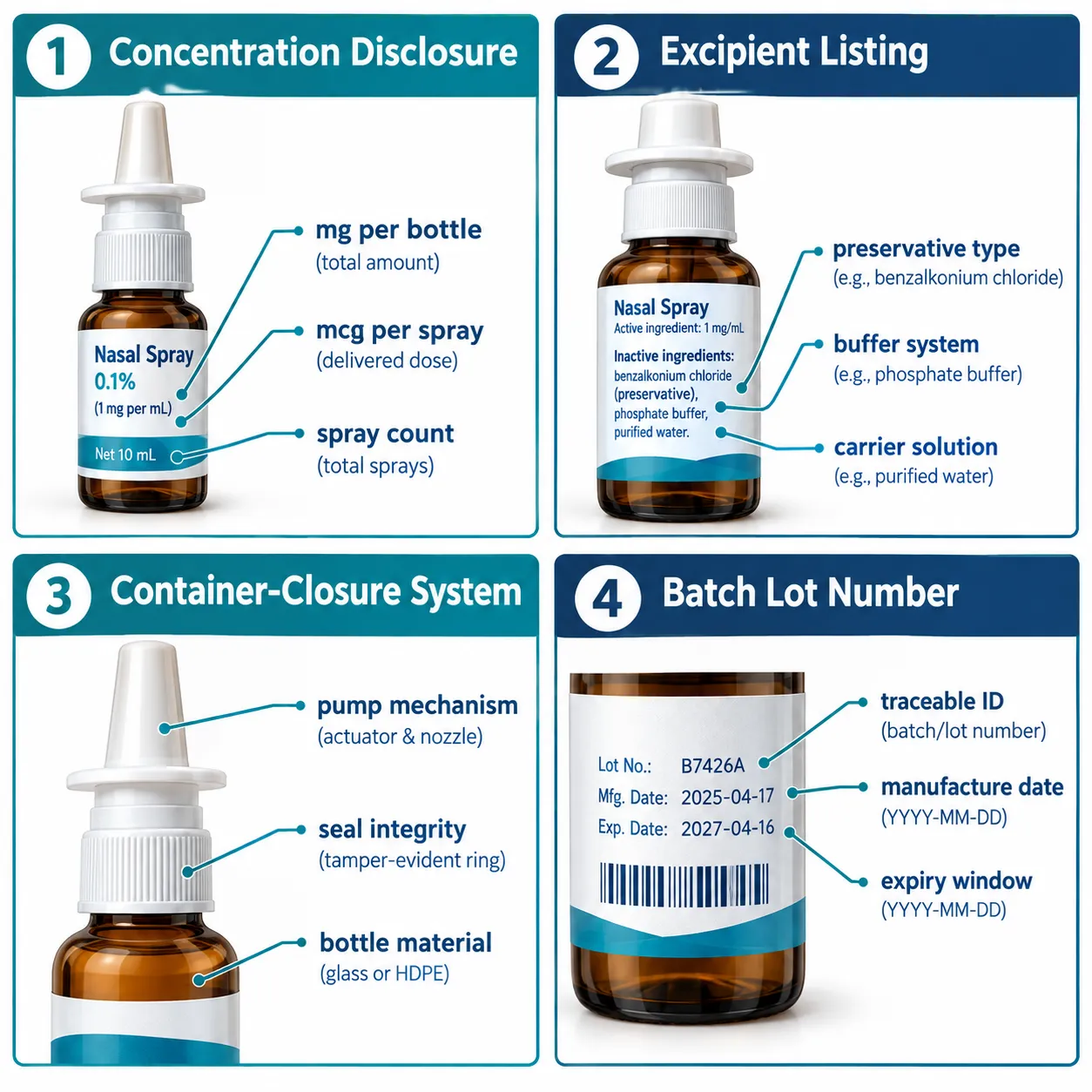
When evaluating Semax nasal spray research products compared across suppliers, the starting point is always the label. Specifically: what is the peptide concentration per spray, how many sprays does the bottle deliver, and what is the total peptide mass?

Why Formulation Transparency Is the First Filter

Current market offerings show significant variation. One 30 mg/10 mL bottle delivers 80 metered sprays at 375 mcg each. A separate product lists 11 mg total with 100 mcg per spray. A compounded pharmacy formulation offers 15 mL at 2.5 mg/mL, yielding 250 mcg per spray. These are not minor differences, a researcher using the first product and then switching to the second without adjustment would deliver less than 27% of the original dose per actuation.

What transparent formulation labeling should include:

Disclosure Element Why It Matters
Total peptide mass (mg) Confirms batch size for cost and protocol planning
Volume (mL) Enables concentration calculation
Per-spray dose (mcg) The critical dosing unit for experimental design
Metered spray count Confirms consistency across the bottle's lifespan
Excipients and preservatives Affects cell viability in in-vitro models
Container-closure system Affects sterility and pump accuracy over time

Excipient disclosure is often overlooked. The buffer system, any antimicrobial preservative, and the carrier solution can all influence outcomes in cell-based assays. A product that lists only the active peptide concentration without naming the excipients leaves a meaningful gap for lab reproducibility.

Researchers exploring other peptides nasal spray formats will find the same transparency hierarchy applies across the category.


Assay Documentation Standards: What Separates Credible Suppliers

The phrase "third-party tested" has become nearly universal in the peptide research market, and nearly meaningless without supporting documentation. A rigorous Semax nasal spray research products comparison must look past marketing language to the actual assay data.

Assay Documentation Standards: What Separates Credible Suppliers

A genuine three-assay quality control standard for Semax includes:

  1. HPLC purity, main peak area should be ≥98%, with the chromatogram available for review
  2. LC-MS identity confirmation, the observed molecular weight should match the theoretical ~799.9 Da for Semax
  3. LAL endotoxin screening, results should fall within research-grade acceptable limits

Some European and North American suppliers now offer selectable COAs for different product strengths (10 mg vs. 30 mg), which is a meaningful step toward the kind of batch-level traceability that serious research programs require. However, a notable portion of the market still provides only a statement of "independently tested" with no downloadable COA, no assay method description, and no lot-specific data.

"A purity claim without a chromatogram is a marketing statement, not a quality standard."

Practical checklist before purchasing:

  • Request the batch-specific COA, not a generic certificate
  • Confirm HPLC method and column specifications are listed
  • Verify LC-MS data shows the correct observed mass
  • Check that endotoxin testing method (LAL or equivalent) is named
  • Confirm the testing laboratory is identified by name

For researchers also evaluating related peptide quality standards, the approach used when sourcing research-grade Glow Blend peptide provides a useful parallel framework for purity and identity verification.


Use-Case Fit: Matching the Product to the Research Model

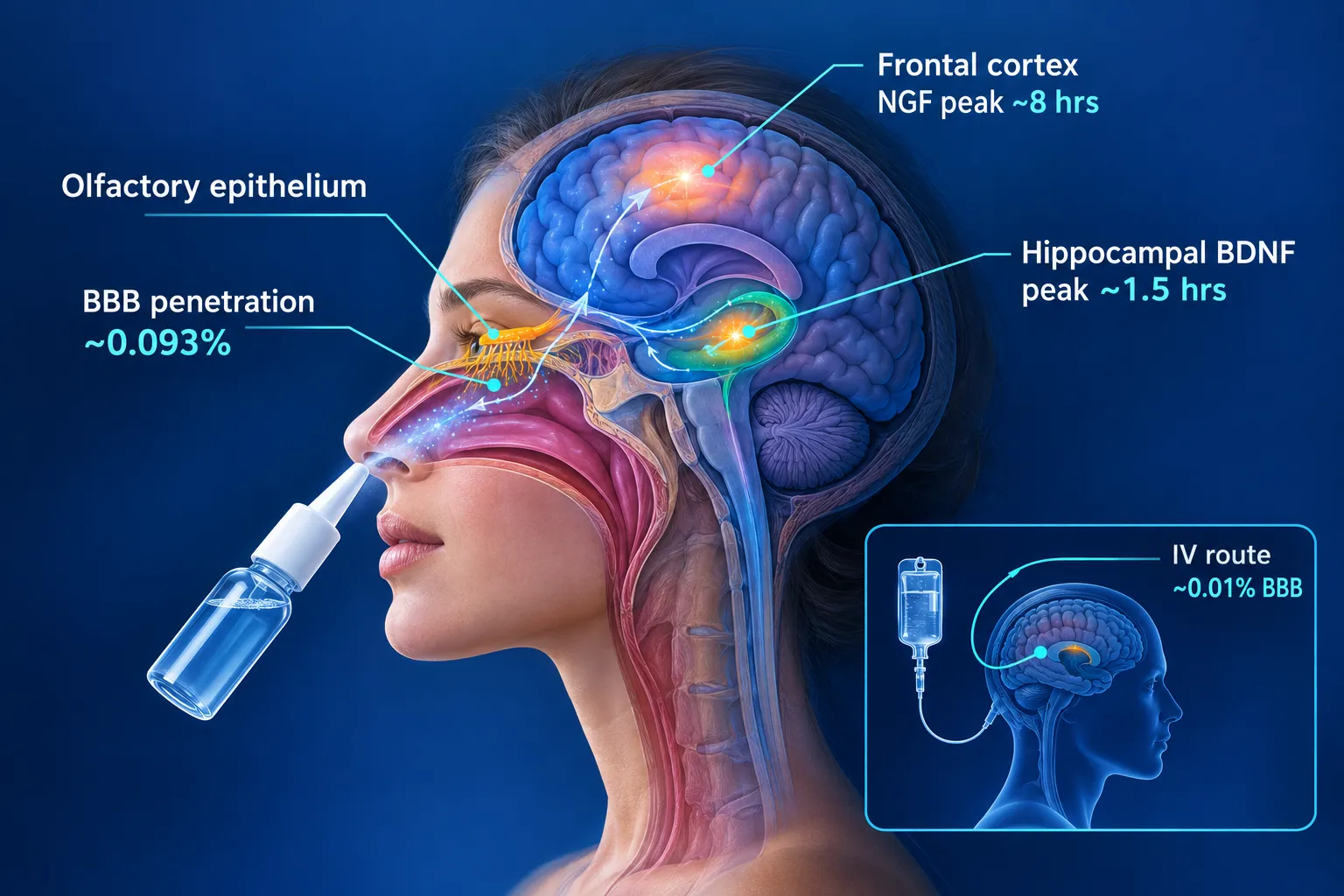
Semax's research utility is grounded in its neurotrophic activity. Studies in rat glial cell models show rapid upregulation of BDNF and NGF mRNA within 30 minutes of exposure. Intranasal dosing in intact rats produces two expression peaks, one at approximately 1.5 hours in the hippocampus and frontal cortex, and a second at roughly 8 hours, supporting its fit for neuroplasticity and cognitive research protocols.

Use-Case Fit: Matching the Product to the Research Model

The pharmacokinetic rationale for the nasal route is clear: approximately 0.093% of intranasally administered Semax crosses the blood-brain barrier, compared with roughly 0.01% via intravenous injection. That nearly tenfold relative increase in CNS exposure underpins the preference for nasal spray formats in CNS-targeted research.

Regulatory and clinical context remains concentrated in Russia, where Semax was originally approved as nasal drops at 0.1% and 1% concentrations for neuroprotection, stroke recovery, and optic nerve atrophy. Western suppliers uniformly frame their products as research-only. In the United States, Semax is not FDA-approved and is accessible only through compounding pharmacies for off-label use.

Dosing protocols from medically reviewed sources converge around 300-600 mcg per nostril once or twice daily in 10-14 day courses with rest periods. A compounded 250 mcg-per-spray product is typically used at one spray per nostril once daily, referencing Russian clinical protocols of 14-21 day courses.

Combination formulations are a growing segment. Semax/Selank combination sprays, standardized at 250 mcg of each peptide per dose, are marketed for overlapping neurocognitive and anxiolytic research use-cases. Researchers considering this niche should review Selank vs Semax comparisons to confirm the mechanistic rationale before selecting a dual-peptide product. Dual-peptide products require COA documentation for both active compounds independently.

Geographic market positioning adds another layer of complexity. Canadian vendors sometimes frame Semax under a natural GH-pathway support angle, while European suppliers emphasize neurocognitive applications. Messaging differences do not change the underlying chemistry, but they can signal which research community a supplier is primarily serving, and whether their documentation standards align with that community's expectations.

Researchers building multi-peptide CNS protocols may also find value in reviewing the IPA Sermorelin stack research framework as a model for how stacked peptide combinations should be documented and evaluated.


Conclusion

Semax nasal spray research products compared across the 2026 market reveal a clear quality divide: suppliers that provide batch-specific HPLC, LC-MS, and endotoxin data versus those relying on generic purity language. Formulation transparency, covering per-spray dose, total peptide mass, excipients, and container-closure details, is the foundational filter. Assay documentation is the differentiator that determines whether a product is fit for reproducible research.

Actionable next steps for researchers:

  1. Build a supplier evaluation checklist using the formulation and assay criteria outlined above
  2. Request lot-specific COA files before placing any order, reject suppliers who cannot provide them
  3. Match the per-spray concentration to your specific experimental protocol before comparing prices
  4. For combination sprays, require independent COA data for each peptide in the formulation
  5. Revisit supplier documentation standards at each new batch, as QC consistency can vary over time
https://www.puretestedpeptides.com/wp-content/uploads/2026/09/semax-nasal-spray-research-products-compared-formulation-transparency-assay-docu.webp 1024 1536 https://www.puretestedpeptides.com/wp-content/uploads/2026/01/buy-peptides-online.jpg 2026-09-25 13:09:272026-09-25 13:09:27Semax Nasal Spray Research Products Compared: Formulation Transparency, Assay Documentation, and Use-Case Fit
5-Amino-1MQ Research Assays: How NNMT-Related Readouts Can Be Separated From MOTS-c and SLU-PP-332 Effects

5-Amino-1MQ Research Assays: How NNMT-Related Readouts Can Be Separated From MOTS-c and SLU-PP-332 Effects

September 25, 2026/0 Comments/in Uncategorized/by

Three metabolically active compounds, 5-Amino-1MQ, MOTS-c, and SLU-PP-332, each touch mitochondrial energy metabolism, yet they do so through entirely different molecular mechanisms. When researchers design experiments involving more than one of these agents, conflated readouts are a genuine risk. Understanding the 5-Amino-1MQ research assays framework, specifically how NNMT-related readouts can be separated from MOTS-c and SLU-PP-332 effects, is essential for generating interpretable, publication-quality preclinical data.

Key Takeaways

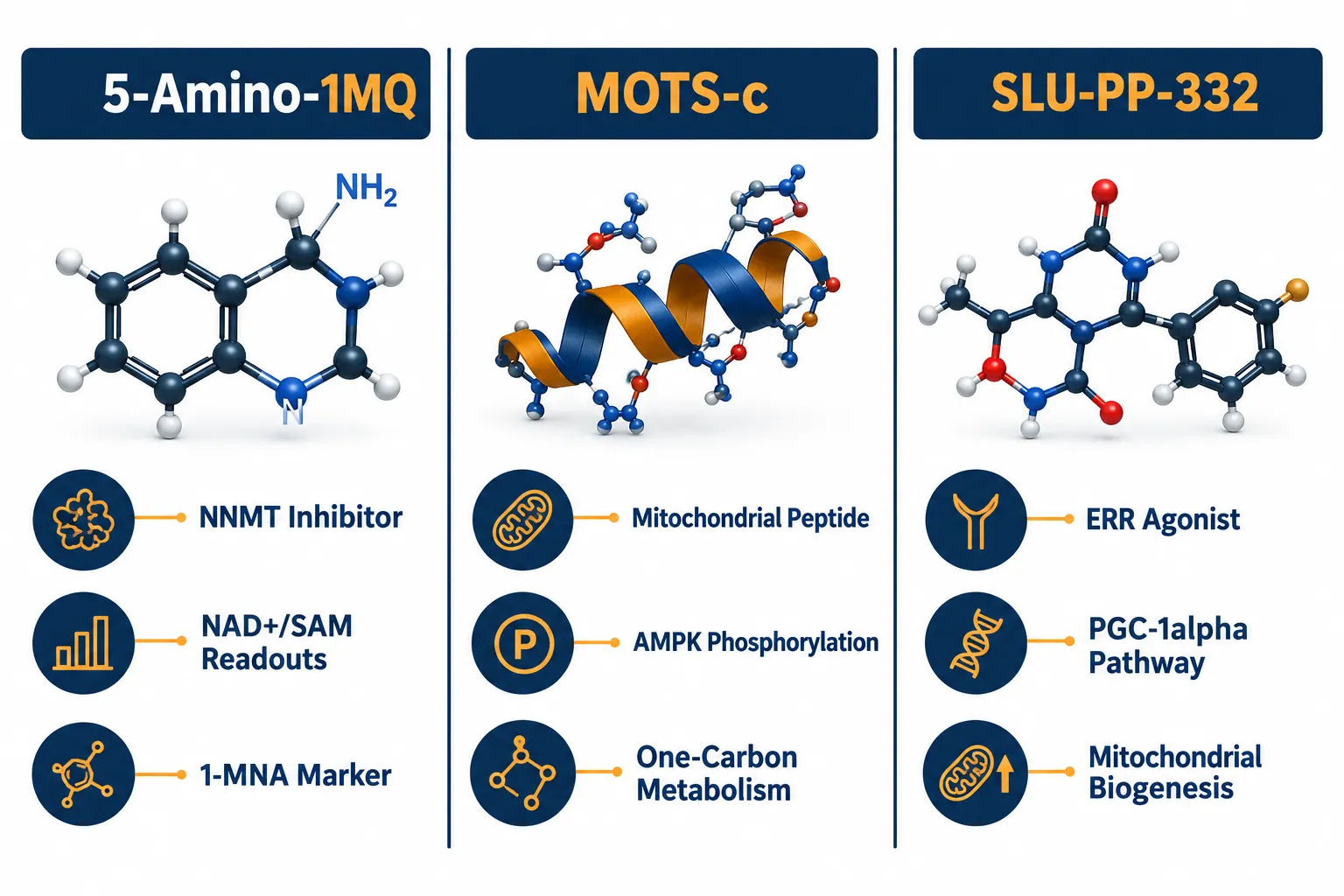
  • 5-Amino-1MQ is a substrate-competitive NNMT inhibitor; its primary assay endpoints are NNMT enzyme activity, 1-methylnicotinamide (1-MNA) levels, and NAD+/SAM pool dynamics.
  • MOTS-c signals through AMPK phosphorylation and one-carbon metabolism remodeling, pathways that are mechanistically orthogonal to NNMT inhibition.
  • SLU-PP-332 activates estrogen-related receptors (ERRs) to drive transcriptional programs for mitochondrial biogenesis, a downstream node distinct from NNMT substrate competition.
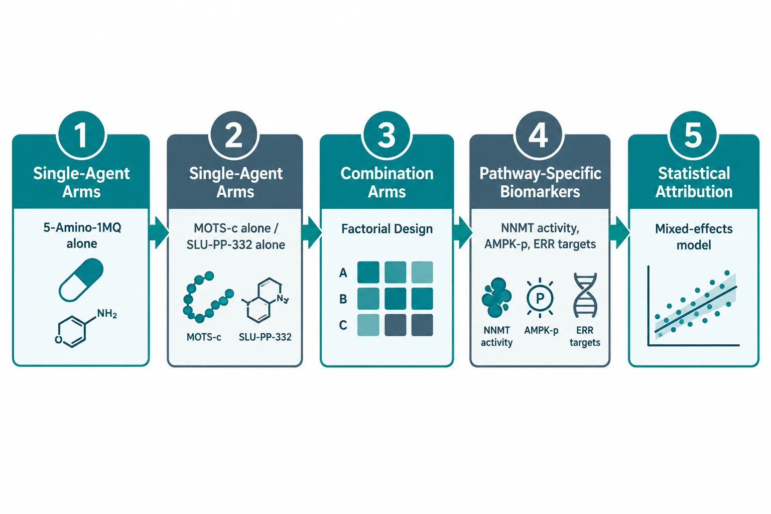
  • Factorial experimental designs with single-agent control arms are the most reliable way to attribute effects to each compound.
  • As of 2026, all available efficacy data for 5-Amino-1MQ remain preclinical; no human trials have been published.

Understanding the NNMT Pathway: The Biochemical Basis of 5-Amino-1MQ Assays

Understanding the NNMT Pathway: The Biochemical Basis of 5-Amino-1MQ Assays

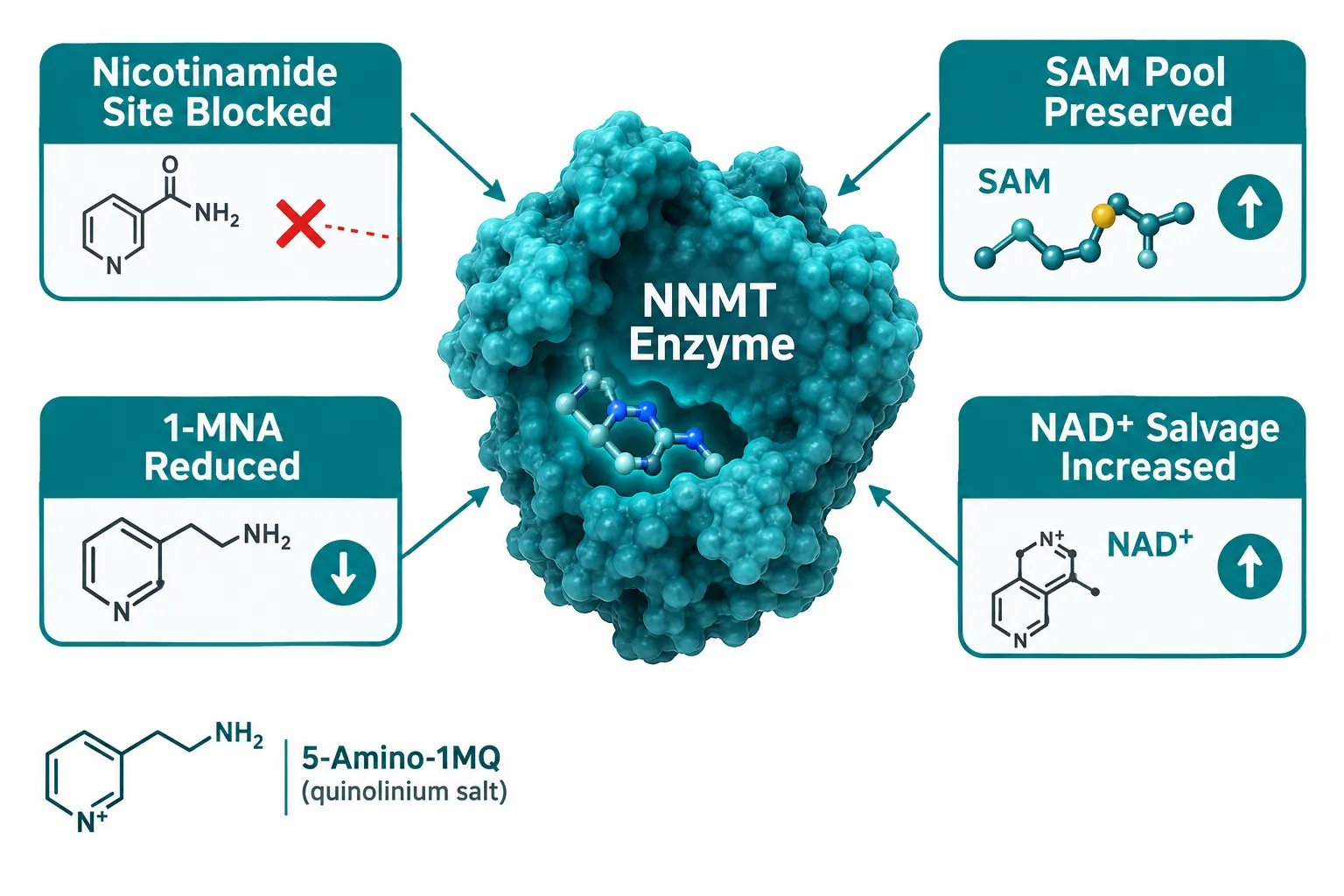
5-Amino-1MQ is a cell-permeable quinolinium salt that competitively inhibits nicotinamide N-methyltransferase (NNMT) at the nicotinamide binding site. NNMT normally consumes S-adenosylmethionine (SAM) to methylate nicotinamide, producing 1-MNA. By blocking this reaction, 5-Amino-1MQ preserves SAM and redirects nicotinamide back into the NAD+ salvage pathway. Reported in vitro data place the IC50 for human NNMT at approximately 1.2 µM, with cellular models showing 1.2-1.6-fold increases in intracellular NAD+.

The most cited preclinical evidence comes from a 2018 mouse study in which 20 mg/kg administered for 11 days reversed diet-induced obesity without reducing food intake, a result attributed to NNMT inhibition shifting adipose tissue toward fat oxidation. More recently, aged-mouse data published in 2026 showed that 5-Amino-1MQ combined with exercise improved grip strength beyond exercise alone, extending the compound's assay relevance into sarcopenia-like models.

Primary NNMT-centric assay endpoints to track:

Endpoint What It Measures Why It Is NNMT-Specific
NNMT enzyme activity Catalytic rate of nicotinamide methylation Direct measure of inhibitor engagement
1-MNA concentration Product of NNMT reaction Falls with effective inhibition
Nicotinamide pool Substrate availability Rises as NNMT is blocked
SAM/SAH ratio Methyl-donor status Increases when NNMT is not consuming SAM
NAD+ salvage flux Isotope-traced NAD+ synthesis Rises as nicotinamide is redirected

These endpoints are naturally separable from MOTS-c and SLU-PP-332 readouts because neither of those agents directly modulates NNMT catalytic activity or 1-MNA output. For researchers sourcing the compound, exploring the 5-amino peptide product category provides context on purity specifications and research-use designations relevant to assay planning.


Mapping the Mechanistic Differences: MOTS-c and SLU-PP-332 Versus NNMT Inhibition

Mapping the Mechanistic Differences: MOTS-c and SLU-PP-332 Versus NNMT Inhibition

The core challenge in multi-agent metabolic research is that MOTS-c, SLU-PP-332, and 5-Amino-1MQ all converge on mitochondrial energy output, but at entirely different upstream nodes.

MOTS-c: AMPK Signaling and One-Carbon Metabolism

MOTS-c is a mitochondria-encoded peptide that activates AMP-activated protein kinase (AMPK) and remodels folate and one-carbon metabolism. Its downstream effects include phosphorylation of ACC (acetyl-CoA carboxylase) and activation of stress-response transcriptional programs. These readouts are mechanistically orthogonal to substrate-competitive NNMT inhibition.

MOTS-c-specific assay markers:

  • AMPK phosphorylation (Thr172)
  • Downstream ACC activity
  • Folate cycle intermediates
  • Stress-response gene expression panels

Researchers pairing MOTS-c with 5-Amino-1MQ, a combination studied alongside related mitochondrial peptides, as discussed in resources on SS31 and MOTS-c research applications, should reserve AMPK phosphorylation readouts exclusively for MOTS-c attribution. Changes in 1-MNA or SAM pools should not be attributed to MOTS-c exposure.

SLU-PP-332: ERR Agonism and Transcriptional Biogenesis

SLU-PP-332 is a pan-ERR (estrogen-related receptor) agonist studied for its exercise-mimetic and mitochondrial biogenesis effects. It drives transcriptional programs through ERR target genes and PGC-1alpha-linked pathways, increasing mitochondrial DNA copy number and oxidative capacity. No published animal or human studies of SLU-PP-332 combined with 5-Amino-1MQ existed as of mid-2026.

"NNMT inhibition increases NAD+/SAM upstream, while ERR agonism drives downstream transcription of oxidative-metabolism genes, these are distinct nodes, not redundant ones."

SLU-PP-332-specific assay markers:

  • ERR target gene expression (ESRRA, ESRRB, ESRRG)
  • PGC-1alpha mRNA and protein levels
  • Mitochondrial DNA copy number
  • Oxygen consumption rate (as an ERR-driven output)

Conceptual analyses in 2026 explicitly frame any combined use of SLU-PP-332 and 5-Amino-1MQ as hypothesis-generating only, recommending parallel single-agent arms to prevent conflating ERR-mediated transcriptional changes with NNMT-driven NAD+/SAM modulation. Researchers interested in related mitochondrial peptide mechanisms can also consult work on SS-31 mechanism and research applications for comparative pathway context.


Designing Assays That Cleanly Separate NNMT-Related Readouts From MOTS-c and SLU-PP-332 Effects

Designing Assays That Cleanly Separate NNMT-Related Readouts From MOTS-c and SLU-PP-332 Effects

Translating mechanistic knowledge into clean experimental design requires structured factorial layouts and pathway-anchored biomarker panels. The following framework reflects current best practices for 5-Amino-1MQ research assays where NNMT-related readouts must be separated from MOTS-c and SLU-PP-332 effects.

Experimental Architecture

A robust multi-agent study should include:

  1. Vehicle control arm, baseline for all endpoints
  2. 5-Amino-1MQ single-agent arm, isolates NNMT inhibition effects
  3. MOTS-c single-agent arm, isolates AMPK/one-carbon effects
  4. SLU-PP-332 single-agent arm, isolates ERR transcriptional effects
  5. Combination arms, factorial pairings and full triple combination

This structure allows statistical attribution of any observed change to a specific mechanism rather than the combination as a whole.

Biomarker Panel Assignment

Compound Primary Readout Category Key Markers
5-Amino-1MQ Enzymology / Metabolomics NNMT activity, 1-MNA, NAD+, SAM/SAH
MOTS-c Phosphoproteomics / Signaling p-AMPK, p-ACC, folate intermediates
SLU-PP-332 Transcriptomics / Biogenesis ERR targets, PGC-1alpha, mtDNA copy number

Controlling for Confounders

Several metabolic outputs, oxygen consumption rate, ATP production, and fatty acid oxidation flux, can be influenced by all three agents through different upstream routes. These shared endpoints should be treated as secondary, integrative readouts rather than attribution markers. Isotope tracing (e.g., 13C-labeled nicotinamide for NAD+ flux) provides the strongest evidence for NNMT-specific pathway engagement and cannot be mimicked by ERR agonism or AMPK activation.

All 5-Amino-1MQ work should be conducted with reagents carrying batch-specific certificates of analysis confirming purity at or above 99%, strictly within in vitro or ex vivo systems where exposure concentration and timing can be precisely controlled. This is consistent with the compound's current status as a research-only agent with no FDA-approved indication and no published human trials as of 2026. For parallel reading on rigorous preclinical peptide assay design, the overview of SS-31 10mg research peptide considerations offers useful methodological context.

A forward-looking note: Expert commentary in 2026 anticipates that the next major step for 5-Amino-1MQ will be formal GLP toxicology studies followed by early-phase human trials targeting obesity, fatty liver, or age-related muscle decline. Future multi-omic panels integrating metabolomics, phosphoproteomics, and transcriptomics will likely become the standard framework for dissecting NNMT versus ERR versus MOTS-c contributions in complex experimental designs. This remains speculative, pending formal study initiation.


Conclusion

Separating NNMT-related readouts from MOTS-c and SLU-PP-332 effects is not merely a technical preference, it is a prerequisite for interpretable data. The 5-Amino-1MQ research assays framework outlined here provides a practical roadmap: anchor NNMT attribution to enzyme activity, 1-MNA, and NAD+/SAM flux; assign AMPK phosphorylation and one-carbon intermediates to MOTS-c; and reserve ERR transcriptional signatures and mitochondrial biogenesis markers for SLU-PP-332.

Actionable next steps for researchers:

  • Build single-agent control arms into every multi-compound study before adding combination arms.
  • Use isotope-traced NAD+ salvage assays as the gold-standard NNMT-specific readout.
  • Treat oxygen consumption rate and ATP output as integrative, not attributive, endpoints.
  • Source 5-Amino-1MQ with verified COA documentation and maintain strict in vitro or ex vivo protocols.
  • Interpret all findings within the current preclinical-only evidence base, no human trial data exist as of 2026.

Rigorous pathway separation today will produce the mechanistic clarity needed for the human-trial assay panels that the field anticipates in the coming years.

https://www.puretestedpeptides.com/wp-content/uploads/2026/09/5-amino-1mq-research-assays-how-nnmt-related-readouts-can-be-separated-from-mots.webp 1024 1536 https://www.puretestedpeptides.com/wp-content/uploads/2026/01/buy-peptides-online.jpg 2026-09-25 13:09:272026-09-25 13:09:275-Amino-1MQ Research Assays: How NNMT-Related Readouts Can Be Separated From MOTS-c and SLU-PP-332 Effects
GLP-3 Retatrutide vs. GLP2-T in Research: Metabolic Signaling Compared With Intestinal Epithelial Models

GLP-3 Retatrutide vs. GLP2-T in Research: Metabolic Signaling Compared With Intestinal Epithelial Models

September 25, 2026/0 Comments/in Uncategorized/by

Fewer than two decades separate the approval of the first GLP-2 analogue for short bowel syndrome and the emergence of a triple receptor agonist that some researchers are already calling the most potent pharmacologic weight-loss agent ever studied. These two compound classes share a naming convention that implies kinship, yet they address entirely different biological questions. Understanding the distinction between GLP-3 Retatrutide vs. GLP2-T in Research: Metabolic Signaling Compared With Intestinal Epithelial Models is not just a matter of terminology — it shapes which experimental models researchers choose, which readouts they measure, and how results should be interpreted.

Key Takeaways

  • The informal label "GLP-3" does not describe a recognized hormone; it is a colloquial term applied to retatrutide, a triple GLP-1/GIP/glucagon receptor agonist.
  • Retatrutide targets systemic metabolic signaling across the pancreas, adipose tissue, liver, and central nervous system.
  • GLP2-T (teduglutide) acts primarily through gut-localized GLP-2 receptors to support intestinal epithelial structure and barrier function.
  • These compounds require different preclinical and clinical model systems, and their readouts do not overlap.
  • Terminology precision matters: conflating these agents can lead to misaligned experimental design and misinterpretation of data.

Clarifying the "GLP-3" Label: What Retatrutide Actually Is

The phrase "GLP-3" circulates widely in media and consumer-facing content, but it does not correspond to a recognized incretin hormone or established drug class. Retatrutide — formally designated LY3437943 — is an investigational, once-weekly synthetic peptide engineered on a GIP backbone and conjugated to a C20 fatty di-acid moiety. That conjugation enables albumin binding, which extends the molecule's half-life and supports weekly dosing.

What makes retatrutide structurally and pharmacologically distinct is its simultaneous agonism at three receptors: the GLP-1 receptor, the GIP receptor, and the glucagon receptor. This triple agonism is not accidental. The molecule was designed through structure-based engineering to optimize activity at all three targets, creating a compound that simultaneously enhances insulin secretion, suppresses appetite through central pathways, and increases energy expenditure via glucagon-driven thermogenesis.

Clarifying the "GLP-3" Label: What Retatrutide Actually Is

For researchers exploring polypeptide peptides in cardiometabolic models, this triple mechanism positions retatrutide as a fundamentally systemic agent. Its receptor targets are distributed across pancreatic islets, adipose tissue, the liver, and the central nervous system — not concentrated in the intestinal epithelium.

Phase 2 clinical data published in the New England Journal of Medicine reported mean weight loss of up to 24.2% after 48 weeks in people with obesity without diabetes, and 16.9% after 36 weeks in those with type 2 diabetes. Emerging phase 3 data as of 2026 suggest average weight loss approaching 28% of baseline body weight. Gastrointestinal symptoms — nausea, diarrhea, vomiting — represent the most common adverse events, a profile broadly comparable to existing GLP-1 receptor agonists.

"Retatrutide's receptor targets span the pancreas, fat tissue, liver, and brain — not the intestinal epithelium."

Researchers sourcing material for preclinical work can review available GLP-3 Reta 10mg and Reta 20mg formats to understand dosing specifications relevant to experimental protocols.


GLP2-T: A Gut-Targeted Epithelial Biology Tool

GLP-2 and its primary clinical analogue teduglutide (GLP2-T) were developed to address a fundamentally different biological problem: intestinal structural failure. Foundational preclinical work established that GLP-2 analogues consistently enhanced mucosal growth, nutrient absorption, and functional recovery in animal models of gut epithelial injury. That work formed the scientific basis for teduglutide's approval in short bowel syndrome.

At the mechanistic level, GLP2-T acts through GLP-2 receptors located on enteric neurons and subepithelial cells. Downstream trophic signaling expands villus height and crypt depth, supporting absorptive surface area. Critically, GLP-2 signaling also helps maintain tight-junction integrity.

GLP2-T: A Gut-Targeted Epithelial Biology Tool

A well-characterized intestinal epithelial barrier study using piglet models and IPEC-J2 monolayer cultures demonstrated that GLP-2 pretreatment dose-dependently prevented lipopolysaccharide-induced increases in myosin light-chain kinase (MLCK) expression and phosphorylated myosin light chain (pMLC) across the duodenum, jejunum, and ileum. These findings point to a direct role for GLP-2 signaling in attenuating mucosal barrier injury at the cellular level.

Relevant model systems for GLP2-T research include:

  • IPEC-J2 intestinal epithelial monolayer cultures
  • Piglet or rodent intestinal injury models
  • Organoid-based villus morphology assays
  • Transepithelial electrical resistance (TEER) measurements for barrier permeability

Key readouts center on villus height, crypt depth, tight-junction protein expression, MLCK activity, and barrier permeability — none of which are primary endpoints in retatrutide trials.


Comparing Model Selection and Readouts Across Both Research Contexts

The core distinction in GLP-3 Retatrutide vs. GLP2-T in Research: Metabolic Signaling Compared With Intestinal Epithelial Models lies in experimental design. Choosing the wrong model for either compound produces data that cannot be meaningfully interpreted.

Feature Retatrutide (GLP-3) GLP2-T (Teduglutide)
Receptor targets GLP-1R, GIPR, GcgR GLP-2R (enteric/subepithelial)
Primary tissue focus Pancreas, adipose, CNS, liver Intestinal epithelium
Preferred preclinical models Islet cell assays, adipocyte cultures, in vivo obesity models IPEC-J2 monolayers, piglet gut injury, organoids
Primary research readouts Body weight, glycemia, lipid profiles Villus height, tight-junction integrity, TEER
Regulatory status (2026) Investigational (Phase 3) Approved (short bowel syndrome)

For retatrutide, the Eli Lilly phase 3 program covers obesity with comorbidities, type 2 diabetes, knee osteoarthritis pain, obstructive sleep apnea, chronic low back pain, cardiovascular outcomes, renal outcomes, and metabolic dysfunction-associated steatotic liver disease. None of these endpoints involve direct measurement of intestinal epithelial morphology.

Comparing Model Selection and Readouts Across Both Research Contexts

Safety profiles also diverge. Retatrutide's adverse-event landscape is dominated by gastrointestinal symptoms tied to its GLP-1 component. GLP-2 analogues carry distinct concerns: fluid retention, intestinal mucosal hyperplasia, and potential neoplastic risk in susceptible patients. These differences drive continued research into epithelial signaling specificity and long-term histological effects for GLP2-T, while retatrutide's ongoing trials focus on cardiovascular and renal safety endpoints.

Researchers interested in broader peptide signaling contexts, including mitochondrial and renal biology, may find relevant background in SS-31 kidney health research and SS-31 mitochondrial research themes, which illustrate how mechanistic specificity shapes model selection across peptide classes.

For those specifically sourcing retatrutide analogs for research, the GLP-3 Reta CAG 10mg and GLP-3 Reta 20mg product pages provide specification details relevant to experimental planning.


Conclusion

The comparison of GLP-3 Retatrutide vs. GLP2-T in Research: Metabolic Signaling Compared With Intestinal Epithelial Models ultimately clarifies that these are not competing agents — they are tools designed for different scientific questions. Retatrutide is a systemic triple agonist built for cardiometabolic research, with clinical endpoints centered on body weight, glycemia, and organ-level outcomes. GLP2-T is a gut-targeted trophic agent built for epithelial biology, with readouts rooted in mucosal structure and barrier function.

Actionable steps for researchers:

  • Confirm receptor target distribution before selecting a model system; systemic metabolic signaling requires different assays than epithelial barrier biology.
  • Avoid applying the informal "GLP-3" label in formal research contexts — use the compound name retatrutide and specify its triple agonist mechanism.
  • Match readouts to mechanism: use TEER, MLCK, and villus morphology for GLP2-T work; use glycemic, lipolytic, and weight-related endpoints for retatrutide studies.
  • Monitor phase 3 data releases for retatrutide, as cardiovascular and renal safety data will significantly shape future research directions.
  • Recognize that teduglutide's approved status versus retatrutide's investigational status reflects different stages of translational development, not relative importance.

Precision in terminology and model selection is not bureaucratic caution — it is the foundation of reproducible, meaningful peptide research.

https://www.puretestedpeptides.com/wp-content/uploads/2026/09/glp-3-retatrutide-vs-glp2-t-in-research-metabolic-signaling-compared-with-intest.webp 1024 1536 https://www.puretestedpeptides.com/wp-content/uploads/2026/01/buy-peptides-online.jpg 2026-09-25 13:03:582026-09-25 13:03:58GLP-3 Retatrutide vs. GLP2-T in Research: Metabolic Signaling Compared With Intestinal Epithelial Models
Intranasal Peptide Research Methods: Deposition, Mucosal Permeability, Excipient Controls, and Brain-Exposure Questions

Intranasal Peptide Research Methods: Deposition, Mucosal Permeability, Excipient Controls, and Brain-Exposure Questions

September 25, 2026/0 Comments/in Uncategorized/by

Fewer than 5% of intranasal peptide doses typically reach systemic circulation when compared with subcutaneous injection, yet the same formulation can produce measurably higher brain concentrations than an intravenous dose of the identical compound. That apparent paradox sits at the heart of modern intranasal peptide research methods: deposition, mucosal permeability, excipient controls, and brain-exposure questions, and it explains why researchers studying peptides such as Semax, Selank, and related neuropeptide formulations need a rigorous, method-specific framework rather than generic pharmacokinetic assumptions.

Key Takeaways

  • Nasal deposition, mucosal absorption, and brain exposure are three distinct, measurable variables that must be characterized separately in any intranasal peptide program.
  • Device geometry and actuation parameters govern whether a peptide deposits in the olfactory zone (relevant to nose-to-brain transport) or the lower turbinates (relevant to systemic absorption).
  • Peptide molecular weight is the strongest single predictor of intranasal systemic bioavailability; peptides under 1 kDa can achieve meaningful absorption, while larger peptides require excipient support.
  • Excipient selection drives both permeability and safety outcomes; regulatory expectations now include cilia function assays and nasal histopathology for any formulation using permeation enhancers.
  • Translating animal nose-to-brain pharmacokinetic data to human CNS exposure remains the field's most significant unsolved methodological problem as of 2026.

Mapping Nasal Deposition: Why Region Matters

Mapping Nasal Deposition: Why Region Matters

The nasal cavity is not a uniform surface. It divides functionally into a lower respiratory region lined with ciliated columnar epithelium and an upper olfactory region that connects directly to the olfactory bulb and, via the trigeminal nerve, to deeper brain structures. For systemic delivery, the respiratory turbinates provide a large vascularized surface. For nose-to-brain targeting, the olfactory epithelium is the critical zone, and it occupies only a small fraction of the total mucosal area.

Why this distinction shapes the entire research design:

  • A peptide deposited on the lower turbinates enters mucociliary clearance within minutes, limiting contact time and absorption.
  • A peptide reaching the olfactory cleft can follow axonal or perineural transport pathways to bypass the blood-brain barrier entirely.
  • Standard commercial nasal sprays are designed for turbinate deposition; nose-to-brain programs require deliberate device selection and validated deposition confirmation.

Deposition Characterization Techniques

Researchers currently use three primary methods to map where a peptide actually lands:

Method What It Measures Key Limitation
3D-printed nasal cast + spectrophotometry Regional drug mass after zone-wise rinsing Anatomical variability across donors
Radiolabeled scintigraphic imaging Real-time deposition in living subjects Regulatory complexity, radiation exposure
Transparent replica + high-resolution imaging Spray plume mechanics linked to deposition Does not capture mucosal biology

Physics-based parametric modeling, using transparent nasal replicas and software-assisted color-mask quantification, is gaining traction as a way to predict regional deposition from device and actuation parameters without animal studies. Studies comparing six commercial spray devices at a standardized 15 mm insertion depth have shown that plume angle and droplet size alone can determine whether olfactory or turbinate deposition dominates, a finding with direct implications for any researcher selecting a delivery device for neuropeptide work.

For researchers exploring nasal spray peptide delivery formats or nasal spray with PT-141, understanding device-specific deposition profiles is a prerequisite, not an afterthought.


Mucosal Permeability and Excipient Controls

Mucosal Permeability and Excipient Controls

Once a peptide reaches the mucosal surface, absorption depends on molecular weight, lipophilicity, enzymatic stability, and the formulation's excipient profile. Molecular weight is the dominant variable for systemic bioavailability: peptides under approximately 1 kDa (roughly ten amino acids or fewer) can achieve meaningful intranasal absorption without excipient support, while larger peptides show steep declines in fractional absorption unless carriers or enhancers are used.

"The inverse relationship between peptide molecular weight and intranasal bioavailability is among the most reproducible findings in nasal pharmacokinetics, and among the most frequently ignored in early-stage formulation decisions."

Excipient Classes and Their Functions

Mucoadhesive polymers such as chitosan and carbopol extend mucosal contact time, slowing mucociliary clearance and increasing the window for absorption. Chitosan also acts as a mild permeation enhancer by transiently loosening tight junctions between epithelial cells.

Cyclodextrins improve peptide solubility and protect against enzymatic degradation at the mucosal surface, which is rich in proteases.

Surfactants and absorption enhancers open paracellular routes more aggressively but carry a corresponding mucosal safety burden.

Polymeric nanoparticles and cell-penetrating peptide (CPP) conjugates represent the most advanced tier, enabling intracellular transport across the epithelial layer. Despite promising animal data, no nanoparticle-based nose-to-brain system had reached clinical trial stage as of early 2026, underscoring the translational gap.

Safety and Regulatory Expectations

Regulatory agencies now expect a defined safety package for any formulation using permeation enhancers or novel excipients:

  • Nasal mucosal histopathology (acute and chronic exposure)
  • Cilia beat frequency and morphology assays
  • Local tolerability studies at the intended dose frequency
  • Reversibility data for any observed mucosal changes

Researchers working with peptides that require enhanced permeability, including those studying peptide dosing protocols or evaluating formulation strategies for compounds like those discussed in SS-31 mechanism and research, should build mucosal safety endpoints into study design from the outset rather than treating them as late-stage add-ons.


Brain-Exposure Questions: Separating CNS from Systemic Pharmacokinetics

Brain-Exposure Questions: Separating CNS from Systemic Pharmacokinetics

The most methodologically complex aspect of intranasal peptide research methods is quantifying brain exposure independently of systemic exposure. The two are not equivalent, and conflating them produces misleading conclusions.

The core distinction:

  • Systemic bioavailability (%F) measures the fraction of the dose entering general circulation, typically assessed via plasma concentration-time curves referenced against subcutaneous or intravenous dosing.
  • Brain exposure is best measured directly via cerebrospinal fluid (CSF) sampling or brain tissue analysis in animal models, and cannot be reliably inferred from plasma data for nose-to-brain programs.

Intranasal GLP-1/GIP dual agonist peptides, intranasal insulin, and interferon-beta-1b have all demonstrated the defining pattern of nose-to-brain delivery: low systemic exposure paired with significantly elevated brain concentrations compared with intravenous or oral routes. This dichotomy is not an artifact, it reflects genuine olfactory and trigeminal transport that bypasses the blood-brain barrier.

Current Methodological Gaps

Despite consistent animal findings, several problems limit translation to human CNS pharmacokinetics:

  1. No validated in vitro model accurately replicates the human olfactory epithelium's transport characteristics.
  2. CSF sampling in humans is invasive, limiting PK characterization to sparse time points.
  3. Standardized animal paradigms for nose-to-brain transport do not yet exist, making cross-study comparisons unreliable.
  4. Anatomical scaling between rodent and human nasal geometry introduces large uncertainty when extrapolating olfactory deposition fractions.

Regulators increasingly expect quantitative deposition imaging linked to PK modeling for nose-to-brain programs, a requirement that makes early investment in nasal cast studies and CSF sampling protocols strategically necessary rather than optional. Researchers evaluating compounds with potential CNS applications, including those reviewing polypeptide peptides in cardiometabolic models or examining CJC-1295 pharmacokinetic comparisons, should treat brain-exposure quantification as a distinct study objective with its own sampling strategy.

Sourcing research-grade material with verified purity is equally foundational. Impurities in peptide formulations can confound both permeability measurements and CNS exposure data. Resources such as evaluating purity for research-grade peptide formulations offer useful frameworks for assessing supplier quality before committing material to in vivo studies.


Conclusion

A rigorous approach to intranasal peptide research requires treating nasal deposition, mucosal permeability, and brain exposure as three distinct measurement problems, not a single pharmacokinetic endpoint. The actionable steps for researchers in 2026 are clear:

  1. Select and validate the delivery device using nasal cast studies before committing to in vivo work; device geometry determines whether olfactory or turbinate deposition dominates.
  2. Match excipient strategy to peptide molecular weight; small peptides may need only mucoadhesives, while larger peptides require carriers or enhancers with a full mucosal safety package.
  3. Design separate PK endpoints for systemic and CNS exposure; do not infer brain concentrations from plasma data in nose-to-brain programs.
  4. Incorporate standardized deposition metrics early, as regulatory expectations for quantitative imaging linked to PK modeling are rising across the field.
  5. Source verified, research-grade peptide material to ensure that formulation variables, not impurity profiles, drive experimental outcomes.

The nose-to-brain route offers genuine advantages for neuropeptide research, but those advantages are only accessible through disciplined, method-specific study design.

https://www.puretestedpeptides.com/wp-content/uploads/2026/09/intranasal-peptide-research-methods-deposition-mucosal-permeability-excipient-co.webp 1024 1536 https://www.puretestedpeptides.com/wp-content/uploads/2026/01/buy-peptides-online.jpg 2026-09-25 13:03:542026-09-25 13:03:54Intranasal Peptide Research Methods: Deposition, Mucosal Permeability, Excipient Controls, and Brain-Exposure Questions
CJC-1295 With DAC vs. Without DAC in Cell and Animal Studies: Sampling Windows, Washout, and Interpretation

CJC-1295 With DAC vs. Without DAC in Cell and Animal Studies: Sampling Windows, Washout, and Interpretation

September 25, 2026/0 Comments/in Uncategorized/by

A single amino acid modification, the Drug Affinity Complex (DAC), extends CJC-1295's plasma half-life from roughly 30 minutes to approximately six to eight days. That difference is not merely pharmacokinetic trivia. It reshapes every decision a researcher makes: when to draw blood, how long to wait before the next treatment cycle, and whether an observed IGF-1 signal belongs to the compound or to residual accumulation from a prior dose. Understanding CJC-1295 With DAC vs. Without DAC in Cell and Animal Studies: Sampling Windows, Washout, and Interpretation is therefore foundational to producing reproducible, attributable preclinical data.

Key Takeaways

  • CJC-1295 without DAC has a half-life near 30 minutes, making it ideal for studying acute, pulsatile GH release; CJC-1295 with DAC sustains elevated GH and IGF-1 for days to weeks.
  • Sampling windows must be matched to the compound's kinetic profile, mismatched timing leads to missed peaks or inflated baseline readings.
  • Washout for DAC-containing protocols requires a minimum of four to six weeks in rodent models to avoid carryover confounding.
  • IGF-1 attribution is the central interpretive challenge: sustained DAC exposure elevates tonic IGF-1, while the non-DAC form produces transient pulses that are easy to miss without frequent sampling.
  • Study design choices, dose interval, sample frequency, and washout length, differ substantially between the two forms and must be pre-specified in the protocol.

Half-Life Determines Everything Downstream

Half-Life Determines Everything Downstream

The pharmacokinetic contrast between the two forms is the logical starting point because every downstream design decision flows from it. CJC-1295 without DAC is a modified GHRH analogue that resists enzymatic cleavage long enough to reach pituitary receptors but is cleared within one to two hours in most rodent models. Its action is therefore discrete and pulse-like, closely mirroring endogenous GHRH activity.

CJC-1295 with DAC covalently binds to circulating albumin through its maleimido-propionyl group. This albumin linkage creates a depot effect, releasing active peptide slowly over days. In rat models, a single subcutaneous dose produces measurable GH elevation for up to five days and elevated IGF-1 for seven to ten days. The practical consequence is that a researcher who draws a single blood sample at 24 hours post-dose will capture very different biological states depending on which form was used.

For researchers exploring multi-peptide combinations, the Tesamorelin, CJC-1295, and Ipamorelin 12mg blend illustrates how stacking long- and short-acting GHRH analogues complicates attribution further, reinforcing the need for precise kinetic mapping before interpreting outcomes.


Sampling Windows in Cell and Animal Studies: Matching Timing to Kinetics

Getting sampling windows right is where most preclinical CJC-1295 studies succeed or fail. The two forms demand fundamentally different schedules.

Without DAC: High-Frequency, Narrow Windows

Because the non-DAC form acts within minutes and clears rapidly, meaningful GH sampling must occur within 15 to 45 minutes post-administration in rodent models. A single time-point study will almost always underestimate peak GH. Recommended practice calls for serial sampling at 15, 30, 60, and 120 minutes to capture the full pulse shape. IGF-1, which responds more slowly to GH stimulation, should be sampled at 4, 8, and 24 hours to capture the downstream hepatic response.

Cell culture studies using the non-DAC form face an analogous challenge: peptide degradation in serum-containing media can reduce effective concentration within hours, so dosing intervals and media refresh schedules must be tightly controlled.

With DAC: Extended Windows, Accumulation Awareness

For the DAC form, single time-point sampling at 24 or 48 hours post-dose is more defensible, but researchers must account for accumulation across repeated doses. In a weekly dosing protocol over four weeks, plasma concentrations do not reach steady state until approximately the third dose. This means IGF-1 readings taken during weeks one and two reflect rising exposure, not steady-state biology. Comparing week-one IGF-1 to week-four IGF-1 without acknowledging this trajectory is a common interpretive error.

Recommended sampling in repeated-dose rodent studies with the DAC form:

  • Trough samples immediately before each dose (to track accumulation)
  • Peak samples 24 to 48 hours after each dose
  • IGF-1 at 72 hours post-dose, where the signal is most stable
  • A terminal bleed at least seven days after the final dose to distinguish residual compound from true washout baseline

For those working with related combination protocols, the Sermorelin, Ipamorelin, and CJC-1295 blend offers context on how multiple secretagogues interact when sampling schedules must account for overlapping kinetic profiles.


Washout Design and IGF-1 Interpretation

Washout Design and IGF-1 Interpretation

Washout is arguably the most underspecified element in published CJC-1295 animal studies. A compound with a six-to-eight-day half-life requires approximately five half-lives, roughly 30 to 40 days, to fall below 5% of peak plasma concentration. In practice, most expert guidance as of 2026 recommends a minimum four-to-six-week washout in rodent models before a crossover arm or a new treatment cycle begins.

Shorter washouts produce carryover elevation of IGF-1 that can be misattributed to a new intervention. This is particularly problematic in crossover designs where the DAC form is tested first.

Attributing GH Pulsatility vs. Sustained Elevation

The interpretive challenge is not just about timing, it is about biology. The non-DAC form preserves the pulsatile nature of GH secretion. Studies examining GH-dependent cellular processes such as lipolysis, protein synthesis, or IGF-1 receptor upregulation benefit from this pulsatility because it more closely models physiological signaling. The DAC form, by contrast, creates a tonic elevation that may saturate GH receptors and alter downstream sensitivity over time.

"Sustained GH elevation and pulsatile GH elevation are not interchangeable experimental conditions, they activate overlapping but distinct receptor dynamics."

Researchers must therefore specify in their protocols whether the biological question requires pulsatile or sustained GH exposure, and select the appropriate form accordingly. Mixing the two forms within a single study without a full washout between them produces data that cannot be cleanly attributed to either kinetic profile.

For additional context on how CJC-1295 is used alongside complementary peptides in research settings, the Tesamorelin, AOD9604, CJC-1295, and Ipamorelin 12mg blend page provides relevant formulation background. Similarly, researchers comparing secretagogue safety profiles may find the discussion on combining Tesamorelin with CJC Ipamorelin useful for contextualizing multi-compound study designs.


Practical Protocol Recommendations

Practical Protocol Recommendations

The following framework consolidates current best-practice guidance for preclinical researchers as of 2026:

Parameter Without DAC With DAC
GH sampling window 15-60 min post-dose 24-48 h post-dose
IGF-1 sampling window 4-24 h post-dose 48-72 h post-dose
Steady-state onset Single dose ~3rd dose (weekly protocol)
Minimum washout 48-72 hours 4-6 weeks
Crossover design risk Low High without full washout

For dose-ranging studies, the Tesamorelin, CJC-1295, and Ipamorelin 12mg blend dosage guide and the corresponding dose reference offer practical starting points when CJC-1295 is part of a multi-peptide protocol.

Key protocol checkpoints:

  • Pre-specify whether the study question requires pulsatile or tonic GH exposure
  • Document accumulation phase separately from steady-state phase in multi-dose designs
  • Include a satellite group for washout verification if the study uses a crossover design
  • In cell studies, refresh media at intervals matched to the peptide's degradation rate in the specific media formulation used

Conclusion

The choice between CJC-1295 with DAC and without DAC is not simply a matter of dosing convenience, it is a fundamental experimental variable that determines when to sample, how long to wait, and how to interpret every GH or IGF-1 data point the study generates. Researchers who treat the two forms as interchangeable risk producing results that are either uninterpretable or actively misleading.

Actionable next steps for preclinical researchers:

  1. Define the biological question first, pulsatile or sustained GH exposure, then select the compound form that matches it.
  2. Build sampling schedules around the compound's actual kinetic profile, not convenience time-points.
  3. Enforce a minimum four-to-six-week washout for any DAC-containing protocol before a crossover or repeat cycle.
  4. Track trough concentrations across a multi-dose DAC study to confirm when steady state is reached before drawing mechanistic conclusions.
  5. Report the form used, the sampling schedule, and the washout length explicitly in methods sections so findings can be accurately compared across studies.

Rigorous attention to these design elements is what separates publishable, reproducible CJC-1295 research from data that raises more questions than it answers.

https://www.puretestedpeptides.com/wp-content/uploads/2026/09/cjc-1295-with-dac-vs-without-dac-in-cell-and-animal-studies-sampling-windows-was.webp 1024 1536 https://www.puretestedpeptides.com/wp-content/uploads/2026/01/buy-peptides-online.jpg 2026-09-25 13:03:292026-09-25 13:03:29CJC-1295 With DAC vs. Without DAC in Cell and Animal Studies: Sampling Windows, Washout, and Interpretation
Page 11 of 81«‹910111213›»
×

Helpful Links

  • My account
  • Cart
  • Checkout
  • Refund and Returns Policy
  • Privacy Policy
  • SMS Privacy Policy
  • Login
  • My Account
  • Logout

USA Made Lab Tested Peptides

All products are sold for research, laboratory, or analytical purposes only, and are not for human consumption


Pure Tested Peptides is a chemical supplier. Pure Tested Peptides is not a compounding / chemical compounding facility as defined under 503A of the Federal Food, Drug, and Cosmetic act. Pure Tested Peptides is not an outsourcing facility as defined under 503B of the Federal Food, Drug, and Cosmetic act.

The statements made within this website have not been evaluated by the US Food and Drug Administration. The products we offer are not intended to diagnose, treat, cure or prevent any disease.

Human/Animal Consumption Prohibited. Laboratory/In-Vitro Experimental Use Only

Scroll to top Scroll to top Scroll to top