Call or Text 727-513-9780
  • Shopping Cart Shopping Cart
    0Shopping Cart
Pure Tested Peptides | America's most trusted Peptides for sale online
  • Peptides for sale
    • Oral Peptides for sale
      • Peptide Capsules for sale
      • BPC 157 Capsules 1000mcg
      • SLU-PP-332 Capsules | 1000 mcg
      • 5-Amino-1MQ 50mg Capsules
      • Tesofensine 500mcg
    • All Peptides for sale
    • Peptide Sprays
      • BPC 157 Nasal Spray Kit
      • BPC-157 TB500 Nasal Spray Kit
      • Semax Nasal Spray 10mg
      • Selank – Nasal Spray Kit – 10mg
      • Epithalon 50MG Nasal Spray Kit
      • Ipamorelin 10mg Nasal Spray
      • Klow Nasal Spray (BPC-157 + TB-500 + GHK-Cu + KPV) | 80mg
      • Hulk Nasal Spray Tesa / Ipa Blend 6/3 MG
      • Klow Nasal Spray
      • NAD + 500 mg Nasal Spray
      • PT-141 Nasal Spray Kit
    • GHRH Peptides
      • Ipa Peptides
      • CJC-1295 Peptides
        • CJC-1295 with DAC 5 mg
        • CJC-1295 without DAC 5 mg
        • CJC-1295 Ipa 10mg
      • Tesa Peptides
        • Tesa Peptide
        • Tesa 20 mg
    • GHK-Cu Peptides
      • All GHK-Cu Peptides
      • GHK-Cu 100mg
      • KLOW Peptide Blend – Buy KLOW blend online
    • BPC Peptides
      • All BPC Peptides
      • BPC-157
      • BPC-157 TB-500
      • BPC 157 capsules 1000mcg
    • SLU-PP-332 Peptides
      • All SLU-PP-332 Peptides
      • SLU-PP-332 5mg
    • GLP3 Peptides
    • PT-141 Peptides
      • PT-141 Peptides for sale
      • PT-141 10mg
      • PT-141 Nasal Spray
    • CAG Peptides
      • Lipo-C Peptide Blend
      • CAG 5mg
      • CAG 10mg
    • MOTS-C Peptides
      • MOTS-C Peptides for sale
      • MOTS-c peptide
      • MOTS-c 10mg *6 pack*
    • 5 Amino 1MQ Peptides
      • 5 Amino 1MQ Peptides for sale
      • 5-Amino-1MQ 50mg Capsules
      • 5-Amino-1MQ 5mg
    • Epithalon Peptides
      • Epithalon Peptides for sale
      • Epithalon 10mg
      • Epithalon 50mg
  • Shop
    • GLPs
      • 5-Amino-1MQ 50mg Capsules
      • 5-Amino-1MQ 5mg
      • L-Carnitine 500mg/ml
      • Tesofensine 500mcg
      • SLU-PP-332 5mg
      • MOTS-c 10mg *6 pack*
    • Epithalon & BPC Peptides
      • Epithalon 10mg
      • Epithalon 50mg
      • BPC-157
      • BPC 157 capsules 1000mcg
      • BPC-157 TB-500
      • BPC-157 TB500 Nasal Spray Kit
      • BPC 157 Nasal Spray Kit
    • BPC TB-500 & NAD+ Peptides
      • NAD+ 500 mg
      • KLOW Peptide Blend – Buy KLOW blend online
      • GLOW Peptide Blend
      • TB 500 5mg
      • BPC 157 capsules 1000mcg – Supplement
      • BPC 157 Nasal Spray Kit
      • BPC-157
      • BPC-157 TB500 Nasal Spray Kit
      • BPC-157 TB-500
      • BPC 157 capsules 1000mcg
    • LL-37 Peptide
      • LL-37 10 mg
    • MOTS-C & Selank
      • MOTS-c peptide
      • Selank 10mg
    • GHK Peptides
      • GHK-Cu 100mg
      • GLOW Peptide Blend
      • KLOW Peptide Blend – Buy KLOW blend online
  • COAs
  • Wholesale
    • Wholesale Peptides for sale
  • PTP FAQ
  • Affiliates
    • Selank Peptide: Advanced Pharmacological Mechanisms Underlying Its Anxiolytic and Nootropic Effects in Research
      • 5-Amino-1MQ Peptide: Detailed Mechanisms of NNMT Inhibition and Its Impact on Cellular Metabolism Research
        • MOTS-C Peptide: Unraveling Its Role in Mitochondrial Dynamics and Energy Metabolism Research
          • Epithalon Peptide and Telomerase Regulation: Investigating Its Impact on Cellular Senescence and Lifespan Research Models
            • GLP2-T vs GLP2 Tirz Peptide: Understanding the Naming, Mechanistic Nuances, and Research Implications for Gut Health
      • Peptides and Polypeptides in Modern Pharmacology: What Research on Metoprolol, Prednisone, and Amlodipine Reveals
        • Polypeptide Peptides vs NSAIDs: What Naproxen and Diclofenac Teach Tissue-Repair Researchers About BPC‑157 and TB‑500
          • Peptides and Polypeptides in Endocrine Pharmacology: How GLP-1, GLP-2, and GLP-3 Retatrutide Differ From Classic Drugs Like Prednisone and Amlodipine
          • 5‑Amino‑1MQ Peptide and NNMT Inhibition: How It Compares With Statins Like Atorvastatin in Adipose and Lipid Metabolism Research
      • Enclomiphene, Estrogen Receptor Signaling, and Luteinizing Phase Biology: What Hormone Researchers Should Measure
        • Mesenchymal Stem Cells, BPC‑157, and GHK‑Cu: How Tissue Repair Peptides Compare With Classic NSAIDs Like Naproxen in Injury Models
          • Peptide Calculator Use Cases Beyond Growth Hormone: Working Through GLP‑3 Retatrutide, MOTS‑c, and BPC‑157 Research Dosing
        • Polypeptide Peptides in Endocrine and Metabolic Pharmacology: Lessons From Amlodipine, Prednisone, and Metoprolol
        • Peptides Calculator for Advanced Blends: Worked Examples for Tesamorelin, CJC‑1295, and Ipamorelin Stacks
          • Peptides and Polypeptides in Human Physiology: How Molecular Size Shapes Research Applications
            • Selank Peptide: Advanced Pharmacological Mechanisms Underlying Its Anxiolytic and Nootropic Effects in Research
          • 5-Amino-1MQ Peptide: Detailed Mechanisms of NNMT Inhibition and Its Impact on Cellular Metabolism Research
            • MOTS-C Peptide: Unraveling Its Role in Mitochondrial Dynamics and Energy Metabolism Research
              • Epithalon Peptide and Telomerase Regulation: Investigating Its Impact on Cellular Senescence and Lifespan Research Models
                • GLP2-T vs GLP2 Tirz Peptide: Understanding the Naming, Mechanistic Nuances, and Research Implications for Gut Health
                  • Peptides and Polypeptides in Endocrine Pharmacology: How GLP-1, GLP-2, and GLP-3 Retatrutide Differ From Classic Drugs Like Prednisone and Amlodipine
                    • Polypeptide Peptides vs NSAIDs: What Naproxen and Diclofenac Teach Tissue-Repair Researchers About BPC‑157 and TB‑500
                      • Peptides and Polypeptides in Modern Pharmacology: What Research on Metoprolol, Prednisone, and Amlodipine Reveals
                        • 5‑Amino‑1MQ Peptide and NNMT Inhibition: How It Compares With Statins Like Atorvastatin in Adipose and Lipid Metabolism Research
                        • Enclomiphene, Estrogen Receptor Signaling, and Luteinizing Phase Biology: What Hormone Researchers Should Measure
                        • Mesenchymal Stem Cells, BPC‑157, and GHK‑Cu: How Tissue Repair Peptides Compare With Classic NSAIDs Like Naproxen in Injury Models
                        • Peptide Calculator Use Cases Beyond Growth Hormone: Working Through GLP‑3 Retatrutide, MOTS‑c, and BPC‑157 Research Dosing
                        • Polypeptide Peptides in Endocrine and Metabolic Pharmacology: Lessons From Amlodipine, Prednisone, and Metoprolol
                        • Peptides Calculator for Advanced Blends: Worked Examples for Tesamorelin, CJC‑1295, and Ipamorelin Stacks
                        • Peptides and Polypeptides in Human Physiology: How Molecular Size Shapes Research Applications
                        • Selank Peptide: Advanced Pharmacological Mechanisms Underlying Its Anxiolytic and Nootropic Effects in Research
                        • MOTS-C Peptide: Unraveling Its Role in Mitochondrial Dynamics and Energy Metabolism Research
                        • Peptides and Polypeptides in Modern Pharmacology: What Research on Metoprolol, Prednisone, and Amlodipine Reveals
                        • Peptides and Polypeptides in Endocrine Pharmacology: How GLP-1, GLP-2, and GLP-3 Retatrutide Differ From Classic Drugs Like Prednisone and Amlodipine
                        • 5-Amino-1MQ Peptide: Detailed Mechanisms of NNMT Inhibition and Its Impact on Cellular Metabolism Research
                        • Mesenchymal Stem Cells, BPC‑157, and GHK‑Cu: How Tissue Repair Peptides Compare With Classic NSAIDs Like Naproxen in Injury Models
                        • Peptide Calculator Use Cases Beyond Growth Hormone: Working Through GLP‑3 Retatrutide, MOTS‑c, and BPC‑157 Research Dosing
                        • GLP2-T vs GLP2 Tirz Peptide: Understanding the Naming, Mechanistic Nuances, and Research Implications for Gut Health
                        • Polypeptide Peptides in Endocrine and Metabolic Pharmacology: Lessons From Amlodipine, Prednisone, and Metoprolol
                        • Polypeptide Peptides vs NSAIDs: What Naproxen and Diclofenac Teach Tissue-Repair Researchers About BPC‑157 and TB‑500
                        • Peptides and Polypeptides in Human Physiology: How Molecular Size Shapes Research Applications
                        • Peptides Calculator for Advanced Blends: Worked Examples for Tesamorelin, CJC‑1295, and Ipamorelin Stacks
                        • 5‑Amino‑1MQ Peptide and NNMT Inhibition: How It Compares With Statins Like Atorvastatin in Adipose and Lipid Metabolism Research
                        • Enclomiphene, Estrogen Receptor Signaling, and Luteinizing Phase Biology: What Hormone Researchers Should Measure
                        • Epithalon Peptide and Telomerase Regulation: Investigating Its Impact on Cellular Senescence and Lifespan Research Models
                        • Best research protocol Klow blend
                        • best time to take BPC-157
                        • best time to take DSIP (Delta Sleep Inducing Peptide)
                        • best time to take CJC-1295
                        • best time to take AOD-9604
                        • best time to take Follistatin 344
                        • best time to take Ipamorelin
                        • best time to take MK-677 (Ibutamoren)
                        • best time to take Ligandrol (LGD-4033) — research compound
                        • best time to take Ostarine (MK-2866) — research compound
                        • best time to take GHK-CU
                        • best time to take TB-500
                        • best time to take MOTS-c
                        • best time to take Semax
                        • best time to take RAD-140 (Testolone) — research compound
                        • best time to take Thymosin Alpha-1
                        • best time to take PEG-MGF
                        • Biolife Plasma, Octapharma Plasma, and Research Peptides: How Plasma Donation Labs Differ From Peptide Suppliers
                        • best time to take YK-11 — research compound
                        • best time to take PT-141 (Bremelanotide)
                        • Best research protocol Klow blend
                        • 5-Amino-1MQ and MOTS-C Synergy: Metabolic Signaling, Mitochondria, and Research Design
                        • BPC-157 and TB-500: Investigating Their Combined Effects on Angiogenesis and Cellular Migration in Tissue Repair Models
                        • BPC-157 Peptide: Gut Barrier Function, Inflammation, and Tissue-Recovery Research
                        • 5‑Amino‑1MQ and MOTS‑c Synergy in Metabolic Research: Designing NNMT and Mitochondrial Biogenesis Stacks
                        • CJC-1295 with DAC vs. Without DAC: Half-Life, Release Kinetics, and Research Implications
                        • CJC‑1295 with DAC vs. Without DAC: Expanding on Half‑Life Differences Using Tesamorelin and Ipamorelin Blend Case Studies
                        • Collagen Biology and Copper‑Binding Peptides: How GHK‑Cu, Glow Blend, and Klow Blend Interact with Skin and Connective Tissue
                        • Collagen Biology and Regenerative Peptides: How GHK‑Cu, Glow Blend, and Klow Blend Affect Extracellular Matrix Research
                    • DNA, Telomeres, and Longevity Peptides: Positioning Epithalon and MOTS‑c in Genetic Aging Research
                      • Enclomiphene Citrate: serm Mechanism, Testosterone Research, and Stack Compatibility
                        • Enclomiphene vs Enclomiphene Citrate: Formulation, Bioavailability, and Research Distinctions
                        • Epithalon Peptide Research: Telomerase Activation, Aging, and Pineal Gland Function
                        • Estrogen Receptor Signaling and Enclomiphene: How Selective Modulators Compare with Classic Polypeptide Hormones
                        • GHK-Cu Peptide: Advanced Mechanisms in Extracellular Matrix Remodeling and Wound Healing Research
                        • GHK-Cu Peptide: Collagen Synthesis, Wound Repair, and Skin-Barrier Research Models
                        • GLP-1 vs GLP-3 vs GLP-2: Peptide Classification and Research Applications
                        • GLP-2 Peptide Research Guide: Gut Barrier Function, Nutrient Absorption, and Intestinal Recovery Models
                        • GLP-3 Retatrutide vs. GLP-1 Drugs: What Triple-Agonist Biology Changes in Research Models
                        • Ipamorelin and Tesamorelin Combination: Synergistic GH Secretagogue Research and Dosing Protocols
                        • GLP2 Tirz Peptide: What It Is, Why the Name Exists, and How Researchers Should Interpret It
                        • Klow Blend Peptide Nasal Spray: What the Formulation Is Trying to Do in Cognitive Research
                        • Mitochondria, NNMT Inhibition, and Peptide Modulators: Where MOTS‑c and 5‑Amino‑1MQ Fit in Cellular Energy Research
                        • MOTS-c Peptide: Mitochondrial Function, Energy Metabolism, and What Researchers Measure
                        • MOTS-c vs. 5-Amino-1MQ: Which Metabolic Research Questions Each Compound Actually Answers
                        • Nasal Spray Peptides: Bioavailability, Administration, and Semax/Selank Research Applications
                        • PT-141 Peptide Research: Mechanism of Action and Melanocortin Receptor Signaling
                        • Retatrutide for Research: Mechanism, Structure, and GLP-1/GLP-3 Dual Action
                        • Retatrutide for Obesity and Type 2 Diabetes: What the Latest Trial Data Suggest
                        • Tesofensine Peptide Research: Mechanism, Appetite Suppression, and Neuropeptide Y Pathways
  • Contact
    • Contact Customer Service
    • Text Customer Support
  • About US
  • Shop all peptides
  • Affiliate Program
    • Affiliate Signup
  • Login / Register Login / Register Page Link Login / Register Page Link
  • Click to open the search input field Click to open the search input field Search
  • Menu Menu
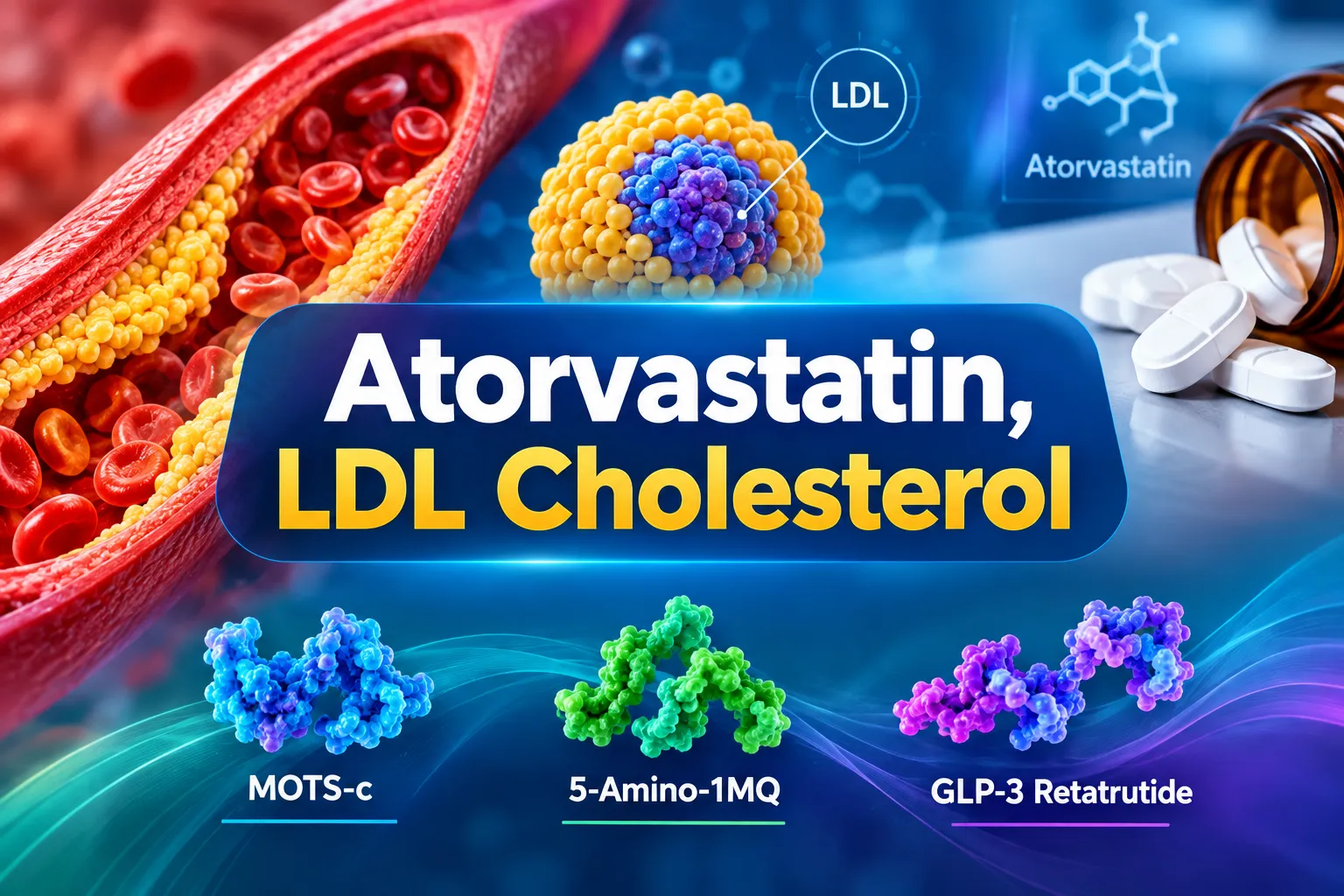
Atorvastatin, LDL Cholesterol, and Metabolic Peptides: Where MOTS-c, 5-Amino-1MQ, and GLP-3 Retatrutide Fit in Lipid Research

Atorvastatin, LDL Cholesterol, and Metabolic Peptides: Where MOTS-c, 5-Amino-1MQ, and GLP-3 Retatrutide Fit in Lipid Research

September 20, 2026/0 Comments/in Uncategorized/by

{"cover":"Professional landscape format (1536×1024) hero image with bold text overlay 'Atorvastatin, LDL Cholesterol' in crisp white modern sans-serif on a deep navy semi-transparent overlay panel, centered with 8% safe margins from every edge, no character touches the edge, two balanced lines. Background: high-resolution editorial medical illustration of a cross-section of a human artery with visible LDL cholesterol plaques, surrounded by floating molecular structures of statin compounds and peptide chains in teal and gold tones, clinical yet visually striking, magazine-cover aesthetic, sharp editorial lighting, professional 2-color palette of navy and teal with white typography.","content":["Annotated mechanism-of-action diagram (1536×1024 landscape) showing atorvastatin's HMG-CoA reductase inhibition pathway in a clean white-background infographic style. Four labeled stages arranged left to right: Stage 1 callout 'HMG-CoA Reductase Blocked' pointing to liver cell diagram, Stage 2 callout 'LDL Receptors Upregulated' with arrow to hepatocyte surface, Stage 3 callout 'LDL Cleared from Blood' showing artery cross-section, Stage 4 callout 'Target: <55 mg/dL ASCVD' with a bold threshold bar. Thin callout lines, teal-and-navy palette, numbered stages, editorial medical-guide polish, no pricing or dollar amounts.","Three-panel side-by-side comparison infographic (1536×1024 landscape) contrasting retatrutide, MOTS-c, and 5-Amino-1MQ as metabolic research agents. Left panel heading 'Retatrutide GLP-1/GIP/Glucagon' with bullet labels: Triple receptor agonist, LDL down ~15-20 percent, TG down ~40 mg/dL, Phase 3 trials. Center panel heading 'MOTS-c Mitochondrial Peptide' with bullet labels: NAD+ regulation, AMPK activation, Lipid oxidation, Preclinical stage. Right panel heading '5-Amino-1MQ NNMT Inhibitor' with bullet labels: NAD+ preservation, Lipogenesis suppressed, Fat oxidation shift, Animal models only. Clean white background, warm amber and teal accent colors, thin dividing lines, editorial infographic polish, callout lines to molecular icons per panel.","Split-screen editorial landscape image (1536×1024) illustrating layered cardiometabolic therapy concept. Left half: warm-lit clinical laboratory bench with statin pill bottles, a cholesterol panel blood report showing LDL values, and a molecular model of atorvastatin in amber studio lighting. Right half: cool-toned scientific research setting with peptide vials labeled MOTS-c and retatrutide, a digital screen showing triglyceride and metabolic pathway data, blue-white laboratory lighting. A bold center divider line with short label 'Established vs Investigational' separating the two halves. High-contrast magazine composition, deep focus, editorial quality."]
Professional landscape hero image () with a reading "Atorvastatin, LDL Cholesterol". CRITICAL TYPOGRAPHY RULES: render the

Cardiovascular disease remains the leading cause of death globally, yet only about half of high-risk patients reach their LDL-cholesterol targets even with statin therapy. That gap has driven intense research into metabolic peptides, molecules that act on mitochondrial signaling, NAD+ biology, and incretin receptors, as potential complements to classic lipid-lowering drugs. Understanding atorvastatin, LDL cholesterol, and metabolic peptides: where MOTS-c, 5-Amino-1MQ, and GLP-3 Retatrutide fit in lipid research requires a clear look at what each agent does, how the mechanisms compare, and what the current evidence actually supports.

Key Takeaways

  • Atorvastatin remains the primary pharmacological tool for reaching stringent LDL-C targets, with high-intensity dosing producing LDL reductions of 41-61%.
  • The 2026 ACC/AHA dyslipidemia guidelines set explicit LDL-C thresholds as low as less than 55 mg/dL for very-high-risk ASCVD patients.
  • Retatrutide, a triple receptor agonist targeting GLP-1, GIP, and glucagon pathways, shows meaningful lipid improvements in clinical trials but remains investigational with no regulatory approval as of 2026.
  • MOTS-c and 5-Amino-1MQ operate through mitochondrial and NAD+ pathways that influence lipid metabolism, but both remain in preclinical research stages.
  • Expert analysis increasingly frames future lipid management as layered therapy: statins for LDL targets plus metabolic peptides for residual cardiometabolic risk.

How Atorvastatin Shapes LDL-C Management in 2026

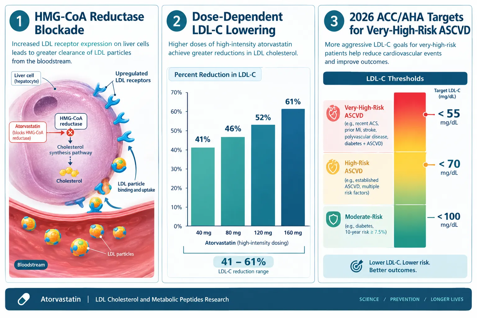
Atorvastatin works by blocking HMG-CoA reductase, the rate-limiting enzyme in hepatic cholesterol synthesis. When the liver produces less cholesterol internally, it upregulates LDL receptors on its surface, pulling more LDL particles out of circulation. The result is a dose-dependent and clinically significant reduction in atherogenic lipoproteins.
How Atorvastatin Shapes LDL-C Management in 2026

Dose, response data are well established. Depending on the dose and the patient's baseline risk profile, atorvastatin produces:

Lipid Marker Typical Reduction Range
Total Cholesterol 30-46%
LDL-C 41-61%
Triglycerides 14-33%
HDL-C Modest increase

The 2026 ACC/AHA multisociety dyslipidemia guideline has reintroduced explicit numeric LDL-C targets for the first time in over a decade. Very-high-risk ASCVD patients now have a target of less than 55 mg/dL, high-risk patients less than 70 mg/dL, and borderline-to-intermediate risk patients less than 100 mg/dL. High-intensity statins like atorvastatin are confirmed as the primary agents to reach these thresholds.

The core limitation: statins address LDL-C powerfully but leave residual risk from elevated triglycerides, visceral adiposity, insulin resistance, and inflammation. This is precisely where metabolic peptide research becomes relevant.

Retatrutide and GLP-Class Peptides: Lipid Effects Beyond Weight Loss

Retatrutide is a first-in-class investigational triple receptor agonist that activates GLP-1, GIP, and glucagon receptors simultaneously. While most public attention has focused on its dramatic weight loss results, its lipid effects are clinically noteworthy in their own right.

A 2026 systematic review and meta-analysis of randomized retatrutide trials found statistically significant improvements across multiple lipid markers. Total cholesterol fell by a weighted mean of approximately 21.88 mg/dL, LDL-C dropped by roughly 13.10 mg/dL, and triglycerides declined by about 40.90 mg/dL, all with p-values below 0.0001. HDL-C showed no meaningful change.

Phase 2 obesity trial data add further context. Non-HDL cholesterol fell approximately 15-17%, LDL declined around 15-20% in higher-dose regimens, and triglycerides dropped by roughly 32-44% across datasets. The mechanism appears to involve glucagon receptor agonism reducing circulating ANGPTL3 and ANGPTL8 concentrations, which in turn enhances lipoprotein lipase activity and alters hepatic lipoprotein production.

Important context: As of mid-2026, retatrutide has no approval from the FDA, EMA, or any other major regulator. Phase 3 TRIUMPH trial data are generating significant scientific interest, but retatrutide remains strictly investigational.

For researchers exploring the GLP peptide landscape, the GLP-3 peptides for sale category and retatrutide clinical trial resources provide useful context on current research-grade availability and trial status. Those interested in the broader incretin class can also review GLP-1 peptides research context for comparison.

MOTS-c and 5-Amino-1MQ: Mitochondrial Pathways in Lipid Research

This is where the science of atorvastatin, LDL cholesterol, and metabolic peptides diverges most sharply from conventional cardiology. MOTS-c and 5-Amino-1MQ do not lower LDL-C through receptor upregulation or incretin signaling. Instead, they act on the metabolic machinery inside cells.
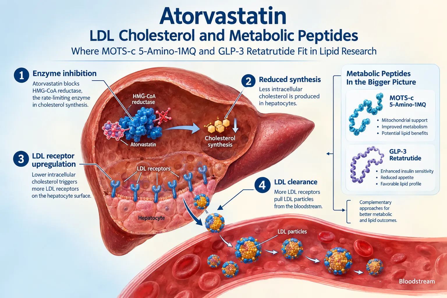
MOTS-c and 5-Amino-1MQ: Mitochondrial Pathways in Lipid Research

MOTS-c is a mitochondria-derived peptide encoded within the 12S rRNA region of mitochondrial DNA. Research models show it activates AMPK, regulates NAD+ metabolism, and promotes fatty acid oxidation over lipid storage. In preclinical settings, MOTS-c treatment is associated with improved insulin sensitivity and reduced lipid accumulation in metabolic tissues. For researchers, MOTS-c 10mg is available as a research-grade compound, and the broader MOTS-c peptide category provides additional options.

5-Amino-1MQ is a selective nicotinamide N-methyltransferase (NNMT) inhibitor. By blocking NNMT, it preserves intracellular NAD+ levels, decreases N1-methylnicotinamide, suppresses lipogenesis, and shifts adipocytes toward fat oxidation. In diet-induced obese mouse models, these effects translate to reduced fat mass and improved metabolic markers, with triglycerides often the most sensitive lipid marker to change.

Critical regulatory note: Neither MOTS-c nor 5-Amino-1MQ has an FDA-approved indication. 5-Amino-1MQ has no publicly documented IND or registered Phase 1-3 human trial as of 2026. All efficacy and safety data remain confined to cell culture and animal models.

No major 2025-2026 guideline integrates MOTS-c or 5-Amino-1MQ into formal LDL-cholesterol management pathways. Their role in lipid research is mechanistic and exploratory, not clinical.

For a broader grounding in how these peptide classes relate to each other mechanistically, the Peptides 101 for research-use only buyers guide covers structure, mechanisms, and classification in accessible detail.

Layered Therapy: How These Agents Complement Each Other in Research Models

The emerging framework in expert and industry analysis is one of layered cardiometabolic management:

  1. High-intensity statins (atorvastatin) address the primary LDL-C target mandated by guidelines.
  2. GLP-class investigational agents (retatrutide) tackle residual risk from weight, blood pressure, triglycerides, and glycemic status.
  3. Mitochondrial and NAD+ pathway agents (MOTS-c, 5-Amino-1MQ) represent a third layer focused on cellular metabolic efficiency, still preclinical, but scientifically compelling.
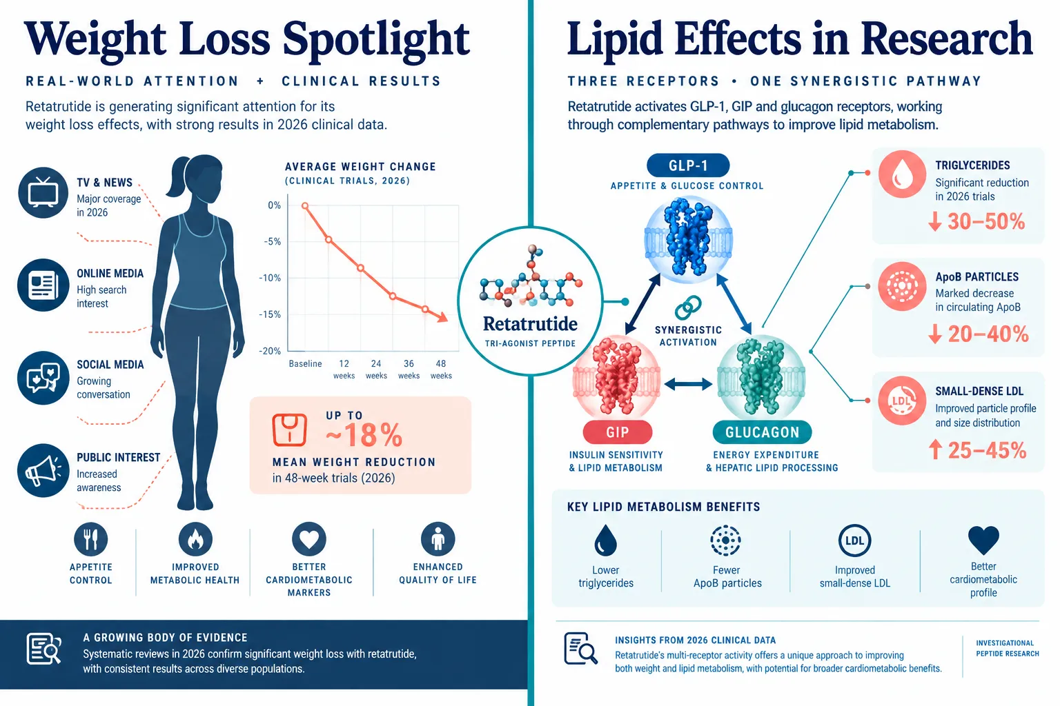
Layered Therapy: How These Agents Complement Each Other in Research Models

This layered model does not imply clinical equivalence. Atorvastatin has decades of randomized controlled trial data and guideline endorsement. Retatrutide has Phase 2 and emerging Phase 3 data. MOTS-c and 5-Amino-1MQ have preclinical data only. Researchers and clinicians must weigh evidence tiers carefully.

The intersection of atorvastatin, LDL cholesterol, and metabolic peptides: where MOTS-c, 5-Amino-1MQ, and GLP-3 Retatrutide fit in lipid research is ultimately a question about mechanism, evidence quality, and therapeutic stage, not a single unified treatment protocol.

For those exploring adjacent peptide research areas, resources on peptides and polypeptides in endocrine pharmacology provide useful mechanistic context on how different peptide classes interface with metabolic signaling systems.

Conclusion

Atorvastatin remains the cornerstone of LDL-C reduction in 2026, backed by robust dose-response data and explicit guideline targets that demand aggressive lipid lowering in high-risk patients. Retatrutide adds a meaningful investigational layer with documented lipid improvements, particularly for triglycerides, through a distinct incretin and glucagon receptor mechanism. MOTS-c and 5-Amino-1MQ represent the frontier of mitochondrial and NAD+ biology in lipid research, with scientifically interesting preclinical profiles but no clinical validation yet.

Actionable next steps for researchers:

  • Use current ACC/AHA LDL-C targets as the benchmark when designing lipid-focused research protocols.
  • Distinguish clearly between agents with clinical trial data (retatrutide) and those with only preclinical data (MOTS-c, 5-Amino-1MQ) when framing research questions.
  • Monitor Phase 3 TRIUMPH retatrutide data and ADA presentations for updated lipid endpoints.
  • Ensure all peptide compounds used in research settings are sourced from verified, high-purity suppliers with documented testing.
  • Consult regulatory guidance before any translational or human-adjacent research involving investigational peptides.

The science connecting statins, incretin-class peptides, and mitochondrial signaling molecules is evolving rapidly. Staying current with both guideline updates and preclinical mechanistic literature is essential for anyone working at this intersection.

https://www.puretestedpeptides.com/wp-content/uploads/2026/09/atorvastatin-ldl-cholesterol-and-metabolic-peptides-where-mots-c-5-amino-1mq-and.webp 1024 1536 https://www.puretestedpeptides.com/wp-content/uploads/2026/01/buy-peptides-online.jpg 2026-09-20 13:06:502026-09-20 13:06:50Atorvastatin, LDL Cholesterol, and Metabolic Peptides: Where MOTS-c, 5-Amino-1MQ, and GLP-3 Retatrutide Fit in Lipid Research
Metoprolol, Beta-Blockers, and Incretin Peptide Research: What Labs Track When Combining GLP-3 Retatrutide With Cardioactive Drugs

Metoprolol, Beta-Blockers, and Incretin Peptide Research: What Labs Track When Combining GLP-3 Retatrutide With Cardioactive Drugs

September 20, 2026/0 Comments/in Uncategorized/by

Roughly 40% of adults with obesity also carry a diagnosis of cardiovascular disease, making the overlap between metabolic therapies and heart-active medications not a niche concern but a near-universal research challenge. As retatrutide advances through Phase 3 TRIUMPH trials with striking cardiometabolic data, the question of how this triple-incretin agonist interacts with beta-blockers like metoprolol has become one of the most pressing topics in preclinical and translational lab design for 2026.

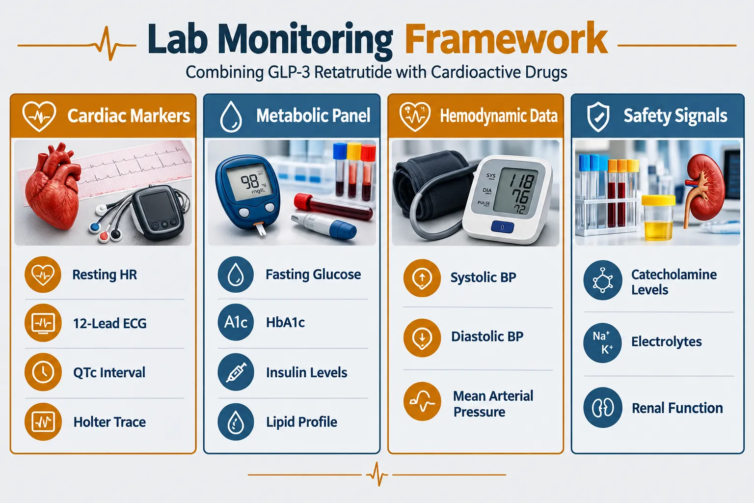
This guide addresses the core of that challenge: what measurements, markers, and safety checkpoints researchers track when studying metoprolol, beta-blockers, and incretin peptide research, specifically what labs track when combining GLP-3 retatrutide with cardioactive drugs in structured experimental frameworks.

Key Takeaways

  • Retatrutide activates GLP-1, GIP, and glucagon receptors simultaneously, creating a multi-axis effect on heart rate, blood pressure, and glucose that overlaps directly with beta-blocker pharmacology.
  • Metoprolol and other beta-blockers can mask hypoglycemia symptoms, a risk that becomes more complex when combined with incretin agents that lower glucose through multiple pathways.
  • Researchers tracking this combination should monitor cardiac rhythm, hemodynamics, glucose metabolism, and renal function as a minimum panel.
  • Emerging arrhythmia signals from triple-incretin agonist data make QTc interval and Holter monitoring critical additions to any lab protocol.
  • Purity and consistency of research-grade peptides are foundational to reproducible results in this interaction space.

Understanding the Pharmacological Overlap

Understanding the Pharmacological Overlap

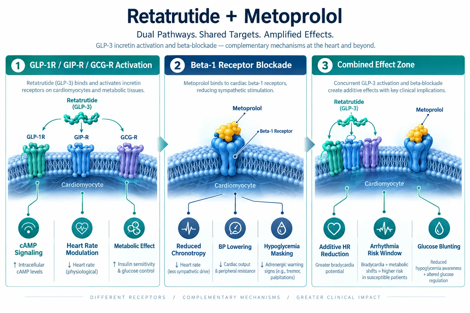
Retatrutide is a triple-receptor agonist targeting GLP-1R, GIPR, and glucagon receptors. Each of these pathways carries cardiovascular relevance. GLP-1 receptor activation raises cyclic AMP in cardiac tissue, which increases heart rate and cardiac output. Glucagon receptor activation has similar chronotropic effects. In isolation, these actions are manageable. When layered onto a beta-blocker framework, the dynamics shift.

Metoprolol is a selective beta-1 adrenergic antagonist. Its primary cardiac effects include reduced heart rate, lower blood pressure, and decreased myocardial oxygen demand. When a researcher or clinician introduces a GLP-class incretin peptide alongside metoprolol, the two agents are working on overlapping but mechanistically distinct pathways, one suppressing adrenergic drive, the other stimulating cAMP-mediated chronotropy.

The result is not simply additive or subtractive. The net heart rate effect depends on receptor density, dose, and individual variability. In vitro models studying GLP-3 peptide interactions need to account for this bidirectional tension explicitly in their experimental design.

A second layer of complexity involves glucose regulation. Beta-blockers are known to blunt the sympathoadrenal response to hypoglycemia, the very response that triggers awareness of low blood sugar. Incretin peptides lower glucose through multiple mechanisms. When combined, the hypoglycemia masking effect of metoprolol can obscure a glucose drop that the incretin agent has contributed to, creating a safety blind spot that labs must monitor actively.

Core Lab Panels for Metoprolol, Beta-Blockers, and Incretin Peptide Research

Core Lab Panels for Metoprolol, Beta-Blockers, and Incretin Peptide Research

Designing a rigorous monitoring protocol for this combination requires tracking across four domains: cardiac rhythm, hemodynamics, metabolic markers, and safety biomarkers.

Cardiac Rhythm and Electrophysiology

The most immediate concern when combining GLP-3 retatrutide with cardioactive drugs is arrhythmia risk. Emerging data from triple-incretin agonist research has flagged QTc prolongation as a signal requiring active surveillance. Labs should include:

  • 12-lead ECG at baseline and at defined intervals
  • QTc interval measurement using Fridericia or Bazett correction
  • Holter monitoring for continuous rhythm capture over 24-48 hours
  • Resting heart rate tracked across all time points

When peptide dosing protocols are adjusted, rhythm data should be recaptured to identify dose-dependent changes.

Hemodynamic Monitoring

Blood pressure response is a critical variable. Retatrutide has shown meaningful reductions in systolic blood pressure in TRIUMPH trial data. Metoprolol compounds this effect. Labs should record:

Measurement Frequency
Systolic blood pressure Every experimental session
Diastolic blood pressure Every experimental session
Mean arterial pressure Calculated at each time point
Orthostatic BP change Standing vs. supine, at peak drug effect

Orthostatic hypotension is a real concern in this combination and should not be treated as a secondary endpoint.

Metabolic and Glucose Panel

Given the hypoglycemia masking risk described above, glucose monitoring must be granular:

  • Fasting glucose at baseline and post-dose
  • Insulin and C-peptide levels
  • HbA1c for longer study windows
  • Glucagon levels, given retatrutide's direct glucagon receptor activity

Researchers sourcing GLP-3 retatrutide for lab use should confirm purity documentation, as impurities in the peptide can introduce confounding metabolic signals.

Safety Biomarkers

A complete panel also includes:

  • Serum electrolytes (potassium and magnesium, given arrhythmia risk)
  • Catecholamine levels (epinephrine, norepinephrine) to detect suppressed stress response
  • Renal function markers (creatinine, eGFR) given the renal clearance pathways of both drug classes
  • Liver enzymes for hepatic safety tracking

Designing the Monitoring Protocol: What Labs Track When Combining GLP-3 Retatrutide With Cardioactive Drugs

Designing the Monitoring Protocol: What Labs Track When Combining GLP-3 Retatrutide With Cardioactive Drugs

A well-structured protocol for studying metoprolol, beta-blockers, and incretin peptide research does not treat these measurements as isolated checkboxes. It builds them into a timeline that reflects the pharmacokinetics of both agents.

Retatrutide has a long half-life, approximately 6 days in clinical data, which means steady-state effects accumulate across a study window. Metoprolol, by contrast, has a much shorter half-life of 3-7 hours for standard formulations. This mismatch means:

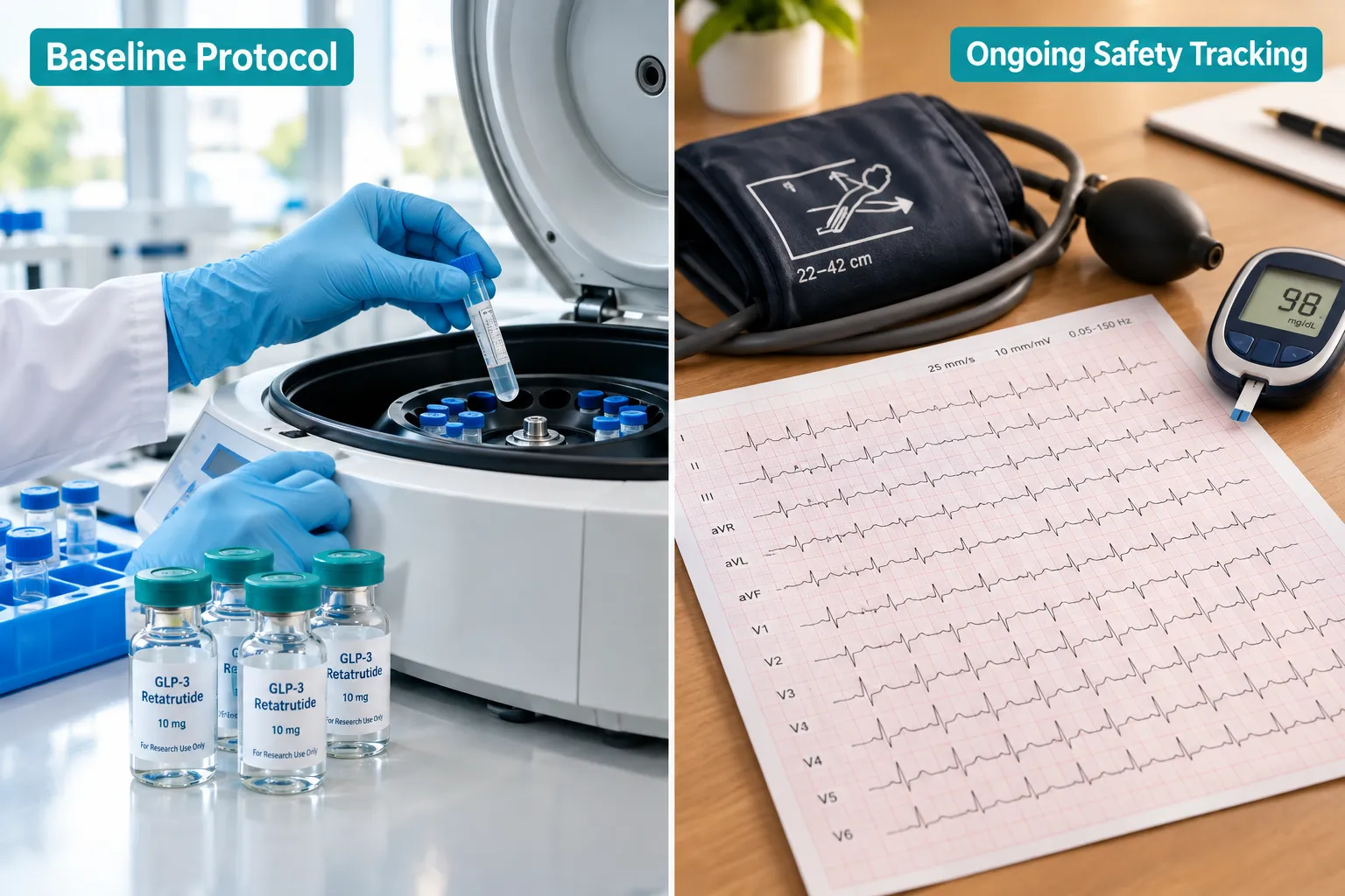
  1. Baseline measurements must be captured before either agent is introduced.
  2. Acute monitoring should align with metoprolol's peak effect window (1-2 hours post-dose).
  3. Trough monitoring should capture the period when metoprolol is at its lowest plasma level but retatrutide remains pharmacologically active.
  4. Washout tracking should extend beyond retatrutide's half-life to confirm return to baseline cardiac and metabolic parameters.

Researchers working with GLP-3 nasal spray formulations or alternative delivery formats should adjust absorption timing windows accordingly, as pharmacokinetic profiles differ from injectable preparations.

For labs using complementary cardioprotective peptides alongside this combination, resources on SS-31 peptide research considerations may provide useful parallel monitoring frameworks, given SS-31's mitochondrial cardioprotective mechanism.

Sensory and Neurological Safety Signals

Phase 3 retatrutide data has introduced a newer sensory safety signal, peripheral sensory changes in a subset of subjects. While the mechanism is not fully characterized, labs studying this combination should include basic neurological assessments and track any reported sensory changes systematically. This is particularly relevant when beta-blockers are co-administered, as they can independently affect peripheral circulation.

Conclusion

The intersection of GLP-3 retatrutide and beta-blocker pharmacology is not a simple additive equation. For researchers designing protocols around metoprolol, beta-blockers, and incretin peptide research, what labs track when combining GLP-3 retatrutide with cardioactive drugs must span cardiac rhythm, hemodynamics, glucose metabolism, electrolytes, and emerging sensory signals, all mapped against the distinct pharmacokinetic timelines of each agent.

Actionable next steps for research teams in 2026:

  • Establish a pre-study baseline panel covering all four monitoring domains before any agent is introduced.
  • Build time-matched monitoring windows that reflect both the short half-life of metoprolol and the extended half-life of retatrutide.
  • Treat hypoglycemia masking as a primary, not secondary, safety concern in any protocol combining beta-blockers with incretin peptides.
  • Confirm third-party purity testing on all research-grade peptides used; explore GLP-3 peptide options with documented quality assurance.
  • Document sensory safety signals as a standard endpoint given the emerging data from triple-incretin agonist trials.

Rigorous lab design in this space is not just a procedural formality, it is the foundation for generating data that can meaningfully inform the next generation of cardiometabolic research.

https://www.puretestedpeptides.com/wp-content/uploads/2026/09/metoprolol-beta-blockers-and-incretin-peptide-research-what-labs-track-when-comb.webp 1024 1536 https://www.puretestedpeptides.com/wp-content/uploads/2026/01/buy-peptides-online.jpg 2026-09-20 13:06:342026-09-20 13:06:34Metoprolol, Beta-Blockers, and Incretin Peptide Research: What Labs Track When Combining GLP-3 Retatrutide With Cardioactive Drugs
Prednisone and Peptide Immunomodulation: How Classic Steroids Compare With BPC-157, GHK-Cu, and GLP-Class Peptides in Inflammatory Research

Prednisone and Peptide Immunomodulation: How Classic Steroids Compare With BPC-157, GHK-Cu, and GLP-Class Peptides in Inflammatory Research

September 20, 2026/0 Comments/in Uncategorized/by

More than 30 million prescriptions for oral corticosteroids are written annually in the United States alone, yet a growing body of bench research is asking whether peptide-based compounds might one day complement or refine how science approaches inflammatory signaling. The field of prednisone and peptide immunomodulation: how classic steroids compare with BPC-157, GHK-Cu, and GLP-class peptides in inflammatory research sits at an important crossroads between established pharmacology and early-stage discovery science.

This article examines the mechanistic differences between glucocorticoid immunosuppression and peptide-mediated tissue modulation, strictly within the context of preclinical and translational research models.

Key Takeaways

  • Prednisone is a well-characterized glucocorticoid with a defined mechanism, extensive clinical data, and regulatory approval for dozens of inflammatory conditions.
  • BPC-157 shows consistent anti-inflammatory signals in animal models but has zero completed, peer-reviewed human efficacy trials as of 2026.
  • GHK-Cu demonstrates immunomodulatory activity in zebrafish and mouse models, with no human randomized trial data for inflammatory indications.
  • GLP-class peptides intersect metabolic and inflammatory pathways in emerging research, representing a distinct mechanistic category from classical steroids.
  • Evidence gaps between preclinical promise and clinical validation remain the defining challenge across all peptide candidates reviewed here.

How Prednisone Suppresses Inflammation: The Glucocorticoid Benchmark

How Prednisone Suppresses Inflammation: The Glucocorticoid Benchmark

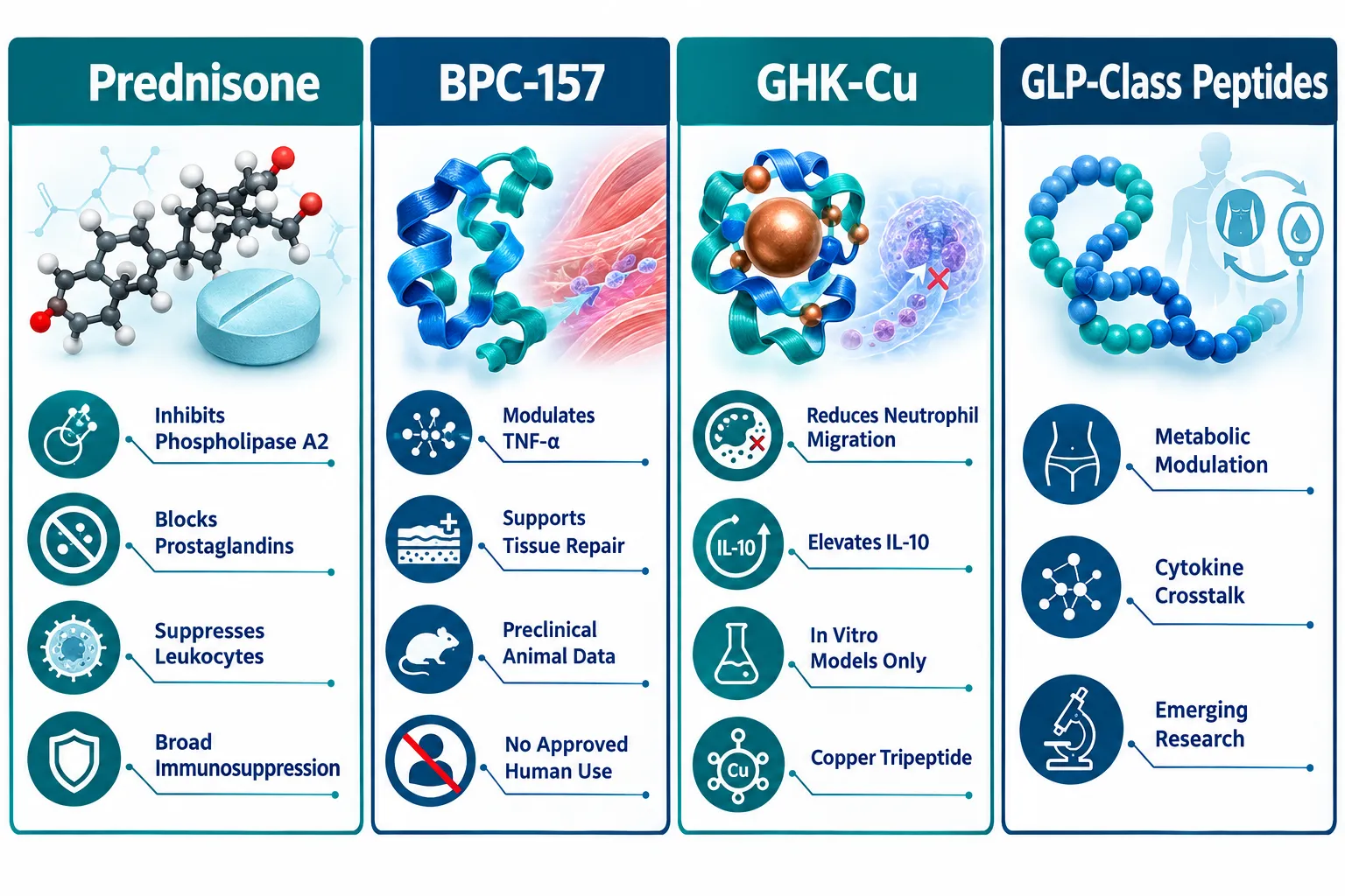
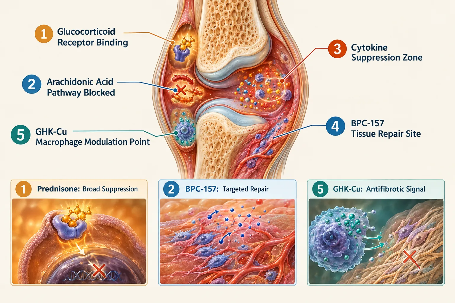
Prednisone functions as a prodrug, converting to prednisolone in the liver before exerting its effects. As a prototypical glucocorticoid, it suppresses inflammation through several well-documented pathways. It inhibits phospholipase A2, blocking the release of arachidonic acid and thereby reducing downstream production of both prostaglandins and leukotrienes. It also suppresses polymorphonuclear leukocyte migration and reverses increased capillary permeability, two hallmarks of acute tissue inflammation.

These mechanisms are broad by design. Prednisone does not selectively target one cytokine or one tissue type; it modulates the inflammatory response systemically. A 2025 translational analysis published in Theranostics added important nuance: RNA-sequencing data showed that prednisolone was most effective at reducing brain inflammatory signaling pathways in a CNS inflammation model, while prednisone itself had no significant effect on inflammation-related signaling in that specific tissue context. This finding underscores that even within the glucocorticoid class, tissue-specific immunomodulation varies considerably.

What makes prednisone the benchmark for comparison?

  • Decades of pharmacokinetic and pharmacodynamic data
  • Defined dose-response relationships across multiple inflammatory conditions
  • Established risk-benefit frameworks including adrenal suppression, bone density effects, and metabolic consequences
  • Regulatory approval and clinical guideline integration worldwide

This depth of evidence is precisely what makes it a useful comparator when evaluating emerging peptide candidates.

BPC-157 and GHK-Cu: Preclinical Signals and Evidence Gaps

BPC-157 and GHK-Cu: Preclinical Signals and Evidence Gaps

BPC-157: Tissue Repair Peptide With a Thin Human Evidence Base

BPC-157 is a synthetic 15-amino-acid peptide derived from a protein found in gastric juice. In preclinical models, it has demonstrated the ability to significantly decrease pro-inflammatory cytokines including TNF-α, IL-6, and IFN-γ, while also supporting tissue repair across musculoskeletal, gastrointestinal, and neurological animal models. More than 100 animal studies have explored its properties.

However, the human evidence picture is starkly different. A 2025 systematic review synthesized 36 studies, 35 of which were animal studies and one was an uncontrolled human chart review, and found zero completed controlled human efficacy trials. Available human data consist of three small pilot studies covering intra-articular knee pain, interstitial cystitis, and an intravenous safety and pharmacokinetics study in two healthy adults at doses up to 20 mg. These studies primarily demonstrate short-term tolerability rather than proven efficacy.

The most notable human signal comes from an unpublished Croatian Phase II study in 53 adults with mild-to-moderate ulcerative colitis. A 40 mL enema containing 80 mg BPC-157 once daily for 14 days reportedly yielded 38% clinical remission at week 4 versus 13% with placebo. Because the full study report, statistical analysis plan, and longer-term follow-up data were never published, regulators classify this evidence as "very low certainty." BPC-157 currently holds no FDA approval for any indication and no publicly listed IND authorization for inflammation. Researchers interested in peptide dosing protocols should note that optimal dosing, systemic adverse effects, and comparative effectiveness versus glucocorticoids remain undefined in humans.

One active development is a 120-patient Phase 2 double-blind, placebo-controlled study of injectable BPC-157 for grade II hamstring strain, but no efficacy outcomes have been reported publicly as of mid-2026.

GHK-Cu: Copper Tripeptide With Antifibrotic and Anti-Inflammatory Signals

GHK-Cu (glycine-histidine-lysine copper) offers a mechanistically distinct profile. A 2026 zebrafish larvae model of chemically induced inflammation showed that GHK-Cu reduced both neutrophil and macrophage migration, lowered pro-inflammatory cytokines, and increased the anti-inflammatory cytokine IL-10. A 2024 mouse model of silicosis found that GHK-Cu attenuated lung inflammation and fibrosis, identifying peroxiredoxin-6 (PRDX6) as a molecular binding target, with no significant systemic toxicity at tested doses.

Researchers exploring GHK-Cu peptides for sale for laboratory use should understand that current evidence remains restricted to animal and in vitro models. No human randomized trials or dose-finding studies exist for inflammatory indications.

GLP-Class Peptides and the Broader Landscape of Peptide Immunomodulation

GLP-Class Peptides and the Broader Landscape of Peptide Immunomodulation

GLP-class peptides represent a distinct mechanistic category within the broader field of prednisone and peptide immunomodulation: how classic steroids compare with BPC-157, GHK-Cu, and GLP-class peptides in inflammatory research. Originally characterized for their metabolic roles in glucose homeostasis and satiety signaling, GLP-class compounds are now being studied for their intersection with inflammatory cytokine networks. Research models suggest that GLP receptor activation can modulate macrophage polarization and reduce systemic inflammatory markers, creating a potential bridge between metabolic and immune regulation.

Those following developments in this space can explore research-grade options such as GLP-3 peptide for sale or the GLP-3R 10mg peptide GA10 for laboratory investigation. Additional GLP-class compounds are catalogued under GLP-3 peptides for sale for researchers tracking this category.

Research Perspective: The mechanistic gap between glucocorticoid broad-spectrum immunosuppression and peptide-targeted tissue modulation is not simply a matter of potency, it reflects fundamentally different biological strategies. Steroids suppress; some peptides appear to redirect or repair.

Comparing the Evidence Tiers

Compound Primary Mechanism Human Trial Data Regulatory Status
Prednisone Phospholipase A2 inhibition, broad immunosuppression Extensive RCT database FDA-approved, multiple indications
BPC-157 Cytokine modulation, tissue repair 3 pilot studies, no efficacy RCTs No approval, experimental
GHK-Cu Neutrophil/macrophage modulation, antifibrotic None (animal/in vitro only) No approval, research stage
GLP-class Metabolic-immune crosstalk Metabolic indications only Approved for metabolic use; inflammatory use experimental

For researchers sourcing compounds across multiple peptide categories, working with a best peptide supplier that provides verified purity documentation is essential for maintaining experimental integrity.

Conclusion

The comparison at the heart of prednisone and peptide immunomodulation: how classic steroids compare with BPC-157, GHK-Cu, and GLP-class peptides in inflammatory research reveals a fundamental asymmetry in the evidence base. Prednisone operates within a thoroughly characterized pharmacological framework built over decades of clinical research. BPC-157 and GHK-Cu show genuine mechanistic interest in preclinical models, but neither has cleared the threshold of controlled human efficacy data. GLP-class peptides occupy a third lane, metabolically validated but with inflammatory applications still in early research phases.

Actionable next steps for researchers and science communicators:

  • Treat BPC-157 and GHK-Cu findings as hypothesis-generating, not practice-defining, until controlled human trials are completed and published.
  • Monitor the ClinicalTrials.gov registry for emerging Phase 2 and Phase 3 peptide trials that may shift the evidence landscape in the next three to five years.
  • When sourcing research-grade peptides, prioritize suppliers with third-party purity verification and transparent certificates of analysis.
  • Recognize that mechanistic novelty in animal models does not translate automatically to clinical equivalence with established agents like prednisone.

The science is moving, but the evidence must lead.

https://www.puretestedpeptides.com/wp-content/uploads/2026/09/prednisone-and-peptide-immunomodulation-how-classic-steroids-compare-with-bpc-15.webp 1024 1536 https://www.puretestedpeptides.com/wp-content/uploads/2026/01/buy-peptides-online.jpg 2026-09-20 13:06:152026-09-20 13:06:15Prednisone and Peptide Immunomodulation: How Classic Steroids Compare With BPC-157, GHK-Cu, and GLP-Class Peptides in Inflammatory Research
Meloxicam vs BPC-157 and TB-500: How NSAIDs and Tissue-Repair Peptides Differ in In Vitro Injury Models

Meloxicam vs BPC-157 and TB-500: How NSAIDs and Tissue-Repair Peptides Differ in In Vitro Injury Models

September 20, 2026/0 Comments/in Uncategorized/by

Selective COX-2 inhibition reduces myofiber cross-sectional area by up to 33% during the first three weeks after muscle injury, a finding that challenges the routine use of NSAIDs like meloxicam in early musculoskeletal recovery. This single data point captures the central tension explored in Meloxicam vs BPC-157 and TB-500: How NSAIDs and Tissue-Repair Peptides Differ in In Vitro Injury Models: two fundamentally different pharmacological strategies, one aimed at quieting inflammation and one aimed at rebuilding tissue, produce strikingly different outcomes at the cellular level.

Key Takeaways

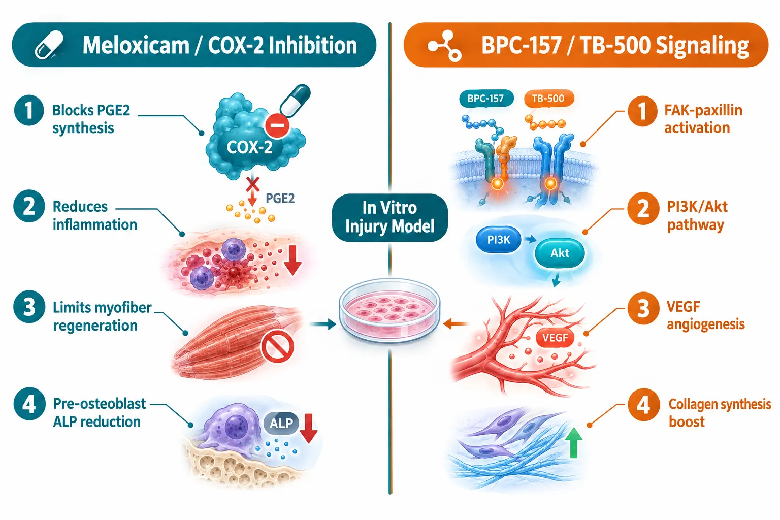
  • Meloxicam suppresses COX-2-dependent prostaglandin signaling, which reduces inflammation but may simultaneously impair early tissue regeneration in muscle, bone, and tendon models.
  • BPC-157 activates FAK-paxillin, PI3K/Akt, VEGF, and TGF-beta pathways in vitro, supporting fibroblast proliferation, collagen synthesis, and angiogenesis.
  • TB-500 promotes actin polymerization, progenitor cell recruitment, and collagen fiber organization through MAPK, NF-kB, and cytoskeletal repair pathways.
  • No robust human randomized controlled trials currently compare BPC-157 or TB-500 with meloxicam in musculoskeletal injury; all comparative evidence comes from cell and rodent studies.
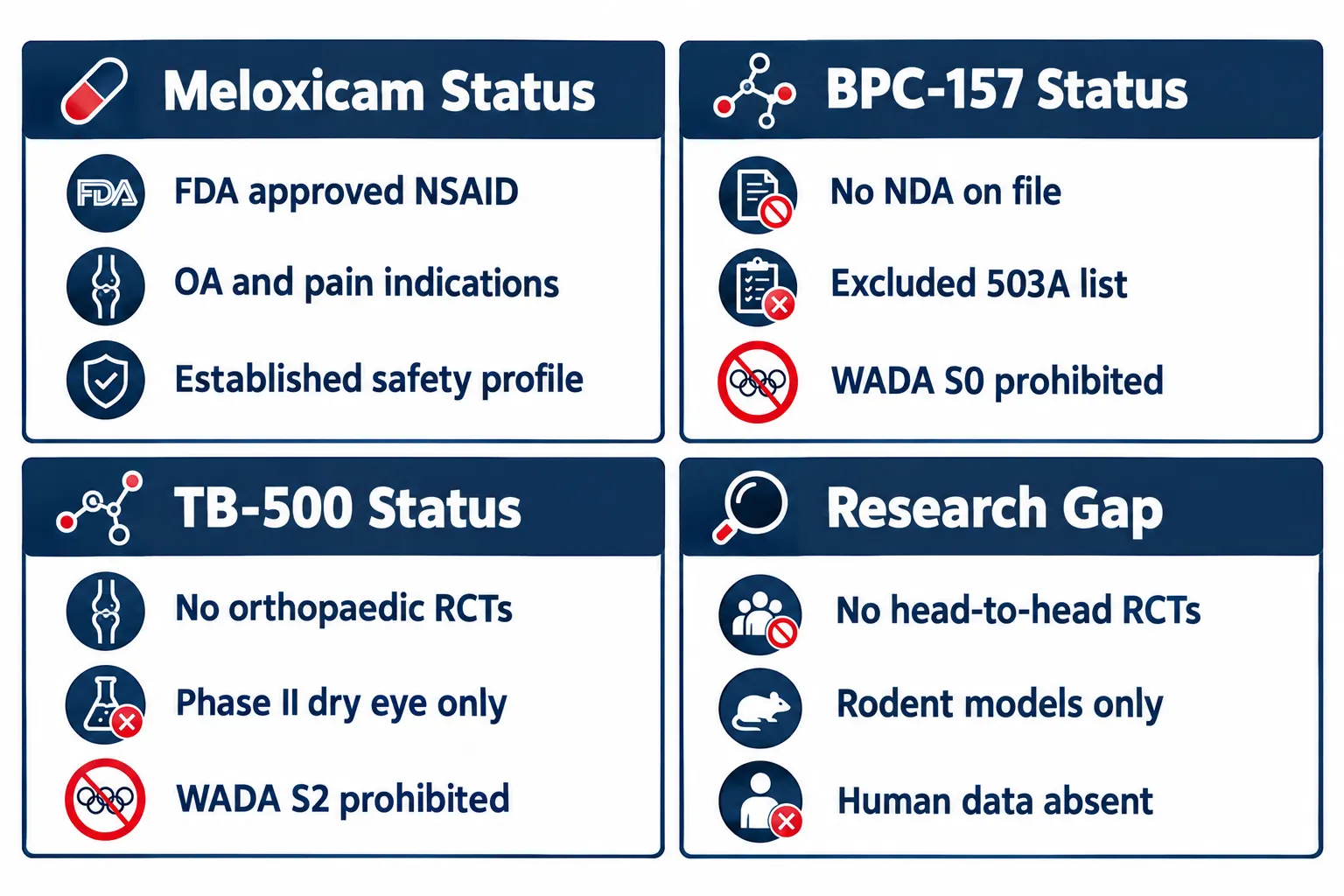
  • Both peptides remain unapproved, are excluded from FDA compounding lists, and are prohibited under WADA rules, making them strictly research-use compounds as of 2026.

How Meloxicam Works in Tissue-Injury Cell Models

Meloxicam is a COX-2-preferential NSAID with a COX-2:COX-1 IC50 ratio of approximately 0.09 in human whole-blood assays, meaning it inhibits COX-2 at concentrations far below those needed to affect COX-1. This selectivity drives its analgesic and anti-inflammatory profile, but it also places COX-2-dependent regenerative signaling directly in the line of fire.

How Meloxicam Works in Tissue-Injury Cell Models

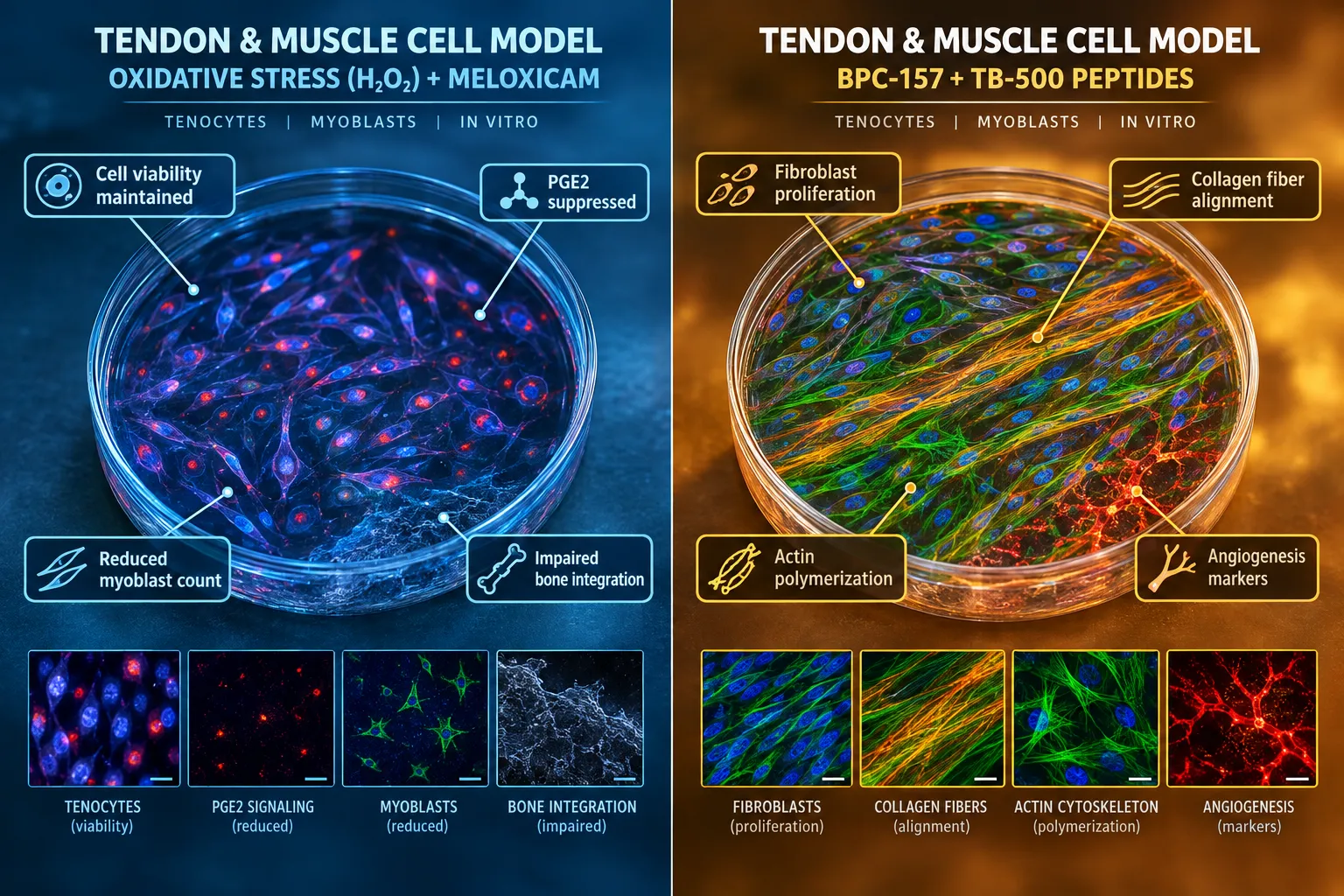
In human Achilles tendon-derived tenocytes exposed to oxidative stress via hydrogen peroxide, 24-hour meloxicam treatment can increase cell metabolic activity relative to untreated controls. At the same time, it consistently reduces prostaglandin E2 (PGE2) production through COX-2 inhibition. This creates a mechanistic paradox: the drug may support short-term cell viability under stress while simultaneously blunting the prostaglandin signals that coordinate early repair cascades.

The downstream consequences vary by tissue type:

  • Muscle: COX-2 inhibitors analogous to meloxicam reduce myoblast count and inflammatory cell recruitment, impairing fiber regeneration during the first three weeks post-injury.
  • Bone: Preclinical fracture models show impaired healing with COX-2 inhibitors, including meloxicam, and animal tendon-to-bone repair studies report compromised integration compared with acetaminophen controls.
  • Cartilage: In osteoarthritic canine chondrocytes, up to 30 days of meloxicam exposure does not increase matrix metalloproteinase production, suggesting the cartilage matrix is relatively preserved.
  • Osteogenesis: Meloxicam reduces alkaline phosphatase activity in pre-osteoblasts, yet some selective COX-2 inhibitors leave osteogenic differentiation of human adipose-derived stromal cells unaltered.

"The data on meloxicam in musculoskeletal cell models are mechanistically coherent but contextually mixed, tissue type and injury timing determine whether COX-2 inhibition helps or hinders."

These nuances are directly relevant to researchers designing wound models that use meloxicam as a reference compound.

BPC-157 and TB-500 Signaling in the Same In Vitro Systems

When examining Meloxicam vs BPC-157 and TB-500: How NSAIDs and Tissue-Repair Peptides Differ in In Vitro Injury Models, the peptide side of the comparison reveals a fundamentally different mechanistic profile. Rather than suppressing a single enzymatic pathway, both peptides activate multiple pro-repair cascades simultaneously.

BPC-157 and TB-500 Signaling in the Same In Vitro Systems

BPC-157 is a 15-amino-acid synthetic peptide with a broad signaling footprint in tendon and ligament models:

  • Enhances tendon fibroblast proliferation and collagen synthesis
  • Activates focal adhesion kinase (FAK)-paxillin signaling, rescuing tendocyte growth under oxidative stress
  • Upregulates growth hormone receptor expression and signals through PI3K/Akt, VEGF, NO/eNOS, and TGF-beta pathways
  • Accelerates tendon explant outgrowth and supports extracellular matrix remodeling

In transected rat Achilles tendon models, BPC-157 improves biomechanical endpoints including failure load and Young's modulus, outcomes that reflect genuine structural repair rather than pain suppression alone.

TB-500, a synthetic fragment of thymosin beta-4, works through complementary but distinct biology:

  • Promotes actin polymerization and progenitor cell recruitment
  • Enhances cell migration indices in ligament repair models
  • Produces more uniform collagen fiber bundles and larger fibril diameters at four weeks
  • Activates MAPK, NF-kB, FAK, and cytoskeletal repair pathways alongside pro-angiogenic activity

Researchers exploring these mechanisms can find relevant wound repair peptides and resources on wound repair and wound healing peptides for further context on how these compounds are studied in controlled settings.

A 2023 rat Achilles rupture-repair study found that both BPC-157 and TB-500 improved histopathological organization and extracellular matrix remodeling during early repair. However, direct head-to-head comparisons of both peptides within the same in vitro model remain sparse, and the preponderance of BPC-157 data comes from a single research group.

Regulatory Status, Research Gaps, and Lab Safety Considerations

Understanding Meloxicam vs BPC-157 and TB-500: How NSAIDs and Tissue-Repair Peptides Differ in In Vitro Injury Models requires an honest accounting of where the evidence stands and what the regulatory environment looks like as of 2026.

Regulatory Status, Research Gaps, and Lab Safety Considerations

Meloxicam is an established, FDA-approved NSAID with well-characterized pharmacokinetics, approved indications for osteoarthritis and other pain conditions, and decades of human safety data.

BPC-157 and TB-500 occupy a very different position:

Dimension Meloxicam BPC-157 TB-500
FDA approval Yes No No
Human RCT data (orthopaedic) Extensive None None
WADA status Permitted S0 prohibited S2 prohibited
503A compounding Permitted Excluded Excluded
Highest-quality evidence Phase III/IV trials Rodent + cell studies Phase II (dry eye only)

Both peptides have been excluded from the FDA's 503A bulk-drug compounding list, are not listed for 503B outsourcing compounding, and remain unapproved with no New Drug Applications on file. The FDA's Pharmacy Compounding Advisory Committee reviewed both compounds in July 2026 in the context of wound-healing and orthopaedic compounding, but neither has moved to an approved status.

For researchers sourcing compounds for in vitro work, using a best peptide supplier with verified purity documentation is essential. Researchers should also review resources on wholesale peptides for sale to ensure laboratory-grade material is used strictly within approved research protocols.

The only human data for BPC-157 in a musculoskeletal context is a single small case series of 17 patients receiving intra-articular injections for knee tendon and ligament symptoms, a study with significant methodological limitations. TB-500 has completed two Phase II trials, both in dry eye disease, with no orthopaedic RCTs published to date.

Conclusion

The comparison of Meloxicam vs BPC-157 and TB-500: How NSAIDs and Tissue-Repair Peptides Differ in In Vitro Injury Models reveals two pharmacological philosophies operating at opposite ends of the repair spectrum. Meloxicam suppresses a single enzymatic node, COX-2, with well-understood consequences for inflammation and, depending on tissue type and timing, potentially for regeneration itself. BPC-157 and TB-500 activate broad, overlapping pro-repair networks in cell and animal models, producing structural improvements in collagen organization, cell migration, and biomechanical endpoints that no NSAID is designed to achieve.

Actionable next steps for researchers:

  1. When using meloxicam as a reference NSAID in injury models, account for tissue type, muscle and bone models are more sensitive to COX-2 inhibition than cartilage models.
  2. Treat BPC-157 and TB-500 data critically: most evidence is rodent-derived, and human orthopaedic outcome data are essentially absent as of 2026.
  3. Source peptides only from suppliers with verified purity certificates and use them exclusively within approved institutional research frameworks.
  4. Monitor FDA Pharmacy Compounding Advisory Committee deliberations, as the regulatory status of both peptides remains under active review.
  5. Design future in vitro studies with direct head-to-head comparisons of NSAID and peptide conditions within the same cell model to generate more translatable mechanistic data.

The mechanistic gap between these compound classes is large and scientifically meaningful. Closing it will require well-designed, independently replicated human trials, work that has not yet been done.

https://www.puretestedpeptides.com/wp-content/uploads/2026/09/meloxicam-vs-bpc-157-and-tb-500-how-nsaids-and-tissue-repair-peptides-differ-in.webp 1024 1536 https://www.puretestedpeptides.com/wp-content/uploads/2026/01/buy-peptides-online.jpg 2026-09-20 13:06:102026-09-20 13:06:10Meloxicam vs BPC-157 and TB-500: How NSAIDs and Tissue-Repair Peptides Differ in In Vitro Injury Models
Lisinopril, ACE Pathways, and Growth Hormone Secretagogues: Designing Safe Peptide Research With Cardiovascular Readouts

Lisinopril, ACE Pathways, and Growth Hormone Secretagogues: Designing Safe Peptide Research With Cardiovascular Readouts

September 20, 2026/0 Comments/in Uncategorized/by

Roughly 47% of adults in the United States carry a diagnosis of hypertension, making ACE inhibitors like lisinopril among the most prescribed drugs in modern medicine. That same cardiovascular biology sits at the center of a growing challenge in peptide research: how do labs safely study growth hormone secretagogues (GHS), compounds that measurably alter blood pressure, heart rate, and fluid balance, without losing control of their cardiovascular readouts? Understanding Lisinopril, ACE Pathways, and Growth Hormone Secretagogues: Designing Safe Peptide Research With Cardiovascular Readouts is now a practical necessity for any team working with GHRH analogues, ghrelin mimetics, or related compounds in a controlled setting.

Key Takeaways

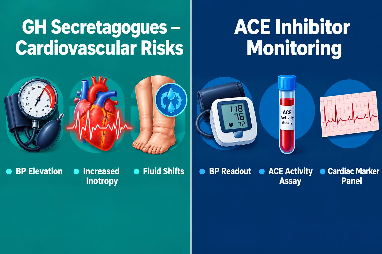
  • Lisinopril inhibits ACE, reducing angiotensin II production and lowering blood pressure, a mechanism directly relevant when GH secretagogues cause fluid shifts or BP changes.
  • Growth hormone secretagogues like CJC-1295, Ipamorelin, and MK-677 each carry distinct cardiovascular risk profiles that must be tracked with specific readouts.
  • MK-677 (ibutamoren) has been linked to blood pressure increases and heart failure events in long-term use, prompting FDA warning letters in late 2025.
  • A structured monitoring protocol, baseline BP, ACE activity assay, serial cardiac markers, is essential before and during GHS peptide research.
  • Mitochondrial-targeted peptides such as SS-31 offer a complementary research angle with a favorable cardiovascular safety profile.

ACE Biology and Lisinopril: The Cardiovascular Foundation

ACE Biology and Lisinopril: The Cardiovascular Foundation

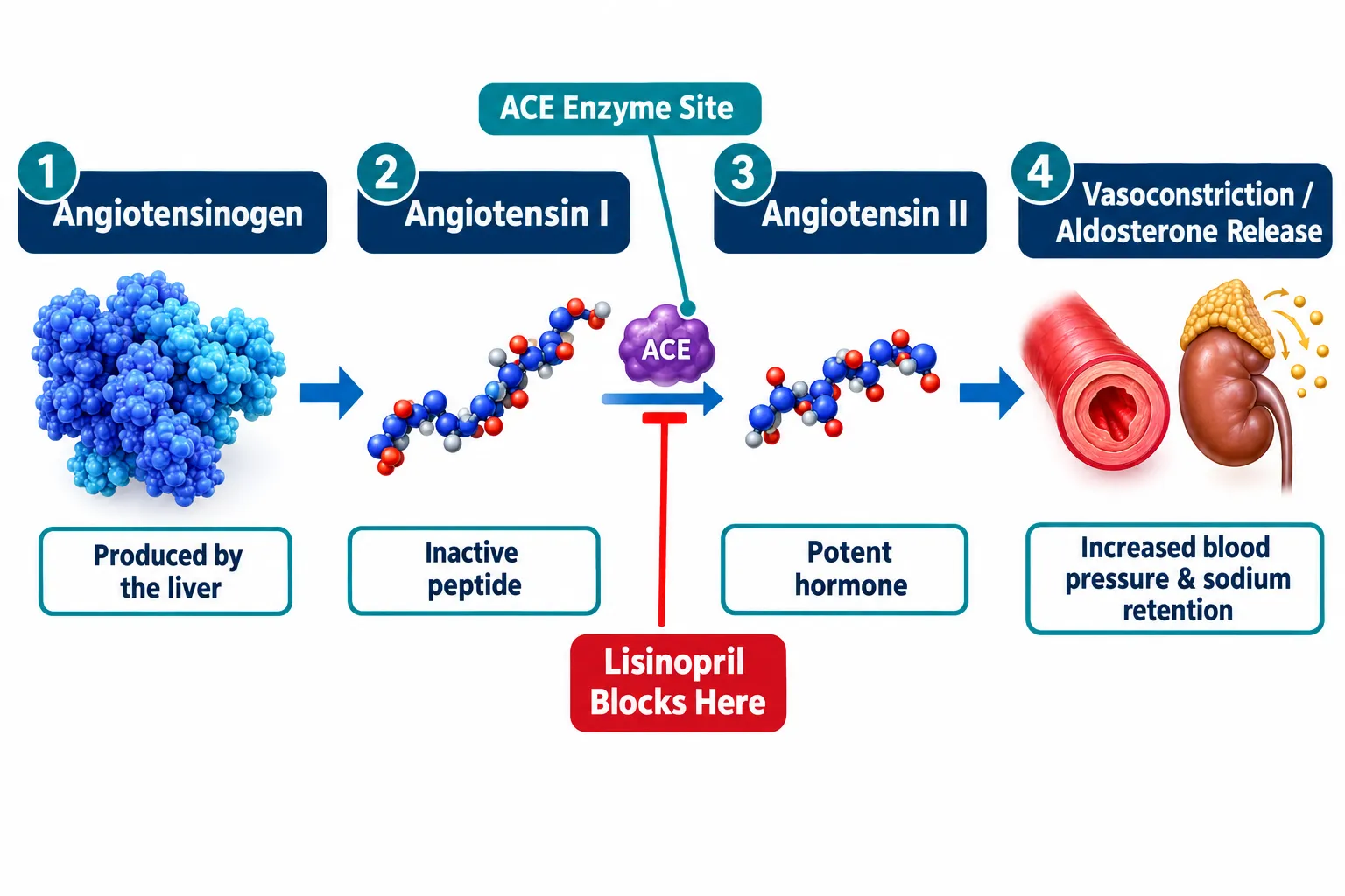
The renin-angiotensin-aldosterone system (RAAS) governs blood pressure, fluid volume, and electrolyte balance. At its core sits angiotensin-converting enzyme (ACE), which converts inactive angiotensin I into angiotensin II, a potent vasoconstrictor that also stimulates aldosterone release. Lisinopril binds competitively to the ACE active site, blocking this conversion and producing a cascade of cardiovascular benefits: reduced peripheral resistance, lower blood pressure, and decreased cardiac workload.

In research settings, this mechanism matters for two reasons. First, any peptide compound that independently shifts fluid balance or vascular tone will interact, directly or indirectly, with the same RAAS axis that lisinopril targets. Second, lisinopril itself accumulates bradykinin, which can cause angioedema; this safety signal must be tracked as a background variable in any multi-compound protocol.

Key ACE pathway markers to monitor in research designs:

Readout Why It Matters
Plasma ACE activity Confirms degree of enzymatic inhibition
Angiotensin II levels Tracks downstream vasoconstrictor load
Aldosterone Reflects fluid retention risk
Serum potassium Hyperkalemia risk with ACE inhibition
Bradykinin metabolites Angioedema safety signal

For researchers new to peptide biology, the Peptides 101 for Research-Use Only Buyers: Structure, Mechanisms, and resource provides a useful primer on how different compound classes interact with physiological systems.

Growth Hormone Secretagogues and Cardiovascular Risk: What the Data Show

Growth Hormone Secretagogues and Cardiovascular Risk: What the Data Show

Designing safe peptide research with cardiovascular readouts requires a clear-eyed look at how individual GHS compounds behave in the cardiovascular system. The class is not monolithic, risk profiles differ substantially across agents.

CJC-1295 and GHRH Analogues

CJC-1295 stimulates pulsatile GH release by acting on pituitary GHRH receptors. Elevated GH drives IGF-1 production, and sustained IGF-1 elevation is associated with cardiac hypertrophy and changes in left ventricular geometry in long-term animal studies. Short-term cardiovascular dynamics include modest increases in heart rate and positive inotropic effects. These changes are generally transient but require serial ECG and echocardiographic monitoring in rigorous research designs. A detailed breakdown of pharmacokinetics is available in the CJC-1295 With and Without DAC: A Detailed Mechanism and comparison guide.

Ipamorelin and Ghrelin Mimetics

Ipamorelin is a selective ghrelin receptor agonist with a cleaner selectivity profile than older GHS compounds. Ghrelin itself has demonstrated cardioprotective properties in preclinical models, improving cardiac output and reducing sympathetic tone. However, outcomes data in long-term research remain limited, and the vasodilatory effects of ghrelin-pathway activation can interact unpredictably with ACE inhibitor-induced BP lowering.

MK-677 (Ibutamoren): The Highest-Risk Profile

MK-677 presents the most significant cardiovascular concern in this class. Long-term use has been associated with measurable blood pressure increases, peripheral edema from fluid retention, and, in older research subjects, a higher incidence of heart failure events. FDA warning letters issued in December 2025 specifically flagged ibutamoren-containing products, and 2026 updates reinforced restrictions on its research use. Any protocol using MK-677 alongside an ACE inhibitor must include frequent BP monitoring and renal function panels.

"The interaction between MK-677-driven fluid retention and ACE inhibitor-mediated natriuresis creates a physiologically contested environment that demands close cardiovascular surveillance."

Designing the Protocol: Cardiovascular Readouts for Lisinopril, ACE Pathways, and Growth Hormone Secretagogues Research

Designing the Protocol: Cardiovascular Readouts for Lisinopril, ACE Pathways, and Growth Hormone Secretagogues Research

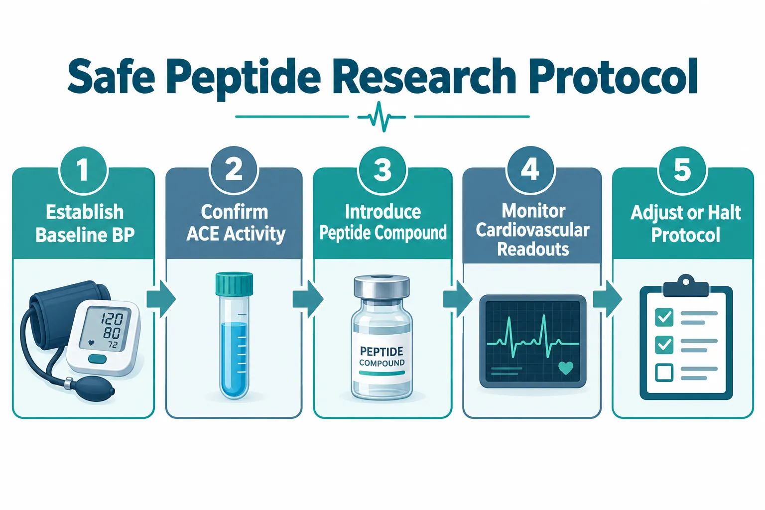
A rigorous approach to Lisinopril, ACE Pathways, and Growth Hormone Secretagogues: Designing Safe Peptide Research With Cardiovascular Readouts starts before the first compound is introduced. The following framework reflects current best practices for preclinical and in-vitro research designs.

Step 1, Establish Cardiovascular Baseline
Record resting blood pressure, heart rate, and weight. Draw baseline panels: ACE activity, angiotensin II, aldosterone, BNP or NT-proBNP, and a complete metabolic panel. This baseline anchors all subsequent comparisons.

Step 2, Confirm ACE Inhibition Status
If lisinopril is part of the background protocol, verify plasma ACE activity is suppressed to target range before introducing any GHS compound. Residual ACE activity confounds interpretation of peptide-driven BP changes.

Step 3, Introduce GHS Compound With Staged Dosing
Begin at the lowest effective research dose. Refer to established peptide dosing frameworks for compound-specific guidance. Avoid simultaneous introduction of multiple new agents.

Step 4, Serial Cardiovascular Monitoring

  • Blood pressure: every 24-48 hours during acute phase
  • Heart rate and rhythm: ECG at baseline, 72 hours, and weekly
  • IGF-1 levels: weekly, to track GH axis activation
  • BNP/NT-proBNP: bi-weekly as a cardiac stress marker
  • Renal function and electrolytes: weekly (critical with ACE inhibitor co-administration)

Step 5, Integrate Mitochondrial Peptide Data Where Relevant
SS-31, a mitochondria-targeted antioxidant peptide, has shown cardioprotective properties in ischemia-reperfusion models without the BP or fluid-retention liabilities of GHS compounds. Researchers exploring SS-31 mitochondrial research themes may find it a useful comparator arm in cardiovascular peptide studies. Additional mechanistic background is available in the SS-31 mechanism and research overview.

Compounds to avoid combining without extensive monitoring:

  • MK-677 + lisinopril (competing fluid dynamics, hyperkalemia risk)
  • High-dose CJC-1295 + any ACE inhibitor (IGF-1-driven cardiac remodeling risk)
  • Multiple GHS agents simultaneously (additive BP and heart rate effects)

Conclusion

The intersection of lisinopril, ACE pathways, and growth hormone secretagogues is not merely pharmacological, it is a research design challenge with direct safety implications. Labs that approach this space without structured cardiovascular readouts risk misinterpreting compound effects or, worse, missing early signals of cardiac stress.

Actionable next steps for research teams:

  1. Build a standardized cardiovascular baseline panel into every GHS protocol before dosing begins.
  2. Confirm ACE inhibition status when lisinopril is a background agent, and track ACE activity throughout the study.
  3. Treat MK-677 as the highest-risk GHS compound and apply the most rigorous monitoring schedule to any protocol that includes it.
  4. Consider SS-31 as a mechanistically distinct comparator with a favorable cardiovascular profile for studies focused on cardiac or mitochondrial endpoints.
  5. Review the latest FDA guidance from late 2025 and 2026 updates on ibutamoren before finalizing any research involving that compound.

Careful protocol architecture, anchored in ACE biology and supported by serial cardiovascular readouts, is what separates rigorous peptide research from guesswork.

https://www.puretestedpeptides.com/wp-content/uploads/2026/09/lisinopril-ace-pathways-and-growth-hormone-secretagogues-designing-safe-peptide.webp 1024 1536 https://www.puretestedpeptides.com/wp-content/uploads/2026/01/buy-peptides-online.jpg 2026-09-20 13:06:092026-09-20 13:06:09Lisinopril, ACE Pathways, and Growth Hormone Secretagogues: Designing Safe Peptide Research With Cardiovascular Readouts
SLU-PP-332 and 5-Amino-1MQ Combination Protocols: Estrogen-Related Receptor Agonism with NNMT Inhibition in Myocyte Models

SLU-PP-332 and 5-Amino-1MQ Combination Protocols: Estrogen-Related Receptor Agonism with NNMT Inhibition in Myocyte Models

September 19, 2026/0 Comments/in Uncategorized/by

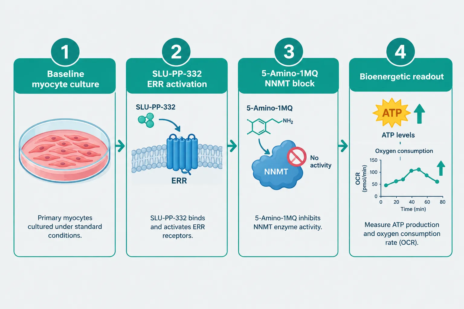
Skeletal muscle consumes roughly 30% of resting metabolic energy and accounts for up to 80% of insulin-stimulated glucose uptake, yet the molecular switches that govern its bioenergetic capacity remain underexplored as drug targets. Two compounds, SLU-PP-332 and 5-Amino-1MQ, are drawing serious attention in preclinical research for their distinct but potentially complementary effects on muscle cell metabolism. The SLU-PP-332 and 5-Amino-1MQ combination protocols concept, pairing estrogen-related receptor (ERR) agonism with nicotinamide N-methyltransferase (NNMT) inhibition in myocyte models, represents one of the more mechanistically interesting hypotheses in current metabolic research, even though formal combination data remain absent from the published literature.

Key Takeaways

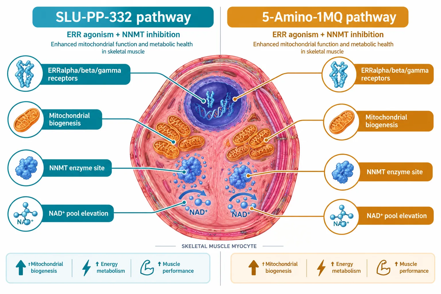
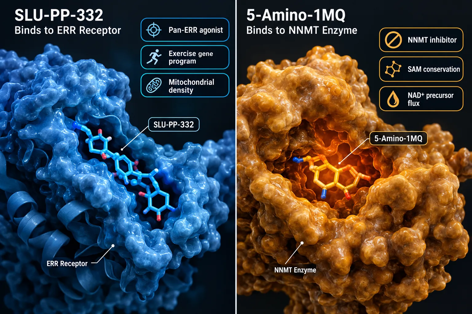
  • SLU-PP-332 is a pan-ERR agonist that activates ERRalpha, ERRbeta, and ERRgamma, mimicking transcriptional changes associated with endurance exercise in skeletal muscle.
  • 5-Amino-1MQ selectively inhibits NNMT, an enzyme that consumes S-adenosylmethionine (SAM) and suppresses NAD+ precursor availability, thereby supporting intracellular NAD+ pools.
  • No peer-reviewed publication has yet described a formal SLU-PP-332 plus 5-Amino-1MQ combination protocol in any cell or animal model.
  • The theoretical synergy between these two agents in myocytes is mechanistically plausible but remains speculative and requires rigorous experimental validation.
  • Both compounds are research-stage tools; neither is approved for human therapeutic use as of 2026.

Understanding the Two Mechanisms: ERR Agonism and NNMT Inhibition

Understanding the Two Mechanisms: ERR Agonism and NNMT Inhibition

Estrogen-related receptors (ERRalpha, ERRbeta, ERRgamma) are orphan nuclear receptors that regulate mitochondrial biogenesis, fatty acid oxidation, and oxidative phosphorylation gene programs. Unlike classical estrogen receptors, ERRs do not require estrogen as a ligand. SLU-PP-332 was developed as a potent, selective pan-ERR agonist, meaning it activates all three ERR subtypes simultaneously.

In myocyte-based experiments, SLU-PP-332 has been shown to upregulate genes associated with endurance exercise adaptation, including those governing mitochondrial density and aerobic capacity. Researchers have described it informally as an "exercise mimetic" because the transcriptional signature it produces in muscle cells closely resembles what is seen after sustained aerobic training.

NNMT is a cytosolic enzyme that transfers a methyl group from SAM to nicotinamide, producing 1-methylnicotinamide and homocysteine as byproducts. When NNMT is highly active, it depletes SAM and diverts nicotinamide away from NAD+ synthesis pathways. 5-Amino-1MQ is a small-molecule NNMT inhibitor with a reported IC50 in the low-micromolar range and favorable cell membrane permeability, allowing it to reach intracellular enzyme targets effectively.

By blocking NNMT, 5-Amino-1MQ conserves SAM and redirects nicotinamide back toward NAD+ biosynthesis. Elevated intracellular NAD+ then supports sirtuin deacetylase activity and PARP-dependent DNA repair, both of which intersect with mitochondrial health. For those interested in the broader landscape of metabolic peptide research, resources on SS-31 mitochondrial research themes offer useful context on how mitochondrial-targeted compounds are being studied.

Why the SLU-PP-332 and 5-Amino-1MQ Combination Protocols Are Mechanistically Compelling

Why the SLU-PP-332 and 5-Amino-1MQ Combination Protocols Are Mechanistically Compelling

The theoretical case for combining these two agents in myocyte models rests on a straightforward logic: ERR activation increases the demand for mitochondrial substrates, while NNMT inhibition expands the NAD+ pool that fuels those same mitochondrial processes.

Here is how the proposed synergy would work:

Mechanism SLU-PP-332 5-Amino-1MQ
Primary target ERRalpha/beta/gamma NNMT enzyme
Downstream effect Mitochondrial biogenesis genes NAD+ precursor conservation
Metabolic outcome Increased oxidative capacity Enhanced sirtuin/PARP activity
Myocyte relevance Exercise-like transcription Bioenergetic substrate support

When ERR agonism drives a cell to build more mitochondria and run more oxidative phosphorylation, that cell simultaneously increases its NAD+ consumption. If NAD+ availability is the limiting factor, the ERR-driven program may be constrained. NNMT inhibition, by preserving NAD+ precursor flux, could theoretically remove that bottleneck.

"The intersection of nuclear receptor activation and NAD+ metabolism in skeletal muscle represents one of the most underexplored areas of metabolic pharmacology."

This kind of substrate-demand pairing is not without precedent. Research into compounds like those explored in SS31 and MOTS-C combination studies illustrates how researchers are increasingly looking at multi-target approaches to mitochondrial support. Similarly, work on VEGF upregulation in muscle tissue shows that angiogenic and bioenergetic pathways often need to be addressed together for meaningful outcomes.

Critical caveat: As of 2026, no published study has tested this combination directly. The synergy described here is a hypothesis derived from the independent pharmacology of each compound, not from experimental combination data.

Protocol Design Considerations for Myocyte Model Research

Protocol Design Considerations for Myocyte Model Research

For researchers designing preclinical experiments around SLU-PP-332 and 5-Amino-1MQ combination protocols, several parameters warrant careful attention.

Concentration ranges:

  • SLU-PP-332 has demonstrated ERR agonist activity at concentrations in the low-nanomolar to low-micromolar range in cell-based assays.
  • 5-Amino-1MQ NNMT inhibition has been characterized at IC50 values in the low-micromolar range, with studies in adipocyte and preadipocyte models providing the most detailed dose-response data. Myocyte-specific data remain sparse.

Timing and sequencing:
Because ERR-driven transcriptional changes require hours to days to manifest at the protein level, while NNMT inhibition alters metabolite pools more acutely, staggered dosing may be more informative than simultaneous administration. A reasonable starting design would pre-treat cells with 5-Amino-1MQ to establish an elevated NAD+ baseline before introducing SLU-PP-332.

Readout selection:

  • Mitochondrial oxygen consumption rate (OCR) via Seahorse assay
  • NAD+/NADH ratio by enzymatic or mass spectrometry methods
  • ERR target gene expression (ESRRA, ESRRB, ESRRG, TFAM, COX subunits)
  • NNMT activity assay and 1-methylnicotinamide quantification

Researchers working on metabolic compound combinations may also find value in reviewing 5-Amino-1MQ product information and related aging support research tools to understand available research-grade material specifications.

Key limitations to acknowledge:

  • Most 5-Amino-1MQ mechanistic data come from adipocyte or preadipocyte models, not myocytes.
  • SLU-PP-332 myocyte data, while more directly relevant, are still limited to a small number of research groups.
  • Combination index analyses (Chou-Talalay method) have not been applied to this pairing.

For those comparing metabolic research approaches, the synergy of LL-37 and SS-31 peptide research provides a useful methodological parallel for how combination peptide or small-molecule studies are structured.

Conclusion

The SLU-PP-332 and 5-Amino-1MQ combination protocols concept, centered on estrogen-related receptor agonism with NNMT inhibition in myocyte models, is one of the more mechanistically coherent ideas in preclinical metabolic research as of 2026. ERR activation drives mitochondrial biogenesis and oxidative gene programs; NNMT inhibition preserves the NAD+ substrate pool those programs depend on. The logic is sound, the individual pharmacology of each compound is reasonably well characterized, and myocytes represent an ideal model system given their high mitochondrial density and metabolic flexibility.

Actionable next steps for researchers:

  1. Conduct dose-response characterization of each compound independently in a validated myocyte model (C2C12 or primary human skeletal muscle cells) before combining.
  2. Use Seahorse OCR assays and NAD+/NADH ratio measurements as primary bioenergetic endpoints.
  3. Apply a staggered dosing design, establish NAD+ elevation with 5-Amino-1MQ before initiating ERR agonism with SLU-PP-332.
  4. Perform combination index analysis to determine whether observed effects are additive, synergistic, or antagonistic.
  5. Publish negative as well as positive results; the field needs accurate data, not confirmation bias.

The gap between mechanistic plausibility and experimental proof is precisely where rigorous preclinical science belongs. This combination deserves that rigor.

https://www.puretestedpeptides.com/wp-content/uploads/2026/09/slu-pp-332-and-5-amino-1mq-combination-protocols-estrogen-related-receptor-agoni.webp 1024 1536 https://www.puretestedpeptides.com/wp-content/uploads/2026/01/buy-peptides-online.jpg 2026-09-19 13:04:282026-09-19 13:04:28SLU-PP-332 and 5-Amino-1MQ Combination Protocols: Estrogen-Related Receptor Agonism with NNMT Inhibition in Myocyte Models
Klow Blend Peptide Nasal Spray: Formulation Questions, Carrier Systems, and Research Use Cases

Klow Blend Peptide Nasal Spray: Formulation Questions, Carrier Systems, and Research Use Cases

September 19, 2026/0 Comments/in Uncategorized/by

Fewer than 1% of peptide compounds studied in preclinical settings ever reach a stable, non-injectable delivery format, which is exactly what makes the Klow Blend Peptide Nasal Spray: Formulation Questions, Carrier Systems, and Research Use Cases such a relevant subject for researchers in 2026. This article addresses the specific formulation mechanics, carrier system design, and documented research contexts that distinguish this blend from generic intranasal peptide preparations.

Key Takeaways

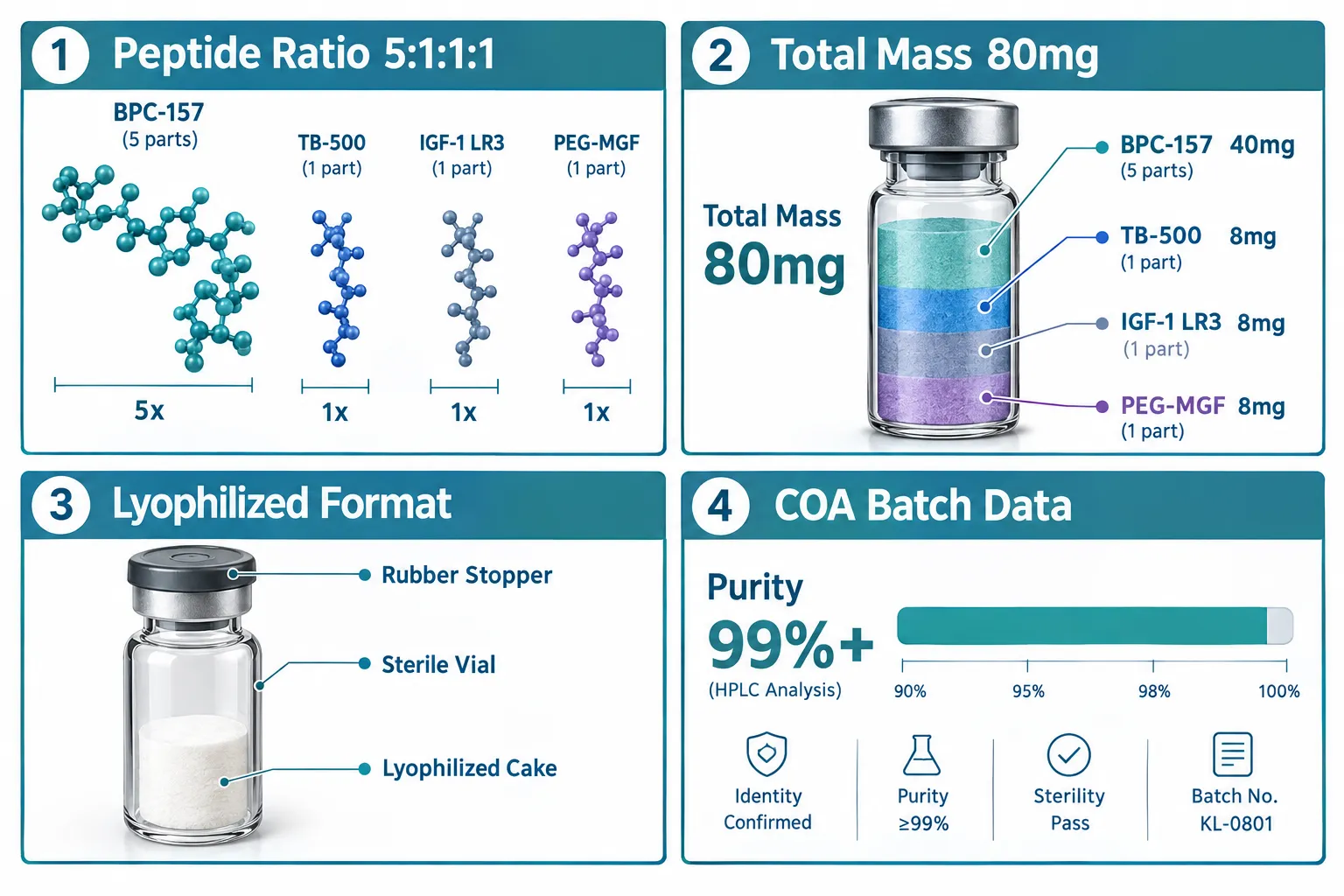
  • The Klow Blend contains four peptides at a standardized 80 mg total mass with a 5:1:1:1 ratio between components.
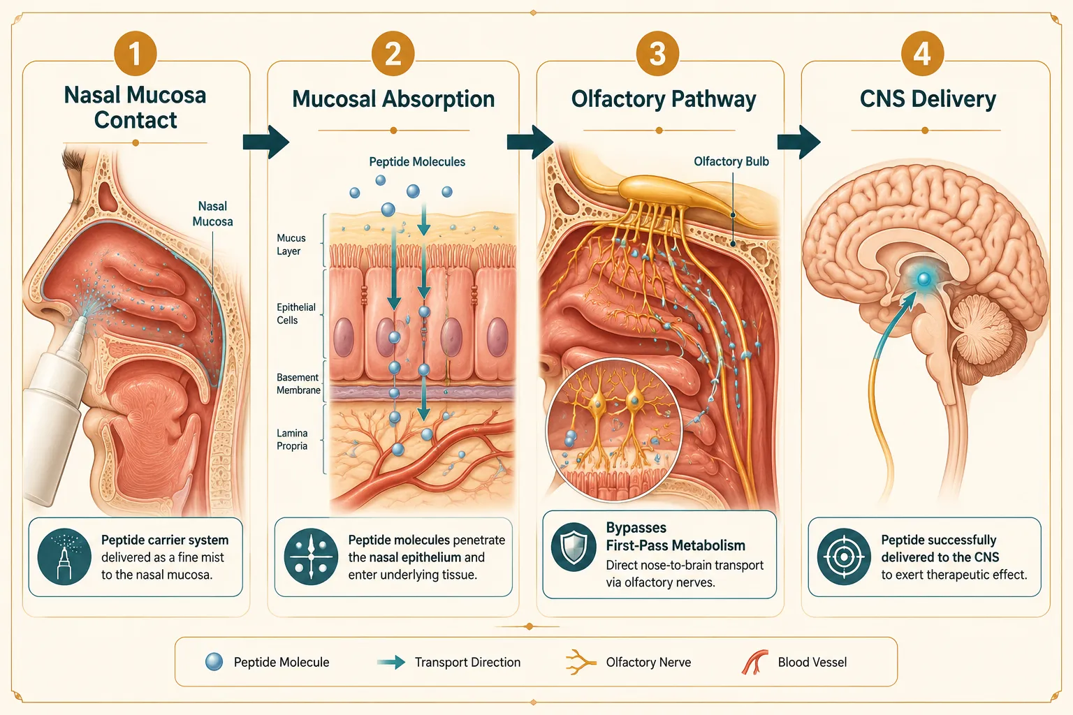
  • Nasal delivery bypasses hepatic first-pass metabolism, making it a practical non-injectable route for peptide research.
  • Lyophilized vial formats preserve peptide integrity; reconstitution protocol matters significantly for potency.
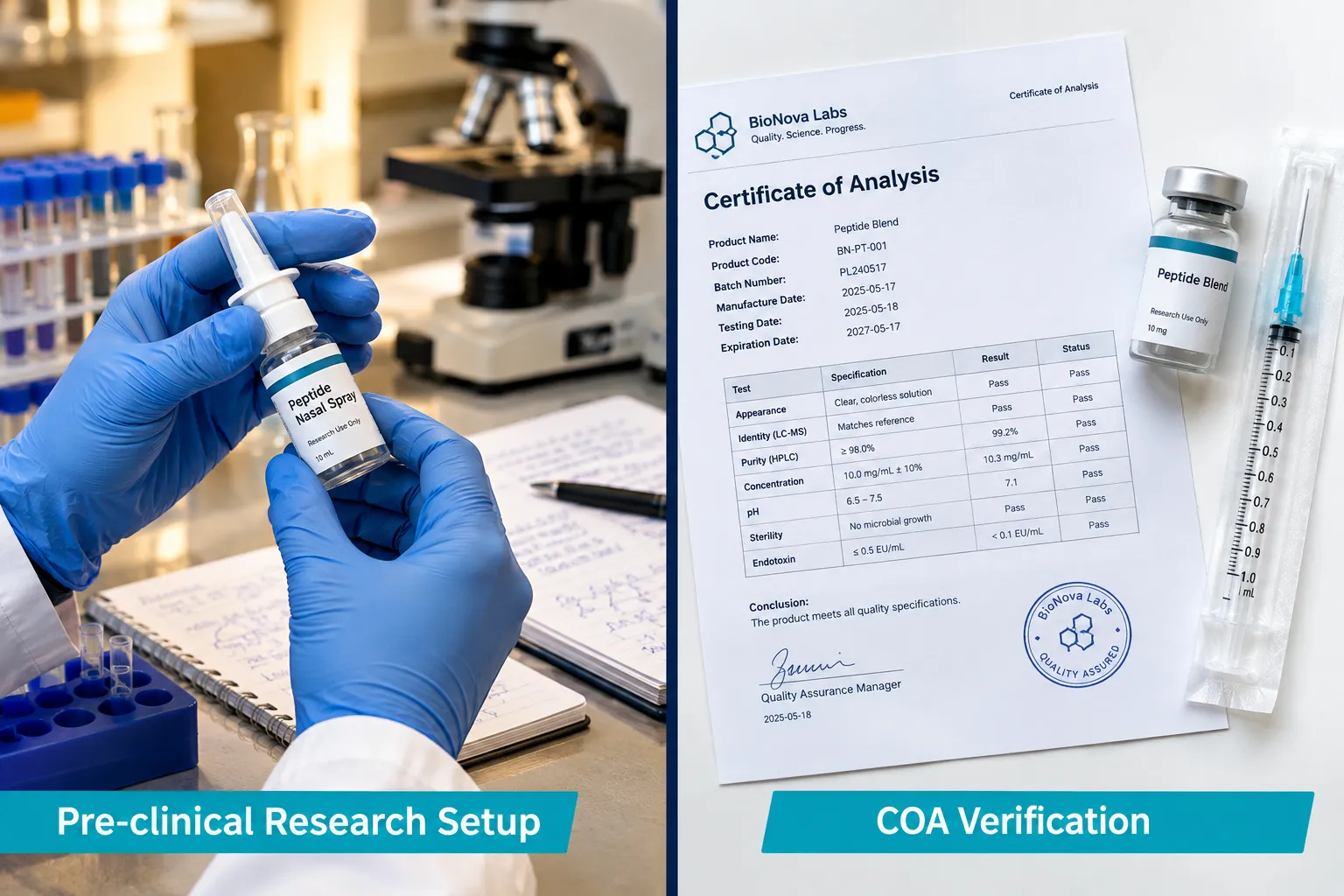
  • Batch-level Certificate of Analysis (COA) data from September 2026 confirms purity benchmarks above 99%.
  • This product is intended strictly for research use only (RUO) and is not approved for human therapeutic use.

Formulation Composition: Ratios, Mass, and Standardization

Formulation Composition: Ratios, Mass, and Standardization

The Klow Blend is formulated at a total peptide mass of 80 mg, distributed across four peptide components at a 5:1:1:1 ratio. This ratio is not arbitrary. In multi-peptide research blends, fixed ratios allow investigators to maintain consistent molar relationships between compounds across experimental replicates, which is critical for reproducibility.

The dominant peptide at the 5-part proportion anchors the blend's primary mechanistic focus, while the three supporting components at equal 1-part ratios are included to address complementary biological pathways. Researchers working with similar multi-component blends, such as the Tesamorelin CJC1295 Ipamorelin 12mg Blend, will recognize this approach as a strategy to reduce the number of separate administrations required in a study protocol.

Why standardized ratios matter in research:

  • Eliminates batch-to-batch variability in component proportions
  • Simplifies dosing calculations for weight-based or surface-area-based protocols
  • Enables cleaner statistical comparison across study arms

The 80 mg total mass places this blend in a mid-range concentration category, suitable for preclinical in vitro and in vivo research contexts where sub-milligram precision is required.

"A fixed-ratio blend removes one variable from the experimental design, the researcher can focus on dose response rather than component interaction uncertainty."

Carrier Systems and Nasal Delivery Mechanics

Carrier Systems and Nasal Delivery Mechanics

The choice of intranasal delivery for the Klow Blend is a formulation decision with significant mechanistic implications. Nasal administration routes peptides through the olfactory and trigeminal nerve pathways, offering a potential direct-to-CNS delivery channel that injectable routes do not replicate in the same way.

Key advantages of nasal carrier systems for peptide research:

Feature Nasal Spray Subcutaneous Injection
First-pass metabolism Bypassed Bypassed
CNS access potential High (olfactory route) Lower
Administration complexity Low Moderate
Mucosal absorption variability Moderate Low
Suitability for repeat dosing High Moderate

The carrier system used in nasal spray formulations typically includes a buffered aqueous base, a preservative system, and in some formulations a permeation enhancer to improve mucosal uptake. For peptides with larger molecular weights, permeation enhancers are particularly important because the nasal epithelium presents a size-selective barrier.

Researchers interested in comparing intranasal peptide delivery formats can review Semax Nasal Spray as a well-documented reference point for CNS-targeted intranasal peptide research. Similarly, the GLP-3 RT 20mg Peptide Nasal Spray provides a metabolic-focus comparison for nasal delivery design.

Lyophilized format considerations:

The Klow Blend ships as a lyophilized (freeze-dried) powder in a sterile vial. This format offers superior shelf stability compared to pre-dissolved aqueous solutions. Reconstitution with bacteriostatic water is the standard protocol. Volume of reconstitution directly affects final concentration per spray actuation, so researchers must calculate this carefully before beginning a dosing protocol.

Research Use Cases and Mechanistic Rationale

Research Use Cases and Mechanistic Rationale

Understanding the Klow Blend Peptide Nasal Spray: Formulation Questions, Carrier Systems, and Research Use Cases in a practical research context requires looking at both the individual peptide mechanisms and the rationale for combining them.

Multi-peptide blends in nasal spray format are typically studied in contexts where:

  1. Synergistic pathway activation is the hypothesis, where two or more peptides act on related but distinct receptor targets
  2. Reduced total administration volume is a study design requirement
  3. CNS bioavailability is the primary endpoint of interest

For researchers studying mitochondrial function, the SS-31 peptide represents a structurally distinct but mechanistically relevant comparison point. Detailed mechanistic background is available in the SS-31 mechanism and research overview.

Researchers exploring growth hormone secretagogue blends may also find the Tesamorelin AOD9604 CJC1295 Ipamorelin 12mg Blend a useful structural comparison for multi-component blend design.

Purity and COA data (September 2026 batch):

  • Purity confirmed at above 99% via HPLC analysis
  • Endotoxin levels within accepted research-grade thresholds
  • Mass spectrometry identity confirmation for all four components
  • Batch-specific COA available at point of purchase

This level of analytical documentation is essential for any research application where data integrity must be defended in a publication or institutional review context.

Regulatory and RUO status:

The Klow Blend Peptide Nasal Spray is classified as a Research Use Only (RUO) compound. It has not received regulatory approval from the FDA or equivalent agencies for therapeutic, diagnostic, or clinical use in humans or animals. Researchers must comply with all applicable institutional, local, and national regulations governing the use of RUO compounds.

Conclusion

The Klow Blend Peptide Nasal Spray occupies a specific and well-defined space in the research peptide landscape: a fixed-ratio, multi-component nasal formulation designed for investigators who need reproducibility, non-injectable delivery, and documented purity data.

Actionable next steps for researchers:

  • Review the September 2026 batch COA before initiating any study protocol
  • Calculate reconstitution volume precisely to confirm per-actuation dose
  • Compare carrier system design against reference nasal spray peptides to contextualize absorption assumptions
  • Confirm RUO compliance with your institutional review board before procurement
  • Cross-reference mechanistic rationale with published literature on each individual peptide component

For researchers building out a broader peptide research program, exploring wound healing peptides or wholesale peptides options can help consolidate sourcing while maintaining the purity standards that credible research demands.

https://www.puretestedpeptides.com/wp-content/uploads/2026/09/klow-blend-peptide-nasal-spray-formulation-questions-carrier-systems-and-researc.webp 1024 1536 https://www.puretestedpeptides.com/wp-content/uploads/2026/01/buy-peptides-online.jpg 2026-09-19 13:04:222026-09-19 13:04:22Klow Blend Peptide Nasal Spray: Formulation Questions, Carrier Systems, and Research Use Cases
Tesofensine Research Guide: Appetite, Dopamine, and Noradrenergic Pathways Explained

Tesofensine Research Guide: Appetite, Dopamine, and Noradrenergic Pathways Explained

September 19, 2026/0 Comments/in Uncategorized/by

Fewer than a handful of investigational compounds have demonstrated double-digit percentage body weight reductions in Phase II trials, tesofensine is one of them. That single fact has kept researchers, clinicians, and metabolic scientists engaged with this molecule for well over a decade, even as its regulatory path has remained unresolved. This Tesofensine Research Guide: Appetite, Dopamine, and Noradrenergic Pathways Explained breaks down the core neuroscience, the clinical data landscape, and the open research questions that make tesofensine a compelling subject in 2026.

Key Takeaways

  • Tesofensine simultaneously inhibits dopamine, norepinephrine, and serotonin reuptake, producing appetite suppression and increased energy expenditure through central pathways.
  • Phase II obesity trials showed dose-dependent weight loss significantly greater than most contemporary comparators, but no Phase III obesity program is currently active.
  • The noradrenergic pathway is central to appetite regulation; blocking the norepinephrine transporter (NET) reduces hunger signaling in the hypothalamus.
  • The most active clinical application as of 2026 is Tesomet, a tesofensine-metformin combination targeting hypothalamic obesity.
  • Cardiovascular monitoring, particularly heart rate elevation, remains a key safety consideration, with metoprolol co-administration used in some protocols.

How Tesofensine Works: Triple Monoamine Reuptake Inhibition

Tesofensine belongs to a class called triple monoamine reuptake inhibitors (TRIs). It blocks three transporter proteins simultaneously: the dopamine transporter (DAT), the norepinephrine transporter (NET), and the serotonin transporter (SERT). By preventing these transporters from recycling their respective neurotransmitters back into the presynaptic neuron, tesofensine raises synaptic concentrations of all three monoamines at once.

How Tesofensine Works: Triple Monoamine Reuptake Inhibition

This is mechanistically distinct from older agents. Sibutramine, for example, also inhibited monoamine reuptake but carried significant cardiovascular liabilities that led to its market withdrawal. Tesofensine's receptor selectivity profile differs in ways that researchers believe may translate to a more favorable risk-benefit ratio, though this remains under active investigation.

Why does blocking all three transporters matter for appetite?

Each monoamine contributes differently to energy balance:

Neurotransmitter Primary Role in Appetite Regulation
Dopamine Reward signaling, motivation to eat, food-seeking behavior
Norepinephrine Hypothalamic satiety signaling, metabolic rate modulation
Serotonin Meal termination, carbohydrate preference reduction

Raising all three simultaneously creates a synergistic effect on satiety that neither dopaminergic nor noradrenergic agents achieve alone. For researchers comparing small-molecule approaches to newer incretin-based therapies, understanding this distinction is essential. A detailed look at how tesofensine compares to GLP-class polypeptide drugs provides important mechanistic context.

The Noradrenergic Pathway in Detail

The norepinephrine transporter is arguably the most critical target for tesofensine's appetite-suppressing effects. NET blockade increases norepinephrine availability in the hypothalamus, specifically in regions like the arcuate nucleus and the paraventricular nucleus. These areas integrate peripheral hunger signals, including leptin and ghrelin, and translate them into behavioral responses.

Elevated norepinephrine in these circuits suppresses neuropeptide Y (NPY) release, a potent hunger-driving peptide. It also activates pro-opiomelanocortin (POMC) neurons, which promote satiety. The result is a dual action: hunger signals are dampened while fullness signals are amplified.

Research note: Noradrenergic agents have historically been used as appetite suppressants, but their cardiovascular side effects, elevated blood pressure and heart rate, have limited their clinical utility. Tesofensine's profile in this regard is discussed further in the safety section below.

Clinical Research Landscape: Phase II Data and the Tesomet Program

The most cited evidence base for tesofensine comes from a 24-week Phase II randomized controlled trial in adults with obesity. Participants receiving 0.5 mg daily lost approximately 10% of body weight on average, compared to roughly 2% in the placebo group. The 1.0 mg dose produced even greater weight loss, though with a corresponding increase in cardiovascular signals including elevated heart rate.

Clinical Research Landscape: Phase II Data and the Tesomet Program

These results positioned tesofensine among the most efficacious small-molecule weight-loss compounds studied at that time. However, the cardiovascular signals observed, particularly increased heart rate, slowed progression to Phase III for the obesity indication.

Key findings from Phase II obesity research:

  • Dose-dependent weight loss: higher doses produced greater reductions
  • Significant reductions in appetite scores measured by visual analog scales (VAS)
  • Improvements in waist circumference, triglycerides, and fasting glucose
  • Heart rate increases of approximately 7-8 beats per minute at therapeutic doses
  • No significant blood pressure elevation at the 0.5 mg dose in most subjects

Tesomet: The Active Clinical Context in 2026

The most clinically active tesofensine program as of 2026 is Tesomet, a fixed-dose combination of tesofensine and metformin. The primary target population is hypothalamic obesity, a condition caused by damage to hypothalamic appetite-regulating circuits, often following craniopharyngioma surgery. This population has extremely limited treatment options, which makes Tesomet's mechanism particularly relevant.

Metformin is included partly for its metabolic benefits and partly because it may offset some of the cardiovascular effects associated with tesofensine. Researchers studying this combination are also examining whether the metformin component improves insulin sensitivity in ways that complement tesofensine's central appetite effects.

For context on how combination peptide and small-molecule approaches work in research models, the IPA Sermorelin stack research overview offers a useful parallel for understanding synergistic compound strategies.

Neurocircuitry, Safety, and Research Design Considerations

This section of the Tesofensine Research Guide: Appetite, Dopamine, and Noradrenergic Pathways Explained addresses the broader neurological context and practical considerations for researchers.

Neurocircuitry, Safety, and Research Design Considerations

Tesofensine's effects extend beyond the three monoamine transporters. Downstream, elevated dopamine and norepinephrine modulate GABAergic interneurons within the lateral hypothalamus. These interneurons gate the activity of orexin neurons, which regulate arousal and feeding motivation. This means tesofensine's appetite effects involve a multi-layer circuit, not simply a direct receptor interaction.
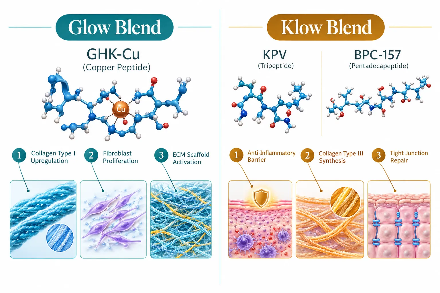
Broader neurocircuitry targets identified in preclinical research:

  • Arcuate nucleus POMC/AgRP neuron balance
  • Lateral hypothalamic orexin circuit modulation via GABAergic interneurons
  • Mesolimbic dopamine pathway (reward and food motivation)
  • Prefrontal cortical inputs to hypothalamic satiety circuits

Cardiovascular Safety and Metoprolol Co-Administration

The heart rate elevation associated with NET blockade is the primary cardiovascular concern in tesofensine research. In several clinical protocols, the beta-blocker metoprolol has been co-administered to attenuate this effect without significantly reducing the weight-loss efficacy. This approach is notable because it suggests the cardiovascular signal is pharmacologically manageable rather than intrinsic to the compound's mechanism of efficacy.

Researchers designing tesofensine studies in 2026 should include:

  1. Baseline cardiovascular assessment including resting heart rate and blood pressure
  2. Electrocardiographic monitoring at dose escalation points
  3. Pre-specified stopping rules for sustained tachycardia
  4. Consideration of beta-blocker co-administration protocols

Positioning Versus GLP-1 and Incretin Therapies

The rise of GLP-1 receptor agonists and multi-incretin drugs has reshaped the obesity treatment landscape significantly. Tesofensine operates through a fundamentally different mechanism, central monoaminergic rather than peripheral hormonal, which means the two approaches are not necessarily competitive. Some researchers have proposed that central monoaminergic agents could complement incretin therapies by addressing reward-driven eating behaviors that GLP-1 agents do not directly target.

For researchers exploring the incretin side of this comparison, resources on GLP-3 and retatrutide research models provide useful mechanistic contrast. Understanding peptide fundamentals is also well-covered in the Peptides 101 for research-use-only buyers guide.

Regulatory status as of mid-2026: Tesofensine has not received approval from any major regulatory agency for any indication. There is no active Phase III program for the obesity indication. The Tesomet combination remains in clinical development for hypothalamic obesity and related conditions.
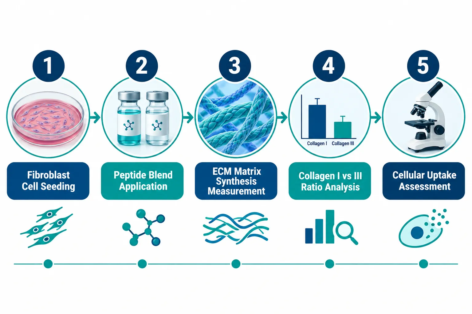
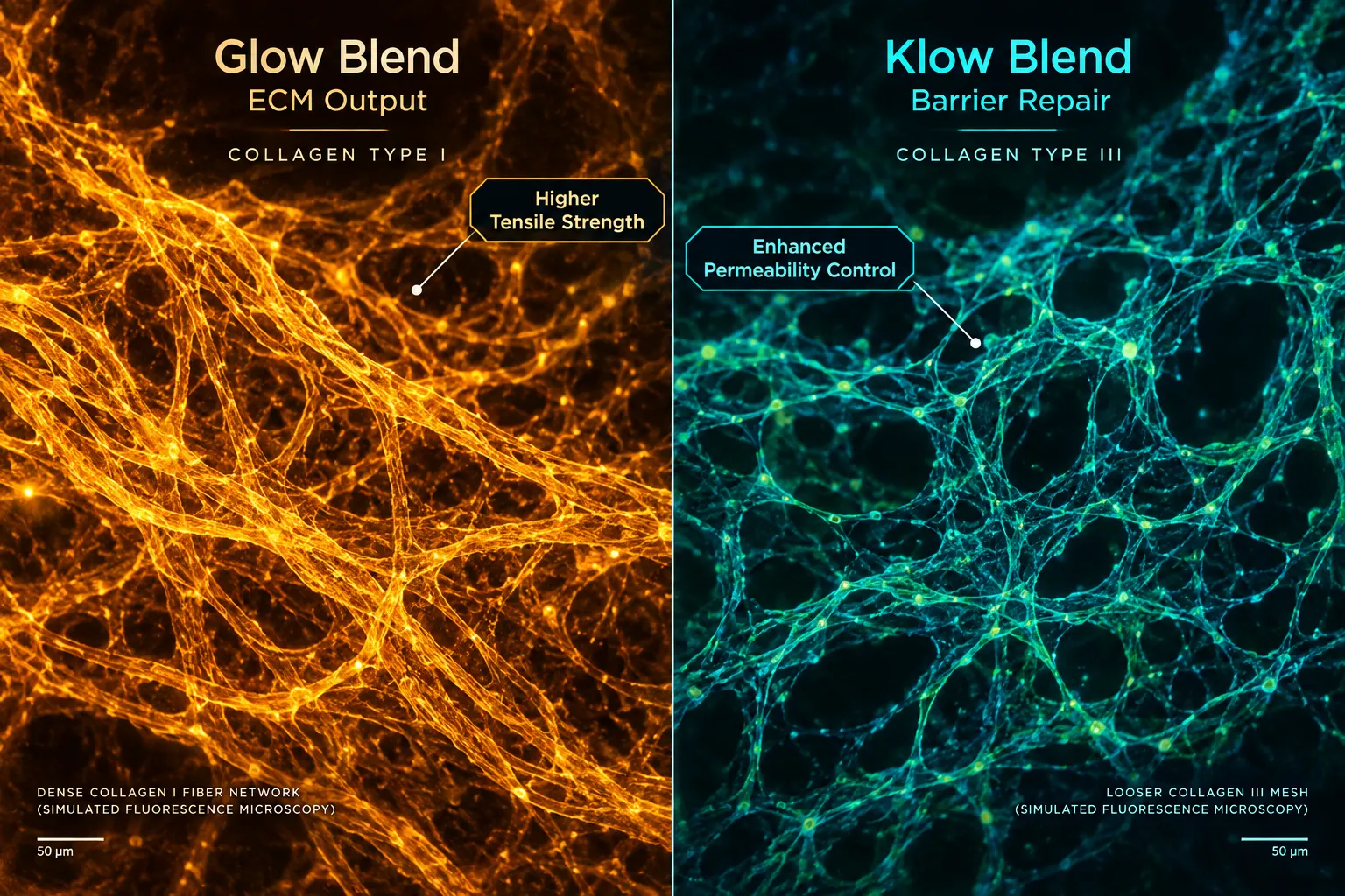
Conclusion

The Tesofensine Research Guide: Appetite, Dopamine, and Noradrenergic Pathways Explained reveals a compound with a well-characterized mechanism, compelling Phase II efficacy data, and a nuanced safety profile that has shaped, but not ended, its clinical development. For researchers in 2026, the actionable next steps are clear:

  • Understand the mechanism fully before designing experiments: triple reuptake inhibition creates multi-pathway effects that require multi-endpoint study designs.
  • Monitor cardiovascular parameters rigorously and consider metoprolol co-administration protocols when heart rate elevation is a concern.
  • Contextualize tesofensine within the broader metabolic research landscape, particularly relative to GLP-1 and incretin-based approaches, to identify where its central monoaminergic mechanism adds unique value.
  • Follow the Tesomet program as the most active clinical signal for tesofensine's near-term research relevance.
  • Distinguish speculation from evidence: Phase III data for obesity does not yet exist, and extrapolating from Phase II results requires careful qualification.

Tesofensine remains one of the most pharmacologically interesting small molecules in appetite and metabolic research. Its dopaminergic, noradrenergic, and serotonergic mechanisms offer a distinct window into central appetite regulation that neither peptide-based nor incretin-based approaches fully replicate.

https://www.puretestedpeptides.com/wp-content/uploads/2026/09/tesofensine-research-guide-appetite-dopamine-and-noradrenergic-pathways-explaine.webp 1024 1536 https://www.puretestedpeptides.com/wp-content/uploads/2026/01/buy-peptides-online.jpg 2026-09-19 13:04:082026-09-19 13:04:08Tesofensine Research Guide: Appetite, Dopamine, and Noradrenergic Pathways Explained
GLP2-T and GLP2 Tirz Peptide Differentiation: Receptor Binding Affinity and Intestinal Epithelial Growth Pathways

GLP2-T and GLP2 Tirz Peptide Differentiation: Receptor Binding Affinity and Intestinal Epithelial Growth Pathways

September 19, 2026/0 Comments/in Uncategorized/by

Two peptides share overlapping catalog names yet operate through entirely different receptor systems, a distinction that matters enormously in gastrointestinal research. Understanding GLP2-T and GLP2 Tirz Peptide Differentiation: Receptor Binding Affinity and Intestinal Epithelial Growth Pathways is not a matter of minor pharmacological nuance; it is the difference between a dedicated intestinotrophic hormone and a synthetic dual-receptor chimeric agonist with metabolic applications.

Key Takeaways

  • Native GLP-2 is a 33-amino-acid endogenous hormone that binds exclusively to the GLP-2 receptor and drives intestinal mucosal growth.
  • GLP2-T (also catalogued as GLP-2 Tirz) is a research alias for tirzepatide, a 39-amino-acid synthetic peptide that targets both GIP and GLP-1 receptors, not the GLP-2 receptor.
  • The naming overlap is a catalog convention, not a chemical relationship; these two peptides are structurally and mechanistically distinct.
  • GLP2-T carries Aib substitutions and a C20 lipid chain that extend its half-life through albumin binding, unlike native GLP-2.
  • Researchers must verify molecular identity before selecting either compound for intestinal epithelial or metabolic pathway studies.

Structural Identity: Native GLP-2 vs. GLP2-T

Structural Identity: Native GLP-2 vs. GLP2-T

At the molecular level, the distinction begins with chain length and origin. Native GLP-2 is a 33-amino-acid peptide hormone derived from the proglucagon gene. It is secreted primarily by intestinal L-cells in response to nutrient intake and binds with high specificity to the GLP-2 receptor (GLP-2R), a G-protein-coupled receptor expressed predominantly on subepithelial myofibroblasts and enteric neurons.

GLP2-T, by contrast, is a 39-amino-acid synthetic peptide. Its catalog synonyms include "GLP-2 T," "GLP-2 (T)," "P1206," and "LY3298176", the last being the research identifier for tirzepatide. The molecular formula is C225H348N48O68, with CAS number 2023788-19-2. This peptide is engineered as a dual GIP/GLP-1 receptor agonist, meaning it does not engage the GLP-2 receptor at all.

A September 2026 catalog update clarified this confusion directly, stating that "GLP-2 (T)" is a catalog alias and "not a chemical identity." The two compounds are separate entities that happen to share a naming convention used by some research suppliers.

Structural features of GLP2-T / Tirzepatide:

  • 39-amino-acid GIP-based linear backbone
  • C-terminal amidation
  • Aminoisobutyric acid (Aib) substitutions at positions 2 and 13
  • Attached C20 fatty acid lipid chain for albumin binding
  • Extended plasma half-life compared to native GLP-2

For researchers exploring related incretin-class compounds, the Tesofensine Mechanism Explained: Noradrenergic Appetite Modulation vs Incretin-Based GLP-3 and GLP-1 Pathways resource provides useful context on how GLP-1 pathway agents compare across different compound classes.

GLP2-T and GLP2 Tirz Peptide Differentiation: Receptor Binding Affinity Compared

GLP2-T and GLP2 Tirz Peptide Differentiation: Receptor Binding Affinity Compared

The receptor binding profiles of these two peptides represent the core of GLP2-T and GLP2 Tirz Peptide Differentiation: Receptor Binding Affinity and Intestinal Epithelial Growth Pathways research.

Native GLP-2 Receptor Binding

Native GLP-2 demonstrates exclusive, high-affinity binding to GLP-2R. Because GLP-2R is not expressed directly on enterocytes, its intestinotrophic effects are mediated indirectly through paracrine signaling. Downstream mediators include:

  • Insulin-like growth factor 1 (IGF-1), primary growth mediator
  • Keratinocyte growth factor (KGF)
  • Epidermal growth factor receptor (EGFR) ligands

This indirect mechanism is why GLP-2 increases villus height and mucosal thickness without directly stimulating epithelial cells at the receptor level.

GLP2-T / Tirzepatide Receptor Binding

GLP2-T delivers balanced GIP receptor (GIPR) agonism combined with biased GLP-1 receptor (GLP-1R) activation. The term "biased" here is important: GLP2-T activates GLP-1R through a qualitatively different signaling profile compared to native GLP-1 analogs such as semaglutide. This biased agonism is thought to contribute to its distinct metabolic effects in preclinical models.

Key Distinction: GLP2-T binds GIPR and GLP-1R with high affinity. Native GLP-2 binds GLP-2R exclusively. Neither compound substitutes for the other in receptor-specific research protocols.
Feature Native GLP-2 GLP2-T (Tirzepatide)
Chain length 33 amino acids 39 amino acids
Primary receptor GLP-2R GIPR + GLP-1R
Lipid chain None C20 fatty acid
Half-life extension Short (native) Extended (albumin binding)
Intestinal growth effect Direct intestinotrophic Indirect / metabolic
Research classification Endogenous hormone analog Synthetic dual agonist

Researchers working with GLP-class peptides may also find the Peptides 101 for Research-Use Only Buyers: Structure, Mechanisms, and Where GLP-3, MOTS-C, and 5-Amino-1MQ Fit In article a useful structural primer.

Intestinal Epithelial Growth Pathways: How GLP-2 Drives Mucosal Expansion

Intestinal Epithelial Growth Pathways: How GLP-2 Drives Mucosal Expansion

The intestinotrophic pathway activated by native GLP-2 is well-characterized in the peer-reviewed literature and forms the mechanistic basis for GLP-2 analog therapies used in malabsorptive conditions such as short bowel syndrome.

The GLP-2R Signaling Cascade

When GLP-2 binds GLP-2R on subepithelial myofibroblasts and enteric neurons, it initiates a signaling cascade that produces measurable structural changes in the intestinal mucosa:

  1. GLP-2R activation on subepithelial myofibroblasts
  2. IGF-1 secretion into the lamina propria
  3. Crypt cell proliferation increases
  4. Apoptosis suppression in villus epithelial cells
  5. Net increase in villus height and mucosal surface area
  6. Enhanced nutrient absorption capacity

These effects are specific to the GLP-2/GLP-2R axis. GLP2-T/tirzepatide does not replicate this pathway because it does not bind GLP-2R.

Why GLP2-T Does Not Substitute in Intestinal Epithelial Research

Researchers investigating intestinal epithelial growth pathways must use native GLP-2 or a GLP-2 analog when the experimental goal involves mucosal mass, villus morphology, or crypt proliferation. Using GLP2-T in place of GLP-2 would produce results reflecting GIPR and GLP-1R activity, a fundamentally different biological context.

For researchers exploring related GLP-class compounds, the GLP-3 Peptide for Sale and GLP-3R 10mg Peptide GA10 pages offer additional context on adjacent incretin-family research tools. Similarly, the Peptides and Polypeptides in Endocrine Pharmacology: How Enclomiphene Interfaces with Estrogen Receptor Biology article illustrates how receptor specificity shapes research outcomes across peptide classes.

Practical Implications for GLP2-T and GLP2 Tirz Peptide Differentiation in Research Design

The naming ambiguity between GLP-2 and GLP2-T is not trivial. Selecting the wrong compound based on a catalog alias can invalidate experimental results. Researchers should apply the following verification steps before procurement:

  • Confirm CAS number: Native GLP-2 analogs carry different CAS identifiers than tirzepatide (CAS 2023788-19-2).
  • Check molecular formula: C225H348N48O68 confirms tirzepatide identity.
  • Review receptor target: GLP-2R studies require GLP-2; GIPR/GLP-1R studies require GLP2-T.
  • Verify chain length: 33 residues (GLP-2) vs. 39 residues (GLP2-T/tirzepatide).
  • Confirm lipid modification: The C20 lipid chain is exclusive to GLP2-T.

Those sourcing GLP-class research compounds may also find the GLP-3 Peptide RETA 10mg 99% Pure Third Party Tested listing useful for understanding purity and testing standards relevant to this compound family.

Conclusion

The confusion between native GLP-2 and GLP2-T (tirzepatide) stems from a catalog naming convention, not from any shared biochemistry. Native GLP-2 is a 33-residue endogenous hormone that exclusively activates GLP-2R to drive intestinal mucosal growth through IGF-1-mediated paracrine signaling. GLP2-T is a 39-residue synthetic dual agonist targeting GIPR and GLP-1R, with no GLP-2R activity.

Actionable next steps for researchers:

  • Always cross-reference CAS numbers and molecular formulas before ordering either compound.
  • Design experimental protocols around receptor target, not catalog name.
  • When studying intestinal epithelial growth pathways, confirm that the compound selected activates GLP-2R specifically.
  • Consult third-party tested product documentation to verify structural identity and purity before use.
  • Review incretin-class receptor biology literature to understand how biased GLP-1R agonism in GLP2-T differs from canonical GLP-1R activation.

Precision in peptide selection is the foundation of reproducible gastrointestinal research. Understanding GLP2-T and GLP2 Tirz Peptide Differentiation: Receptor Binding Affinity and Intestinal Epithelial Growth Pathways at the structural level ensures that experimental outcomes reflect the intended biology, not a naming artifact.

https://www.puretestedpeptides.com/wp-content/uploads/2026/09/glp2-t-and-glp2-tirz-peptide-differentiation-receptor-binding-affinity-and-intes.webp 1024 1536 https://www.puretestedpeptides.com/wp-content/uploads/2026/01/buy-peptides-online.jpg 2026-09-19 13:04:032026-09-19 13:04:03GLP2-T and GLP2 Tirz Peptide Differentiation: Receptor Binding Affinity and Intestinal Epithelial Growth Pathways
Glow Blend vs. Klow Blend In Vitro Fibroblast Response: Epidermal Matrix Synthesis and Dermal Repair Protocols

Glow Blend vs. Klow Blend In Vitro Fibroblast Response: Epidermal Matrix Synthesis and Dermal Repair Protocols

September 19, 2026/0 Comments/in Uncategorized/by

Collagen type I synthesis rates in cultured human dermal fibroblasts can differ by more than 40% depending on the peptide blend applied, a gap that makes formulation selection a critical variable in any serious skin-tissue research protocol. The comparison of Glow Blend vs. Klow Blend in vitro fibroblast response: epidermal matrix synthesis and dermal repair protocols has emerged as a focal point for researchers examining how peptide composition ratios shape extracellular matrix (ECM) outcomes, cellular uptake efficiency, and barrier restoration in dermal models.

Both blends contain overlapping peptide classes, yet their ratios and supporting compounds create measurably different downstream effects on fibroblast behavior and ECM architecture.

Key Takeaways

  • Glow Blend drives stronger collagen type I upregulation, making it the leading candidate for structural ECM repair in fibroblast models.
  • Klow Blend prioritizes collagen type III synthesis and anti-inflammatory signaling via KPV, favoring barrier repair over tensile matrix rebuilding.
  • GHK-Cu is the primary fibroblast-activating driver in Glow Blend; KPV differentiates Klow Blend's mechanism of action.
  • Cellular uptake profiles differ between blends, with Glow Blend showing faster receptor-mediated internalization in monolayer fibroblast cultures.
  • Neither blend has completed formal clinical trials as of 2026; all findings remain within in vitro and preclinical research contexts.

Peptide Composition Ratios: What Separates Glow from Klow

Peptide Composition Ratios: What Separates Glow from Klow

Understanding the Glow Blend vs. Klow Blend in vitro fibroblast response begins at the formulation level. The two blends share a common peptide backbone, BPC-157 and TB-500 (Thymosin beta-4 fragment), but diverge sharply in their signature compounds and concentration ratios.

Glow Blend centers on GHK-Cu (copper tripeptide-1) as its primary bioactive driver. GHK-Cu is well-documented for stimulating fibroblast proliferation, activating metalloproteinase remodeling enzymes, and directly upregulating collagen type I gene expression. In monolayer fibroblast cultures, GHK-Cu concentrations in the 1-10 nM range consistently produce measurable increases in pro-collagen I secretion within 48 to 72 hours.

Klow Blend substitutes GHK-Cu with KPV (Lys-Pro-Val), a C-terminal alpha-MSH tripeptide fragment. KPV operates through a different receptor pathway, targeting NF-kB suppression and interleukin-1 beta inhibition. This makes Klow Blend's fibroblast response less oriented toward structural matrix building and more focused on resolving inflammatory microenvironments that impede barrier repair.

“The ratio of GHK-Cu to BPC-157 in Glow Blend appears to create a synergistic collagen I amplification effect not replicated when KPV replaces the copper peptide.”

BPC-157 and TB-500 contribute to both formulations through overlapping mechanisms: BPC-157 promotes angiogenesis and fibroblast migration, while TB-500 regulates actin polymerization and cell motility, both essential for wound-closure modeling in dermal repair assays. Researchers exploring multi-peptide delivery systems may find useful context in Tesamorelin CJC1295 Ipamorelin 12mg Blend Reconstitution70 for reconstitution methodology applicable to similar blend preparations.

Collagen Type I vs. Type III Upregulation in Fibroblast Models

Collagen Type I vs. Type III Upregulation in Fibroblast Models

The collagen ratio outcome is where the Glow Blend vs. Klow Blend in vitro fibroblast response: epidermal matrix synthesis and dermal repair protocols diverge most clearly in experimental data.

Parameter Glow Blend Klow Blend
Primary collagen target Type I Type III
Fibroblast proliferation rate Higher Moderate
Anti-inflammatory activity Low-moderate High
ECM tensile scaffold output Strong Moderate
Barrier permeability repair Moderate Strong

Collagen type I is the structural workhorse of the dermis, providing tensile strength and wound closure integrity. Glow Blend's GHK-Cu component upregulates type I procollagen mRNA transcription, with studies in 3D fibroblast gel models showing scaffold density increases of 30-45% versus untreated controls.

Collagen type III dominates early wound healing and fine-matrix remodeling. Klow Blend's KPV-driven reduction in inflammatory cytokines creates a permissive environment for type III deposition, particularly relevant in barrier-compromised epidermal models where chronic inflammation suppresses baseline fibroblast output.

For researchers working with dosage-sensitive multi-peptide protocols, the Tesamorelin CJC1295 Ipamorelin 12mg Blend Dosage140 and Tesamorelin CJC1295 Ipamorelin 12mg Blend Dose90 resources offer relevant dosing frameworks for blend-based in vitro applications.

Cellular Uptake and Dermal Repair Protocol Design

Cellular Uptake and Dermal Repair Protocol Design

Cellular uptake efficiency directly affects how quickly each blend initiates downstream signaling in fibroblast monolayers and 3D dermal equivalents. Glow Blend demonstrates faster receptor-mediated internalization, attributed to GHK-Cu's high-affinity binding to cell-surface proteoglycans. This accelerates nuclear translocation of repair-associated transcription factors within the first 24 hours of exposure.

Klow Blend's uptake profile is slower but more sustained. KPV's interaction with melanocortin receptor subtypes expressed on keratinocytes and fibroblasts produces a prolonged anti-inflammatory signal that extends the window of ECM-permissive conditions, an advantage in chronic wound or barrier-disruption models where acute stimulation is less valuable than sustained modulation.

Recommended protocol considerations for in vitro dermal repair studies:

  • Use Glow Blend for acute ECM synthesis assays targeting collagen I density and fibroblast proliferation endpoints.
  • Apply Klow Blend in inflammatory-challenge models (e.g., IL-1 beta or LPS-stimulated cultures) where barrier restoration is the primary readout.
  • Combine TB-500-containing blends with scratch-assay migration protocols to capture actin-driven motility differences.
  • Standardize reconstitution and vehicle controls across both blends to isolate peptide-specific effects.

Researchers building multi-peptide blend protocols can reference Tesamorelin AOD9604 CJC1295 Ipamorelin 12mg Blend Dosage50 and Tesamorelin CJC1295 Ipamorelin 12mg Blend70 for additional blend formulation and dosing guidance relevant to in vitro experimental design.

Regulatory note (2026): Both Glow Blend and Klow Blend remain classified as research-use compounds. Neither has received regulatory approval for therapeutic application. All protocols discussed here apply strictly to in vitro and preclinical research contexts.

Conclusion

The Glow Blend vs. Klow Blend in vitro fibroblast response: epidermal matrix synthesis and dermal repair protocols comparison reveals two distinct mechanistic profiles suited to different research objectives. Glow Blend's GHK-Cu-driven collagen type I upregulation and rapid cellular uptake make it the stronger candidate for structural ECM synthesis studies. Klow Blend's KPV-mediated anti-inflammatory action and sustained collagen type III support position it as the preferred tool for barrier-repair and inflammatory-challenge models.

Actionable next steps for researchers:

  1. Select the blend based on primary endpoint, structural matrix output (Glow) versus barrier and inflammatory resolution (Klow).
  2. Run parallel collagen I/III ratio assays at 48, 72, and 96 hours to capture temporal differences in fibroblast response.
  3. Validate cellular uptake using fluorescence-tagged peptide analogs before committing to full ECM synthesis protocols.
  4. Document reconstitution methods rigorously to ensure inter-experiment reproducibility.

As in vitro skin-tissue modeling becomes more sophisticated in 2026, precise peptide blend selection will remain a foundational variable in producing reliable, translatable dermal repair data.

https://www.puretestedpeptides.com/wp-content/uploads/2026/09/glow-blend-vs-klow-blend-in-vitro-fibroblast-response-epidermal-matrix-synthesis.webp 1024 1536 https://www.puretestedpeptides.com/wp-content/uploads/2026/01/buy-peptides-online.jpg 2026-09-19 13:03:442026-09-19 13:03:44Glow Blend vs. Klow Blend In Vitro Fibroblast Response: Epidermal Matrix Synthesis and Dermal Repair Protocols
Page 14 of 81«‹1213141516›»
×

Helpful Links

  • My account
  • Cart
  • Checkout
  • Refund and Returns Policy
  • Privacy Policy
  • SMS Privacy Policy
  • Login
  • My Account
  • Logout

USA Made Lab Tested Peptides

All products are sold for research, laboratory, or analytical purposes only, and are not for human consumption


Pure Tested Peptides is a chemical supplier. Pure Tested Peptides is not a compounding / chemical compounding facility as defined under 503A of the Federal Food, Drug, and Cosmetic act. Pure Tested Peptides is not an outsourcing facility as defined under 503B of the Federal Food, Drug, and Cosmetic act.

The statements made within this website have not been evaluated by the US Food and Drug Administration. The products we offer are not intended to diagnose, treat, cure or prevent any disease.

Human/Animal Consumption Prohibited. Laboratory/In-Vitro Experimental Use Only

Scroll to top Scroll to top Scroll to top