Call or Text 727-513-9780
  • Shopping Cart Shopping Cart
    0Shopping Cart
Pure Tested Peptides | America's most trusted Peptides for sale online
  • Peptides for sale
    • Oral Peptides for sale
      • Peptide Capsules for sale
      • BPC 157 Capsules 1000mcg
      • SLU-PP-332 Capsules | 1000 mcg
      • 5-Amino-1MQ 50mg Capsules
      • Tesofensine 500mcg
    • All Peptides for sale
    • Peptide Sprays
      • BPC 157 Nasal Spray Kit
      • BPC-157 TB500 Nasal Spray Kit
      • Semax Nasal Spray 10mg
      • Selank – Nasal Spray Kit – 10mg
      • Epithalon 50MG Nasal Spray Kit
      • Ipamorelin 10mg Nasal Spray
      • Klow Nasal Spray (BPC-157 + TB-500 + GHK-Cu + KPV) | 80mg
      • Hulk Nasal Spray Tesa / Ipa Blend 6/3 MG
      • Klow Nasal Spray
      • NAD + 500 mg Nasal Spray
      • PT-141 Nasal Spray Kit
    • GHRH Peptides
      • Ipa Peptides
      • CJC-1295 Peptides
        • CJC-1295 with DAC 5 mg
        • CJC-1295 without DAC 5 mg
        • CJC-1295 Ipa 10mg
      • Tesa Peptides
        • Tesa Peptide
        • Tesa 20 mg
    • GHK-Cu Peptides
      • All GHK-Cu Peptides
      • GHK-Cu 100mg
      • KLOW Peptide Blend – Buy KLOW blend online
    • BPC Peptides
      • All BPC Peptides
      • BPC-157
      • BPC-157 TB-500
      • BPC 157 capsules 1000mcg
    • SLU-PP-332 Peptides
      • All SLU-PP-332 Peptides
      • SLU-PP-332 5mg
    • GLP3 Peptides
    • PT-141 Peptides
      • PT-141 Peptides for sale
      • PT-141 10mg
      • PT-141 Nasal Spray
    • CAG Peptides
      • Lipo-C Peptide Blend
      • CAG 5mg
      • CAG 10mg
    • MOTS-C Peptides
      • MOTS-C Peptides for sale
      • MOTS-c peptide
      • MOTS-c 10mg *6 pack*
    • 5 Amino 1MQ Peptides
      • 5 Amino 1MQ Peptides for sale
      • 5-Amino-1MQ 50mg Capsules
      • 5-Amino-1MQ 5mg
    • Epithalon Peptides
      • Epithalon Peptides for sale
      • Epithalon 10mg
      • Epithalon 50mg
  • Shop
    • GLPs
      • 5-Amino-1MQ 50mg Capsules
      • 5-Amino-1MQ 5mg
      • L-Carnitine 500mg/ml
      • Tesofensine 500mcg
      • SLU-PP-332 5mg
      • MOTS-c 10mg *6 pack*
    • Epithalon & BPC Peptides
      • Epithalon 10mg
      • Epithalon 50mg
      • BPC-157
      • BPC 157 capsules 1000mcg
      • BPC-157 TB-500
      • BPC-157 TB500 Nasal Spray Kit
      • BPC 157 Nasal Spray Kit
    • BPC TB-500 & NAD+ Peptides
      • NAD+ 500 mg
      • KLOW Peptide Blend – Buy KLOW blend online
      • GLOW Peptide Blend
      • TB 500 5mg
      • BPC 157 capsules 1000mcg – Supplement
      • BPC 157 Nasal Spray Kit
      • BPC-157
      • BPC-157 TB500 Nasal Spray Kit
      • BPC-157 TB-500
      • BPC 157 capsules 1000mcg
    • LL-37 Peptide
      • LL-37 10 mg
    • MOTS-C & Selank
      • MOTS-c peptide
      • Selank 10mg
    • GHK Peptides
      • GHK-Cu 100mg
      • GLOW Peptide Blend
      • KLOW Peptide Blend – Buy KLOW blend online
  • COAs
  • Wholesale
    • Wholesale Peptides for sale
  • PTP FAQ
  • Affiliates
    • Selank Peptide: Advanced Pharmacological Mechanisms Underlying Its Anxiolytic and Nootropic Effects in Research
      • 5-Amino-1MQ Peptide: Detailed Mechanisms of NNMT Inhibition and Its Impact on Cellular Metabolism Research
        • MOTS-C Peptide: Unraveling Its Role in Mitochondrial Dynamics and Energy Metabolism Research
          • Epithalon Peptide and Telomerase Regulation: Investigating Its Impact on Cellular Senescence and Lifespan Research Models
            • GLP2-T vs GLP2 Tirz Peptide: Understanding the Naming, Mechanistic Nuances, and Research Implications for Gut Health
      • Peptides and Polypeptides in Modern Pharmacology: What Research on Metoprolol, Prednisone, and Amlodipine Reveals
        • Polypeptide Peptides vs NSAIDs: What Naproxen and Diclofenac Teach Tissue-Repair Researchers About BPC‑157 and TB‑500
          • Peptides and Polypeptides in Endocrine Pharmacology: How GLP-1, GLP-2, and GLP-3 Retatrutide Differ From Classic Drugs Like Prednisone and Amlodipine
          • 5‑Amino‑1MQ Peptide and NNMT Inhibition: How It Compares With Statins Like Atorvastatin in Adipose and Lipid Metabolism Research
      • Enclomiphene, Estrogen Receptor Signaling, and Luteinizing Phase Biology: What Hormone Researchers Should Measure
        • Mesenchymal Stem Cells, BPC‑157, and GHK‑Cu: How Tissue Repair Peptides Compare With Classic NSAIDs Like Naproxen in Injury Models
          • Peptide Calculator Use Cases Beyond Growth Hormone: Working Through GLP‑3 Retatrutide, MOTS‑c, and BPC‑157 Research Dosing
        • Polypeptide Peptides in Endocrine and Metabolic Pharmacology: Lessons From Amlodipine, Prednisone, and Metoprolol
        • Peptides Calculator for Advanced Blends: Worked Examples for Tesamorelin, CJC‑1295, and Ipamorelin Stacks
          • Peptides and Polypeptides in Human Physiology: How Molecular Size Shapes Research Applications
            • Selank Peptide: Advanced Pharmacological Mechanisms Underlying Its Anxiolytic and Nootropic Effects in Research
          • 5-Amino-1MQ Peptide: Detailed Mechanisms of NNMT Inhibition and Its Impact on Cellular Metabolism Research
            • MOTS-C Peptide: Unraveling Its Role in Mitochondrial Dynamics and Energy Metabolism Research
              • Epithalon Peptide and Telomerase Regulation: Investigating Its Impact on Cellular Senescence and Lifespan Research Models
                • GLP2-T vs GLP2 Tirz Peptide: Understanding the Naming, Mechanistic Nuances, and Research Implications for Gut Health
                  • Peptides and Polypeptides in Endocrine Pharmacology: How GLP-1, GLP-2, and GLP-3 Retatrutide Differ From Classic Drugs Like Prednisone and Amlodipine
                    • Polypeptide Peptides vs NSAIDs: What Naproxen and Diclofenac Teach Tissue-Repair Researchers About BPC‑157 and TB‑500
                      • Peptides and Polypeptides in Modern Pharmacology: What Research on Metoprolol, Prednisone, and Amlodipine Reveals
                        • 5‑Amino‑1MQ Peptide and NNMT Inhibition: How It Compares With Statins Like Atorvastatin in Adipose and Lipid Metabolism Research
                        • Enclomiphene, Estrogen Receptor Signaling, and Luteinizing Phase Biology: What Hormone Researchers Should Measure
                        • Mesenchymal Stem Cells, BPC‑157, and GHK‑Cu: How Tissue Repair Peptides Compare With Classic NSAIDs Like Naproxen in Injury Models
                        • Peptide Calculator Use Cases Beyond Growth Hormone: Working Through GLP‑3 Retatrutide, MOTS‑c, and BPC‑157 Research Dosing
                        • Polypeptide Peptides in Endocrine and Metabolic Pharmacology: Lessons From Amlodipine, Prednisone, and Metoprolol
                        • Peptides Calculator for Advanced Blends: Worked Examples for Tesamorelin, CJC‑1295, and Ipamorelin Stacks
                        • Peptides and Polypeptides in Human Physiology: How Molecular Size Shapes Research Applications
                        • Selank Peptide: Advanced Pharmacological Mechanisms Underlying Its Anxiolytic and Nootropic Effects in Research
                        • MOTS-C Peptide: Unraveling Its Role in Mitochondrial Dynamics and Energy Metabolism Research
                        • Peptides and Polypeptides in Modern Pharmacology: What Research on Metoprolol, Prednisone, and Amlodipine Reveals
                        • Peptides and Polypeptides in Endocrine Pharmacology: How GLP-1, GLP-2, and GLP-3 Retatrutide Differ From Classic Drugs Like Prednisone and Amlodipine
                        • 5-Amino-1MQ Peptide: Detailed Mechanisms of NNMT Inhibition and Its Impact on Cellular Metabolism Research
                        • Mesenchymal Stem Cells, BPC‑157, and GHK‑Cu: How Tissue Repair Peptides Compare With Classic NSAIDs Like Naproxen in Injury Models
                        • Peptide Calculator Use Cases Beyond Growth Hormone: Working Through GLP‑3 Retatrutide, MOTS‑c, and BPC‑157 Research Dosing
                        • GLP2-T vs GLP2 Tirz Peptide: Understanding the Naming, Mechanistic Nuances, and Research Implications for Gut Health
                        • Polypeptide Peptides in Endocrine and Metabolic Pharmacology: Lessons From Amlodipine, Prednisone, and Metoprolol
                        • Polypeptide Peptides vs NSAIDs: What Naproxen and Diclofenac Teach Tissue-Repair Researchers About BPC‑157 and TB‑500
                        • Peptides and Polypeptides in Human Physiology: How Molecular Size Shapes Research Applications
                        • Peptides Calculator for Advanced Blends: Worked Examples for Tesamorelin, CJC‑1295, and Ipamorelin Stacks
                        • 5‑Amino‑1MQ Peptide and NNMT Inhibition: How It Compares With Statins Like Atorvastatin in Adipose and Lipid Metabolism Research
                        • Enclomiphene, Estrogen Receptor Signaling, and Luteinizing Phase Biology: What Hormone Researchers Should Measure
                        • Epithalon Peptide and Telomerase Regulation: Investigating Its Impact on Cellular Senescence and Lifespan Research Models
                        • Best research protocol Klow blend
                        • best time to take BPC-157
                        • best time to take DSIP (Delta Sleep Inducing Peptide)
                        • best time to take CJC-1295
                        • best time to take AOD-9604
                        • best time to take Follistatin 344
                        • best time to take Ipamorelin
                        • best time to take MK-677 (Ibutamoren)
                        • best time to take Ligandrol (LGD-4033) — research compound
                        • best time to take Ostarine (MK-2866) — research compound
                        • best time to take GHK-CU
                        • best time to take TB-500
                        • best time to take MOTS-c
                        • best time to take Semax
                        • best time to take RAD-140 (Testolone) — research compound
                        • best time to take Thymosin Alpha-1
                        • best time to take PEG-MGF
                        • Biolife Plasma, Octapharma Plasma, and Research Peptides: How Plasma Donation Labs Differ From Peptide Suppliers
                        • best time to take YK-11 — research compound
                        • best time to take PT-141 (Bremelanotide)
                        • Best research protocol Klow blend
                        • 5-Amino-1MQ and MOTS-C Synergy: Metabolic Signaling, Mitochondria, and Research Design
                        • BPC-157 and TB-500: Investigating Their Combined Effects on Angiogenesis and Cellular Migration in Tissue Repair Models
                        • BPC-157 Peptide: Gut Barrier Function, Inflammation, and Tissue-Recovery Research
                        • 5‑Amino‑1MQ and MOTS‑c Synergy in Metabolic Research: Designing NNMT and Mitochondrial Biogenesis Stacks
                        • CJC-1295 with DAC vs. Without DAC: Half-Life, Release Kinetics, and Research Implications
                        • CJC‑1295 with DAC vs. Without DAC: Expanding on Half‑Life Differences Using Tesamorelin and Ipamorelin Blend Case Studies
                        • Collagen Biology and Copper‑Binding Peptides: How GHK‑Cu, Glow Blend, and Klow Blend Interact with Skin and Connective Tissue
                        • Collagen Biology and Regenerative Peptides: How GHK‑Cu, Glow Blend, and Klow Blend Affect Extracellular Matrix Research
                    • DNA, Telomeres, and Longevity Peptides: Positioning Epithalon and MOTS‑c in Genetic Aging Research
                      • Enclomiphene Citrate: serm Mechanism, Testosterone Research, and Stack Compatibility
                        • Enclomiphene vs Enclomiphene Citrate: Formulation, Bioavailability, and Research Distinctions
                        • Epithalon Peptide Research: Telomerase Activation, Aging, and Pineal Gland Function
                        • Estrogen Receptor Signaling and Enclomiphene: How Selective Modulators Compare with Classic Polypeptide Hormones
                        • GHK-Cu Peptide: Advanced Mechanisms in Extracellular Matrix Remodeling and Wound Healing Research
                        • GHK-Cu Peptide: Collagen Synthesis, Wound Repair, and Skin-Barrier Research Models
                        • GLP-1 vs GLP-3 vs GLP-2: Peptide Classification and Research Applications
                        • GLP-2 Peptide Research Guide: Gut Barrier Function, Nutrient Absorption, and Intestinal Recovery Models
                        • GLP-3 Retatrutide vs. GLP-1 Drugs: What Triple-Agonist Biology Changes in Research Models
                        • Ipamorelin and Tesamorelin Combination: Synergistic GH Secretagogue Research and Dosing Protocols
                        • GLP2 Tirz Peptide: What It Is, Why the Name Exists, and How Researchers Should Interpret It
                        • Klow Blend Peptide Nasal Spray: What the Formulation Is Trying to Do in Cognitive Research
                        • Mitochondria, NNMT Inhibition, and Peptide Modulators: Where MOTS‑c and 5‑Amino‑1MQ Fit in Cellular Energy Research
                        • MOTS-c Peptide: Mitochondrial Function, Energy Metabolism, and What Researchers Measure
                        • MOTS-c vs. 5-Amino-1MQ: Which Metabolic Research Questions Each Compound Actually Answers
                        • Nasal Spray Peptides: Bioavailability, Administration, and Semax/Selank Research Applications
                        • PT-141 Peptide Research: Mechanism of Action and Melanocortin Receptor Signaling
                        • Retatrutide for Research: Mechanism, Structure, and GLP-1/GLP-3 Dual Action
                        • Retatrutide for Obesity and Type 2 Diabetes: What the Latest Trial Data Suggest
                        • Tesofensine Peptide Research: Mechanism, Appetite Suppression, and Neuropeptide Y Pathways
  • Contact
    • Contact Customer Service
    • Text Customer Support
  • About US
  • Shop all peptides
  • Affiliate Program
    • Affiliate Signup
  • Login / Register Login / Register Page Link Login / Register Page Link
  • Click to open the search input field Click to open the search input field Search
  • Menu Menu
Cardiovascular Stress Markers in Growth Hormone Research: Monitoring Lisinopril and ACE Pathways Alongside Secretagogues

Cardiovascular Stress Markers in Growth Hormone Research: Monitoring Lisinopril and ACE Pathways Alongside Secretagogues

September 16, 2026/0 Comments/in Uncategorized/by

Fewer than 30% of researchers tracking growth hormone secretagogue protocols routinely measure ACE-related cardiovascular endpoints, a gap that carries real physiological consequences. As interest in GHRH analogs and synthetic secretagogues accelerates in 2026, the intersection of cardiovascular stress markers in growth hormone research and blood pressure regulation via the renin-angiotensin-aldosterone system (RAAS) has become one of the most clinically relevant monitoring challenges in the field.

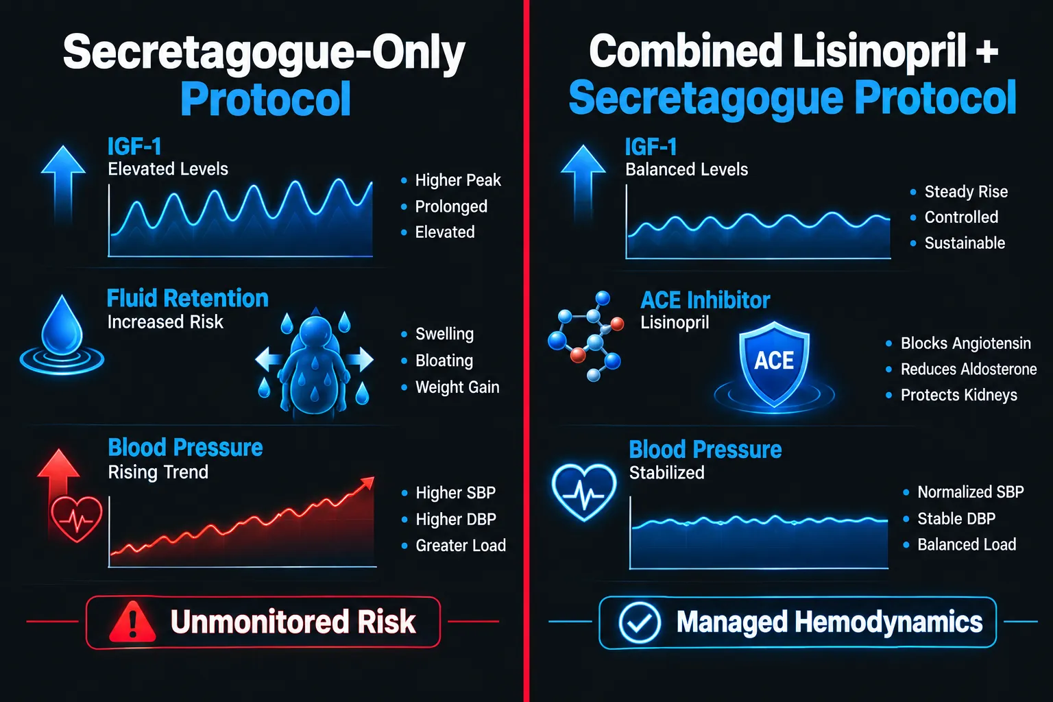
When secretagogues elevate IGF-1 and promote fluid retention, the cardiovascular system absorbs much of that load. Lisinopril, an ACE inhibitor, is frequently co-administered to manage that burden, but the interaction between these two pathways demands a structured, marker-driven approach.

Key Takeaways

  • Growth hormone secretagogues raise IGF-1 and can promote sodium and fluid retention, increasing cardiac preload and blood pressure.
  • Lisinopril and other ACE inhibitors blunt RAAS activation but introduce their own monitoring needs, including potassium elevation and renal function shifts.
  • Tracking cardiovascular stress markers, hemodynamic, structural, biomarker, and vascular, is essential when combining these agents in research protocols.
  • NT-proBNP, hs-troponin, seated and ambulatory blood pressure, and echocardiographic data form the core monitoring panel.
  • Integrated monitoring of the ACE pathway alongside secretagogue use reduces the risk of undetected cardiac stress.

How Secretagogues Interact With the Cardiovascular System
How Secretagogues Interact With the Cardiovascular System

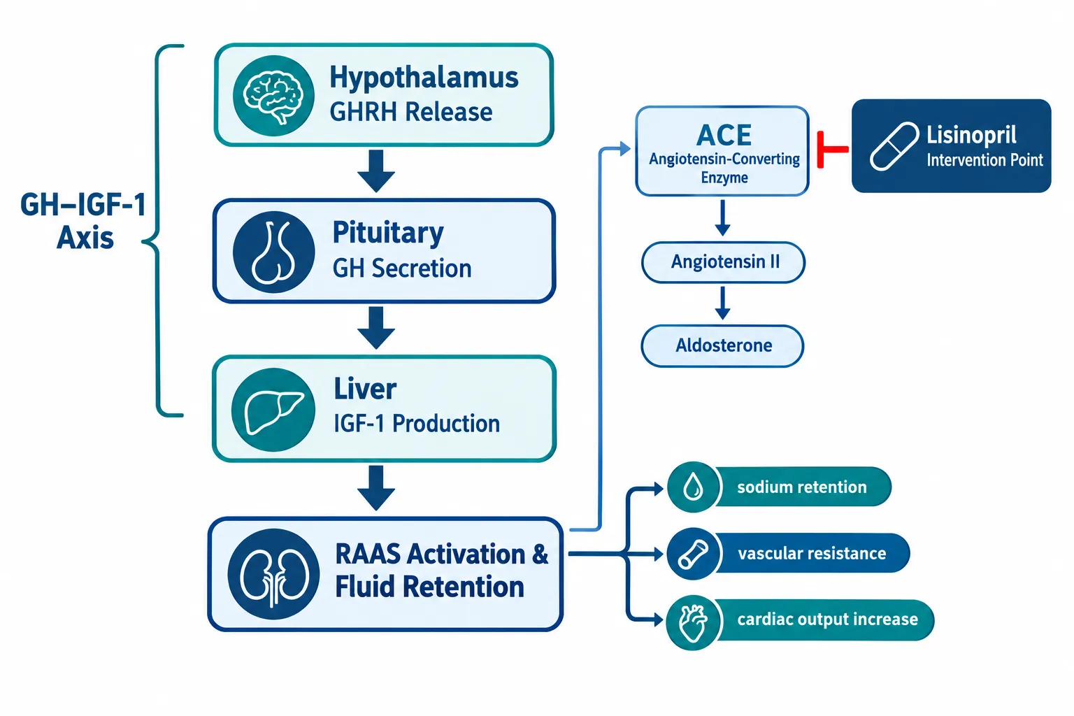
Growth hormone secretagogues, including GHRH analogs, ghrelin mimetics, and synthetic GH-releasing peptides, stimulate pituitary GH release, which in turn drives hepatic IGF-1 production. This cascade carries meaningful cardiovascular effects that researchers must account for.

IGF-1 elevation promotes myocardial growth, increases cardiac output, and enhances vascular smooth muscle responsiveness. In the short term, these effects can look beneficial. Over longer exposures, however, elevated IGF-1 contributes to left ventricular hypertrophy and arterial stiffness, both established markers of cardiovascular stress.

Ghrelin and its synthetic analogs add another layer. Research consistently shows that ghrelin-type secretagogues exert vasodilatory, anti-ischemic, and anti-apoptotic effects on cardiac tissue. They reduce peripheral vascular resistance and may lower blood pressure acutely. However, this vasodilation can interact unpredictably with ACE inhibitors, raising the risk of orthostatic hypotension.

Fluid retention is a particularly important concern. GH directly stimulates renal sodium reabsorption via the IGF-1 receptor, expanding plasma volume. This increases cardiac preload and, over time, elevates systolic blood pressure. Researchers working with IPA Sermorelin stack protocols should factor this mechanism into their baseline cardiovascular assessments.

The RAAS responds to this expanded volume by modulating aldosterone and angiotensin II levels. This is precisely where lisinopril enters the picture.

Lisinopril and ACE Pathway Dynamics in Secretagogue Protocols
Lisinopril and ACE Pathway Dynamics in Secretagogue Protocols

Lisinopril blocks ACE, the enzyme that converts angiotensin I to the potent vasoconstrictor angiotensin II. By reducing angiotensin II, lisinopril lowers peripheral resistance, reduces aldosterone-driven sodium retention, and decreases cardiac afterload. These are exactly the mechanisms that counteract GH-induced fluid retention.

"The ACE pathway and the GH/IGF-1 axis do not operate in isolation, co-administration of lisinopril alongside secretagogues creates a dynamic cardiovascular environment that requires active, structured monitoring."

However, ACE inhibition introduces its own variables. Potassium levels rise because aldosterone suppression reduces urinary potassium excretion. Renal perfusion pressure can drop, particularly in subjects with any pre-existing renal sensitivity. Bradykinin accumulates, which may amplify the vasodilatory effects already present from ghrelin-type secretagogues.

Key interaction points to monitor:

  • Blood pressure response may be exaggerated when ghrelin-type secretagogues and lisinopril are combined
  • Potassium can climb into hyperkalemic ranges without regular electrolyte testing
  • Renal function (creatinine, eGFR) may shift as RAAS suppression alters glomerular filtration dynamics
  • Fluid balance changes can mask or amplify biomarker readings

Researchers exploring peptide combinations that modulate metabolic and cardiovascular pathways, such as those reviewed in tesofensine noradrenergic and incretin-based pathway research, will recognize that multi-pathway interactions demand multi-marker monitoring frameworks.

Core Cardiovascular Stress Markers for Combined RAAS-GH Axis Monitoring
Core Cardiovascular Stress Markers for Combined RAAS-GH Axis Monitoring

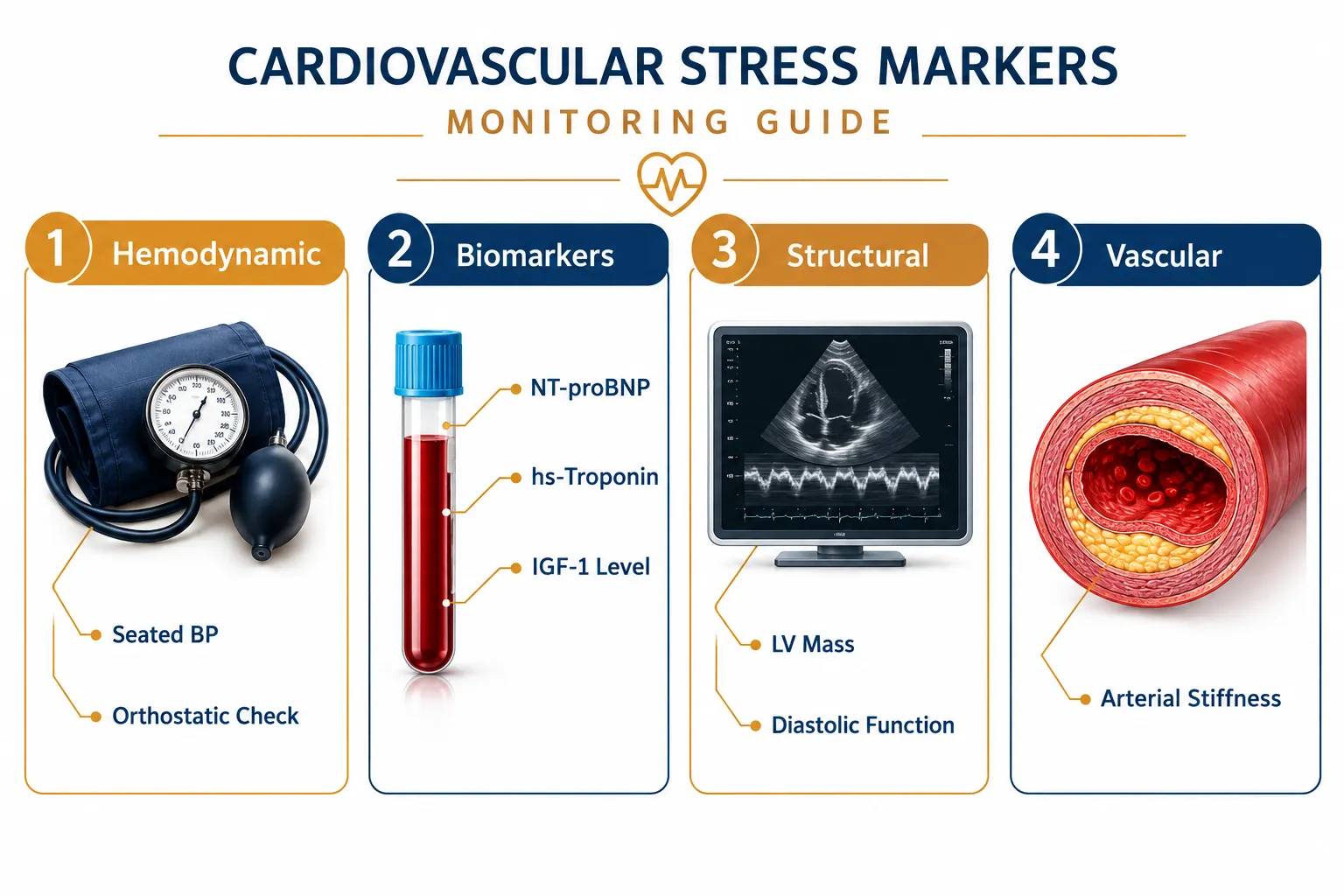
Monitoring cardiovascular stress markers in growth hormone research alongside lisinopril and ACE pathway modulation requires a structured, four-domain approach.

Hemodynamic Markers

Marker Measurement Method Key Concern
Seated blood pressure Manual or automated sphygmomanometry Hypertension from GH fluid load
Ambulatory blood pressure 24-hour ABPM device Masked hypertension patterns
Orthostatic blood pressure Supine to standing protocol Hypotension risk with ACE + ghrelin
Resting heart rate ECG or pulse oximetry Tachycardia from volume shifts

Structural Markers

Echocardiography provides the most direct window into cardiac remodeling. Left ventricular mass index, wall thickness, and both systolic and diastolic function should be assessed at baseline and at defined intervals. Diastolic dysfunction often precedes systolic changes and can appear early in GH-excess states.

Biomarker Panel

NT-proBNP is the most sensitive biomarker for detecting early cardiac wall stress and volume overload. Elevated levels signal that the heart is under pressure before symptoms appear. High-sensitivity troponin (hs-troponin) identifies subclinical myocardial injury.

Additional biomarkers include:

  • IGF-1 (to calibrate secretagogue dosing and exposure)
  • Serum creatinine and eGFR (renal safety on ACE inhibitor)
  • Potassium and sodium (electrolyte balance under RAAS suppression)
  • Fasting glucose and lipid panel (metabolic context)

Research on mitochondrial cardioprotection compounds such as those examined in SS-31 mitochondrial research highlights how cellular stress markers can complement systemic biomarker panels in cardiovascular monitoring.

Vascular Markers

Arterial stiffness indices, measured via pulse wave velocity or augmentation index, capture changes in large-vessel compliance that echocardiography and biomarkers may miss. Endothelial function assessment adds further resolution in longer-duration protocols.

Practical Monitoring Frequency and Protocol Design

Effective cardiovascular stress monitoring in growth hormone research is not a one-time assessment. A tiered frequency model works best:

Baseline (before protocol initiation): Full panel, hemodynamic, echocardiographic, biomarker, and vascular measures.

Early phase (weeks 2-4): Blood pressure, heart rate, potassium, creatinine, and eGFR. This window captures the most acute RAAS-GH interaction effects.

Mid-protocol (weeks 6-12): Repeat biomarker panel including NT-proBNP and hs-troponin. Reassess IGF-1 to confirm secretagogue response is within target range.

End of protocol: Full repeat of baseline panel plus echocardiographic comparison for structural changes.

Researchers working with GLP-based peptide compounds, including those available as GLP-3 Reta 10mg or GLP-3 Reta 20mg, should apply similarly structured cardiovascular monitoring, as GLP receptor pathways also influence cardiac output and vascular tone.

Conclusion

Cardiovascular stress markers in growth hormone research represent a non-negotiable component of responsible secretagogue protocol design, particularly when lisinopril and ACE pathway modulation are part of the picture. The interaction between GH-driven fluid retention, IGF-1-mediated cardiac remodeling, and ACE inhibitor-induced RAAS suppression creates a layered hemodynamic environment that no single marker can fully capture.

Actionable next steps for researchers:

  1. Establish a full cardiovascular baseline before initiating any secretagogue-lisinopril combination protocol.
  2. Prioritize NT-proBNP, hs-troponin, potassium, and eGFR as the minimum biomarker panel throughout the study period.
  3. Use ambulatory blood pressure monitoring to detect masked hypertension or exaggerated hypotensive responses.
  4. Schedule echocardiographic assessment at baseline and protocol end to quantify structural changes.
  5. Adjust secretagogue dosing based on IGF-1 levels and correlate with hemodynamic trends rather than treating them as independent variables.

Structured monitoring transforms cardiovascular risk from an unknown variable into a manageable, measurable research parameter.

https://www.puretestedpeptides.com/wp-content/uploads/2026/09/cardiovascular-stress-markers-in-growth-hormone-research-monitoring-lisinopril-a.webp 1024 1536 https://www.puretestedpeptides.com/wp-content/uploads/2026/01/buy-peptides-online.jpg 2026-09-16 13:07:362026-09-16 13:07:36Cardiovascular Stress Markers in Growth Hormone Research: Monitoring Lisinopril and ACE Pathways Alongside Secretagogues
Statin Therapy and Liver Enzymatic Readouts in Metabolic Peptide Models: Atorvastatin vs. MOTS-c and 5-Amino-1MQ

Statin Therapy and Liver Enzymatic Readouts in Metabolic Peptide Models: Atorvastatin vs. MOTS-c and 5-Amino-1MQ

September 16, 2026/0 Comments/in Uncategorized/by

Atorvastatin reduces cardiovascular events in millions of patients worldwide, yet its interaction with mitochondrial lipid oxidation pathways, the same pathways targeted by emerging research peptides, creates a hepatic monitoring challenge that most clinical protocols have not yet addressed. As researchers and clinicians increasingly encounter subjects using both statin therapy and novel metabolic compounds, understanding how liver enzymatic readouts shift across these combined models has become a pressing question in 2026.

This article examines the intersection of statin-driven cholesterol suppression and the mitochondrial and enzymatic mechanisms of two research peptides: MOTS-c and 5-Amino-1MQ. The goal is to give researchers a clear, evidence-grounded framework for interpreting ALT, AST, GGT, and related liver markers in these combined metabolic models.

Key Takeaways

  • Atorvastatin at high doses (40-80 mg) carries a recognized risk of transaminase elevation, requiring baseline and interval liver function monitoring.
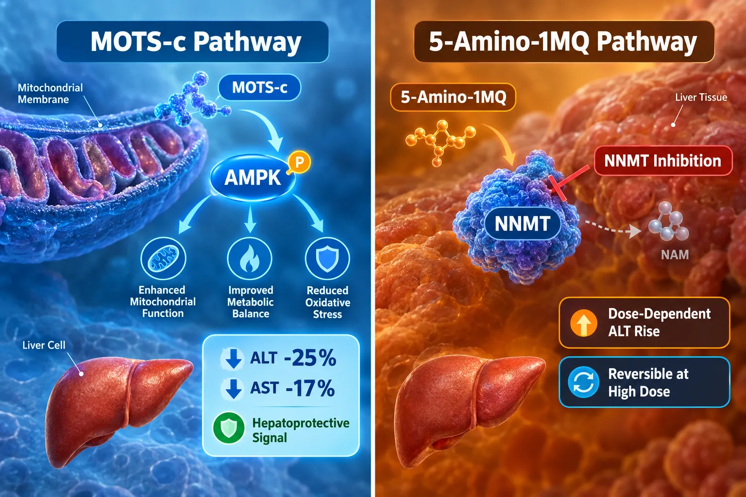
  • MOTS-c analogs have shown early signals of hepatoprotection, with one analog reducing plasma ALT by approximately 25% and AST by 17% versus placebo in early-phase research.
  • 5-Amino-1MQ demonstrates no short-term hepatotoxicity signal at standard doses, but high-dose rodent studies show dose-dependent, reversible ALT/AST rises of 2-3 times the upper limit of normal.
  • No formal drug-interaction profile exists for MOTS-c or 5-Amino-1MQ combined with statins; monitoring protocols should mirror established statin hepatic surveillance guidelines.
  • Human safety data for both peptides remains limited; all liver risk assessments are currently extrapolated from animal models.

How Atorvastatin Affects Liver Enzymatic Readouts

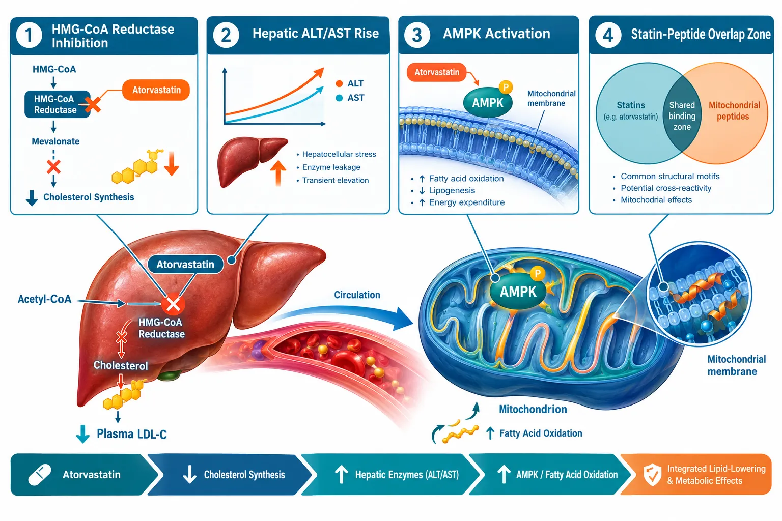
Atorvastatin works by inhibiting HMG-CoA reductase, the rate-limiting enzyme in hepatic cholesterol synthesis. This mechanism is primarily hepatic, meaning the liver bears the greatest pharmacological burden. In most patients, liver enzymes remain stable. However, high-dose regimens, specifically 40 mg and 80 mg daily, are consistently flagged in clinical guidance for a higher risk of transaminase elevation.

How Atorvastatin Affects Liver Enzymatic Readouts

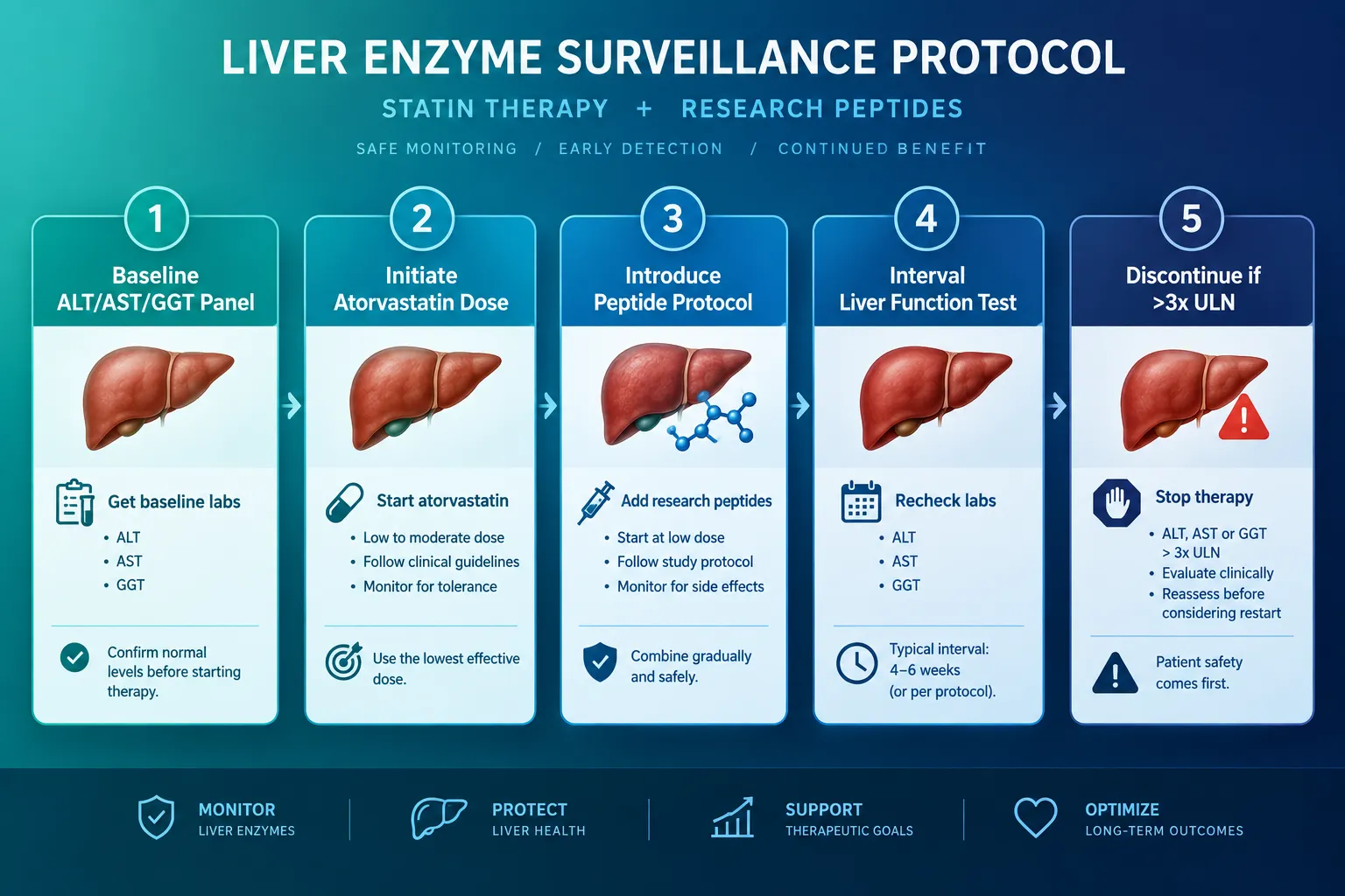
The standard threshold for clinical concern is a rise in ALT or AST exceeding three times the upper limit of normal (ULN). At that point, dose reduction or discontinuation is recommended. Baseline liver panels before starting atorvastatin are considered standard practice, and interval testing is advised when doses increase or when additional hepatically metabolized compounds are introduced.

Key liver markers to track with atorvastatin:

Marker Baseline Role Concern Threshold
ALT Hepatocellular injury >3x ULN
AST Broader hepatic/muscle signal >3x ULN
GGT Cholestatic marker Elevated with dose escalation
Bilirubin Hepatic function Rising with severe injury

The lipid-lowering mechanism of atorvastatin also indirectly touches mitochondrial function. Statins can reduce coenzyme Q10 synthesis, a downstream consequence of HMG-CoA reductase inhibition, which may mildly impair mitochondrial electron transport. This is relevant because both MOTS-c and 5-Amino-1MQ act on overlapping mitochondrial and metabolic pathways, creating a potential zone of interaction that warrants careful enzyme surveillance.

MOTS-c in the Context of Statin Therapy and Liver Enzymatic Readouts in Metabolic Peptide Models

MOTS-c is a mitochondrial-derived peptide that activates AMPK signaling, promoting fatty acid oxidation and glucose uptake. Its mechanism is fundamentally mitochondrial, which places it in direct functional proximity to the mitochondrial stress that high-dose statins can produce.

Early clinical research on a MOTS-c-derived analog (CB4211) reported a reduction in liver triglyceride levels alongside a 25% decrease in plasma ALT and a 17% decrease in AST compared to placebo. These findings suggest a hepatoprotective rather than hepatotoxic profile, at least in subjects with hepatic steatosis. This is a meaningful distinction: while atorvastatin may nudge liver enzymes upward at high doses, MOTS-c analogs appear to exert a counterbalancing effect in fatty liver models.

Researchers exploring MOTS-c and elamipretide combinations should note that no published cases of MOTS-c increasing statin-related hepatotoxicity have been reported. However, the absence of a formal drug-interaction profile means caution is still warranted. MOTS-c is not FDA-approved and is available only as a research-grade MOTS-c peptide within registered trials or research settings.

"MOTS-c's AMPK activation pathway overlaps with the mitochondrial stress zone created by statin-induced CoQ10 suppression, making liver enzyme monitoring essential, not optional."

MOTS-c + Atorvastatin: Current Evidence Summary

  • No published adverse interaction cases as of 2026
  • Theoretical concern: combined AMPK/mitochondrial pathway activity at high statin doses
  • Recommended monitoring: baseline ALT/AST, interval testing at 4-8 weeks
  • Human data: limited to early Phase I safety signals only

5-Amino-1MQ Liver Enzymatic Readouts in Metabolic Peptide Models: Atorvastatin Comparison

5-Amino-1MQ inhibits nicotinamide N-methyltransferase (NNMT), an enzyme highly expressed in liver tissue. By blocking NNMT, the compound alters methionine cycle flux and shifts cellular energy metabolism toward fat oxidation. This hepatic expression profile makes liver enzyme monitoring especially relevant for this compound.

5-Amino-1MQ Liver Enzymatic Readouts in Metabolic Peptide Models: Atorvastatin Comparison

Acute animal studies using doses ranging from 50 to 5,000 mg/kg reported no mortality and no observable adverse reactions over 48 hours. A 14-day subacute study found AST and GGT unchanged, with only a transient CRP elevation at the highest intravenous dose. These findings suggest no short-term hepatotoxicity signal at standard research doses.

However, the picture changes at high doses over longer periods. Rodent studies using 5 to 10 times the proposed therapeutic dose found that approximately 8-12% of animals developed ALT rises of 2-3 times the upper limit of normal after 4-6 weeks of daily dosing. Critically, these elevations normalized within 2-3 weeks of discontinuation, indicating dose-dependent but reversible hepatotoxicity.

A 2024 diet-induced obesity mouse study also reported improved liver histology over 28 days with standard dosing, aligning with the hepatoprotective potential seen in MOTS-c models. Yet a 2025 dosing synthesis found that 100 mg/kg doses did not produce proportionally greater fat loss than lower doses but did produce elevated ALT/AST in a subset of subjects, a clear dose ceiling signal.

Those researching 5-Amino-1MQ 60 capsules for metabolic research should also review available 5-Amino-1MQ capsule formulations and ensure sourcing from lab-tested peptide suppliers to maintain research integrity.

5-Amino-1MQ Liver Enzyme Profile at a Glance:

  • Standard doses: No hepatotoxicity signal in acute and subacute animal studies
  • High doses (5-10x therapeutic): 8-12% of animals show ALT/AST 2-3x ULN
  • Recovery: Enzyme normalization within 2-3 weeks post-discontinuation
  • Human data: None; all liver risk assessments are extrapolated from animal models
  • Chronic toxicology (28-day and 90-day studies): Not publicly reported as of 2026

When combining 5-Amino-1MQ with atorvastatin, the shared hepatic burden is the primary concern. Both compounds are processed in liver tissue, and both can elevate transaminases at high doses. The recommended approach mirrors statin clinical practice: obtain a full baseline hepatic panel (ALT, AST, GGT, bilirubin), monitor at defined intervals, and discontinue with further evaluation if ALT or AST exceeds 2-3 times the upper limit of normal.

Monitoring Protocol for Combined Metabolic Peptide and Statin Models

Researchers working with statin therapy and liver enzymatic readouts in metabolic peptide models need a structured surveillance approach. The following framework integrates guidance from statin clinical practice and available peptide safety data.

Monitoring Protocol for Combined Metabolic Peptide and Statin Models

Recommended Monitoring Steps:

  1. Baseline panel, ALT, AST, GGT, total bilirubin before initiating any compound
  2. Atorvastatin initiation, Recheck at 6-12 weeks, especially at 40-80 mg doses
  3. Peptide introduction, Introduce one compound at a time where possible; recheck liver panel at 4 weeks
  4. Interval surveillance, Every 8-12 weeks during active combined use
  5. Threshold action, Discontinue the most recently added compound and retest if ALT/AST exceeds 3x ULN; evaluate both compounds if elevation persists

This stepwise approach allows researchers to isolate which compound is driving any enzymatic change, a critical distinction when both atorvastatin and a research peptide are active simultaneously.

Conclusion

The intersection of statin therapy and liver enzymatic readouts in metabolic peptide models represents one of the more nuanced monitoring challenges in current research settings. Atorvastatin's hepatic mechanism creates a baseline enzymatic risk that compounds when combined with peptides acting on overlapping mitochondrial and metabolic pathways.

MOTS-c presents an intriguing counterpoint: early data suggests hepatoprotective rather than hepatotoxic effects, with ALT and AST reductions in fatty liver models. 5-Amino-1MQ shows a clean short-term liver safety profile at standard doses, but dose-dependent and reversible transaminase elevations at high doses demand the same disciplined monitoring applied to statins.

Actionable next steps for researchers:

  • Establish a full baseline hepatic panel before combining any statin with MOTS-c or 5-Amino-1MQ
  • Use the lowest effective dose of each compound and escalate only with documented enzyme stability
  • Apply the 3x ULN discontinuation threshold consistently across all compounds in the model
  • Source compounds exclusively from verified, lab-tested peptide suppliers to ensure purity and dose accuracy
  • Treat all current liver risk assessments for both peptides as preliminary until formal human clinical trial data is available

The field is advancing rapidly, but the absence of peer-reviewed human trials for both MOTS-c and 5-Amino-1MQ means that rigorous enzymatic monitoring remains the single most important safeguard in these combined metabolic models.

https://www.puretestedpeptides.com/wp-content/uploads/2026/09/statin-therapy-and-liver-enzymatic-readouts-in-metabolic-peptide-models-atorvast.webp 1024 1536 https://www.puretestedpeptides.com/wp-content/uploads/2026/01/buy-peptides-online.jpg 2026-09-16 13:07:052026-09-16 13:07:05Statin Therapy and Liver Enzymatic Readouts in Metabolic Peptide Models: Atorvastatin vs. MOTS-c and 5-Amino-1MQ
Hydration and Osmolality in Intensive Peptide Studies: The Role of Electrolyte Solutions and Liquid IV Protocols

Hydration and Osmolality in Intensive Peptide Studies: The Role of Electrolyte Solutions and Liquid IV Protocols

September 16, 2026/0 Comments/in Uncategorized/by

Fluid balance is rarely the headline variable in peptide research, yet it quietly determines whether a study produces clean, reproducible data or confounded results. When researchers investigate GLP-class metabolic peptides or mitochondria-targeting compounds, shifts in cellular hydration status and plasma osmolality can alter receptor binding, hormone signaling, and tissue distribution in ways that standard protocols often fail to account for. Understanding hydration and osmolality in intensive peptide studies — and the role of electrolyte solutions and liquid IV protocols in managing those variables — is therefore a foundational concern, not an afterthought.

Key Takeaways

  • Plasma osmolality targets near 285 mOsm/kg represent the physiological benchmark that electrolyte solutions in peptide research should support, not disrupt.
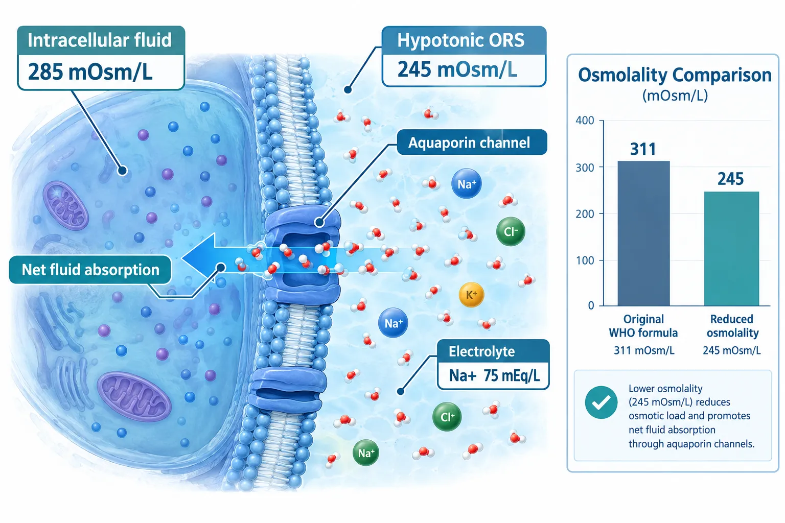
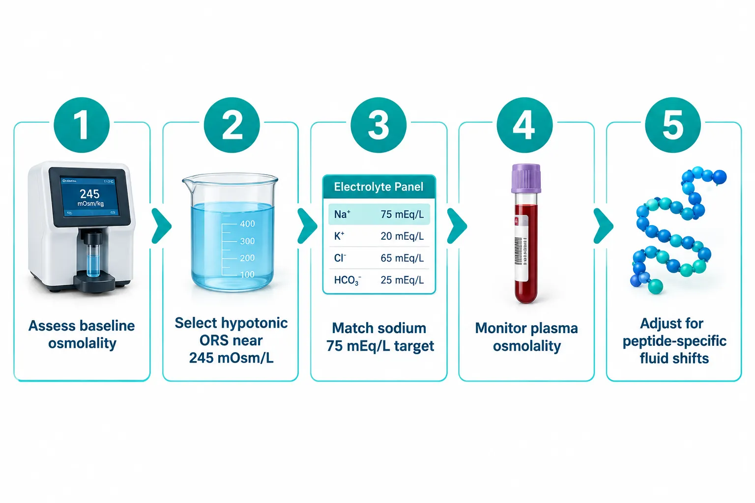
  • Current international guidelines converge on hypotonic oral rehydration solutions near 245 mOsm/L as the most effective standard for rapid, efficient rehydration.
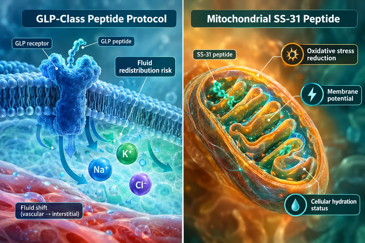
  • GLP-class and mitochondrial peptides each carry distinct fluid-shift risks that demand osmolality-aware hydration protocols.
  • Liquid IV-style products show theoretical promise but currently lack peer-reviewed clinical evidence demonstrating superiority over properly formulated electrolyte solutions.
  • Selecting the right electrolyte solution means checking sodium content, carbohydrate load, and total osmolarity before integrating it into any intensive protocol.

Why Osmolality Matters in Intensive Peptide Research

Why Osmolality Matters in Intensive Peptide Research

Osmolality measures the concentration of dissolved particles in a fluid, expressed in milliosmoles per kilogram of water (mOsm/kg). In a living system, plasma osmolality is tightly regulated around 285 to 295 mOsm/kg. Even modest deviations — as little as 10 mOsm/kg above or below that range — trigger compensatory hormonal responses involving vasopressin, aldosterone, and the renin-angiotensin system.

For researchers working with peptides such as SS-31, which targets mitochondrial cardiolipin to reduce oxidative stress, or with GLP-receptor agonists like those explored in GLP-3R peptide formulations, these hormonal cascades are not background noise. They directly interact with the pathways under investigation. A subject or model system that enters a protocol in a mildly hypertonic or hypotonic state introduces a confounding variable that no downstream statistical correction can fully remove.

Three osmolality-related risks in peptide studies:

  • Hypertonic conditions slow gastric emptying, reduce net fluid absorption, and can falsely elevate plasma peptide concentrations by reducing distribution volume.
  • Hypotonic conditions dilute electrolytes, alter membrane potential, and may blunt receptor-mediated responses that depend on sodium-potassium gradients.
  • Fluctuating osmolality across study visits creates inter-session variability that inflates standard deviations and reduces statistical power.
Standardizing hydration inputs is therefore as important as standardizing peptide dose and timing.

Electrolyte Solutions and Osmolality Standards: What the Evidence Supports

The global benchmark for oral rehydration solution (ORS) osmolality has shifted significantly over the past two decades. The original WHO formula carried an osmolarity of approximately 311 mOsm/L with sodium at 90 mEq/L. Clinical evidence accumulated showing that this formulation, while effective at replacing electrolytes, was not optimal for net fluid absorption. The revised WHO/UNICEF standard specifies a reduced-osmolality ORS with sodium at 75 mEq/L and total osmolarity at 245 mOsm/L — a hypotonic formulation that demonstrably improves net fluid absorption and reduces gastrointestinal side effects compared with its predecessor.

Health Canada's ORS monograph reinforces this direction, specifying that total osmolarity should not exceed 280 mOsm/L and that hypotonic solutions improve clinical outcomes. Peer-reviewed pharmacotechnical analysis supports an optimal absorption window between 200 and 260 mOsm/kg, with 245 mOsm/L representing the current evidence-based sweet spot.

Key benchmark: Solutions in the 200-260 mOsm/kg range yield the greatest net fluid absorption. Hypertonic solutions above this range slow gastric emptying — a critical consideration when pairing electrolyte solutions with intensive peptide regimens.

For intensive peptide studies, this has direct implications. Commercially available ORS products span a carbohydrate content of 13.5 to 40 g/L, sodium of 45 to 75 mEq/L, and osmolarity ranging from roughly 200 to 305 mOsm/L. Selecting a product toward the upper end of that range — or using heavily sweetened sports drinks with osmolarity above 300 mOsm/L — risks slowing gastric emptying and creating transient hypertonicity that interferes with study conditions.

Practical selection criteria for electrolyte solutions in peptide protocols:

Parameter Target Range Rationale
Total osmolarity 225-260 mOsm/L Maximizes net fluid absorption
Sodium 60-75 mEq/L Matches WHO reduced-ORS standard
Glucose/carbohydrate 13.5-20 g/L Supports sodium co-transport without hypertonicity
Potassium 15-25 mEq/L Supports intracellular balance

GLP-Class and Mitochondrial Peptides: Specific Fluid-Shift Considerations

GLP-Class and Mitochondrial Peptides: Specific Fluid-Shift Considerations

Not all peptides interact with fluid balance in the same way. Understanding hydration and osmolality in intensive peptide studies requires mapping the specific fluid-shift risks of each peptide class.

GLP-receptor peptides — including agents studied alongside compounds like GLP-3R 30mg formulations and broader cardiometabolic peptide models — influence gastric emptying rate, gut motility, and fluid secretion in the gastrointestinal tract. These effects mean that subjects in GLP-focused protocols may absorb oral fluids at altered rates, making the osmolality of any co-administered electrolyte solution especially consequential. A hypertonic solution that would merely slow absorption in a resting subject could produce meaningful fluid redistribution in a GLP-stimulated gut.

Mitochondria-targeting peptides such as SS-31 operate at the level of the inner mitochondrial membrane, modulating oxidative phosphorylation and reactive oxygen species. Research on SS-31 peptide benefits and SS-31 research considerations highlights that mitochondrial function is sensitive to cellular hydration status. Dehydration reduces mitochondrial membrane potential and amplifies oxidative stress — the very pathology SS-31 is designed to study. Running an SS-31 protocol without a controlled hydration baseline risks confounding the primary endpoint.

Growth hormone-releasing peptides like tesa influence body composition and fluid compartmentalization through IGF-1-mediated pathways. Sodium and water retention are recognized downstream effects of growth hormone axis activation, meaning that plasma osmolality monitoring should be built into any extended tesa protocol.

Liquid IV Protocols: Promise, Evidence Gaps, and Practical Guidance

Liquid IV Protocols: Promise, Evidence Gaps, and Practical Guidance

Liquid IV-style products — high-sodium, glucose-containing sachets marketed on cellular transport technology — have attracted attention as rapid rehydration tools for intensive protocols. The theoretical basis is sound: sodium-glucose co-transport (SGLT1) in the small intestine can accelerate fluid uptake when the sodium-to-glucose ratio is optimized, and a well-formulated product near 245 mOsm/L could theoretically outperform plain water in restoring plasma osmolality after exercise-induced dehydration.

The clinical evidence, however, remains thin. A registered randomized crossover trial (NCT06063655) is tracking body weight, urine osmolality, plasma osmolality, and blood electrolytes following exercise-induced dehydration with Liquid I.V. rehydration, but as of 2026 no peer-reviewed results have been published. An earlier poster study from Washington State University Vancouver compared plasma osmolarity after plain water versus Liquid I.V. in mildly dehydrated participants and predicted no significant difference between groups — though this remains an undergraduate-level poster rather than a peer-reviewed clinical trial.

For peptide researchers, the practical takeaway is straightforward: any liquid IV protocol should be evaluated on its actual osmolarity value, sodium content, and carbohydrate load against the 245 mOsm/L benchmark before adoption. A product that clusters near that target with sodium around 75 mEq/L is defensible. A heavily sweetened product above 300 mOsm/L is not, regardless of marketing claims.

Conclusion

Hydration and osmolality in intensive peptide studies — and the role of electrolyte solutions and liquid IV protocols — deserve the same methodological rigor applied to dosing, timing, and endpoint selection. The evidence base is clear: hypotonic electrolyte solutions near 245 mOsm/L, with sodium around 75 mEq/L and modest glucose content, provide the most efficient and gastrointestinally tolerable rehydration platform currently available.

Actionable next steps for researchers:

  1. Measure baseline plasma osmolality in all subjects before peptide administration and flag any value outside 280-295 mOsm/kg.
  2. Select an electrolyte solution with documented osmolarity at or below 260 mOsm/L — check the product specification sheet, not just the label claims.
  3. For GLP-class protocols, account for altered gastric emptying when timing oral fluid administration relative to peptide dosing.
  4. For mitochondrial peptide studies, treat cellular hydration status as a primary covariate, not a background variable.
  5. Treat liquid IV-style products as potentially useful tools but require osmolarity data before incorporating them into any standardized protocol.

Fluid balance is not a peripheral concern in peptide research. It is a core experimental variable — and managing it precisely is what separates reproducible science from noise.

https://www.puretestedpeptides.com/wp-content/uploads/2026/09/hydration-and-osmolality-in-intensive-peptide-studies-the-role-of-electrolyte-so.webp 1024 1536 https://www.puretestedpeptides.com/wp-content/uploads/2026/01/buy-peptides-online.jpg 2026-09-16 13:06:542026-09-16 13:06:54Hydration and Osmolality in Intensive Peptide Studies: The Role of Electrolyte Solutions and Liquid IV Protocols
Evaluating Antiemetic Protocols in Incretin Research: Ondansetron Mechanisms During GLP-3 Retatrutide Administration

Evaluating Antiemetic Protocols in Incretin Research: Ondansetron Mechanisms During GLP-3 Retatrutide Administration

September 16, 2026/0 Comments/in Uncategorized/by

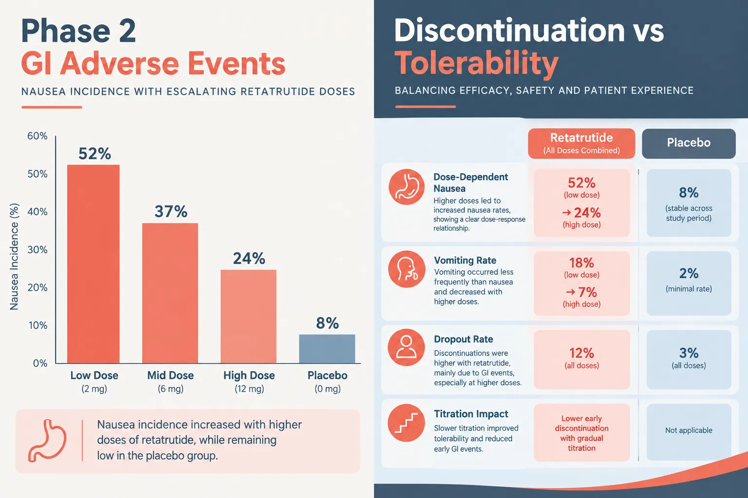
Nausea affects more than 40% of participants in high-dose retatrutide arms during Phase 2 trials, a rate that places gastrointestinal (GI) tolerability at the center of every research design decision. As investigators push toward maximizing metabolic efficacy with this potent multi-incretin agonist, evaluating antiemetic protocols in incretin research, including the role of ondansetron mechanisms during GLP-3 retatrutide administration, has become a critical and underexplored frontier.

Key Takeaways

  • Retatrutide (LY3437943) is a triple incretin agonist targeting GLP-1, GIP, and glucagon receptors, producing dose-dependent GI side effects.
  • Nausea and vomiting rates in high-dose retatrutide cohorts are among the highest observed in the incretin drug class.
  • Ondansetron, a selective 5-HT3 receptor antagonist, targets both central and peripheral serotonin pathways implicated in incretin-induced emesis.
  • No formal ondansetron-specific antiemetic protocols have been published for retatrutide trials as of 2026; current management relies on dose titration.
  • Structured antiemetic research protocols represent a significant opportunity to improve participant retention and trial outcomes.

Understanding Retatrutide and Its GI Burden

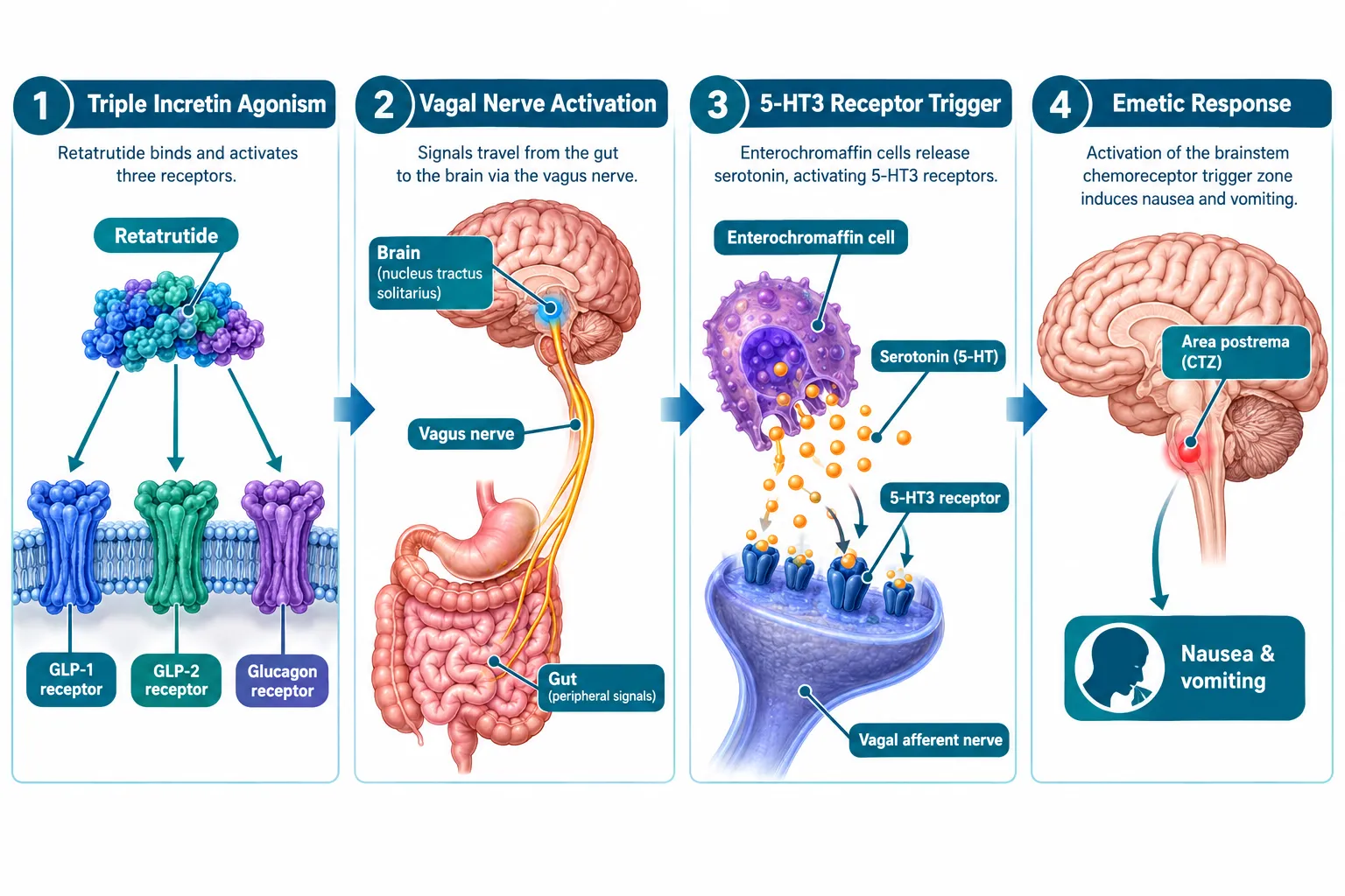
Retatrutide, developed under the designation LY3437943, is a single-molecule triple agonist that simultaneously activates glucagon-like peptide-1 (GLP-1), glucose-dependent insulinotropic polypeptide (GIP), and glucagon receptors. This multi-receptor engagement drives impressive reductions in body weight and improvements in metabolic markers, but it also amplifies the GI signaling cascades that produce nausea, vomiting, and early satiety.

Understanding Retatrutide and Its GI Burden

In Phase 2 dose-ranging studies, nausea incidence climbed steeply with dose escalation. At the highest tested doses, nausea was reported in over 40% of participants, with vomiting affecting a substantial subset. These figures exceed rates typically seen with GLP-1 receptor agonists alone, reflecting the additive GI burden of triple incretin engagement. Researchers exploring retatrutide clinical trials have consistently flagged GI tolerability as the primary dose-limiting factor.

The timing of adverse events follows a predictable pattern: symptoms peak during dose escalation phases and tend to attenuate as participants acclimate. However, the severity during escalation is sufficient to drive meaningful discontinuation rates, which in turn compromises the statistical integrity of long-term efficacy data.

Key GI adverse event characteristics in retatrutide research:

Adverse Event Onset Timing Dose Relationship Primary Mechanism
Nausea Early escalation Dose-dependent GLP-1 vagal activation, 5-HT3 signaling
Vomiting Concurrent with nausea Dose-dependent Central emetic pathway activation
Early satiety Persistent Moderate Gastric motility slowing
Discontinuation Peak escalation High-dose cohorts Cumulative GI burden

The 5-HT3 Pathway: Why Ondansetron Is Relevant

The central question in evaluating antiemetic protocols in incretin research involving ondansetron mechanisms during GLP-3 retatrutide administration lies in the biology of serotonin signaling. GLP-1 receptor activation in the gut stimulates enterochromaffin cells to release serotonin (5-hydroxytryptamine, or 5-HT). This serotonin binds to 5-HT3 receptors on vagal afferent neurons, transmitting an emetic signal to the brainstem's chemoreceptor trigger zone (CTZ) and the nucleus tractus solitarius (NTS).

Ondansetron is a highly selective 5-HT3 receptor antagonist. By blocking these receptors at both peripheral (gut-vagal) and central (CTZ) sites, it interrupts the serotonin-mediated emetic cascade before it reaches full activation. This dual-site mechanism makes it mechanistically well-suited to incretin-induced nausea, which originates peripherally but is amplified centrally.

Ondansetron's selectivity for the 5-HT3 receptor makes it a pharmacologically logical candidate for managing the serotonin-driven nausea associated with potent multi-incretin agonists like retatrutide.

The triple-agonist profile of retatrutide likely intensifies this cascade compared to single-receptor agents. Glucagon receptor activation adds motility effects, while GIP receptor engagement may further modulate gut hormone signaling. The cumulative result is a more complex emetic environment than ondansetron was originally designed to address, but one in which 5-HT3 blockade still targets a primary mechanistic node.

The 5-HT3 Pathway: Why Ondansetron Is Relevant

Current Protocol Gaps and the Path Forward

Despite the clear mechanistic rationale, a critical evidence gap exists. As of 2026, no published retatrutide trial has incorporated a formal, pre-specified ondansetron antiemetic protocol. Current management strategies rely almost entirely on conservative dose titration schedules and general supportive care guidance. Participants are typically advised to eat smaller meals, avoid high-fat foods, and wait out the escalation period.

This approach has limitations. Dose titration slows the research timeline and may prevent some participants from reaching target doses. Supportive care guidance is inconsistently applied across sites. And without standardized antiemetic co-administration data, researchers cannot determine whether pharmacological intervention would meaningfully reduce discontinuation rates.

For those sourcing research-grade compounds, the GLP-3 Reta 10mg research peptide represents one avenue for preclinical investigation, and resources on buying GLP-3 Reta for research purposes are available for qualified investigators. Similarly, researchers exploring related peptide tolerability profiles may find value in SS-31 mitochondrial research themes as a parallel framework for understanding cellular stress responses during peptide administration.

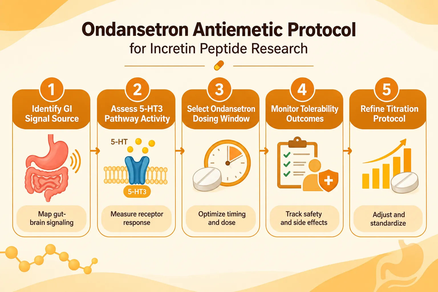
Proposed elements of a structured ondansetron protocol for incretin research:

  • Timing: Administer ondansetron 30-60 minutes prior to retatrutide injection during dose escalation phases.
  • Dosing window: Standard antiemetic dosing (4-8 mg oral) aligned with escalation schedules.
  • Duration: Protocol-defined use limited to escalation periods, with reassessment at maintenance doses.
  • Outcome tracking: Standardized nausea severity scales (e.g., FLIE, VAS) recorded at consistent intervals.
  • Discontinuation monitoring: Compare dropout rates between antiemetic-supported and standard-care arms.

Researchers interested in buying retatrutide peptide for preclinical work should also consider how antiemetic co-administration variables will be documented in their protocols. Parallel work in Semax research demonstrates how neuropeptide tolerability frameworks can inform protocol design across compound classes.

Current Protocol Gaps and the Path Forward

The risk-benefit calculus here is straightforward: if ondansetron reduces early discontinuation by even a modest percentage, the downstream gains in data completeness and statistical power justify its inclusion in trial designs. The question is no longer whether to study this intervention, but how to structure that study rigorously.

Conclusion

Evaluating antiemetic protocols in incretin research, specifically ondansetron mechanisms during GLP-3 retatrutide administration, is no longer a secondary concern. It is a prerequisite for generating reliable, high-quality data from one of the most promising metabolic research compounds in development.

Actionable next steps for research teams:

  1. Incorporate pre-specified 5-HT3 antagonist co-administration arms into upcoming retatrutide Phase 3 sub-studies.
  2. Standardize nausea outcome measurement tools across all sites to enable meaningful cross-trial comparison.
  3. Publish tolerability sub-analyses that isolate the effect of antiemetic support on dose completion rates.
  4. Engage regulatory bodies early on antiemetic co-administration as a protocol variable, not a post-hoc intervention.

The serotonin pathway is not a minor footnote in incretin pharmacology, it is a central driver of the GI burden that limits these compounds' research potential. Addressing it with the same rigor applied to efficacy endpoints will define the next generation of incretin trial design.

https://www.puretestedpeptides.com/wp-content/uploads/2026/09/evaluating-antiemetic-protocols-in-incretin-research-ondansetron-mechanisms-duri.webp 1024 1536 https://www.puretestedpeptides.com/wp-content/uploads/2026/01/buy-peptides-online.jpg 2026-09-16 13:06:492026-09-16 13:06:49Evaluating Antiemetic Protocols in Incretin Research: Ondansetron Mechanisms During GLP-3 Retatrutide Administration
Meloxicam and Tissue-Repair Peptides: Comparing Nonsteroidal Anti-Inflammatory Drugs with BPC-157 in In Vitro Models

Meloxicam and Tissue-Repair Peptides: Comparing Nonsteroidal Anti-Inflammatory Drugs with BPC-157 in In Vitro Models

September 16, 2026/0 Comments/in Uncategorized/by

COX-2 inhibition and growth factor upregulation are not two sides of the same coin, they are mechanistically opposite strategies for managing tissue injury. This fundamental difference sits at the center of any rigorous comparison of meloxicam and tissue-repair peptides: comparing nonsteroidal anti-inflammatory drugs with BPC-157 in in vitro models. As cell culture research grows more sophisticated in 2026, the contrast between these two approaches has become sharper, more clinically relevant, and more debated.

Key Takeaways

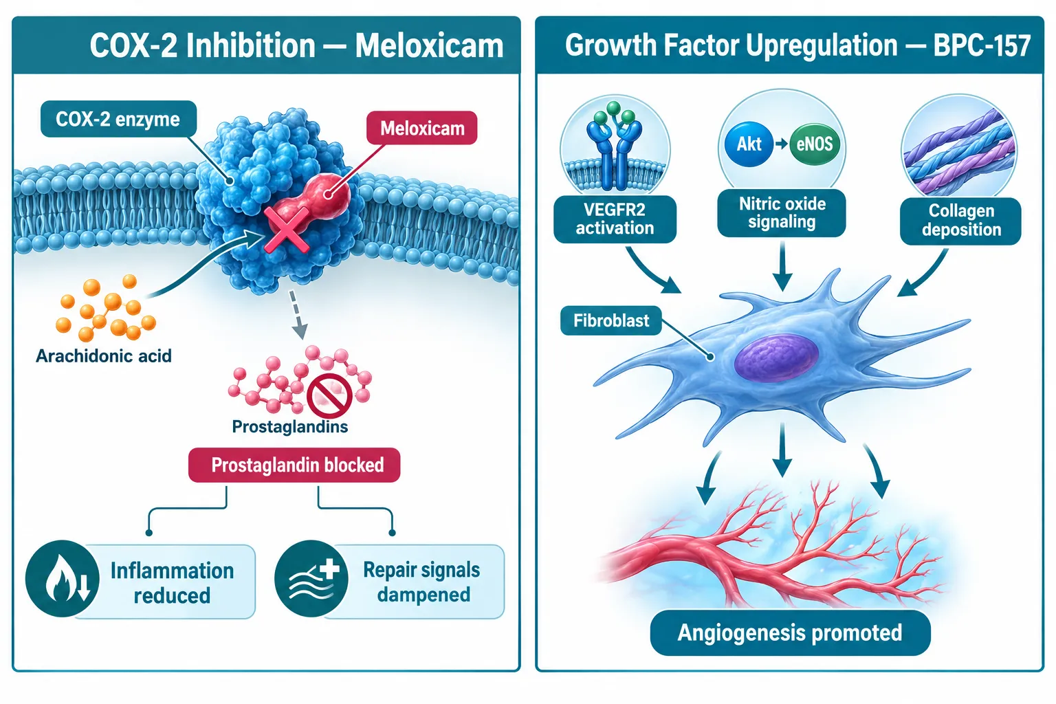
  • Meloxicam reduces inflammation primarily by blocking COX-2 enzymes and suppressing prostaglandin synthesis, which may also dampen early repair signaling.
  • BPC-157 is a pleiotropic tissue-repair peptide that activates VEGFR2, the Akt-eNOS nitric oxide axis, and promotes fibroblast activity, collagen deposition, and angiogenesis.
  • In vitro musculoskeletal models consistently show BPC-157 enhancing tenocyte survival, fibroblast migration, and growth hormone receptor expression.
  • NSAIDs such as meloxicam have been repeatedly associated with reduced collagen synthesis in preclinical models, contrasting with BPC-157's repair-promoting profile.
  • BPC-157 remains a research-only compound without major regulatory approval; meloxicam is a fully approved NSAID with no clinical indication for tissue regeneration.

How COX-2 Inhibition and Peptide Signaling Differ at the Cellular Level

Understanding the mechanistic gap between meloxicam and BPC-157 starts at the receptor level.

Meloxicam is a COX-2-preferential NSAID. It works by blocking cyclooxygenase-2 enzymes, which halts the conversion of arachidonic acid into prostaglandins. This reduces pain and swelling efficiently. However, prostaglandins also serve as early-phase messengers that recruit repair cells to damaged tissue. When that signal is suppressed, the downstream cascade of fibroblast recruitment, collagen synthesis, and angiogenesis can be partially blunted.

How COX-2 Inhibition and Peptide Signaling Differ at the Cellular Level

BPC-157, by contrast, operates through an entirely different set of molecular targets. It activates VEGFR2 (vascular endothelial growth factor receptor 2) and the Akt-eNOS nitric oxide pathway, which directly promotes new blood vessel formation and cell survival. It also upregulates growth hormone receptors on fibroblasts and tenocytes, enhancing their proliferative and migratory capacity. Rather than silencing an inflammatory cascade, BPC-157 accelerates the transition from injury to active repair.

Key distinction: Meloxicam turns down the inflammatory signal. BPC-157 turns up the repair signal. These are not equivalent actions.

This mechanistic separation is why researchers studying meloxicam and tissue-repair peptides in cell culture systems often find these two compounds occupying non-overlapping functional roles rather than competing for the same outcome.

What In Vitro Models Reveal About BPC-157 and NSAID Effects on Tissue Repair

Cell culture studies have produced some of the clearest evidence for BPC-157's repair-promoting properties.

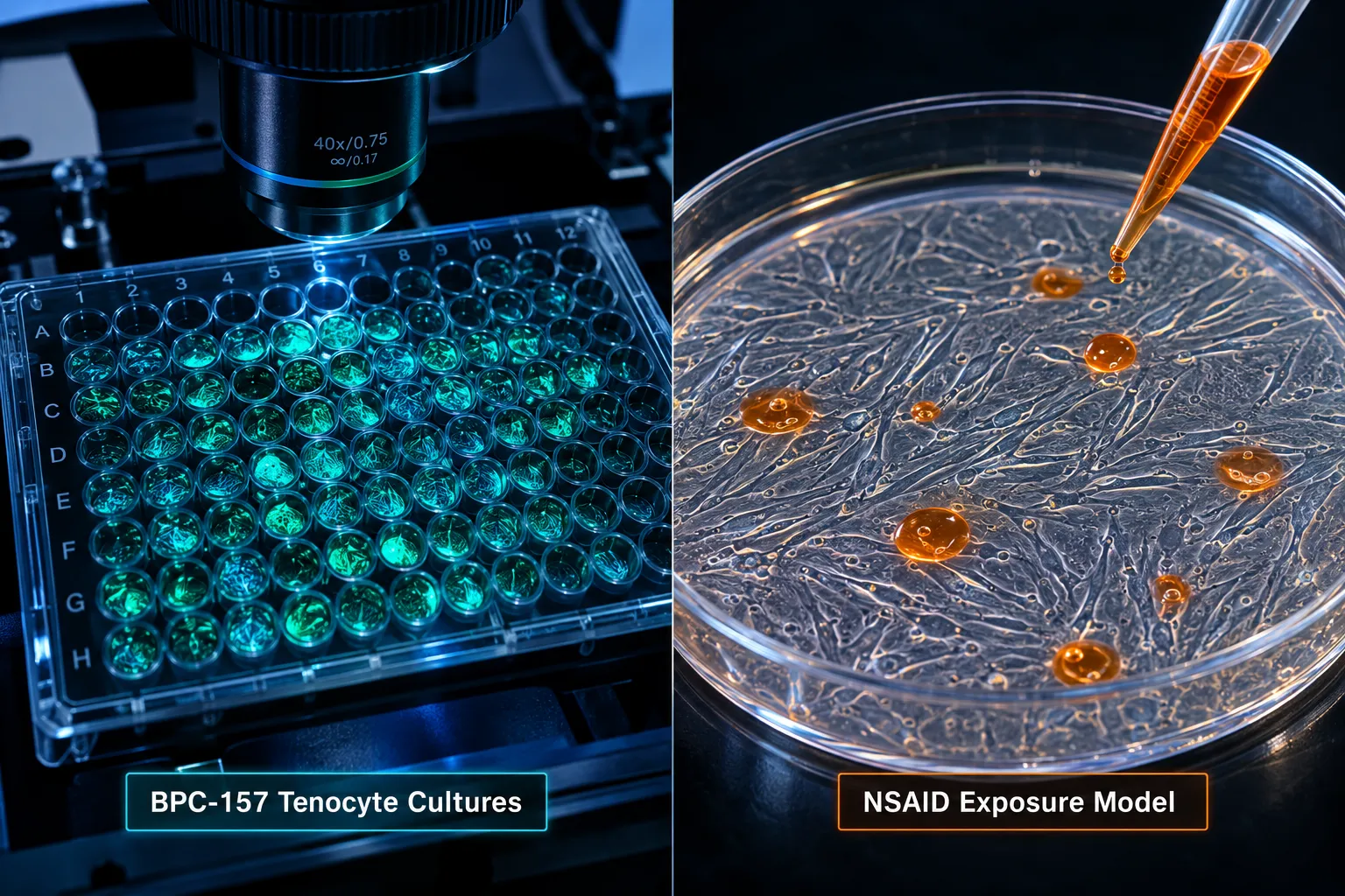
In rat Achilles tenocyte cultures, BPC-157 exposure consistently produces enhanced cell survival and proliferation. Fibroblast cultures treated with BPC-157 show increased growth hormone receptor expression, which correlates with improved collagen fiber organization. Wound-healing scratch assays demonstrate accelerated cell migration into the injury zone, and angiogenesis assays confirm increased tubule formation in endothelial cell models.

What In Vitro Models Reveal About BPC-157 and NSAID Effects on Tissue Repair

NSAIDs tell a different story in the same types of models. Multiple preclinical musculoskeletal studies, including a 2026 systematic overview of 36 studies, have linked conventional NSAID use to reduced collagen synthesis and slower long-term structural healing. Ibuprofen and naproxen are the most cited examples, but the COX-2 inhibition mechanism shared with meloxicam raises similar theoretical concerns for repair-focused endpoints.

Summary of in vitro findings:

Compound Primary Mechanism Effect on Collagen Effect on Angiogenesis Effect on Fibroblast Activity
Meloxicam COX-2 inhibition Potentially reduced Neutral to negative Minimal direct effect
BPC-157 VEGFR2 / Akt-eNOS activation Enhanced Strongly promoted Significantly increased

A particularly compelling area of in vitro research involves gastrointestinal cell models. BPC-157 has repeatedly protected gastric and intestinal mucosal cells from NSAID-induced damage in rat studies, positioning it as a cytoprotective reference compound rather than an NSAID substitute. For researchers exploring SS-31 peptides for sale or other mitochondria-targeted compounds, this cytoprotective angle offers a useful parallel framework.

Practical Research Considerations When Comparing NSAIDs with BPC-157 in Cell Culture Systems

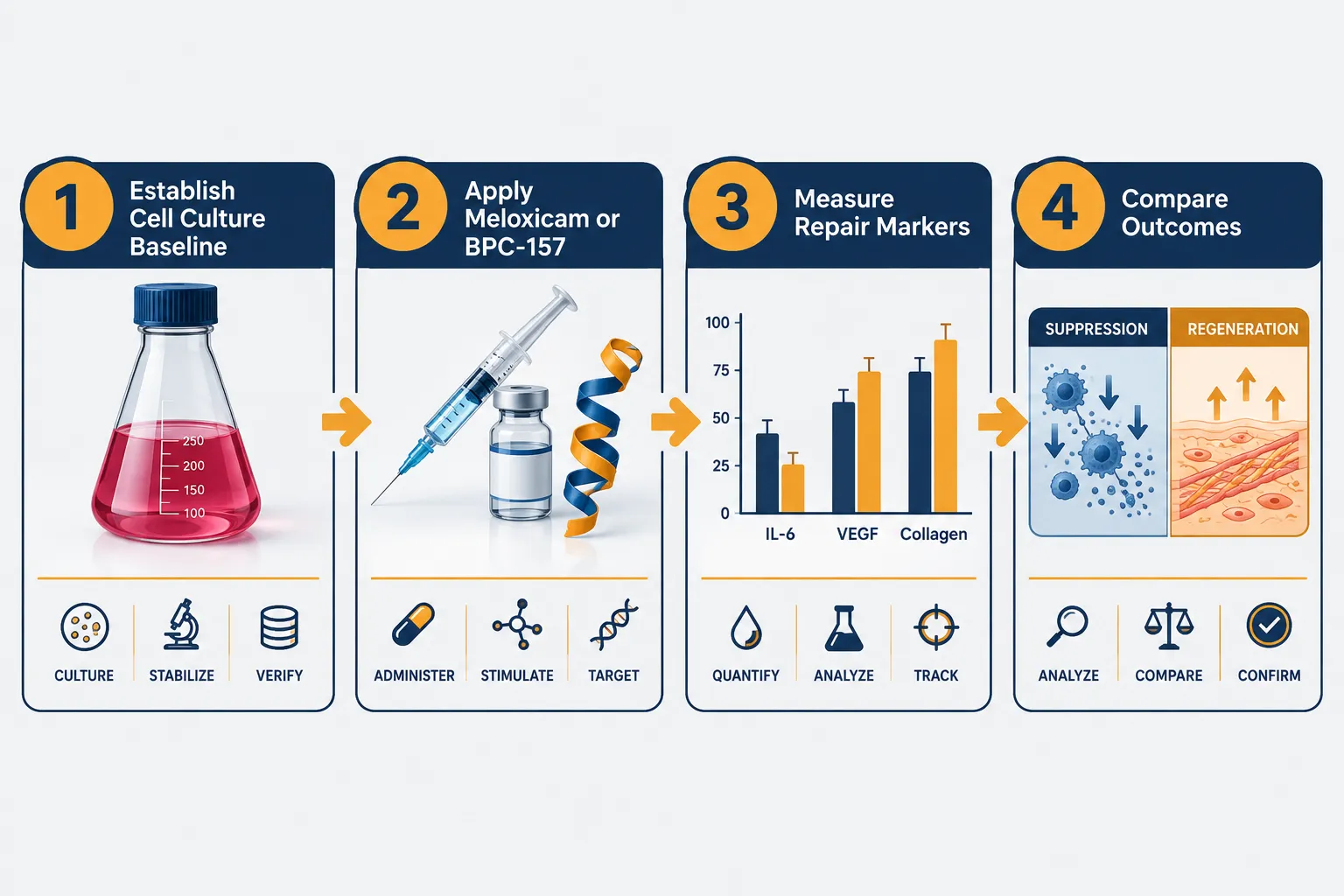
Designing in vitro experiments that meaningfully compare meloxicam and tissue-repair peptides: comparing nonsteroidal anti-inflammatory drugs with BPC-157 in in vitro models requires careful attention to endpoint selection, dosing protocols, and timing.

Practical Research Considerations When Comparing NSAIDs with BPC-157 in Cell Culture Systems

Key design considerations include:

  • Endpoint selection: If the primary endpoint is inflammation suppression (IL-6, TNF-alpha, PGE2), meloxicam performs reliably. If the endpoint is structural repair (collagen density, cell migration rate, VEGF expression), BPC-157 is the mechanistically appropriate comparator.
  • Timing of compound exposure: COX-2 inhibition is most relevant in the early inflammatory phase. BPC-157's growth-factor-driven effects are most active during the proliferative and remodeling phases. Applying both simultaneously may produce conflicting signals.
  • Concentration calibration: BPC-157 research protocols for gut and NSAID-induced damage models typically span 4-6 weeks in animal studies; in vitro timelines should account for the compound's mechanism of action rather than simply mirroring NSAID dosing schedules.

Researchers working with multiple peptide classes, such as those exploring GHK-Cu peptide for skin and connective tissue models, or reviewing CJC-1295 pharmacokinetic comparisons for growth hormone axis research, will recognize that peptide-driven repair signaling requires different experimental frameworks than small-molecule anti-inflammatory drugs.

One 2026 molecular docking study identified FER, TUBA1B, and MICAL2 as proteins where meloxicam shows strong in silico binding affinity, suggesting possible cytoskeletal and signaling roles beyond prostaglandin suppression. However, these findings remain computational and have not yet been validated in dedicated cell culture assays.

Regulatory context matters: BPC-157 is currently a research-only compound under ongoing regulatory review, while meloxicam is a fully approved NSAID. Researchers combining them in experimental settings should note that continuous COX-2 inhibition may theoretically blunt BPC-157's growth-factor-driven repair signaling. Using the lowest effective NSAID dose and avoiding around-the-clock administration during BPC-157 protocols is a commonly recommended precaution when structural repair is the primary endpoint.

Those exploring complementary peptide combinations may also find value in reviewing the synergy of LL-37 and SS-31 for additional context on how peptide combinations interact in repair-focused models. Similarly, researchers studying metabolic and hormonal contexts alongside tissue repair may reference Tesamorelin and Ipamorelin combination safety considerations as a model for responsible multi-compound research design.

For those sourcing research-grade compounds, wholesale peptides with verified purity documentation are essential for reproducible in vitro results.

Conclusion

The comparison of meloxicam and tissue-repair peptides: comparing nonsteroidal anti-inflammatory drugs with BPC-157 in in vitro models ultimately reveals two compounds with fundamentally different roles in tissue biology. Meloxicam is a well-characterized, clinically approved tool for reducing prostaglandin-mediated inflammation, effective, predictable, but limited in its capacity to actively drive structural repair. BPC-157 is a pleiotropic research peptide that promotes angiogenesis, collagen deposition, and fibroblast activity through growth factor pathways, offering a mechanistically distinct and potentially complementary profile.

Actionable next steps for researchers:

  1. Define repair-specific endpoints (VEGF, collagen, cell migration) separately from inflammation endpoints (IL-6, COX-2, PGE2) in study design.
  2. Avoid applying NSAID dosing logic to BPC-157 protocols; align exposure timing with the compound's mechanism of action.
  3. Treat computational findings (such as meloxicam's docking affinity to cytoskeletal proteins) as hypothesis-generating, not conclusive.
  4. Source third-party tested, purity-verified peptides to ensure experimental reproducibility.
  5. Monitor the regulatory landscape for BPC-157, as its status continues to evolve in 2026.

The field is moving toward a clearer understanding that analgesia with suppression and analgesia with repair are not interchangeable goals, and that in vitro models are the most precise tool available for distinguishing between them.

https://www.puretestedpeptides.com/wp-content/uploads/2026/09/meloxicam-and-tissue-repair-peptides-comparing-nonsteroidal-anti-inflammatory-dr.webp 1024 1536 https://www.puretestedpeptides.com/wp-content/uploads/2026/01/buy-peptides-online.jpg 2026-09-16 13:06:352026-09-16 13:06:35Meloxicam and Tissue-Repair Peptides: Comparing Nonsteroidal Anti-Inflammatory Drugs with BPC-157 in In Vitro Models
CJC-1295 With DAC vs Without DAC: Half-Life, Release Profile, and Research Selection Guide

CJC-1295 With DAC vs Without DAC: Half-Life, Release Profile, and Research Selection Guide

September 15, 2026/0 Comments/in Uncategorized/by

{"cover":"Professional landscape format (1536×1024) hero image with bold text overlay 'CJC-1295 With DAC vs Without DAC' in crisp white sans-serif on a deep navy semi-transparent overlay panel, centered with 8% safe margins from every edge. Background: high-resolution editorial split composition showing two glass laboratory vials side by side on a clean white lab bench, one labeled DAC and one labeled No DAC, soft studio lighting with cool blue and teal tones, shallow depth of field, molecular structure silhouettes faintly visible in background, pharmaceutical research magazine cover aesthetic, professional 2-color palette of navy and teal.","content":["Annotated mechanism diagram (1536×1024) illustrating half-life extension: left panel titled 'Without DAC, Mod GRF 1-29' shows a short curved arrow labeled '30 min half-life' with callout lines pointing to 'rapid clearance', 'pulsatile GH spike', and 'enzyme degradation sites'; right panel titled 'With DAC' shows a long curved arc labeled '6-8 day half-life' with callout lines pointing to 'albumin binding site', 'sustained GH elevation', and 'DAC moiety attachment point'; clean white background, teal and navy palette, thin callout lines, editorial medical-diagram polish, landscape 1536×1024.","Four-panel labelled comparison guide (1536×1024) for GH release profiles: Panel 1 heading 'Pulsatile Release' with bullet labels 'sharp GH peak', 'mimics natural rhythm', 'short dosing window'; Panel 2 heading 'Sustained Release' with bullet labels 'blunted peak', 'prolonged elevation', 'less frequent dosing'; Panel 3 heading 'Research Use: No DAC' with bullet labels 'stack flexibility', 'timing control', 'GHRP pairing'; Panel 4 heading 'Research Use: With DAC' with bullet labels 'once-weekly dosing', 'steady baseline', 'trough monitoring'; bright clinical white background, coral and navy accent colors, thin panel dividers, editorial infographic polish, landscape format.","Numbered step-by-step research selection flow diagram (1536×1024) titled 'Selecting the Right CJC-1295 Form': Step 1 labeled 'Define Research Goal' with sub-label 'pulsatile vs steady GH'; Step 2 labeled 'Assess Dosing Frequency' with sub-label 'daily vs weekly protocol'; Step 3 labeled 'Consider Stack Partners' with sub-label 'GHRP, ipamorelin, tesa'; Step 4 labeled 'Evaluate Safety Profile' with sub-label 'tachyphylaxis risk, DAC caution'; Step 5 labeled 'Source Verified Peptide' with sub-label 'purity testing required'; connected by bold directional arrows on a clean slate-gray background, teal step nodes, white label boxes, professional research-guide aesthetic, landscape 1536×1024."]

Professional landscape hero image () with a reading "CJC-1295 With DAC vs Without DAC". CRITICAL TYPOGRAPHY RULES: render

Two peptides share nearly identical names, yet their pharmacokinetic behavior is so different that swapping one for the other in a research protocol can produce completely opposite GH release patterns. Understanding the CJC-1295 with DAC vs without DAC distinction, including half-life, release profile, and research selection, is one of the most practically important decisions in growth hormone secretagogue research today.

Key Takeaways

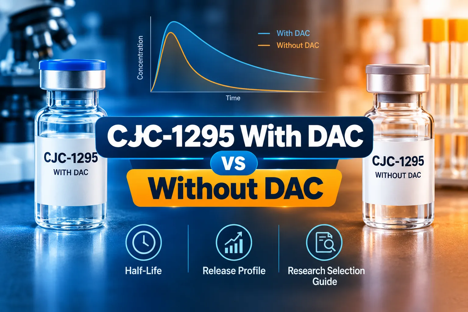
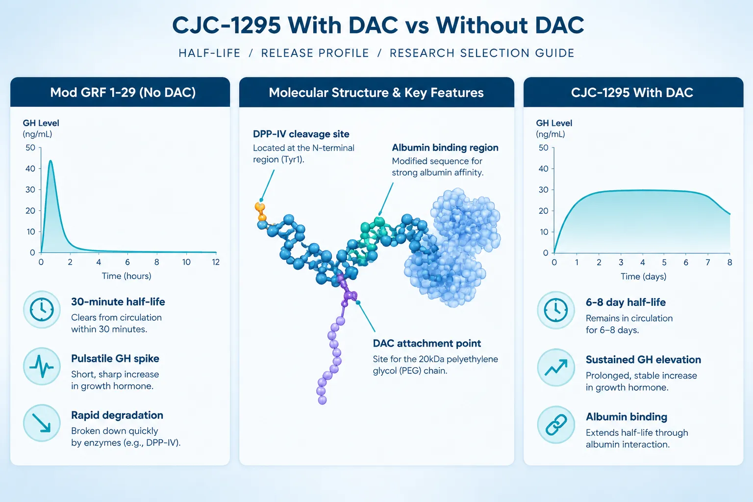
  • CJC-1295 without DAC (also called Mod GRF 1-29) has a half-life of roughly 30 minutes, producing a sharp, pulsatile GH spike.
  • CJC-1295 with DAC binds to albumin in plasma, extending its half-life to approximately 6-8 days and producing a sustained, blunted GH elevation.
  • The two variants are not interchangeable; each suits different research designs and stacking strategies.
  • Nomenclature confusion is common, "CJC-1295 no DAC" and "Mod GRF 1-29" refer to the same peptide.
  • Both remain research-only compounds in 2026, with no approved clinical indications.

What Is CJC-1295 and Why Does DAC Change Everything

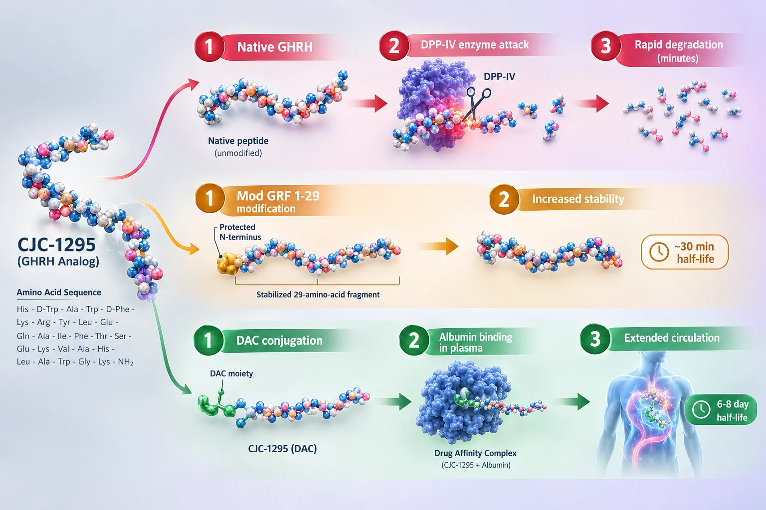
CJC-1295 is a synthetic analogue of growth hormone-releasing hormone (GHRH). Native GHRH is rapidly degraded by dipeptidyl peptidase IV (DPP-IV) enzymes in plasma, giving it a half-life of only a few minutes. Early modifications produced Mod GRF 1-29, a stabilized 29-amino-acid fragment with a half-life extended to roughly 30 minutes. This version is widely sold as "CJC-1295 without DAC" or simply "CJC-1295 no DAC."

The Drug Affinity Complex (DAC) technology takes stabilization a step further. A lysine residue is modified with a maleimidoproprionic acid group that forms a covalent bond with circulating albumin. Because albumin itself has a half-life of roughly 19 days, the DAC-conjugated peptide is shielded from clearance, extending its effective half-life to approximately 6-8 days in research models.

For a deeper look at the structural and pharmacokinetic differences between these two forms, the CJC-1295 with and without DAC mechanism and pharmacokinetic comparison covers the underlying science in detail.

What Is CJC-1295 and Why Does DAC Change Everything

The core structural difference:

Feature Mod GRF 1-29 (No DAC) CJC-1295 With DAC
Half-life ~30 minutes ~6-8 days
Albumin binding No Yes (covalent)
GH release pattern Pulsatile spike Sustained elevation
Dosing frequency Daily or per-session Once or twice weekly
DPP-IV resistance Partial High

Release Profile: Pulsatile vs Sustained GH Stimulation

The half-life gap between these two variants directly determines their GH release profiles, and this distinction sits at the heart of the CJC-1295 with DAC vs without DAC research selection guide.

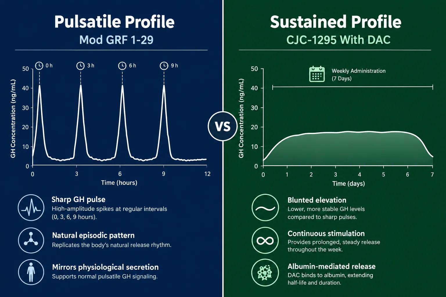
Mod GRF 1-29 (no DAC) produces a sharp, high-amplitude GH pulse that mirrors the body's natural episodic secretion pattern. Peak GH levels appear within 15-30 minutes of administration and return to baseline within a few hours. This pulsatile pattern is considered physiologically favorable by many researchers because it preserves the natural on-off rhythm of the somatotropic axis. It also offers precise timing control, making it easier to pair with ghrelin mimetics like ipamorelin or GHRP-2 for synergistic GH release.

Researchers studying GH secretagogue stacks often combine Mod GRF 1-29 with ipamorelin, as explored in resources on sermorelin, ipamorelin, and CJC-1295 combination protocols.

CJC-1295 with DAC produces a blunted but prolonged GH elevation. The Teichman 2006 Phase 1 study remains the most-cited pharmacokinetic anchor for this variant, demonstrating dose-dependent increases in IGF-1 lasting several days after a single injection. This sustained profile reduces the need for daily dosing but also raises concerns about tachyphylaxis, a desensitization of pituitary receptors caused by continuous GHRH stimulation rather than intermittent pulses.

"A sustained GH elevation is not automatically superior to a pulsatile one. The research question determines which profile is appropriate."

Release Profile: Pulsatile vs Sustained GH Stimulation

Researchers interested in body composition outcomes, including visceral fat reduction, may find relevant context in visceral fat research protocols that examine GH secretagogue effects on adipose tissue.

Research Selection Guide: Choosing Between CJC-1295 With DAC vs Without DAC

Selecting the correct variant depends on three primary research variables: the desired GH release pattern, the dosing schedule, and the peptide stack being used.

Choose Mod GRF 1-29 (no DAC) when:

  • The protocol requires mimicking natural pulsatile GH secretion
  • Daily or per-session administration is feasible
  • The peptide will be stacked with a GHRP or ipamorelin for amplified pulse height
  • Researchers want fine-grained control over timing and amplitude

Choose CJC-1295 with DAC when:

  • The protocol benefits from less frequent dosing (once or twice weekly)
  • A sustained IGF-1 elevation is the primary endpoint
  • The research design does not require precise pulse timing
  • Stacking with other secretagogues is not a primary concern

For multi-peptide research designs, blended formulations such as tesa, CJC-1295, and ipamorelin combination protocols offer an alternative approach worth reviewing.

Researchers should also note that the DAC variant carries a more cautious safety profile in 2026 consensus literature. Continuous GHRH receptor stimulation raises questions about receptor downregulation, and some protocols now include structured off-weeks when using the DAC form. The no-DAC variant's short half-life makes receptor rest automatic between doses.

For context on how other peptide classes interact with endocrine pathways, the overview of peptides and polypeptides in endocrine pharmacology provides useful background on receptor biology.

Research Selection Guide: Choosing Between CJC-1295 With DAC vs Without DAC

Nomenclature note: The label "CJC-1295 no DAC" is a vendor convention, not an official chemical name. The correct scientific designation is Modified GRF 1-29 (Mod GRF 1-29). Researchers sourcing peptides should confirm which compound is actually present, as mislabeling remains a documented issue in the research peptide supply chain.

Both compounds remain strictly research-use-only substances in 2026 with no approved human therapeutic applications.

Conclusion

The CJC-1295 with DAC vs without DAC comparison is not a question of which peptide is better, it is a question of which release profile matches the research objective. Mod GRF 1-29 delivers a short, sharp GH pulse ideal for pulsatile protocols and multi-peptide stacks. CJC-1295 with DAC delivers sustained GH elevation suited to low-frequency dosing designs, but demands greater attention to receptor desensitization risk.

Actionable next steps for researchers in 2026:

  1. Confirm the exact compound identity before designing any protocol, verify whether the supplier is providing Mod GRF 1-29 or the DAC-conjugated form.
  2. Match the release profile to the research endpoint: pulsatile for physiological mimicry, sustained for steady IGF-1 elevation studies.
  3. Review stacking compatibility before combining either variant with GHRPs or other secretagogues.
  4. Source only from suppliers with documented third-party purity testing to ensure compound integrity.
  5. Monitor current literature, as mid-2026 consensus continues to favor the no-DAC variant for most stacked research designs due to its more controllable pharmacokinetic profile.
https://www.puretestedpeptides.com/wp-content/uploads/2026/09/cjc-1295-with-dac-vs-without-dac-half-life-release-profile-and-research-selectio.webp 1024 1536 https://www.puretestedpeptides.com/wp-content/uploads/2026/01/buy-peptides-online.jpg 2026-09-15 13:05:092026-09-15 13:05:09CJC-1295 With DAC vs Without DAC: Half-Life, Release Profile, and Research Selection Guide
PT-141 Peptide Research Safety Guide: Dosing, Timing, and Tolerability Questions Labs Ask

PT-141 Peptide Research Safety Guide: Dosing, Timing, and Tolerability Questions Labs Ask

September 15, 2026/0 Comments/in Uncategorized/by

Roughly 40% of participants in early PT-141 clinical trials experienced nausea significant enough to require protocol adjustments, yet the compound still advanced through multiple phases of human study and reached regulatory approval. That tension between biological activity and tolerability management sits at the heart of every serious PT-141 Peptide Research Safety Guide: Dosing, Timing, and Tolerability Questions Labs Ask.

This article moves past mechanism basics. It focuses on the practical protocol questions that researchers, study designers, and compounding labs raise once they already understand how PT-141 (bremelanotide) activates melanocortin receptors.

Key Takeaways

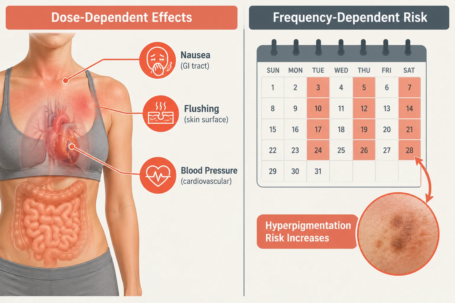
  • PT-141 has a well-characterized dose-dependent tolerability profile; nausea is the primary limiting adverse event and is strongly tied to dose size.
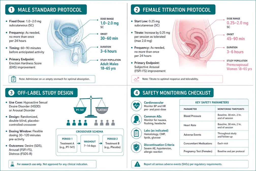
  • Approved subcutaneous dosing sits at 1.75 mg, but research protocols often start lower (0.5-1.0 mg) and titrate carefully.
  • Frequency of administration is a critical variable, hyperpigmentation risk rises sharply with repeated, closely spaced dosing.
  • Cardiovascular monitoring, especially blood pressure, is a non-negotiable safety checkpoint in any responsible study design.
  • Male and female research populations show meaningfully different tolerability patterns that labs must account for in study design.

What the Approved Dose Framework Tells Researchers

The FDA-approved subcutaneous dose of 1.75 mg, administered approximately 45 minutes before anticipated activity, forms the baseline reference point for any PT-141 Peptide Research Safety Guide: Dosing, Timing, and Tolerability Questions Labs Ask. Clinical monographs specify a maximum of one dose per 24 hours and no more than one dose every 72 hours in standard use.

What the Approved Dose Framework Tells Researchers

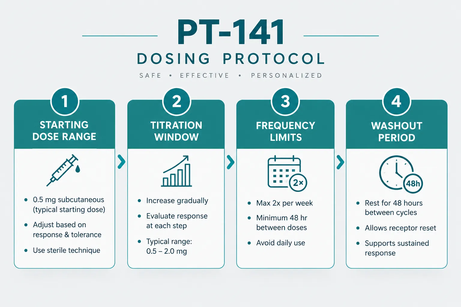
For research settings, these boundaries matter because they define the outer edge of a well-studied safety envelope. Most investigational protocols use this approved framework as a ceiling, not a starting point. Common research titration approaches include:

  • Starting dose: 0.5 mg subcutaneous to assess individual response
  • Intermediate dose: 1.0 mg if the 0.5 mg dose is well tolerated after two to three exposures
  • Standard research dose: 1.75 mg, consistent with the approved label
  • Frequency limit: No more than twice weekly in most current study designs, with a minimum 48-hour gap between doses

"The dose is only half the equation. How often a subject receives PT-141 determines a different set of risks entirely."

Intranasal administration was studied in earlier trials at doses up to 20 mg. Maximum-tolerated dose data from that work helped define the nausea threshold that now informs subcutaneous titration strategies. Subcutaneous delivery produces a more controlled absorption curve and is the standard route in current research.

For labs sourcing compounds for controlled studies, working with lab tested peptides is a baseline quality requirement before any dosing protocol can be considered valid.

Tolerability Profile: The Questions Labs Must Answer First

Every credible PT-141 Peptide Research Safety Guide: Dosing, Timing, and Tolerability Questions Labs Ask places tolerability assessment before efficacy measurement. The adverse event profile is well-characterized and dose-dependent.

Tolerability Profile: The Questions Labs Must Answer First

Nausea and Gastrointestinal Effects

Nausea is the most commonly reported adverse event across all PT-141 trials. Key findings from clinical data include:

  • Nausea incidence rises sharply above 1.75 mg
  • Onset typically occurs within 30-60 minutes of administration
  • Duration is usually under two hours at approved doses
  • Pre-dosing with a light meal reduces severity in most subjects

Labs designing multi-dose studies should build nausea scoring into every session log. Dropout rates in trials that did not manage nausea proactively were meaningfully higher than in those with structured tolerability support.

Cardiovascular and Blood Pressure Monitoring

PT-141 produces a transient, dose-dependent decrease in blood pressure followed by a rebound increase. This biphasic cardiovascular effect is one of the most important safety questions labs must address. Standard monitoring requirements include:

Monitoring Point Timing Relative to Dose
Baseline blood pressure 10 minutes before administration
First post-dose check 30 minutes after administration
Peak effect window 45-90 minutes after administration
Return-to-baseline check 3 hours after administration

Subjects with pre-existing cardiovascular conditions are typically excluded from PT-141 research protocols for this reason.

Skin Hyperpigmentation

This adverse effect is strongly frequency-dependent rather than dose-dependent. Melanocortin receptor activation drives melanin production, and repeated stimulation accumulates the effect. Labs running extended or repeat-dose studies must:

  • Document baseline skin tone and any existing pigmentation changes
  • Limit dosing frequency to no more than twice weekly
  • Include washout periods of at least two weeks between study phases
  • Monitor focal injection sites separately from systemic pigmentation changes

Male vs. Female Research Populations and Off-Label Study Design

The approved indication for PT-141 is hypoactive sexual desire disorder in premenopausal women. However, a significant portion of current research examines male populations and other off-label applications, which raises distinct study design questions.

Male vs. Female Research Populations and Off-Label Study Design

In female populations, the tolerability profile aligns closely with label data. Hormonal cycle phase at time of dosing is a variable some protocols now track, as receptor sensitivity may fluctuate across the cycle.

In male populations, research data suggest comparable efficacy signals at similar doses, but the tolerability curve differs slightly. Flushing and transient blood pressure changes appear more pronounced in some male cohorts. Labs designing male-focused studies often use a more conservative titration schedule.

Off-label research considerations for labs in 2026 include:

  • Clearly defined primary endpoints that do not rely solely on subjective self-report
  • Structured washout periods between study phases (minimum two weeks)
  • Exclusion criteria that address cardiovascular risk, active skin conditions, and concurrent melanocortin-affecting medications
  • Ethics board documentation that explicitly addresses the off-label nature of the research

Researchers comparing PT-141 to other peptides with central nervous system or receptor-mediated mechanisms may also find value in reviewing Semax research and Semax peptide literature for parallel study design frameworks. Similarly, labs building multi-peptide research panels sometimes reference SS-31 mitochondrial research themes for safety monitoring methodology applicable across peptide classes. For labs sourcing multiple compounds, wholesale peptides for sale options can support broader research programs, and GHK-Cu peptide studies offer additional reference points for skin-related adverse event monitoring protocols.

Conclusion

The PT-141 Peptide Research Safety Guide: Dosing, Timing, and Tolerability Questions Labs Ask comes down to three operational priorities: start low and titrate deliberately, treat frequency as a distinct risk variable from dose size, and build cardiovascular monitoring into every session regardless of subject health status.

Actionable next steps for labs:

  1. Establish a written titration protocol starting at 0.5 mg before moving to the 1.75 mg reference dose.
  2. Cap dosing frequency at twice weekly and document all inter-dose intervals.
  3. Implement a standardized blood pressure monitoring timeline at every administration session.
  4. Include skin pigmentation photography at baseline and at each study phase checkpoint.
  5. Differentiate male and female cohort protocols rather than applying a single design across populations.
  6. Source compounds only from suppliers providing documented purity and sterility testing.

Research into PT-141 continues to generate useful safety data in 2026. Labs that build rigorous tolerability frameworks from the start produce cleaner datasets and reduce the risk of protocol-disrupting adverse events mid-study.

https://www.puretestedpeptides.com/wp-content/uploads/2026/09/pt-141-peptide-research-safety-guide-dosing-timing-and-tolerability-questions-la.webp 1024 1536 https://www.puretestedpeptides.com/wp-content/uploads/2026/01/buy-peptides-online.jpg 2026-09-15 13:04:362026-09-15 13:04:36PT-141 Peptide Research Safety Guide: Dosing, Timing, and Tolerability Questions Labs Ask
BPC-157 and TB-500 Synergy: What Researchers Should Know About Combined Tissue Repair Models

BPC-157 and TB-500 Synergy: What Researchers Should Know About Combined Tissue Repair Models

September 15, 2026/0 Comments/in Uncategorized/by

A 2026 Phase 2 clinical trial launched to evaluate BPC-157 as a standalone agent for hamstring repair, with no TB-500 arm included. That design choice speaks volumes about where the science actually stands on combined peptide protocols.

Researchers and clinicians increasingly ask whether pairing BPC-157 and TB-500 produces better tissue repair outcomes than either peptide alone. The concept of BPC-157 and TB-500 synergy in combined tissue repair models is compelling on paper, but the evidence base demands careful reading before any research protocol is designed around it.

Key Takeaways

  • No peer-reviewed human trial has tested BPC-157 and TB-500 together; all synergy claims remain mechanistic extrapolations.
  • A 2026 rat Achilles tendon study found the combination did not outperform either single agent on any measured endpoint.
  • BPC-157 is proposed to act locally on microcirculation and matrix stability; TB-500 is proposed to support broader cytoskeletal remodeling, complementary roles, but unvalidated as a pair.
  • Neither peptide has FDA approval for any indication, and no reference dose or ratio exists for the combination.
  • Researchers designing combination studies should treat the "synergy" framing as a hypothesis requiring controlled testing, not an established outcome.

What the Research Actually Says About BPC-157 and TB-500 Synergy

The term "synergy" implies that two agents together produce a measurably greater effect than the sum of their individual contributions. For BPC-157 and TB-500 synergy in combined tissue repair models, that bar has not been cleared in any controlled study as of 2026.

A systematic review covering 36 BPC-157 studies found only one human trial, and none involving TB-500 co-administration. A parallel scoping review of thymosin-β4 and its synthetic fragment TB-500 confirmed that direct human evidence for TB-500 is limited to a single study in a corneal or wound context. Musculoskeletal and tendon repair claims for TB-500 remain extrapolations from animal work.

What the Research Actually Says About BPC-157 and TB-500 Synergy

The most direct test of the combination to date comes from a 2026 exploratory rat Achilles tendon model. Results were instructive, and sobering:

Group Median Load-to-Failure Statistical Significance vs. Control
Control 26.91 N ,
TB-500 (60 µg/kg/day) 37.41 N p = 0.041
BPC-157 37.16 N Not significant
Combination 34.54 N Not significant vs. any group

Both single-agent arms improved histopathology and extracellular matrix organization. The combination arm did not outperform either peptide alone on any measured endpoint, biomechanical or histological. This does not prove antagonism, but it does refute additive benefit in this model.

"Synergistic, neutral, or antagonistic outcomes are all plausible and currently indistinguishable given the available data."

Researchers exploring wound repair peptides in preclinical settings should treat this finding as a design signal: the combination arm needs its own hypothesis and endpoints, not borrowed assumptions.

The Mechanistic Rationale, and Its Limits

The theoretical case for combining BPC-157 and TB-500 rests on their proposed complementary mechanisms:

BPC-157 (Body Protection Compound-157)

  • Enhances local microcirculation at the injury site
  • Modulates growth factor balance, including VEGF and EGF pathways
  • Supports extracellular matrix stability and collagen organization
  • Primarily acts at or near the site of administration

TB-500 (Thymosin-Beta 4 fragment)

  • Binds actin and regulates cytoskeletal dynamics
  • Promotes cell migration and tissue remodeling systemically
  • Supports broader structural repair beyond the local injury zone
  • Proposed to complement BPC-157's local action with systemic reach

This complementary framing is mechanistically plausible. However, there is no pharmacokinetic data on how the two peptides interact when co-administered, no dose-response or ratio-finding study for the blend, and no validated interaction endpoint in any model system.

The Mechanistic Rationale, and Its Limits

Researchers studying other peptide combinations, such as those examining the synergy of LL-37 and SS-31, will recognize this pattern: mechanistic complementarity is a starting point, not a conclusion. The same standard applies here.

For context on how wound models are typically structured to test such hypotheses, established preclinical frameworks require defined endpoints, dose-ranging arms, and controls for each agent alone before a combination arm can be interpreted.

What Researchers Should Know About Designing Combined Tissue Repair Models

Given the evidence gaps, researchers approaching BPC-157 and TB-500 synergy in combined tissue repair models need a structured framework. The following considerations are essential:

What Researchers Should Know About Designing Combined Tissue Repair Models

1. Regulatory and Approval Status
Neither BPC-157 nor TB-500 holds FDA approval for any indication. There is no established reference dose, approved ratio, or standardized clinical use for either peptide alone, let alone the combination. All research use must account for applicable institutional and regulatory requirements.

2. Dosing Protocols in Research Literature
Clinician-reviewed protocol guides describe a commonly referenced research dosing framework, sometimes called the "Wolverine stack", as follows:

  • BPC-157: 250-500 µg subcutaneously per day, administered near the injury site
  • TB-500: 2-2.5 mg subcutaneously twice weekly
  • Loading phase: 4-6 weeks
  • Maintenance phase: reduced frequency for an additional 4-8 weeks

These regimens are not supported by controlled trials. They are observational and protocol-style frameworks, not validated clinical schedules.

3. Endpoint Selection
The 2026 Achilles tendon study demonstrated that histological improvement and biomechanical improvement do not always move together. Researchers should pre-specify both types of endpoints and include single-agent control arms to allow meaningful interpretation of any combination result.

4. The Human Evidence Gap
The most advanced formal BPC-157 program as of 2026 is a randomized, double-blind, placebo-controlled Phase 2 trial (NCT07437547) evaluating BPC-157 alone for acute grade II hamstring strain. This trial does not include TB-500. It signals institutional interest in BPC-157 as a regulated agent but cannot inform synergy questions.

TB-500's human evidence base is similarly thin, concentrated in corneal and wound healing contexts, with no controlled musculoskeletal trial completed. Researchers reviewing other peptide science programs, such as Semax research protocols, will note that even well-studied peptides face significant translational gaps from animal to human data.

5. Long-Term Safety
No long-term safety dataset exists for either peptide, individually or in combination. Researchers should not assume that tolerability in short-duration animal studies translates to human safety profiles.

Conclusion

The concept of BPC-157 and TB-500 synergy in combined tissue repair models is scientifically interesting and mechanistically coherent, but it remains a hypothesis in 2026, not a validated outcome. The most recent animal data suggests the combination may not add benefit over either agent alone, at least in tendon repair models. No human combination trial exists, no reference dosing ratio has been established, and no pharmacokinetic interaction data is available.

Actionable next steps for researchers:

  • Design combination studies with single-agent control arms and pre-specified endpoints for both histological and biomechanical outcomes.
  • Treat published protocol dosing frameworks as starting hypotheses, not validated regimens.
  • Monitor the Phase 2 BPC-157 trial (NCT07437547) for safety, tolerability, and endpoint data that may inform future combination study design.
  • Review the broader wound repair peptides literature to contextualize BPC-157 and TB-500 findings within established tissue repair frameworks.
  • Document regulatory status clearly in all research materials, neither peptide is approved for any clinical indication.

The combination question is worth investigating rigorously. The current evidence simply has not answered it yet.

https://www.puretestedpeptides.com/wp-content/uploads/2026/09/bpc-157-and-tb-500-synergy-what-researchers-should-know-about-combined-tissue-re.webp 1024 1536 https://www.puretestedpeptides.com/wp-content/uploads/2026/01/buy-peptides-online.jpg 2026-09-15 13:04:352026-09-15 13:04:35BPC-157 and TB-500 Synergy: What Researchers Should Know About Combined Tissue Repair Models
5-Amino-1MQ and MOTS-c Synergy: What Makes the Combination Interesting in Metabolic Research

5-Amino-1MQ and MOTS-c Synergy: What Makes the Combination Interesting in Metabolic Research

September 15, 2026/0 Comments/in Uncategorized/by

Metabolic disease now affects more than one billion people worldwide, yet most approved interventions target only a single pathway. That single-target limitation is precisely why researchers are turning toward compound combinations that work on different parts of the same system simultaneously. The study of 5-Amino-1MQ and MOTS-c synergy: what makes the combination interesting in metabolic research sits at the center of this shift, drawing attention for its mechanistic logic even before formal clinical trials have begun.

Key Takeaways

  • 5-Amino-1MQ inhibits the NNMT enzyme, preserving NAD+ and driving thermogenesis in preclinical fat models.
  • MOTS-c is a mitochondria-derived peptide that activates AMPK and modulates mTOR signaling, with stronger human evidence than 5-Amino-1MQ.
  • The two compounds target complementary, non-redundant pathways, which is the core rationale for pairing them.
  • Neither compound is approved for human therapeutic use; both remain research-only, and MOTS-c is banned in competitive sport.
  • Triple mitochondrial stacks combining NAD+ precursors, MOTS-c, and 5-Amino-1MQ are emerging in 2026 research discussions, though human data is absent.

How Each Compound Works Independently

Understanding the synergy starts with understanding each agent on its own terms.

5-Amino-1MQ is a small-molecule inhibitor of nicotinamide N-methyltransferase (NNMT), an enzyme highly expressed in fat tissue. When NNMT is active, it consumes S-adenosylmethionine (SAM) and depletes the NAD+ pool. By blocking NNMT, 5-Amino-1MQ preserves cellular NAD+, raises the SAM-to-SAH ratio, and shifts white adipocytes toward a more thermogenic phenotype. In obese mouse models reported through 2024-2026, NNMT inhibition with this compound limited weight gain, reduced fat mass, and improved liver pathology markers associated with non-alcoholic fatty liver disease (NAFLD). Researchers sourcing this compound can review options under 5-amino peptide research products or 5-Amino-1MQ 60 capsule formulations.

MOTS-c is a 16-amino-acid peptide encoded within the mitochondrial genome, specifically the 12S rRNA region. It functions as a mitochondrial-derived signaling molecule that translocates to the nucleus under metabolic stress. Its primary downstream effect is activation of AMP-activated protein kinase (AMPK), the cell's master energy sensor. Secondary effects include modulation of mTOR signaling and improvements in insulin sensitivity. MOTS-c has a more mature evidence base than 5-Amino-1MQ, with data spanning rodent models, aging studies, and early human observations in exercise physiology. Those researching this peptide can explore MOTS-c from Peptide Sciences.

How Each Compound Works Independently

The Mechanistic Case for 5-Amino-1MQ and MOTS-c Synergy in Metabolic Research

"Complementary, not redundant" is the phrase researchers use most often when describing why these two compounds are paired.

The logic is straightforward. 5-Amino-1MQ works upstream in the NAD+ biosynthesis and methylation axis. MOTS-c works at the AMPK/mTOR node. These are distinct steps in the same broader metabolic network, which means:

Feature 5-Amino-1MQ MOTS-c
Primary target NNMT enzyme AMPK activation
Key substrate NAD+ / SAM pool Mitochondrial stress signals
Main tissue effect White adipose thermogenesis Skeletal muscle, liver, cardiac
Evidence stage Preclinical (rodent, 2024-2026) Preclinical + early human
Regulatory status Research only Research only; banned in sport

When NAD+ is preserved by NNMT inhibition, mitochondrial function improves. When AMPK is simultaneously activated by MOTS-c, the cell is signaled to increase fatty acid oxidation and reduce anabolic mTOR activity. The two signals reinforce each other without competing for the same receptor or enzyme. This is the mechanistic core of the 5-Amino-1MQ and MOTS-c synergy argument.

Researchers studying related mitochondria-targeted peptides, such as those reviewed in the SS-31 mechanism and research overview, will recognize a similar logic: compounds that protect mitochondrial membrane integrity can amplify the effects of signaling peptides that depend on healthy mitochondrial function.

The Mechanistic Case for 5-Amino-1MQ and MOTS-c Synergy in Metabolic Research

Emerging Stacking Protocols and What Makes This Combination Interesting in Metabolic Research

In 2026, research community discussions have moved beyond single-compound protocols toward triple mitochondrial stacks that combine an NAD+ precursor (such as NMN or NR), MOTS-c, and 5-Amino-1MQ. The rationale for the three-way combination is layered:

  1. NAD+ precursors provide raw substrate for sirtuin activation and mitochondrial repair.
  2. 5-Amino-1MQ prevents NNMT from consuming that NAD+ before it can be used.
  3. MOTS-c activates AMPK to ensure the cell actually burns the available energy rather than storing it.

For the two-compound pairing specifically, practical research protocols in 2026 emphasize staggered dosing rather than simultaneous administration. The reasoning is pharmacokinetic: allowing 5-Amino-1MQ to elevate NAD+ levels before MOTS-c is introduced may create a more favorable intracellular environment for AMPK signaling. Endpoint monitoring in such protocols typically tracks fasting glucose, insulin sensitivity markers, body composition changes, and liver enzyme panels.

Researchers interested in peptide stacking logic more broadly may find useful context in the IPA Sermorelin stack research article and the detailed CJC-1295 pharmacokinetic comparison, both of which illustrate how sequencing affects compound performance. For a broader view of how peptides compare to small-molecule drugs in cardiometabolic models, the polypeptide peptides in cardiometabolic models review provides relevant background.

Emerging Stacking Protocols and What Makes This Combination Interesting in Metabolic Research

Safety Considerations and the Limits of Current Evidence

Enthusiasm for the combination must be balanced against what is not yet known.

Known unknowns include:

  • No published human pharmacokinetic data for the combination
  • No dose-ranging safety studies for the pairing in any species
  • Unknown interaction effects at the NAD+/AMPK convergence point under chronic dosing
  • MOTS-c is classified as a prohibited substance in competitive sport by WADA, creating legal and ethical considerations for athlete-adjacent research

The evidence asymmetry between the two compounds is also worth noting. MOTS-c has a more developed research profile, including cardiac metabolism studies and aging-related data. 5-Amino-1MQ's most compelling data comes from the 2024-2026 wave of NNMT-inhibition studies in obese rodent models. Extrapolating preclinical findings to human applications remains speculative for both, and doubly so for their combination.

Predicted future applications in obesity, NAFLD, metabolic syndrome, and aging-related metabolic decline are scientifically plausible given the mechanisms involved. However, plausibility is not evidence, and researchers should treat current protocols as hypothesis-generating rather than therapeutically validated.

Conclusion

The scientific interest in 5-Amino-1MQ and MOTS-c synergy: what makes the combination interesting in metabolic research rests on a sound mechanistic foundation. NAD+ preservation through NNMT inhibition and AMPK activation through mitochondrial peptide signaling are genuinely complementary processes. The preclinical data for each compound independently is promising, particularly the 2024-2026 NNMT-inhibition findings for liver and adipose outcomes.

Actionable next steps for researchers:

  • Review the primary NNMT-inhibition literature before designing combination protocols.
  • Apply staggered dosing sequences and document pharmacokinetic windows carefully.
  • Select validated endpoints (glucose, insulin, body composition, liver enzymes) rather than relying on subjective outcomes.
  • Monitor regulatory updates on MOTS-c status, particularly in sport and clinical research contexts.
  • Treat any human-adjacent findings as preliminary until peer-reviewed combination studies exist.

The combination is not yet proven. It is, however, one of the more rationally designed pairings in current metabolic peptide research, and that distinction alone makes it worth watching closely.

https://www.puretestedpeptides.com/wp-content/uploads/2026/09/5-amino-1mq-and-mots-c-synergy-what-makes-the-combination-interesting-in-metabol.webp 1024 1536 https://www.puretestedpeptides.com/wp-content/uploads/2026/01/buy-peptides-online.jpg 2026-09-15 13:04:162026-09-15 13:04:165-Amino-1MQ and MOTS-c Synergy: What Makes the Combination Interesting in Metabolic Research
Enclomiphene Citrate vs Enclomiphene: Stability, Storage, and Research Handling

Enclomiphene Citrate vs Enclomiphene: Stability, Storage, and Research Handling

September 15, 2026/0 Comments/in Uncategorized/by

Most researchers who order enclomiphene for laboratory work assume the citrate salt and the free base are the same compound with a different label. That assumption can compromise data, shorten shelf life, and introduce variables that are difficult to trace back to the source material. Understanding the distinctions in Enclomiphene Citrate vs Enclomiphene: Stability, Storage, and Research Handling is not a minor technical footnote, it is foundational to reproducible science.

Key Takeaways

  • Enclomiphene citrate is a salt form of enclomiphene; the citrate counterion meaningfully changes solubility, crystalline stability, and handling requirements.
  • Research-grade powder in either form is significantly more stable than aqueous or organic solutions, which degrade faster under ambient conditions.
  • Both forms are sensitive to light, moisture, and heat; amber vials, desiccants, and cold storage are non-negotiable for long-term integrity.
  • Finished formulations such as capsules and liquid suspensions show acceptable stability at or below 25 °C when properly packaged.
  • Vendor quality documentation, including certificates of analysis and HPLC purity data, is a critical checkpoint before any research use.

What the Citrate Salt Actually Changes

What the Citrate Salt Actually Changes

Enclomiphene is the trans-isomer of clomiphene, a selective estrogen receptor modulator with growing research interest in male reproductive endocrinology. When enclomiphene is converted to its citrate salt, a citric acid molecule pairs ionically with the enclomiphene base. This is not a cosmetic change.

The citrate counterion delivers three practical advantages:

Property Enclomiphene (Free Base) Enclomiphene Citrate
Water solubility Low Moderately higher
Crystalline stability Moderate Greater
Formulation flexibility Limited Broader
Hygroscopicity High Reduced

The increased water solubility of the citrate form matters when preparing stock solutions for in vitro work. The free base requires organic solvents such as DMSO or ethanol to dissolve reliably, and those solvents introduce their own biological variables at higher concentrations. The citrate salt can often be dissolved in aqueous buffers at research-relevant concentrations, which simplifies assay design.

"The choice between enclomiphene base and enclomiphene citrate is not arbitrary, it directly affects dissolution behavior, solution stability, and the reproducibility of downstream assays."

The molecular weight difference between the two forms is also worth noting. Enclomiphene free base has a molecular weight of approximately 406 g/mol, while the citrate salt sits closer to 598 g/mol. Researchers calculating molar concentrations must account for this difference to avoid systematic dosing errors across experiments.

This distinction mirrors well-established patterns seen with the parent compound clomiphene citrate, where the salt form has long been preferred for pharmaceutical manufacturing because of its superior stability profile and more predictable dissolution kinetics.

Stability Factors: Light, Moisture, Heat, and Form

Stability Factors: Light, Moisture, Heat, and Form

Both enclomiphene and enclomiphene citrate are susceptible to the same primary degradation drivers: ultraviolet light, ambient moisture, and elevated temperature. However, the degree of vulnerability differs between solid powder, aqueous solution, and organic solution.

Solid Powder Stability

Research-grade powder, whether free base or citrate, is the most stable form. When stored correctly, properly manufactured powder can maintain integrity for 24 months or longer. Correct storage means:

  • Temperature at or below -20 °C for long-term archival
  • Desiccant packs inside a sealed, airtight container
  • Amber or opaque vials to block UV exposure
  • Minimal handling that introduces ambient air or humidity

The citrate form benefits from reduced hygroscopicity relative to the free base. In practical terms, this means the citrate powder is less likely to absorb atmospheric moisture during brief handling windows, which is a meaningful advantage in non-controlled lab environments.

Solution Stability

Aqueous solutions of enclomiphene citrate are markedly less stable than the solid form. At room temperature, degradation can become measurable within days. Refrigerated aqueous solutions (2-8 °C) extend working life but should still be used within a short window, typically 48 to 72 hours for research applications.

Organic solvent solutions (DMSO, ethanol) offer moderate stability but are not suitable for extended storage. Freeze-thaw cycling accelerates degradation in both aqueous and organic solutions, which is why aliquoting into single-use volumes before freezing is standard practice.

Finished Formulations

Capsule and liquid suspension formulations of enclomiphene citrate demonstrate extended stability at or below 25 °C when packaged with appropriate moisture barriers. Regulatory stability expectations for finished dosage forms generally require data supporting a shelf life of 24 months under controlled conditions, and enclomiphene citrate formulations have been evaluated under those frameworks in clinical development programs.

Practical Research Handling for Enclomiphene Citrate vs Enclomiphene

Practical Research Handling for Enclomiphene Citrate vs Enclomiphene

Applying the stability science above to day-to-day laboratory practice requires a clear protocol. The following guidance applies to both forms but is especially relevant for enclomiphene citrate given its broader use in research pipelines.

Before Use

  • Verify the certificate of analysis (COA) from the supplier, confirming HPLC purity of 98% or above
  • Confirm the compound is labeled correctly as citrate salt or free base, the two are not interchangeable in concentration calculations
  • Inspect the desiccant seal; a broken seal is grounds for rejection

During Reconstitution

  • Warm sealed vials to room temperature before opening to prevent condensation on the powder
  • Use the appropriate solvent for the form: aqueous buffer for citrate, DMSO or ethanol for free base
  • Prepare only the volume needed; do not reconstitute bulk quantities

During Storage

  • Store reconstituted solutions at 2-8 °C and use within 48-72 hours
  • Store dry powder at -20 °C in amber vials with desiccant
  • Log every freeze-thaw cycle; limit to no more than two per aliquot

Researchers sourcing serms for comparative studies should apply the same handling discipline to related compounds. For those working with peptide-based research compounds alongside small-molecule serms, foundational guidance on compound classes is available in resources covering peptides 101 for research-use only buyers.

Vendor selection is equally important. A vendor scoring rubric that evaluates third-party testing, cold-chain shipping, and documentation practices will reduce the risk of receiving degraded or mislabeled material. Researchers running multi-compound protocols, for example, pairing a serm with a growth hormone secretagogue, can find relevant product context in resources such as the IPA Sermorelin stack research overview.

For those evaluating research-grade serm products specifically, the serm 10mg product page provides purity and specification details relevant to procurement decisions.

Conclusion

The distinction between enclomiphene citrate and enclomiphene free base is not semantic. The citrate salt offers measurably better crystalline stability, reduced hygroscopicity, and broader formulation options, but it also carries a different molecular weight that must be factored into every concentration calculation. Both forms demand cold, dark, dry storage in the solid state, and both degrade significantly faster once dissolved.

Actionable next steps for researchers in 2026:

  1. Audit current stock: confirm whether materials on hand are labeled as citrate salt or free base and recalculate molar concentrations accordingly.
  2. Implement single-use aliquoting before freezing to eliminate freeze-thaw degradation.
  3. Require HPLC-verified COAs from all suppliers before accepting shipments.
  4. Establish a 48-72 hour use window for all reconstituted solutions stored at 2-8 °C.
  5. Apply the same vendor documentation standards to all research compounds, not just serms.

Reproducibility in enclomiphene research depends on treating the compound form as a controlled variable, not an afterthought.

https://www.puretestedpeptides.com/wp-content/uploads/2026/09/enclomiphene-citrate-vs-enclomiphene-stability-storage-and-research-handling.webp 1024 1536 https://www.puretestedpeptides.com/wp-content/uploads/2026/01/buy-peptides-online.jpg 2026-09-15 13:04:102026-09-15 13:04:10Enclomiphene Citrate vs Enclomiphene: Stability, Storage, and Research Handling
Page 16 of 81«‹1415161718›»
×

Helpful Links

  • My account
  • Cart
  • Checkout
  • Refund and Returns Policy
  • Privacy Policy
  • SMS Privacy Policy
  • Login
  • My Account
  • Logout

USA Made Lab Tested Peptides

All products are sold for research, laboratory, or analytical purposes only, and are not for human consumption


Pure Tested Peptides is a chemical supplier. Pure Tested Peptides is not a compounding / chemical compounding facility as defined under 503A of the Federal Food, Drug, and Cosmetic act. Pure Tested Peptides is not an outsourcing facility as defined under 503B of the Federal Food, Drug, and Cosmetic act.

The statements made within this website have not been evaluated by the US Food and Drug Administration. The products we offer are not intended to diagnose, treat, cure or prevent any disease.

Human/Animal Consumption Prohibited. Laboratory/In-Vitro Experimental Use Only

Scroll to top Scroll to top Scroll to top