Call or Text 727-513-9780
  • Shopping Cart Shopping Cart
    0Shopping Cart
Pure Tested Peptides | America's most trusted Peptides for sale online
  • Peptides for sale
    • Oral Peptides for sale
      • Peptide Capsules for sale
      • BPC 157 Capsules 1000mcg
      • SLU-PP-332 Capsules | 1000 mcg
      • 5-Amino-1MQ 50mg Capsules
      • Tesofensine 500mcg
    • All Peptides for sale
    • Peptide Sprays
      • BPC 157 Nasal Spray Kit
      • BPC-157 TB500 Nasal Spray Kit
      • Semax Nasal Spray 10mg
      • Selank – Nasal Spray Kit – 10mg
      • Epithalon 50MG Nasal Spray Kit
      • Ipamorelin 10mg Nasal Spray
      • Klow Nasal Spray (BPC-157 + TB-500 + GHK-Cu + KPV) | 80mg
      • Hulk Nasal Spray Tesa / Ipa Blend 6/3 MG
      • Klow Nasal Spray
      • NAD + 500 mg Nasal Spray
      • PT-141 Nasal Spray Kit
    • GHRH Peptides
      • Ipa Peptides
      • CJC-1295 Peptides
        • CJC-1295 with DAC 5 mg
        • CJC-1295 without DAC 5 mg
        • CJC-1295 Ipa 10mg
      • Tesa Peptides
        • Tesa Peptide
        • Tesa 20 mg
    • GHK-Cu Peptides
      • All GHK-Cu Peptides
      • GHK-Cu 100mg
      • KLOW Peptide Blend – Buy KLOW blend online
    • BPC Peptides
      • All BPC Peptides
      • BPC-157
      • BPC-157 TB-500
      • BPC 157 capsules 1000mcg
    • SLU-PP-332 Peptides
      • All SLU-PP-332 Peptides
      • SLU-PP-332 5mg
    • GLP3 Peptides
      • GLP3-R
      • GLP3-R CAG 10mg
      • GLP3-R 20mg
    • PT-141 Peptides
      • PT-141 Peptides for sale
      • PT-141 10mg
      • PT-141 Nasal Spray
    • CAG Peptides
      • Lipo-C Peptide Blend
      • CAG 5mg
      • CAG 10mg
    • MOTS-C Peptides
      • MOTS-C Peptides for sale
      • MOTS-c peptide
      • MOTS-c 10mg *6 pack*
    • 5 Amino 1MQ Peptides
      • 5 Amino 1MQ Peptides for sale
      • 5-Amino-1MQ 50mg Capsules
      • 5-Amino-1MQ 5mg
    • Epithalon Peptides
      • Epithalon Peptides for sale
      • Epithalon 10mg
      • Epithalon 50mg
  • Shop
    • GLPs
      • 5-Amino-1MQ 50mg Capsules
      • 5-Amino-1MQ 5mg
      • GLP3-Reta
      • L-Carnitine 500mg/ml
      • Tesofensine 500mcg
      • SLU-PP-332 5mg
      • MOTS-c 10mg *6 pack*
    • Epithalon & BPC Peptides
      • Epithalon 10mg
      • Epithalon 50mg
      • BPC-157
      • BPC 157 capsules 1000mcg
      • BPC-157 TB-500
      • BPC-157 TB500 Nasal Spray Kit
      • BPC 157 Nasal Spray Kit
    • BPC TB-500 & NAD+ Peptides
      • NAD+ 500 mg
      • KLOW Peptide Blend – Buy KLOW blend online
      • GLOW Peptide Blend
      • TB 500 5mg
      • BPC 157 capsules 1000mcg – Supplement
      • BPC 157 Nasal Spray Kit
      • BPC-157
      • BPC-157 TB500 Nasal Spray Kit
      • BPC-157 TB-500
      • BPC 157 capsules 1000mcg
    • LL-37 Peptide
      • LL-37 10 mg
    • MOTS-C & Selank
      • MOTS-c peptide
      • Selank 10mg
    • GHK Peptides
      • GHK-Cu 100mg
      • GLOW Peptide Blend
      • KLOW Peptide Blend – Buy KLOW blend online
  • COAs
  • Wholesale
    • Wholesale Peptides for sale
  • PTP FAQ
  • Affiliates
    • Selank Peptide: Advanced Pharmacological Mechanisms Underlying Its Anxiolytic and Nootropic Effects in Research
      • 5-Amino-1MQ Peptide: Detailed Mechanisms of NNMT Inhibition and Its Impact on Cellular Metabolism Research
        • MOTS-C Peptide: Unraveling Its Role in Mitochondrial Dynamics and Energy Metabolism Research
          • Epithalon Peptide and Telomerase Regulation: Investigating Its Impact on Cellular Senescence and Lifespan Research Models
            • GLP2-T vs GLP2 Tirz Peptide: Understanding the Naming, Mechanistic Nuances, and Research Implications for Gut Health
      • Peptides and Polypeptides in Modern Pharmacology: What Research on Metoprolol, Prednisone, and Amlodipine Reveals
        • Polypeptide Peptides vs NSAIDs: What Naproxen and Diclofenac Teach Tissue-Repair Researchers About BPC‑157 and TB‑500
          • Peptides and Polypeptides in Endocrine Pharmacology: How GLP-1, GLP-2, and GLP-3 Retatrutide Differ From Classic Drugs Like Prednisone and Amlodipine
          • 5‑Amino‑1MQ Peptide and NNMT Inhibition: How It Compares With Statins Like Atorvastatin in Adipose and Lipid Metabolism Research
      • Enclomiphene, Estrogen Receptor Signaling, and Luteinizing Phase Biology: What Hormone Researchers Should Measure
        • Mesenchymal Stem Cells, BPC‑157, and GHK‑Cu: How Tissue Repair Peptides Compare With Classic NSAIDs Like Naproxen in Injury Models
          • Peptide Calculator Use Cases Beyond Growth Hormone: Working Through GLP‑3 Retatrutide, MOTS‑c, and BPC‑157 Research Dosing
        • Polypeptide Peptides in Endocrine and Metabolic Pharmacology: Lessons From Amlodipine, Prednisone, and Metoprolol
        • Peptides Calculator for Advanced Blends: Worked Examples for Tesamorelin, CJC‑1295, and Ipamorelin Stacks
          • Peptides and Polypeptides in Human Physiology: How Molecular Size Shapes Research Applications
            • Selank Peptide: Advanced Pharmacological Mechanisms Underlying Its Anxiolytic and Nootropic Effects in Research
          • 5-Amino-1MQ Peptide: Detailed Mechanisms of NNMT Inhibition and Its Impact on Cellular Metabolism Research
            • MOTS-C Peptide: Unraveling Its Role in Mitochondrial Dynamics and Energy Metabolism Research
              • Epithalon Peptide and Telomerase Regulation: Investigating Its Impact on Cellular Senescence and Lifespan Research Models
                • GLP2-T vs GLP2 Tirz Peptide: Understanding the Naming, Mechanistic Nuances, and Research Implications for Gut Health
                  • Peptides and Polypeptides in Endocrine Pharmacology: How GLP-1, GLP-2, and GLP-3 Retatrutide Differ From Classic Drugs Like Prednisone and Amlodipine
                    • Polypeptide Peptides vs NSAIDs: What Naproxen and Diclofenac Teach Tissue-Repair Researchers About BPC‑157 and TB‑500
                      • Peptides and Polypeptides in Modern Pharmacology: What Research on Metoprolol, Prednisone, and Amlodipine Reveals
                        • 5‑Amino‑1MQ Peptide and NNMT Inhibition: How It Compares With Statins Like Atorvastatin in Adipose and Lipid Metabolism Research
                        • Enclomiphene, Estrogen Receptor Signaling, and Luteinizing Phase Biology: What Hormone Researchers Should Measure
                        • Mesenchymal Stem Cells, BPC‑157, and GHK‑Cu: How Tissue Repair Peptides Compare With Classic NSAIDs Like Naproxen in Injury Models
                        • Peptide Calculator Use Cases Beyond Growth Hormone: Working Through GLP‑3 Retatrutide, MOTS‑c, and BPC‑157 Research Dosing
                        • Polypeptide Peptides in Endocrine and Metabolic Pharmacology: Lessons From Amlodipine, Prednisone, and Metoprolol
                        • Peptides Calculator for Advanced Blends: Worked Examples for Tesamorelin, CJC‑1295, and Ipamorelin Stacks
                        • Peptides and Polypeptides in Human Physiology: How Molecular Size Shapes Research Applications
                        • Selank Peptide: Advanced Pharmacological Mechanisms Underlying Its Anxiolytic and Nootropic Effects in Research
                        • MOTS-C Peptide: Unraveling Its Role in Mitochondrial Dynamics and Energy Metabolism Research
                        • Peptides and Polypeptides in Modern Pharmacology: What Research on Metoprolol, Prednisone, and Amlodipine Reveals
                        • Peptides and Polypeptides in Endocrine Pharmacology: How GLP-1, GLP-2, and GLP-3 Retatrutide Differ From Classic Drugs Like Prednisone and Amlodipine
                        • 5-Amino-1MQ Peptide: Detailed Mechanisms of NNMT Inhibition and Its Impact on Cellular Metabolism Research
                        • Mesenchymal Stem Cells, BPC‑157, and GHK‑Cu: How Tissue Repair Peptides Compare With Classic NSAIDs Like Naproxen in Injury Models
                        • Peptide Calculator Use Cases Beyond Growth Hormone: Working Through GLP‑3 Retatrutide, MOTS‑c, and BPC‑157 Research Dosing
                        • GLP2-T vs GLP2 Tirz Peptide: Understanding the Naming, Mechanistic Nuances, and Research Implications for Gut Health
                        • Polypeptide Peptides in Endocrine and Metabolic Pharmacology: Lessons From Amlodipine, Prednisone, and Metoprolol
                        • Polypeptide Peptides vs NSAIDs: What Naproxen and Diclofenac Teach Tissue-Repair Researchers About BPC‑157 and TB‑500
                        • Peptides and Polypeptides in Human Physiology: How Molecular Size Shapes Research Applications
                        • Peptides Calculator for Advanced Blends: Worked Examples for Tesamorelin, CJC‑1295, and Ipamorelin Stacks
                        • 5‑Amino‑1MQ Peptide and NNMT Inhibition: How It Compares With Statins Like Atorvastatin in Adipose and Lipid Metabolism Research
                        • Enclomiphene, Estrogen Receptor Signaling, and Luteinizing Phase Biology: What Hormone Researchers Should Measure
                        • Epithalon Peptide and Telomerase Regulation: Investigating Its Impact on Cellular Senescence and Lifespan Research Models
                        • Best research protocol Klow blend
                        • best time to take BPC-157
                        • best time to take DSIP (Delta Sleep Inducing Peptide)
                        • best time to take CJC-1295
                        • best time to take AOD-9604
                        • best time to take Follistatin 344
                        • best time to take Ipamorelin
                        • best time to take MK-677 (Ibutamoren)
                        • best time to take Ligandrol (LGD-4033) — research compound
                        • best time to take Ostarine (MK-2866) — research compound
                        • best time to take GHK-CU
                        • best time to take TB-500
                        • best time to take MOTS-c
                        • best time to take Semax
                        • best time to take RAD-140 (Testolone) — research compound
                        • best time to take Thymosin Alpha-1
                        • best time to take PEG-MGF
                        • Biolife Plasma, Octapharma Plasma, and Research Peptides: How Plasma Donation Labs Differ From Peptide Suppliers
                        • best time to take YK-11 — research compound
                        • best time to take PT-141 (Bremelanotide)
                        • Best research protocol Klow blend
                        • 5-Amino-1MQ and MOTS-C Synergy: Metabolic Signaling, Mitochondria, and Research Design
                        • BPC-157 and TB-500: Investigating Their Combined Effects on Angiogenesis and Cellular Migration in Tissue Repair Models
                        • BPC-157 Peptide: Gut Barrier Function, Inflammation, and Tissue-Recovery Research
                        • 5‑Amino‑1MQ and MOTS‑c Synergy in Metabolic Research: Designing NNMT and Mitochondrial Biogenesis Stacks
                        • CJC-1295 with DAC vs. Without DAC: Half-Life, Release Kinetics, and Research Implications
                        • CJC‑1295 with DAC vs. Without DAC: Expanding on Half‑Life Differences Using Tesamorelin and Ipamorelin Blend Case Studies
                        • Collagen Biology and Copper‑Binding Peptides: How GHK‑Cu, Glow Blend, and Klow Blend Interact with Skin and Connective Tissue
                        • Collagen Biology and Regenerative Peptides: How GHK‑Cu, Glow Blend, and Klow Blend Affect Extracellular Matrix Research
                    • DNA, Telomeres, and Longevity Peptides: Positioning Epithalon and MOTS‑c in Genetic Aging Research
                      • Enclomiphene Citrate: serm Mechanism, Testosterone Research, and Stack Compatibility
                        • Enclomiphene vs Enclomiphene Citrate: Formulation, Bioavailability, and Research Distinctions
                        • Epithalon Peptide Research: Telomerase Activation, Aging, and Pineal Gland Function
                        • Estrogen Receptor Signaling and Enclomiphene: How Selective Modulators Compare with Classic Polypeptide Hormones
                        • GHK-Cu Peptide: Advanced Mechanisms in Extracellular Matrix Remodeling and Wound Healing Research
                        • GHK-Cu Peptide: Collagen Synthesis, Wound Repair, and Skin-Barrier Research Models
                        • GLP-1 vs GLP-3 vs GLP-2: Peptide Classification and Research Applications
                        • GLP-2 Peptide Research Guide: Gut Barrier Function, Nutrient Absorption, and Intestinal Recovery Models
                        • GLP-3 Retatrutide vs. GLP-1 Drugs: What Triple-Agonist Biology Changes in Research Models
                        • Ipamorelin and Tesamorelin Combination: Synergistic GH Secretagogue Research and Dosing Protocols
                        • GLP2 Tirz Peptide: What It Is, Why the Name Exists, and How Researchers Should Interpret It
                        • Klow Blend Peptide Nasal Spray: What the Formulation Is Trying to Do in Cognitive Research
                        • Mitochondria, NNMT Inhibition, and Peptide Modulators: Where MOTS‑c and 5‑Amino‑1MQ Fit in Cellular Energy Research
                        • MOTS-c Peptide: Mitochondrial Function, Energy Metabolism, and What Researchers Measure
                        • MOTS-c vs. 5-Amino-1MQ: Which Metabolic Research Questions Each Compound Actually Answers
                        • Nasal Spray Peptides: Bioavailability, Administration, and Semax/Selank Research Applications
                        • PT-141 Peptide Research: Mechanism of Action and Melanocortin Receptor Signaling
                        • Retatrutide for Research: Mechanism, Structure, and GLP-1/GLP-3 Dual Action
                        • Retatrutide for Obesity and Type 2 Diabetes: What the Latest Trial Data Suggest
                        • Tesofensine Peptide Research: Mechanism, Appetite Suppression, and Neuropeptide Y Pathways
  • Contact
    • Contact Customer Service
    • Text Customer Support
  • About US
  • Shop all peptides
  • Affiliate Program
    • Affiliate Signup
  • Login / Register Login / Register Page Link Login / Register Page Link
  • Click to open the search input field Click to open the search input field Search
  • Menu Menu
Enclomiphene, Estrogen Receptor Signaling, and Luteinizing Phase Biology: What Hormone Researchers Should Measure

Enclomiphene, Estrogen Receptor Signaling, and Luteinizing Phase Biology: What Hormone Researchers Should Measure

July 18, 2026/0 Comments/by Pure Tested

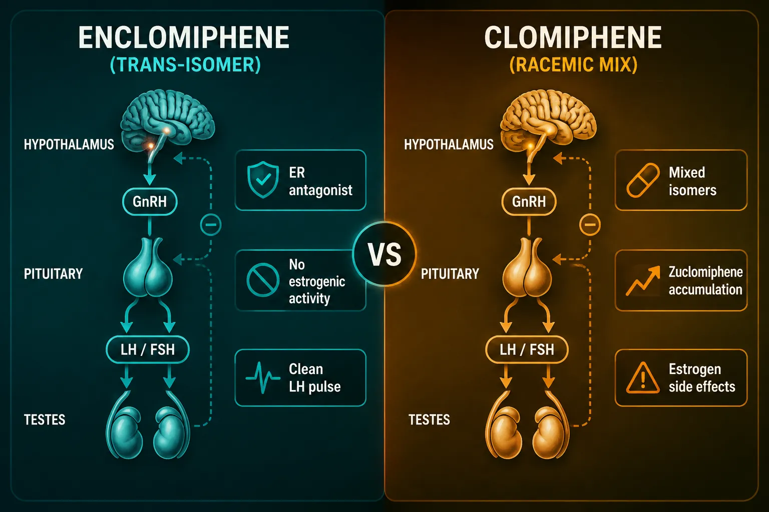
Only one isomer of clomiphene citrate drives the hypothalamic-pituitary-gonadal (HPG) axis upward, and that isomer is enclomiphene. Understanding Enclomiphene, Estrogen Receptor Signaling, and Luteinizing Phase Biology: What Hormone Researchers Should Measure requires bridging classical reproductive endocrinology with modern selective estrogen receptor modulator (serm) pharmacology. For researchers designing rigorous in vitro or preclinical protocols in 2026, knowing which endpoints to track, and why, is the difference between publishable data and noise.

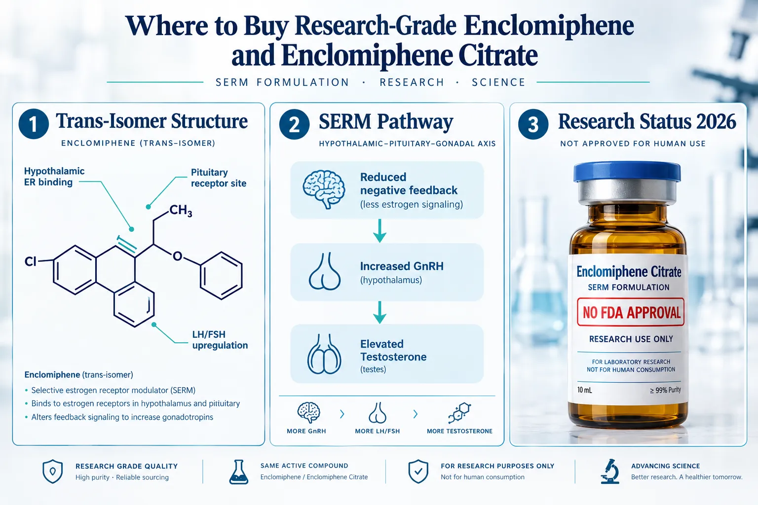
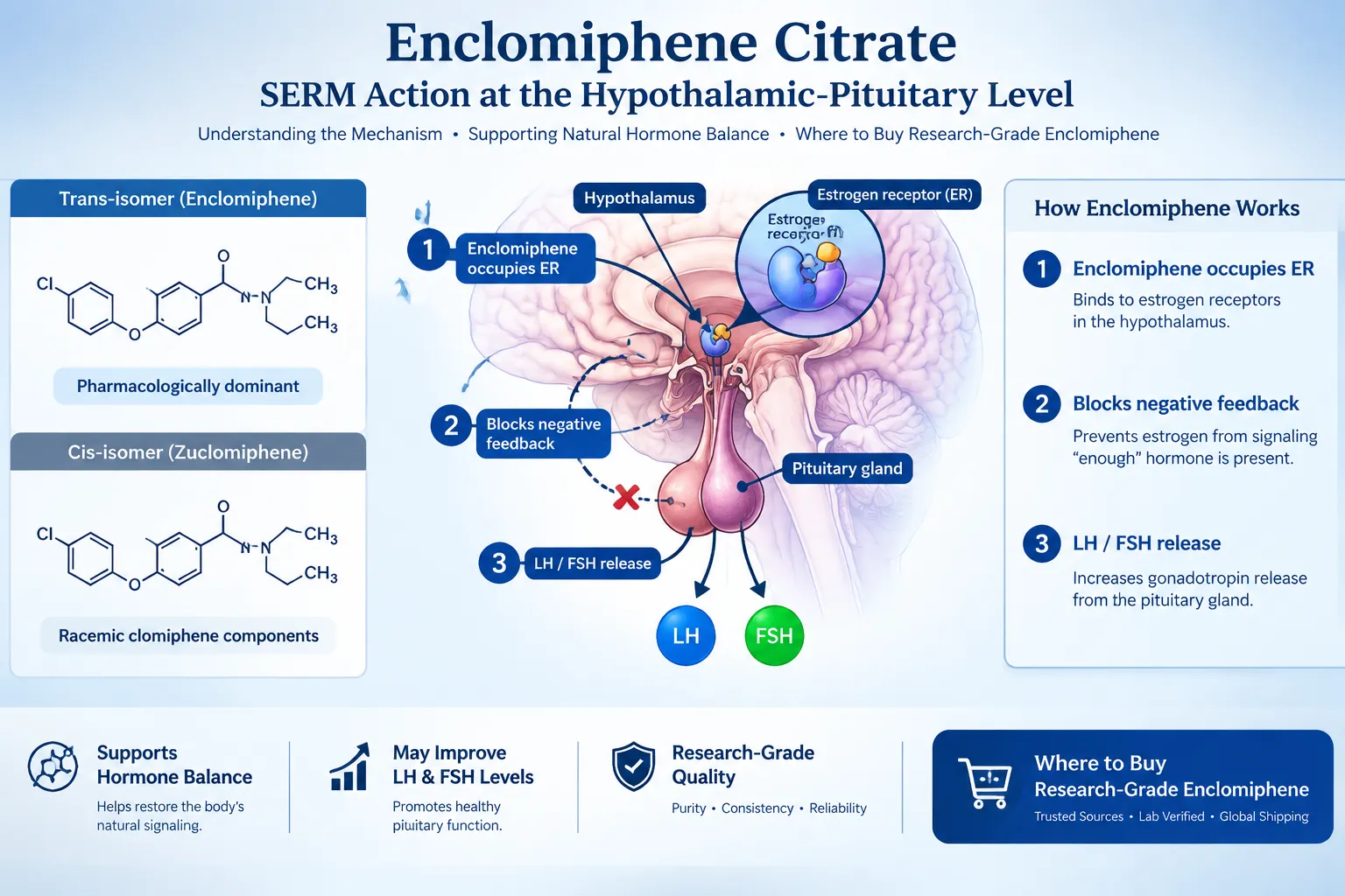
Bright editorial infographic-style landscape image () showing the hypothalamic-pituitary-gonadal axis as a clean vertical

Key Takeaways

  • Enclomiphene is the trans-isomer of clomiphene and acts as a selective estrogen receptor antagonist at the hypothalamic level.
  • Blocking estrogen receptor alpha (ERa) in the hypothalamus removes negative feedback, elevating GnRH pulse frequency and downstream LH and FSH secretion.
  • The luteinizing phase is the primary hormonal window where LH surge dynamics are most measurable and most relevant to serm research.
  • Core endpoints for enclomiphene experiments include LH, FSH, total testosterone, free testosterone, and estradiol (E2).
  • Researchers should also monitor sex hormone-binding globulin (SHBG) and LH pulse frequency as secondary markers.

The HPG Axis and Luteinizing Phase Biology

The HPG axis operates through a precise feedback loop. The hypothalamus releases gonadotropin-releasing hormone (GnRH) in pulses. Those pulses stimulate the anterior pituitary to secrete luteinizing hormone (LH) and follicle-stimulating hormone (FSH). LH then acts on Leydig cells (in males) or theca cells (in females) to drive steroidogenesis.

The luteinizing phase, the period surrounding the LH surge, is the most dynamic window in this cycle. During this phase:

  • LH concentrations can spike 5- to 10-fold above baseline
  • Estradiol peaks just before the LH surge, triggering positive feedback at the pituitary
  • Progesterone begins rising post-surge

This feedback architecture is exactly where enclomiphene exerts its effect. By occupying estrogen receptors at the hypothalamus without activating them, enclomiphene prevents estradiol from signaling "enough hormone, slow down." The result is sustained GnRH pulsatility and elevated gonadotropin output.

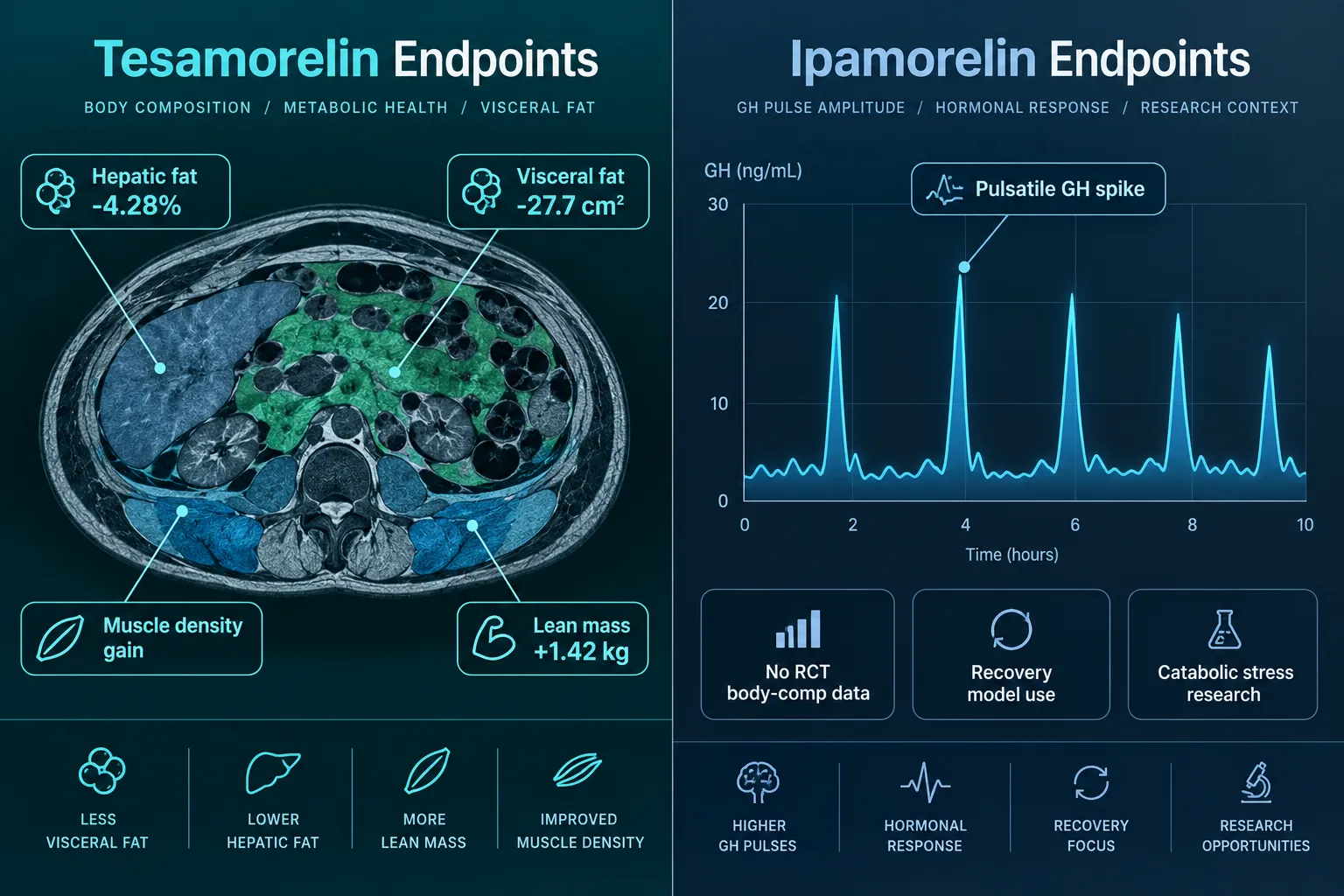
Researchers studying body composition peptides, such as those exploring tesa and its somatotropic mechanisms, will recognize this axis-level thinking as foundational to any endocrine research design.


How Enclomiphene Modulates Estrogen Receptor Signaling

How Enclomiphene Modulates Estrogen Receptor Signaling

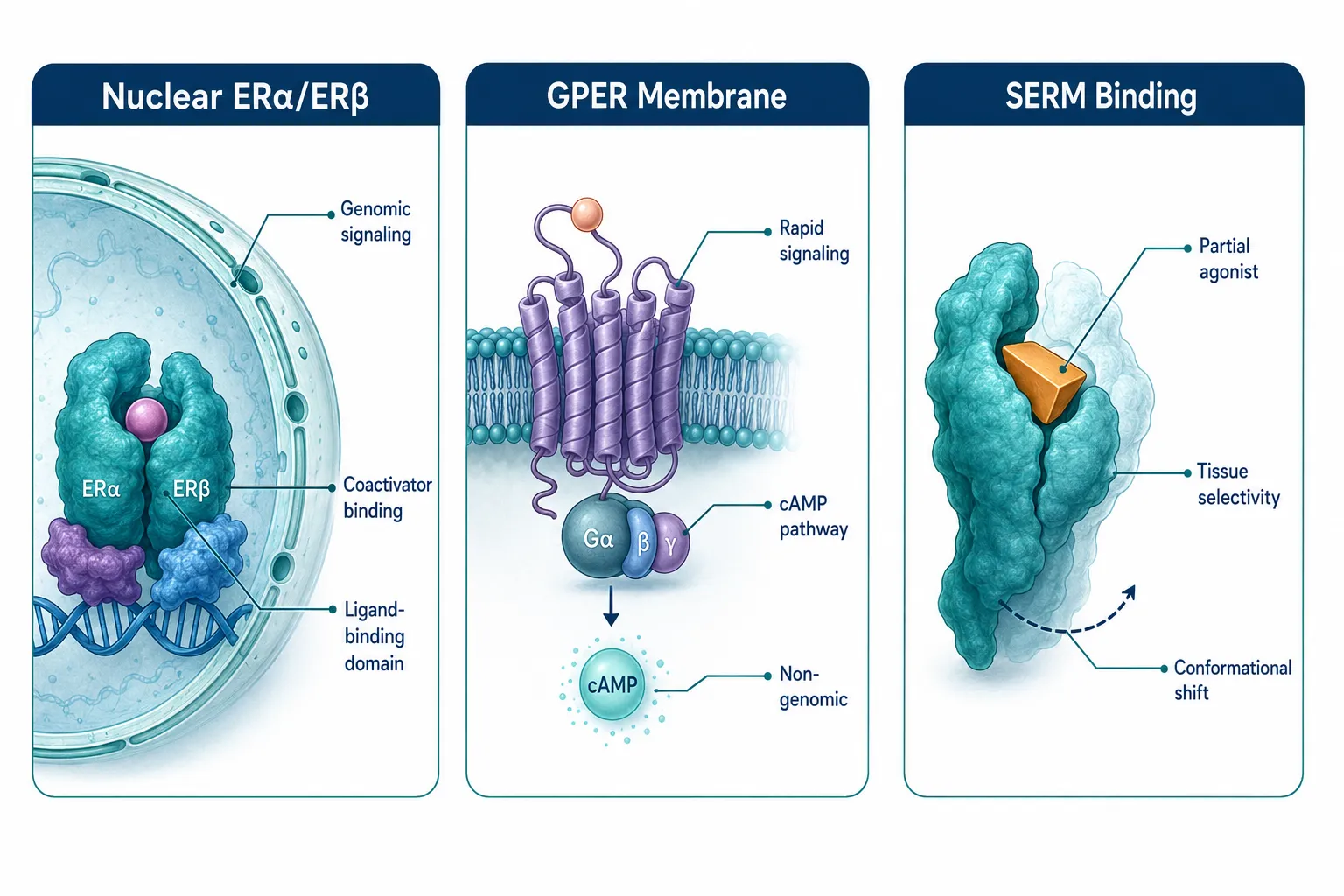
Enclomiphene's selectivity is its defining research value. Unlike its sister isomer zuclomiphene, which carries partial agonist activity and a longer half-life, enclomiphene acts predominantly as a pure antagonist at hypothalamic ERa receptors.

Receptor-Level Mechanism

Receptor Site Enclomiphene Action Research Implication
Hypothalamic ERa Antagonist Removes negative feedback; raises GnRH pulse rate
Pituitary ER Weak antagonist Amplifies LH and FSH response
Peripheral ER (bone, liver) Minimal activity Reduces confounding estrogenic effects

This tissue-selective profile makes enclomiphene a cleaner research tool than full clomiphene citrate for isolating HPG axis dynamics. Researchers studying mitochondrial and cellular signaling cascades, such as those working with SS-31 and its mitochondrial dynamics, will appreciate how receptor selectivity reduces experimental confounders.

"The value of enclomiphene in preclinical models lies not just in what it activates, but in what it leaves undisturbed."

Because enclomiphene does not strongly activate peripheral estrogen receptors, downstream effects on hepatic SHBG production are less pronounced than with full clomiphene. This is a critical variable to measure in any serm protocol.


Enclomiphene, Estrogen Receptor Signaling, and Luteinizing Phase Biology: What Hormone Researchers Should Measure

Enclomiphene, Estrogen Receptor Signaling, and Luteinizing Phase Biology: What Hormone Researchers Should Measure

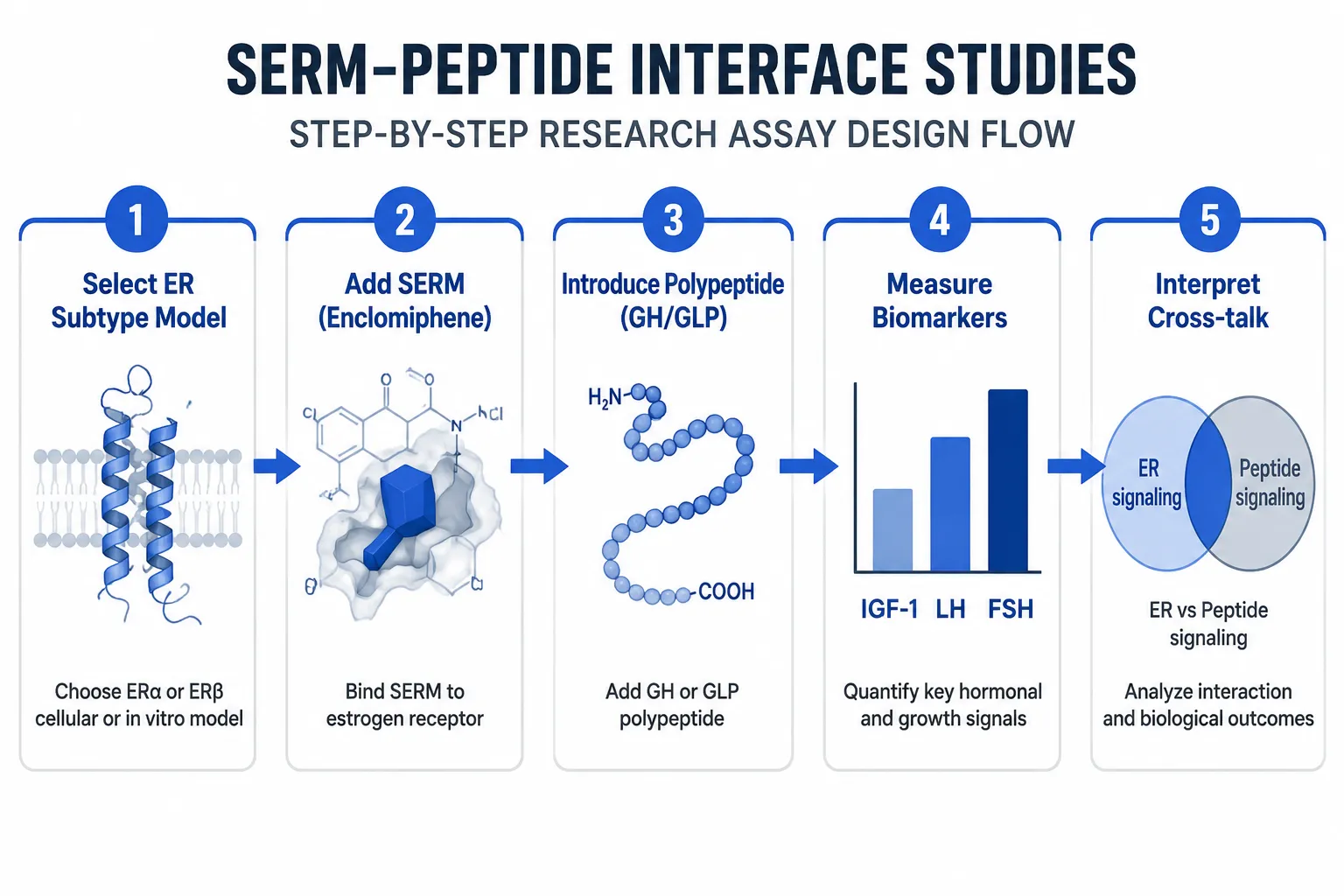
Designing a research-grade enclomiphene experiment requires a structured panel of endpoints. Below are the primary and secondary markers researchers should capture.

Primary Endpoints

1. Luteinizing Hormone (LH)
Measure both basal LH and pulsatile LH frequency. Enclomiphene's primary mechanism should produce measurable increases in LH pulse amplitude within 24-72 hours of administration in most preclinical models.

2. Follicle-Stimulating Hormone (FSH)
FSH rises alongside LH but with different kinetics. Tracking FSH independently confirms HPG axis activation rather than isolated LH secretion.

3. Total and Free Testosterone
Downstream steroidogenesis is the functional output of LH signaling. Both total and free testosterone should be measured to account for SHBG-binding changes.

4. Estradiol (E2)
As testosterone rises, aromatase activity converts a fraction to estradiol. Monitoring E2 is essential for understanding the feedback loop's re-equilibration point.

Secondary Endpoints

  • SHBG, Enclomiphene's limited hepatic ER activity means SHBG changes are smaller than with full clomiphene, but still measurable
  • LH pulse frequency, Requires frequent sampling (every 10-20 minutes) over a 4-8 hour window; more informative than single-point LH values
  • Progesterone, Relevant in female models to confirm ovulatory response post-LH surge

Researchers exploring multi-peptide endocrine protocols, including those examining GLP-1 incretin research themes or longevity-focused compound blends, should note that hormonal cross-talk between metabolic and reproductive axes can influence these endpoints.

Timing Considerations

Endpoint timing matters as much as endpoint selection. Recommended sampling windows:

  • Baseline: 7 days pre-administration
  • Acute response: 24, 48, and 72 hours post-first dose
  • Steady-state: Day 14 and Day 28
  • Washout: 14 days post-cessation

For researchers also examining growth hormone secretagogue interactions, resources like tesa body composition research themes offer parallel frameworks for longitudinal hormonal tracking.


Conclusion

Understanding Enclomiphene, Estrogen Receptor Signaling, and Luteinizing Phase Biology: What Hormone Researchers Should Measure is not purely academic, it directly shapes protocol quality. Enclomiphene's clean antagonism at hypothalamic ERa makes it one of the most targeted tools available for studying HPG axis dynamics without the confounding estrogenic noise of full clomiphene.

Actionable next steps for researchers:

  1. Build a baseline hormonal panel (LH, FSH, total testosterone, free testosterone, E2, SHBG) before any serm administration
  2. Use pulsatile LH sampling, not single-point measurements, to capture true axis activation
  3. Track E2 and SHBG in parallel to understand feedback re-equilibration
  4. Pre-register sampling timepoints to prevent post-hoc endpoint selection bias
  5. Cross-reference findings with metabolic axis data, particularly if co-administering peptides that influence GH or insulin signaling

Researchers seeking high-documentation research compounds to pair with endocrine studies can review BPC-157 core peptides documentation and AOD-9604 research method notes for complementary protocol frameworks.

https://www.puretestedpeptides.com/wp-content/uploads/2026/07/enclomiphene-estrogen-receptor-signaling-and-luteinizing-phase-biology-what-horm-1.webp 1024 1536 Pure Tested https://www.puretestedpeptides.com/wp-content/uploads/2026/01/buy-peptides-online.jpg Pure Tested2026-07-18 13:05:052026-07-20 14:59:48Enclomiphene, Estrogen Receptor Signaling, and Luteinizing Phase Biology: What Hormone Researchers Should Measure
Mesenchymal Stem Cells, BPC‑157, and GHK‑Cu: How Tissue Repair Peptides Compare With Classic NSAIDs Like Naproxen in Injury Models

Mesenchymal Stem Cells, BPC‑157, and GHK‑Cu: How Tissue Repair Peptides Compare With Classic NSAIDs Like Naproxen in Injury Models

July 18, 2026/0 Comments/by Pure Tested

Roughly 30 million tendon and ligament injuries occur in the United States every year, yet the most common treatment response remains the same: reach for an anti-inflammatory pill. That reflex is now being challenged by a growing body of preclinical research examining whether regenerative agents, including mesenchymal stem cells, BPC‑157, and GHK‑Cu, can do something naproxen fundamentally cannot: rebuild damaged tissue rather than simply quiet the pain signal.

Peptides vs NSAIDs tissue repair comparison hero

Key Takeaways

  • NSAIDs like naproxen suppress inflammation by blocking COX enzymes but do not stimulate tissue regeneration and may actively impair mesenchymal stem cell activity.
  • BPC‑157 promotes tendon, ligament, and muscle healing by upregulating VEGF and nitric oxide pathways, driving angiogenesis in injured tissue.
  • GHK‑Cu accelerates wound closure through collagen synthesis and anti-inflammatory signaling, offering a complementary regenerative mechanism.
  • Preclinical data suggest naproxen can reduce the therapeutic efficacy of MSC-based treatments and interfere with osteogenic differentiation.
  • The mechanistic gap between these two approaches, suppression versus regeneration, is the central research question driving interest in peptide-based injury protocols in 2026.

How NSAIDs and Regenerative Peptides Work at the Cellular Level

Understanding the contrast between regenerative peptide approaches and conventional anti-inflammatory molecules starts with basic cell biology.

NSAIDs such as naproxen and diclofenac inhibit cyclooxygenase (COX-1 and COX-2) enzymes. This reduces prostaglandin synthesis, which lowers pain and swelling. The mechanism is well understood and clinically validated. However, prostaglandins also play a role in initiating the healing cascade. By suppressing them broadly, NSAIDs can blunt the early inflammatory phase that tissues need to begin repair.

Mesenchymal stem cells (MSCs) are multipotent stromal cells capable of differentiating into bone, cartilage, and connective tissue. They also secrete paracrine factors that modulate local inflammation and recruit other repair cells. Research has shown that naproxen can reduce the therapeutic efficacy of human mesenchymal stromal cell therapy in posttraumatic osteoarthritis models. A separate study found that naproxen disrupts osteogenic differentiation of MSCs by interfering with Indian hedgehog signaling, a pathway critical for bone and cartilage formation.

BPC‑157 (Body Protection Compound 157) is a synthetic pentadecapeptide derived from a gastric protein. Its primary tissue-repair mechanisms include upregulation of vascular endothelial growth factor (VEGF), promotion of nitric oxide synthesis, and enhancement of tendon cell outgrowth, survival, and migration. In rat models of transected medial collateral ligaments, BPC‑157 improved both functional and biomechanical recovery. A 2019 review confirmed consistently positive effects across tendon, ligament, and muscle injury models.

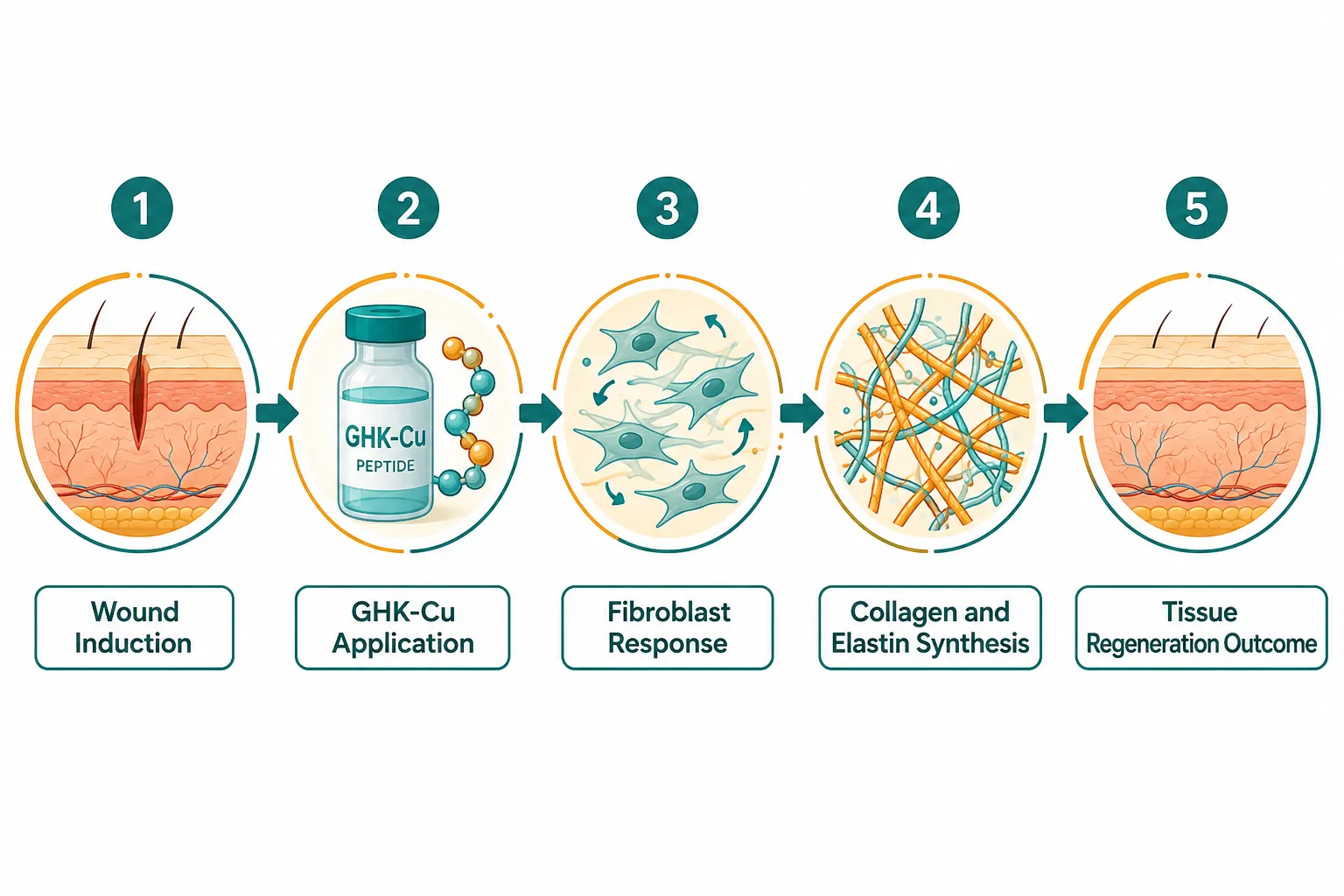
GHK‑Cu (copper peptide glycyl-L-histidyl-L-lysine) works through a distinct but complementary pathway. It stimulates collagen and glycosaminoglycan synthesis, promotes angiogenesis, and modulates inflammatory cytokines. These properties make it particularly relevant in wound healing and soft-tissue remodeling research. For researchers exploring topical and systemic peptide applications, GHK-Cu peptides for sale are among the most studied copper-based compounds in the field.

How NSAIDs and Regenerative Peptides Work at the Cellular Level


BPC‑157, GHK‑Cu, and the Mechanistic Gap With Naproxen in Injury Models

The phrase "Mesenchymal Stem Cells, BPC‑157, and GHK‑Cu: How Tissue Repair Peptides Compare With Classic NSAIDs Like Naproxen in Injury Models" captures a genuine scientific tension. These two categories of compounds are not simply different doses of the same idea, they operate on fundamentally different biological logic.

Feature NSAIDs (Naproxen) BPC‑157 / GHK‑Cu
Primary action COX inhibition, anti-inflammatory VEGF upregulation, collagen synthesis
Effect on MSCs May impair differentiation Supports paracrine repair signaling
Tissue rebuilding No direct effect Documented in preclinical models
GI safety profile Known mucosal risk BPC‑157 shown to counteract NSAID GI damage

One particularly striking finding: BPC‑157 has been shown to counteract gastrointestinal, liver, and brain toxicity caused by diclofenac in animal models. This positions BPC‑157 not only as a tissue-repair agent but potentially as a protective compound against NSAID-induced organ stress.

For researchers interested in the broader landscape of peptide mechanisms, the ultimate guide to peptide therapy benefits and uses provides a useful reference framework. Additionally, TB-500 muscle recovery research themes explore another regenerative peptide with overlapping soft-tissue applications.

BPC‑157 also demonstrates neuroprotective effects in animal models of traumatic brain injury and spinal cord compression, a range of activity that no NSAID replicates. This breadth suggests a systemic repair orientation rather than localized symptom suppression.

GHK‑Cu's role is more focused on extracellular matrix remodeling. Its ability to upregulate collagen synthesis while simultaneously reducing inflammatory cytokines makes it a candidate for both acute injury and chronic tissue degeneration research. Those sourcing research-grade material can review the GHK-Cu peptide research and sourcing guide for purity and procurement considerations.

BPC‑157, GHK‑Cu, and the Mechanistic Gap With Naproxen in Injury Models


What the Research Signals for Future Injury Protocols

The comparison of Mesenchymal Stem Cells, BPC‑157, and GHK‑Cu with classic NSAIDs like naproxen in injury models is not yet a clinical story, it remains largely preclinical. Human trials are limited, and no regulatory body has approved BPC‑157 or GHK‑Cu as therapeutic drugs for musculoskeletal injury. That context matters.

What preclinical data do support is a mechanistic argument: agents that promote angiogenesis, stimulate MSC activity, and rebuild extracellular matrix are doing something categorically different from COX inhibition. The two approaches are not mutually exclusive in theory, but the evidence that NSAIDs can impair MSC-based treatments suggests caution about combining them without careful protocol design.

Researchers and clinicians evaluating these compounds should also consider delivery systems. Innovative peptide delivery systems continue to evolve, with oral, injectable, and topical formats each showing different bioavailability profiles. For those examining purity standards before sourcing, peptide purity testing explained simply is a practical starting point.

Other regenerative peptides worth examining alongside BPC‑157 and GHK‑Cu include MOTS-c for its mitochondrial and metabolic repair signaling, see MOTS-c the mitochondrial peptide, and the broader category of aging support peptides that intersect with tissue longevity research.

What the Research Signals for Future Injury Protocols


Conclusion

The mechanistic contrast between tissue repair peptides and classic NSAIDs like naproxen is sharper than most injury management discussions acknowledge. NSAIDs suppress inflammation efficiently but do not rebuild tissue and may actively interfere with MSC-based repair. BPC‑157 and GHK‑Cu, by contrast, work upstream, promoting angiogenesis, collagen synthesis, and cellular survival in injured connective tissue.

Actionable next steps for researchers and practitioners:

  • Review preclinical injury model data for BPC‑157 and GHK‑Cu before designing protocols that also involve NSAID use.
  • Evaluate whether concurrent NSAID administration is necessary, given evidence of MSC impairment.
  • Prioritize purity-verified peptide sources and consult current delivery system research for optimal bioavailability.
  • Monitor emerging human trial data, as the field is moving quickly in 2026.

The question is no longer whether regenerative peptides differ from NSAIDs, they clearly do. The research priority now is understanding when, how, and for whom those differences matter most.

https://www.puretestedpeptides.com/wp-content/uploads/2026/07/mesenchymal-stem-cells-bpc-157-and-ghk-cu-how-tissue-repair-peptides-compare-wit.webp 1024 1536 Pure Tested https://www.puretestedpeptides.com/wp-content/uploads/2026/01/buy-peptides-online.jpg Pure Tested2026-07-18 13:04:472026-07-20 14:59:48Mesenchymal Stem Cells, BPC‑157, and GHK‑Cu: How Tissue Repair Peptides Compare With Classic NSAIDs Like Naproxen in Injury Models
Peptide Calculator Use Cases Beyond Growth Hormone: Working Through GLP‑3 Retatrutide, MOTS‑c, and BPC‑157 Research Dosing

Peptide Calculator Use Cases Beyond Growth Hormone: Working Through GLP‑3 Retatrutide, MOTS‑c, and BPC‑157 Research Dosing

July 18, 2026/0 Comments/by Pure Tested

Most researchers reach for a peptide calculator when reconstituting a growth hormone secretagogue blend, then stop there. Yet the same arithmetic logic that converts a Tesamorelin vial into syringe units applies equally to metabolic triple agonists, mitochondrial peptides, and tissue-repair compounds. Peptide Calculator Use Cases Beyond Growth Hormone: Working Through GLP-3 Retatrutide, MOTS-c, and BPC-157 Research Dosing is a topic that deserves its own worked framework, because each compound carries unique concentration targets, titration schedules, and reconstitution constraints that a generic calculator must accommodate.

Bright infographic-style editorial landscape (): isometric illustration of three distinct peptide molecules — GLP-3

Key Takeaways

  • Peptide calculators are not limited to GH secretagogues, they handle any lyophilized compound requiring reconstitution math.
  • Retatrutide (GLP-3) uses slow titration schedules that demand week-by-week dose recalculation.
  • MOTS-c reconstitution targets are typically low-volume and require precise unit conversion.
  • BPC-157 research often involves both injectable and oral formats, each with different concentration logic.
  • GHK-Cu and other repair peptides follow the same calculator inputs: vial mass, diluent volume, and desired dose.

Why Peptide Calculator Use Cases Extend Well Beyond Growth Hormone

Growth hormone peptides like Ipamorelin and CJC-1295 popularized the reconstitution calculator because their dosing windows are narrow and their blends are common. A Tesamorelin dosage calculator works on the same three-input model every other peptide uses:

Input Example Value
Vial mass (mg) 5 mg
Diluent added (mL) 2 mL bacteriostatic water
Desired dose (mcg) 250 mcg

Result: concentration = 2,500 mcg/mL; draw = 0.10 mL (10 units on a U-100 syringe).

That formula is universal. The only variable is the peptide itself, and that is where researchers working with newer metabolic and tissue-repair compounds need a more expanded mental model.


Worked Examples: Peptide Calculator Use Cases Beyond Growth Hormone for Retatrutide, MOTS-c, and BPC-157

Retatrutide (GLP-3): Titration Math Week by Week

Retatrutide is a triple receptor agonist targeting GLP-1, GIP, and glucagon receptors simultaneously. It remains investigational, with Phase 3 trials ongoing as of 2026. Because it uses a slow titration schedule, commonly starting at 2 mg per week and stepping up over several weeks, the calculator must be re-run at each dose change.

Example scenario:

  • Vial: 10 mg retatrutide
  • BAC water added: 2 mL
  • Concentration: 5,000 mcg/mL (5 mg/mL)
  • Week 1 dose: 2 mg = draw 0.40 mL (40 units)
  • Week 4 dose: 4 mg = draw 0.80 mL (80 units)

Tools like PeptiTools and PeptideDeck provide live syringe diagrams that update as the dose field changes, which is especially useful for multi-week titration. For background on the incretin research context, see the GLP-3 retatrutide incretin research themes overview, and for a broader generational comparison, the generations of GLP-1 differences resource is instructive.

"The arithmetic never changes, only the target dose does. Running the calculator fresh at each titration step prevents cumulative dosing errors."

MOTS-c: Low-Volume Precision

MOTS-c, the mitochondrial peptide, is typically studied at doses in the 5-10 mg range. Because vials are often supplied at 5 mg, researchers frequently add only 1 mL of BAC water to achieve a 5 mg/mL concentration, meaning a 5 mg dose draws a full 1 mL, while a 2.5 mg dose draws 0.50 mL (50 units).

Key consideration: At low diluent volumes, measurement error is amplified. A 0.02 mL miscalculation at 5 mg/mL equals a 100 mcg dosing error. Dedicated MOTS-c calculators, such as those offered by MOTS-c Research, handle the unit conversion explicitly, displaying results in both mL and U-100 syringe units side by side. Researchers interested in MOTS-c metabolic stress applications can explore the MOTS-c metabolic stress research page for additional context.

BPC-157: Injectable vs. Oral Concentration Logic

BPC-157 is unique because it appears in both injectable and oral research formats. For injectable use, a common reconstitution is 5 mg into 2.5 mL BAC water, yielding 2 mg/mL. A 250 mcg research dose then draws 0.125 mL (12.5 units).

For oral BPC-157 formats, concentration logic shifts entirely, volume is less relevant than total mass per capsule or solution. Platforms like PeptideCalcs allow researchers to toggle between injectable and oral modes, keeping the math format-appropriate. The BPC-157 10mg vial research themes page provides additional reconstitution reference points.

BPC-157: Injectable vs. Oral Concentration Logic


Applying the Same Framework to GHK-Cu and Multi-Peptide Protocols

The calculator framework extends cleanly to copper peptides and combination protocols. GHK-Cu longevity research typically involves doses of 1-2 mg, often reconstituted in 1 mL of sterile water for a 1-2 mg/mL concentration. At 1 mg/mL, a 1 mg dose draws exactly 1 mL, straightforward, but only if the researcher has confirmed the vial mass and diluent volume before calculating.

Multi-peptide protocols, for example, combining BPC-157 with a mitochondrial support compound like SS-31 elamipretide, require running the calculator independently for each compound. Shared syringes or combined vials change the concentration of both peptides and invalidate pre-calculated draw volumes. Each compound must retain its own reconstitution record.

Best practices for multi-peptide calculator use:

  • Label each vial with concentration (mg/mL) and reconstitution date.
  • Store calculator outputs alongside vial records, not just in memory.
  • Re-run calculations if a vial is partially used and diluent volume has changed.
  • Use platforms that support custom vial inputs rather than locked preset values.

Platforms such as PeptiTools, PepExact, and Blackwell BioLabs offer free, no-signup calculators that accommodate custom inputs across a wide range of peptides, including Retatrutide, BPC-157, MOTS-c, and GHK-Cu, making them practical choices for researchers managing diverse compound libraries.

Applying the Same Framework to GHK-Cu and Multi-Peptide Protocols


Conclusion

Peptide Calculator Use Cases Beyond Growth Hormone: Working Through GLP-3 Retatrutide, MOTS-c, and BPC-157 Research Dosing confirms a straightforward principle: the reconstitution formula is universal, but each peptide demands context-specific inputs and awareness of format, titration schedule, and concentration sensitivity.

Actionable next steps for researchers in 2026:

  1. Identify the vial mass and intended diluent volume for each compound before touching a syringe.
  2. Use a calculator that displays results in both mL and U-100 syringe units simultaneously.
  3. For titrating compounds like Retatrutide, bookmark the calculator and re-run it at each dose step.
  4. Maintain a written reconstitution log per vial, do not rely on memory for concentration values.
  5. Consult a qualified clinician before applying any calculated dose in a research context.

The math is accessible. The discipline around it is what separates reliable research from avoidable error.

https://www.puretestedpeptides.com/wp-content/uploads/2026/07/peptide-calculator-use-cases-beyond-growth-hormone-working-through-glp-3-retatru.webp 1024 1536 Pure Tested https://www.puretestedpeptides.com/wp-content/uploads/2026/01/buy-peptides-online.jpg Pure Tested2026-07-18 13:04:442026-07-20 14:59:49Peptide Calculator Use Cases Beyond Growth Hormone: Working Through GLP‑3 Retatrutide, MOTS‑c, and BPC‑157 Research Dosing
Polypeptide Peptides in Endocrine and Metabolic Pharmacology: Lessons From Amlodipine, Prednisone, and Metoprolol

Polypeptide Peptides in Endocrine and Metabolic Pharmacology: Lessons From Amlodipine, Prednisone, and Metoprolol

July 17, 2026/0 Comments/by Pure Tested

Three drugs, amlodipine, prednisone, and metoprolol, have shaped cardiovascular and endocrine medicine for decades. Yet their well-documented off-target effects on glucose metabolism, adrenal function, and mitochondrial signaling now serve as a compelling argument for why polypeptide peptides in endocrine and metabolic pharmacology deserve serious research attention in 2026.

Bright editorial split-screen infographic landscape (): left half shows a clean white-background molecular diagram of a

Key Takeaways

  • Amlodipine, prednisone, and metoprolol each interact with endocrine pathways in ways that go beyond their primary targets, producing metabolic side effects that peptide-based agents may avoid.
  • Polypeptide peptides in endocrine and metabolic pharmacology offer receptor selectivity, shorter off-target profiles, and tissue-specific action that small molecules often cannot match.
  • GLP-1 receptor agonists and multi-agonist peptides represent the most clinically advanced examples of this shift, with GLP-3 retatrutide research extending the frontier.
  • Mitochondrial peptides such as MOTS-c address metabolic dysregulation at the cellular energy level, a target unreachable by classic small molecules.
  • Understanding the pharmacological gaps left by legacy drugs helps researchers identify where peptide-based tools offer the greatest research value.

How Classic Small Molecules Interact With Endocrine Pathways

Amlodipine blocks L-type calcium channels in vascular smooth muscle, reducing blood pressure and myocardial oxygen demand. However, calcium signaling is also central to pancreatic beta-cell insulin secretion. Disrupting this pathway even modestly can impair glucose-stimulated insulin release, a finding that has been observed in long-term hypertension management research.

Prednisone, a synthetic glucocorticoid, binds glucocorticoid receptors with broad tissue distribution. Its anti-inflammatory power comes at a metabolic cost: stimulation of hepatic gluconeogenesis, suppression of peripheral insulin sensitivity, and disruption of the hypothalamic-pituitary-adrenal axis. These are not rare side effects, they are mechanistic consequences of how the drug binds.

Metoprolol, a beta-1 selective adrenergic blocker, reduces heart rate and cardiac output effectively. Its endocrine liability lies in masking hypoglycemic symptoms and blunting the catecholamine-driven recovery from low blood glucose, a clinically relevant concern in diabetic patients.

The pattern is consistent: each drug achieves its primary goal through a mechanism that inevitably touches endocrine or metabolic circuitry.

"The off-target metabolic effects of classic small molecules are not design flaws, they are the predictable result of targeting signaling pathways that evolution never isolated."


Polypeptide Peptides in Endocrine and Metabolic Pharmacology: The Receptor Targeting Advantage

Polypeptide Peptides in Endocrine and Metabolic Pharmacology: The Receptor Targeting Advantage

Where small molecules bind with high affinity but low tissue selectivity, polypeptide peptides in endocrine and metabolic pharmacology operate through receptor systems that are more anatomically restricted. This distinction is not merely theoretical.

Proglucagon-derived peptides, including GLP-1, GLP-2, glucagon, and oxyntomodulin, each act on distinct receptor populations across the gut, pancreas, brain, and liver. GLP-1 receptor agonists lower blood glucose by enhancing insulin secretion only when glucose is already elevated, a glucose-dependent mechanism that eliminates the hypoglycemia risk associated with metoprolol-class drugs.

The next generation goes further. Multi-agonist peptides combine amino acid sequences from GLP-1, glucagon, and GIP hormones into single molecules with enhanced potency and extended half-lives. Research into GLP-3 retatrutide represents this frontier, targeting multiple incretin receptors simultaneously to address obesity and type 2 diabetes with a precision that prednisone-driven metabolic disruption cannot approach.

The GIP receptor plays a particularly important role here. GIP works synergistically with GLP-1 to amplify insulin secretion and may also support bone metabolism and fat storage regulation, a multi-system effect achieved without the adrenal suppression that defines glucocorticoid pharmacology.

Key differences between small molecules and peptide agents:

Feature Small Molecules (e.g., Prednisone) Peptide Agents (e.g., GLP-1 agonists)
Receptor selectivity Broad Tissue-restricted
Metabolic off-target effects Common Reduced
Half-life engineering Limited Highly modifiable
Glucose-dependent action No Yes (GLP-1 class)

Adrenomedullin, a 52-amino acid peptide hormone, further illustrates the endocrine complexity peptides can address. It regulates cardiovascular tone and lymphatic function while also inhibiting insulin secretion in a dose-dependent manner, a finding that positions it as both a research target and a cautionary example of peptide pleiotropy.


Mitochondrial Peptides and the Metabolic Gap Left by Legacy Drugs

Mitochondrial Peptides and the Metabolic Gap Left by Legacy Drugs

Neither amlodipine, prednisone, nor metoprolol addresses cellular energy metabolism at the mitochondrial level. This is a significant gap. Chronic glucocorticoid use, in particular, impairs mitochondrial biogenesis and increases reactive oxygen species production, effects that accelerate metabolic aging.

This is precisely where mitochondrial-derived peptides enter the research conversation. MOTS-c, encoded within mitochondrial DNA, regulates glucose uptake, fatty acid oxidation, and insulin sensitivity through AMPK activation. Its mechanism operates entirely outside the receptor systems targeted by classic cardiovascular drugs, making it a complementary rather than competing research tool.

SS-31 peptide research addresses a related problem: mitochondrial membrane integrity under oxidative stress. Where prednisone-induced metabolic disruption increases oxidative burden, SS-31 targets cardiolipin on the inner mitochondrial membrane to preserve electron transport chain function.

For researchers exploring body composition and visceral adiposity, conditions worsened by long-term glucocorticoid exposure, tesa offers a growth hormone-releasing hormone analog that specifically reduces visceral fat without the broad hormonal disruption of steroid-class drugs.

Non-incretin peptide systems are also gaining traction. Apelin, spexin, and meteorin-like protein (METRNL) each interact with energy balance pathways that small molecules have historically ignored, opening new drug discovery targets for metabolic disease research.

For those examining AOD-9604 metabolic research, the lipolytic fragment of growth hormone provides another example of how peptide engineering can isolate a single metabolic function, fat mobilization, without replicating the full hormonal cascade of its parent molecule.


Conclusion

The lessons from amlodipine, prednisone, and metoprolol are not arguments against small-molecule pharmacology. They are a precise map of where that pharmacology ends and where polypeptide peptides in endocrine and metabolic pharmacology begin. Each classic drug reveals a metabolic vulnerability, impaired insulin secretion, adrenal suppression, blunted glycemic recovery, that modern peptide research is systematically designed to address.

Actionable next steps for researchers and clinicians:

  • Review the receptor selectivity profiles of any metabolic intervention against the endocrine off-target effects documented in glucocorticoid and beta-blocker literature.
  • Explore mitochondrial peptide tools such as MOTS-c and SS-31 for research models involving oxidative stress or insulin resistance secondary to classic drug exposure.
  • Track multi-agonist peptide development, particularly GLP-1/GIP/glucagon tri-agonists, as the most clinically proximate evolution of endocrine peptide pharmacology.
  • Use the pharmacological gaps in legacy drugs as a framework for identifying where peptide-based research tools add the most mechanistic value.

The field is not replacing its foundations. It is building precisely where those foundations show their limits.

https://www.puretestedpeptides.com/wp-content/uploads/2026/07/polypeptide-peptides-in-endocrine-and-metabolic-pharmacology-lessons-from-amlodi.webp 1024 1536 Pure Tested https://www.puretestedpeptides.com/wp-content/uploads/2026/01/buy-peptides-online.jpg Pure Tested2026-07-17 13:07:122026-07-20 14:59:49Polypeptide Peptides in Endocrine and Metabolic Pharmacology: Lessons From Amlodipine, Prednisone, and Metoprolol
Peptides Calculator for Advanced Blends: Worked Examples for Tesamorelin, CJC‑1295, and Ipamorelin Stacks

Peptides Calculator for Advanced Blends: Worked Examples for Tesamorelin, CJC‑1295, and Ipamorelin Stacks

July 17, 2026/0 Comments/by Pure Tested

A miscalculated peptide dose, even by a single decimal place, can mean delivering ten times the intended amount. For researchers working with multi-peptide blends, precision is not optional. This guide applies a practical peptides calculator for advanced blends: worked examples for Tesamorelin, CJC-1295, and Ipamorelin stacks to walk through real reconstitution math, dose conversions, and error-prevention strategies that protect both data quality and research integrity.

Key Takeaways

  • Combining CJC-1295 (a GHRH analog) with Ipamorelin (a GHRP) stimulates growth hormone release through two complementary pathways, producing a stronger GH pulse than either peptide alone.
  • Accurate peptide calculator math starts with knowing vial mass (mcg), diluent volume (mL), and target dose (mcg) before drawing any syringe.
  • Tesamorelin, CJC-1295, and Ipamorelin can be stacked in a single blend or dosed separately; each approach requires its own reconstitution calculation.
  • Timing injections on an empty stomach, ideally 90 minutes after the last meal or before sleep, aligns with natural GH secretion rhythms.
  • Cycling protocols (commonly 8 weeks on, 12 weeks off) help maintain receptor sensitivity over time.

Why Stack Tesamorelin, CJC-1295, and Ipamorelin

Why Stack Tesamorelin, CJC-1295, and Ipamorelin

Growth hormone secretion is governed by two main signals: growth hormone-releasing hormone (GHRH) and growth hormone-releasing peptides (GHRPs). Tesamorelin and CJC-1295 are both GHRH analogs, while Ipamorelin is a selective GHRP. When a GHRH analog and a GHRP are administered together, they act on different receptors simultaneously, producing a synergistic GH pulse that exceeds what either compound generates alone.

Tesamorelin is an FDA-approved GHRH analog with a well-characterized mechanism. CJC-1295 (without DAC, also called Mod GRF 1-29) offers a shorter half-life that mimics a natural pulsatile release. Ipamorelin is favored in research for its selectivity, it stimulates GH release with minimal effect on cortisol or prolactin. For a deeper look at how these mechanisms compare, see this Ipamorelin vs Tesamorelin research overview.

Researchers also explore triple-component blends. The Tesamorelin, CJC-1295, and Ipamorelin 12mg blend combines all three peptides in a single vial, simplifying logistics while maintaining the synergistic rationale. Safety considerations for combining these compounds are covered in this guide on combining Tesamorelin with CJC and Ipamorelin.


Using a Peptides Calculator for Advanced Blends: Worked Examples for Tesamorelin, CJC-1295, and Ipamorelin Stacks

Using a Peptides Calculator for Advanced Blends: Worked Examples for Tesamorelin, CJC-1295, and Ipamorelin Stacks

The core peptide calculator formula is straightforward:

Injection volume (mL) = Target dose (mcg) / Concentration (mcg/mL)

Concentration is determined during reconstitution:

Concentration (mcg/mL) = Vial mass (mcg) / Diluent volume (mL)

Worked Example 1: Separate Vials

A researcher has three separate 5 mg (5,000 mcg) vials, one each of Tesamorelin, CJC-1295, and Ipamorelin, and adds 2 mL of bacteriostatic water to each.

Peptide Vial Mass Diluent Concentration
Tesamorelin 5,000 mcg 2 mL 2,500 mcg/mL
CJC-1295 5,000 mcg 2 mL 2,500 mcg/mL
Ipamorelin 5,000 mcg 2 mL 2,500 mcg/mL

Target doses per injection: Tesamorelin 500 mcg, CJC-1295 100 mcg, Ipamorelin 100 mcg.

  • Tesamorelin: 500 / 2,500 = 0.20 mL (20 units on a 100-unit insulin syringe)
  • CJC-1295: 100 / 2,500 = 0.04 mL (4 units)
  • Ipamorelin: 100 / 2,500 = 0.04 mL (4 units)

For protocol-specific dosage guidance, the Tesamorelin dosage calculator provides additional reference values.

Worked Example 2: Pre-Mixed 12mg Blend

Using a 12mg blend vial dosed at 140 mcg with 2 mL bacteriostatic water added:

  • Total vial mass: 12,000 mcg
  • Concentration: 12,000 / 2 = 6,000 mcg/mL
  • Target dose: 140 mcg
  • Injection volume: 140 / 6,000 = 0.023 mL (~2.3 units)

For lower-dose protocols, the 90 mcg dosing variant follows the same formula with a smaller draw.

Error-Prevention Checklist

  • Confirm vial label units (mg vs. mcg) before calculating
  • Use a fresh insulin syringe for each draw
  • Never shake vials, roll gently to mix
  • Administer subcutaneously, at least 90 minutes after the last meal
  • Log every reconstitution date; discard after 28 days refrigerated

Cycle Protocols and Timing Strategies

Cycle Protocols and Timing Strategies

Standard research protocols for CJC-1295 and Ipamorelin use 100-200 mcg per peptide per injection, administered 2-3 times daily. Tesamorelin is commonly studied at 500-2,000 mcg per day depending on the research objective. The Tesamorelin dosage for fat loss page outlines dose ranges used in published research.

Injection timing matters. Administering doses before sleep aligns with the body's natural nocturnal GH surge, potentially amplifying the peptide-induced pulse. A widely used research cycle runs 8 weeks on, followed by 12 weeks off to preserve receptor sensitivity and avoid desensitization.

For researchers exploring related secretagogue combinations, the Sermorelin, Ipamorelin, and CJC-1295 stack overview provides a useful point of comparison. Staying current with evolving protocols is also supported by resources like what is new in peptide research.


Conclusion

Accurate dose math is the foundation of credible peptide research. By applying the peptides calculator for advanced blends: worked examples for Tesamorelin, CJC-1295, and Ipamorelin stacks shown above, researchers can eliminate the most common reconstitution errors before they occur.

Actionable next steps:

  1. Identify your vial mass and choose a diluent volume that yields a workable concentration for your target dose.
  2. Use the formula (dose / concentration = volume) before every draw, never estimate.
  3. Follow a documented cycle protocol (8 weeks on, 12 weeks off) and log biomarker data throughout.
  4. Cross-reference dose ranges with established resources such as the Tesamorelin dosage reference guide before finalizing any research protocol.

Precision, documentation, and consistent timing transform a promising peptide stack into reproducible, trustworthy research data.

https://www.puretestedpeptides.com/wp-content/uploads/2026/07/peptides-calculator-for-advanced-blends-worked-examples-for-tesa-cjc-1295.webp 1024 1536 Pure Tested https://www.puretestedpeptides.com/wp-content/uploads/2026/01/buy-peptides-online.jpg Pure Tested2026-07-17 13:06:202026-07-20 14:59:50Peptides Calculator for Advanced Blends: Worked Examples for Tesamorelin, CJC‑1295, and Ipamorelin Stacks
Peptides and Polypeptides in Human Physiology: How Molecular Size Shapes Research Applications

Peptides and Polypeptides in Human Physiology: How Molecular Size Shapes Research Applications

July 17, 2026/0 Comments/by Pure Tested

Over 80 peptide-based drugs have received clinical approval worldwide, yet the structural logic that separates a two-amino-acid dipeptide from a 200-residue polypeptide hormone still escapes most research summaries. That gap matters enormously. In the study of peptides and polypeptides in human physiology, molecular size is not a minor detail, it determines receptor binding geometry, metabolic stability, delivery route, and ultimately which research models are even viable.

This article moves beyond introductory definitions to examine how chain length and molecular weight shape endocrine signaling, immune modulation, and mitochondrial biology, with direct implications for researchers working with compounds like GLP-1 analogs, MOTS-c, and BPC-157.

Key Takeaways

  • Peptides range from 2 to ~50 amino acid residues (500-5,000 daltons); polypeptides exceed 50 residues and can fold into functional proteins.
  • Molecular size directly governs pharmacokinetics: shorter peptides degrade faster but penetrate tissues more readily than larger polypeptides.
  • Proglucagon-derived peptides (GLP-1, GLP-2, glucagon) illustrate how small sequence variations in the same precursor polypeptide produce radically different physiological effects.
  • Mitochondria-targeted peptides such as MOTS-c and SS-31 demonstrate that even very short chains can exert organelle-level regulatory effects.
  • Machine learning and AI-driven design tools are accelerating the identification of novel peptide sequences with optimized size-to-function ratios.

Key Takeaways

Defining the Size Spectrum: From Dipeptides to Polypeptides

The boundary between a peptide and a polypeptide is a matter of chain length and, by extension, structural complexity.

Category Residue Range Approximate MW Example
Dipeptide 2 < 300 Da Carnosine
Oligopeptide 3-10 300-1,000 Da GHK-Cu (tripeptide)
Peptide 10-50 1,000-5,000 Da BPC-157 (15 aa)
Polypeptide 50-200+ 5,000-25,000 Da GLP-1 precursor fragments

Peptide hormones sit within the 3-to-200 amino acid window and act as water-soluble signaling molecules that bind cell-surface receptors with high selectivity. Their water solubility is a direct consequence of size: chains short enough to remain in solution without hydrophobic collapse can reach membrane-bound targets efficiently.

Micropeptides, polypeptides with fewer than 100-150 amino acids encoded by short open reading frames, represent a newer research frontier. Unlike peptides produced by post-translational cleavage of larger precursors, micropeptides are primary gene products, which changes how researchers model their synthesis and regulation.

For researchers exploring simple peptides at the shorter end of this spectrum, understanding where a compound sits on the size continuum is the first step in predicting its behavior in a biological system.


How Molecular Size Shapes Research Applications in Endocrine and Metabolic Models

The proglucagon gene is one of the clearest demonstrations of how a single polypeptide precursor can be cleaved into multiple functionally distinct peptides. Glucagon, GLP-1, GLP-2, and oxyntomodulin all derive from the same precursor but differ in length and sequence. Each regulates a distinct axis, glucose homeostasis, appetite, gastrointestinal motility, and lipid metabolism, because each binds a different receptor with a different affinity profile shaped by its specific residue count and tertiary structure.

This is why the study of peptides and polypeptides in human physiology: how molecular size shapes research applications cannot be reduced to "bigger is more potent." A longer chain introduces more folding possibilities, which can increase receptor selectivity but also increase susceptibility to proteolytic degradation.

GLP-1 peptide research exemplifies this tension. Native GLP-1 has a plasma half-life of under two minutes due to rapid cleavage by dipeptidyl peptidase-4 (DPP-4). Analog development has focused on modifying the N-terminal residues, a size and sequence intervention, to resist that cleavage without disrupting receptor binding geometry.

"Molecular size is not just a classification tool, it is the primary engineering variable in peptide drug design."

Similarly, cagrilintide and GLP-1 synergy research explores dual-receptor agonism, where two peptides of different lengths act on complementary metabolic pathways simultaneously.

How Molecular Size Shapes Research Applications in Endocrine and Metabolic Models

Mitochondrial and Immune Research: Where Small Chains Carry Large Consequences

Two research areas illustrate the outsized physiological impact that short peptide chains can have: mitochondrial biology and innate immune modulation.

MOTS-c is a 16-amino acid peptide encoded within mitochondrial DNA, an unusual origin that places it outside the nuclear genome entirely. Research models examining MOTS-c and mitochondrial dynamics have linked this short chain to metabolic flexibility, insulin sensitivity, and stress response regulation. Its small size allows rapid intracellular transit, a pharmacokinetic advantage that larger polypeptides cannot replicate.

SS-31 (elamipretide) is a tetrapeptide, just four amino acids, that targets the inner mitochondrial membrane. Despite its minimal chain length, SS-31 research has examined its role in cardiolipin stabilization and mitochondrial membrane potential. Four residues, precisely arranged, are sufficient to engage a highly specific subcellular target.

On the immune side, BPC-157 at 15 amino acids sits in the mid-peptide range. BPC-157 research themes have investigated tissue repair signaling and mucosal integrity, with its moderate chain length providing a balance between tissue penetration and receptor engagement duration.

Epithalon, a tetrapeptide derived from the thymus, represents another short-chain compound with broad research interest. Epithalon research has explored telomere biology and cellular aging models, a reminder that four residues can carry significant biological information when the sequence is precise.


Pharmacokinetics, Delivery, and the Size-Stability Trade-Off

Peptides face a fundamental pharmacokinetic challenge: the same structural features that make them potent and selective also make them vulnerable. Proteases and peptidases in the gastrointestinal tract and bloodstream degrade most unmodified peptides within minutes. Oral bioavailability is typically low, which is why most research-grade peptides are administered parenterally.

Key size-related pharmacokinetic principles include:

  • Shorter chains (< 10 residues) are cleared faster but distribute into tissues more readily.
  • Mid-range peptides (10-50 residues) offer a window of improved stability with retained receptor specificity.
  • Polypeptides (> 50 residues) may require structural modification (PEGylation, cyclization) to achieve clinically relevant half-lives.

Machine learning models are now being applied to predict which sequence modifications at specific residue positions will improve stability without altering receptor binding. This computational approach treats molecular size as a tunable parameter rather than a fixed property.

For researchers sourcing compounds like tesa, a 44-amino acid GHRH analog, or ipamorelin, a 5-amino acid ghrelin mimetic, understanding the size-stability relationship is essential for designing valid experimental protocols.


Pharmacokinetics, Delivery, and the Size-Stability Trade-Off

Conclusion

The study of peptides and polypeptides in human physiology: how molecular size shapes research applications is ultimately a study in precision. Chain length determines folding behavior, receptor compatibility, metabolic half-life, and delivery feasibility. Researchers who treat molecular size as a primary variable, rather than a background specification, gain a more predictive framework for designing experiments and interpreting results.

Actionable next steps for researchers:

  1. Map each compound in a study to its residue count and molecular weight before selecting an administration route.
  2. Cross-reference size data with known protease cleavage sites to anticipate degradation timelines.
  3. When working with polypeptide-derived fragments (e.g., proglucagon products), account for the parent precursor's folding behavior when modeling fragment activity.
  4. Explore AI-assisted sequence screening tools to identify size-optimized analogs for target pathways.
  5. Source compounds from verified suppliers with documented purity data to ensure that molecular weight specifications match actual product composition.

As the field advances in 2026, the intersection of structural biochemistry, computational design, and rigorous sourcing standards will define which peptide research programs yield reproducible, translatable findings.

https://www.puretestedpeptides.com/wp-content/uploads/2026/07/peptides-and-polypeptides-in-human-physiology-how-molecular-size-shapes-research.webp 1024 1536 Pure Tested https://www.puretestedpeptides.com/wp-content/uploads/2026/01/buy-peptides-online.jpg Pure Tested2026-07-17 13:05:522026-07-20 14:59:50Peptides and Polypeptides in Human Physiology: How Molecular Size Shapes Research Applications
DNA, Telomeres, and Epithalon: How Longevity‑Focused Peptides Interface With Genomic Stability in Research Models

DNA, Telomeres, and Epithalon: How Longevity‑Focused Peptides Interface With Genomic Stability in Research Models

July 17, 2026/0 Comments/by Pure Tested

Every time a human cell divides, its chromosomes lose a small fragment from their protective ends. After enough divisions, those ends, called telomeres, erode to a critical threshold, triggering cellular senescence or death. This biological clock ticks inside every tissue, and slowing it has become one of the most active frontiers in longevity research. The study of DNA, Telomeres, and Epithalon: How Longevity-Focused Peptides Interface With Genomic Stability in Research Models sits at the center of that frontier, asking whether short synthetic peptides can meaningfully alter genomic aging trajectories in controlled experimental settings.

Key Takeaways

  • Epithalon is a synthetic tetrapeptide (Ala-Glu-Asp-Gly) that has been shown in research models to activate telomerase and promote telomere elongation in human cell lines.
  • Normal cells and cancer cells appear to use different telomere-lengthening pathways when exposed to Epithalon, suggesting cell-type-specific mechanisms.
  • MOTS-c, a mitochondria-derived peptide, complements Epithalon research by targeting nuclear gene expression and DNA repair signaling rather than telomerase directly.
  • Preclinical rodent studies report a 10-25% increase in median lifespan with Epithalon, though human evidence remains observational and limited.
  • As of 2026, Epithalon is not FDA-approved and is restricted to research use only; independent replication of findings is still needed.

Key Takeaways


The Molecular Architecture of Telomere Biology and Epithalon

Telomeres are repetitive nucleotide sequences (TTAGGG in humans) that cap chromosome ends, preventing degradation and illegitimate recombination. The enzyme telomerase, specifically its catalytic subunit hTERT, rebuilds these sequences after division. In most somatic cells, telomerase activity is low or absent, which means telomeres shorten with each replication cycle.

Epithalon enters this picture as a four-amino-acid chain (alanine-glutamic acid-aspartic acid-glycine) that mimics a peptide naturally produced by the pineal gland. Research published in 2025 demonstrated that Epithalon induces measurable telomerase activity in human cell lines, including upregulation of hTERT mRNA expression. The result was documented telomere elongation, a finding that directly links a short peptide to one of the most studied molecular clocks in biology.

A particularly notable detail from that same research: the mechanism differed by cell type. In normal human cells, Epithalon promoted telomere extension through telomerase activation. In cancer cell lines, elongation occurred instead via the Alternative Lengthening of Telomeres (ALT) pathway, a recombination-based mechanism that bypasses telomerase entirely. This distinction matters enormously for research design, since it implies Epithalon does not simply amplify telomerase indiscriminately.

For researchers exploring Epithalon's research profile and sourcing, understanding this cell-type specificity is essential context when designing experimental protocols.

Key structural fact: Epithalon's tetrapeptide sequence is small enough to cross cellular membranes with relative ease, which may explain its ability to influence nuclear gene expression, including hTERT transcription.


How DNA, Telomeres, and Epithalon Research Extends Into Broader Genomic Pathways

The study of DNA, Telomeres, and Epithalon: How Longevity-Focused Peptides Interface With Genomic Stability in Research Models does not stop at telomerase. Genomic stability involves a wider network: base-excision repair, double-strand break repair, chromatin remodeling, and the regulation of age-related gene expression. Several longevity-focused peptides are now being studied for their roles across these overlapping systems.

MOTS-c is a prime example. Encoded within mitochondrial DNA, this peptide translocates to the nucleus under metabolic stress and directly modulates nuclear gene expression. Research on MOTS-c mitochondrial and metabolic research themes shows it activates AMPK pathways and influences the expression of genes tied to oxidative stress response and DNA damage repair, functions that are complementary to, rather than redundant with, Epithalon's telomerase-focused action.

How DNA, Telomeres, and Epithalon Research Extends Into Broader Genomic Pathways

This distinction is worth mapping clearly:

Peptide Primary Genomic Target Key Pathway
Epithalon Telomere length / hTERT Telomerase activation, ALT
MOTS-c Nuclear gene expression AMPK, oxidative stress response
GHK-Cu DNA repair gene upregulation Chromatin remodeling

GHK-Cu, a copper-binding tripeptide, has been studied for its ability to upregulate genes involved in DNA repair and antioxidant defense. Researchers interested in this angle can explore GHK-Cu peptide research and sourcing for additional context on its genomic activity.

By contrast, SS-31 (Elamipretide) focuses primarily on mitochondrial membrane integrity rather than nuclear DNA. The SS-31 mechanism and research overview provides a useful comparison point: SS-31 has undergone more extensive clinical trials and received FDA approval for certain conditions, illustrating the disparity in evidence depth between peptides targeting mitochondria versus those targeting telomeres.


Evidence Quality, Limitations, and Research Outlook in 2026

Preclinical data on Epithalon includes rodent lifespan studies reporting a 10-25% increase in median survival with administration. Observational studies in elderly human subjects have noted improvements in melatonin secretion and antioxidant biomarkers. Epithalon may also modulate circadian rhythms through its influence on the pineal gland axis, with downstream effects on sleep regulation and systemic inflammatory tone.

However, the evidence base carries significant caveats:

  • Single-source concentration: A substantial portion of Epithalon research originates from one research group, raising reproducibility concerns.
  • Non-randomized human data: Observational studies lack control groups, limiting causal inference.
  • Regulatory status: As of 2026, Epithalon holds no FDA approval for any medical indication and is classified for research use only, with noted immunogenicity considerations.

Experts consistently call for independent, large-scale randomized controlled trials before any clinical conclusions can be drawn.

For researchers building broader longevity-focused protocols, mitochondrial longevity research themes and MOTS-c research data offer complementary genomic angles. Those examining thymic and immune-aging connections may also find Thymalin thymus bioregulation research relevant to the wider genomic stability picture.

Evidence Quality, Limitations, and Research Outlook in 2026

"The most rigorous research programs treat Epithalon not as a standalone answer but as one variable within a multi-pathway model of genomic aging."


Conclusion

The intersection of DNA, Telomeres, and Epithalon: How Longevity-Focused Peptides Interface With Genomic Stability in Research Models represents one of the most scientifically layered areas in current peptide research. Epithalon's documented ability to activate telomerase in normal human cells, while engaging the ALT pathway in cancer cells, signals a degree of mechanistic sophistication that warrants serious continued investigation. When placed alongside MOTS-c's nuclear gene regulation and GHK-Cu's DNA repair activity, a picture emerges of peptides operating across complementary genomic nodes rather than a single target.

Actionable next steps for researchers:

  1. Design cell-type-specific assays that distinguish telomerase-dependent from ALT-dependent telomere changes.
  2. Pair Epithalon studies with MOTS-c protocols to assess whether mitochondrial and telomere pathways show additive effects on genomic stability markers.
  3. Prioritize sourcing from lab-tested, verified peptide suppliers to ensure compound purity in experimental models.
  4. Track hTERT mRNA expression as a primary endpoint alongside telomere length measurements.
  5. Monitor the independent replication literature closely, as 2026 is an active year for longevity peptide research publication.
https://www.puretestedpeptides.com/wp-content/uploads/2026/07/dna-telomeres-and-epithalon-how-longevity-focused-peptides-interface-with-genomi.webp 1024 1536 Pure Tested https://www.puretestedpeptides.com/wp-content/uploads/2026/01/buy-peptides-online.jpg Pure Tested2026-07-17 13:05:232026-07-20 14:59:51DNA, Telomeres, and Epithalon: How Longevity‑Focused Peptides Interface With Genomic Stability in Research Models
Mesenchymal Stem Cells and Peptide‑Driven Tissue Repair: Comparing BPC‑157, TB‑500, and GHK‑Cu in Regeneration Studies

Mesenchymal Stem Cells and Peptide‑Driven Tissue Repair: Comparing BPC‑157, TB‑500, and GHK‑Cu in Regeneration Studies

July 17, 2026/0 Comments/by Pure Tested

Roughly 50 million musculoskeletal injuries are treated in the United States each year, yet tendons and ligaments remain notoriously slow to heal, largely because their resident stem cell populations receive weak biochemical signals after damage. That gap has pushed researchers toward a compelling question: can short-chain peptides amplify what mesenchymal stem cells (MSCs) already do naturally? The field of mesenchymal stem cells and peptide-driven tissue repair: comparing BPC-157, TB-500, and GHK-Cu in regeneration studies is now producing some of the most actionable preclinical data in regenerative biology.

Key Takeaways

  • MSCs drive repair through migration, differentiation, and paracrine signaling, all three pathways can be modulated by targeted peptides.
  • BPC-157 enhances MSC migration and angiogenesis, making it particularly relevant for tendon and ligament models.
  • TB-500 (Thymosin Beta-4) promotes actin cytoskeleton remodeling, directly supporting MSC motility and engraftment at injury sites.
  • GHK-Cu activates gene expression linked to collagen synthesis and anti-inflammatory signaling in dermal MSC models.
  • Peptide purity and validated sourcing are critical variables when interpreting or replicating regeneration study results.

Key Takeaways


How MSCs Orchestrate Tissue Repair

Mesenchymal stem cells are multipotent stromal cells found in bone marrow, adipose tissue, and connective tissue niches. In healthy tissue, they remain largely quiescent. After injury, damage-associated signals recruit MSCs to the wound site, where they contribute through three core mechanisms:

  1. Migration, chemotactic movement toward injury signals (SDF-1, VEGF, growth factors).
  2. Differentiation, commitment to tenocyte, fibroblast, or chondrocyte lineages depending on local cues.
  3. Paracrine signaling, secretion of cytokines, exosomes, and growth factors that modulate inflammation and stimulate resident cells.

Understanding these three pathways is essential for evaluating how peptides interact with MSC biology. For a broader overview of how tissue biology underpins recovery, the recovery and tissue biology overview provides useful foundational context.


Comparing BPC-157, TB-500, and GHK-Cu in Regeneration Studies: MSC-Level Mechanisms

BPC-157: Angiogenesis and MSC Recruitment

BPC-157 (Body Protection Compound-157) is a 15-amino-acid peptide derived from a gastric protein. In tendon and ligament models, it upregulates VEGF receptor expression and activates the FAK-paxillin pathway, both critical for MSC chemotaxis toward injury zones.

Key findings from preclinical research:

  • Accelerated tendon-to-bone healing in rat rotator cuff models
  • Increased fibroblast and MSC density at repair sites
  • Reduced pro-inflammatory cytokine load (TNF-alpha, IL-6), creating a more permissive environment for MSC engraftment

The BPC-157 research overview and detailed data on BPC-157 nasal and oral delivery formats expand on delivery considerations relevant to tissue-level dosing.

TB-500: Actin Dynamics and MSC Motility

TB-500 is a synthetic analog of Thymosin Beta-4, a 43-amino-acid peptide that sequesters G-actin monomers. Its relevance to MSC biology centers on actin cytoskeleton remodeling, the physical process that allows cells to extend lamellipodia and migrate through extracellular matrix.

"Thymosin Beta-4 does not simply accelerate healing, it changes the cellular architecture that makes directed migration possible."

In muscle and ligament repair models, TB-500 has been shown to:

  • Enhance MSC spreading and adhesion on collagen substrates
  • Upregulate MMP-2 (matrix metalloproteinase-2), facilitating matrix remodeling
  • Promote anti-apoptotic signaling in transplanted MSC populations

Detailed compound data is available on the TB-500 product and research page. Researchers comparing stacking strategies will also find the BPC-157 and TB-500 combination research directly relevant.

TB-500: Actin Dynamics and MSC Motility

GHK-Cu: Gene Activation and Dermal MSC Signaling

GHK-Cu (glycyl-L-histidyl-L-lysine copper complex) operates through a distinct mechanism. Rather than driving cell motility, it functions primarily as a gene expression modulator, activating over 4,000 human genes in microarray studies, many of them tied to collagen I and III synthesis, anti-inflammatory pathways, and antioxidant defense.

In dermal regeneration models, GHK-Cu:

  • Stimulates fibroblast proliferation and MSC-derived collagen deposition
  • Downregulates TGF-beta-1 (associated with fibrosis) while upregulating TGF-beta-3 (associated with scarless repair)
  • Activates the ubiquitin-proteasome pathway to clear damaged proteins from the extracellular matrix

This makes GHK-Cu particularly valuable in skin and wound-healing contexts, where dermal MSC paracrine output determines scar quality and tissue architecture.


Comparing the Three Peptides: A Functional Summary

Peptide Primary MSC Target Key Tissue Model Dominant Pathway
BPC-157 Migration, angiogenesis Tendon, ligament VEGF / FAK-paxillin
TB-500 Motility, matrix remodeling Muscle, ligament Actin / MMP-2
GHK-Cu Paracrine gene activation Dermis, wound healing TGF-beta / ubiquitin

These peptides are not interchangeable, they target different nodes of the MSC repair cascade. Researchers exploring broader regenerative peptide categories can also review longevity peptide research for adjacent mechanistic context.


Research Quality and Sourcing Considerations

Research Quality and Sourcing Considerations

Reproducibility in MSC and peptide-driven tissue repair studies depends heavily on compound purity. Contaminated or degraded peptides introduce confounding variables that distort migration assays, gene expression data, and histological outcomes. Reference-grade benchmarking, as outlined in resources on Bachem and reference standards for peptide benchmarks, is considered best practice in serious regeneration research.

Researchers sourcing compounds for in vitro or in vivo work should also consult all peptides available for research to evaluate purity specifications before designing studies.


Conclusion

The intersection of mesenchymal stem cells and peptide-driven tissue repair: comparing BPC-157, TB-500, and GHK-Cu in regeneration studies reveals a nuanced picture. Each peptide engages a distinct MSC mechanism, BPC-157 drives recruitment and vascularization, TB-500 enables physical cell migration through matrix remodeling, and GHK-Cu reshapes the paracrine signaling environment at the gene expression level. No single compound covers all three nodes simultaneously.

Actionable next steps for researchers in 2026:

  • Design studies that distinguish MSC migration endpoints from differentiation and paracrine outputs to avoid conflating mechanisms.
  • Use validated, purity-certified peptide sources to ensure reproducible results across tendon, ligament, and dermal models.
  • Consider sequential or combinatorial peptide protocols that address all three MSC repair pathways, informed by the mechanistic distinctions outlined above.
  • Cross-reference findings against established tissue biology frameworks before drawing translational conclusions.

The stem cell biology foregrounded here offers a more precise lens than general "healing peptide" narratives, and that precision is exactly what rigorous regeneration research demands.

https://www.puretestedpeptides.com/wp-content/uploads/2026/07/mesenchymal-stem-cells-and-peptide-driven-tissue-repair-comparing-bpc-157-tb-500.webp 1024 1536 Pure Tested https://www.puretestedpeptides.com/wp-content/uploads/2026/01/buy-peptides-online.jpg Pure Tested2026-07-17 13:05:072026-07-20 14:59:51Mesenchymal Stem Cells and Peptide‑Driven Tissue Repair: Comparing BPC‑157, TB‑500, and GHK‑Cu in Regeneration Studies
The Role of 5-Amino-1MQ Peptide in Adipose Tissue Metabolism and Fat Loss Research

The Role of 5-Amino-1MQ Peptide in Adipose Tissue Metabolism and Fat Loss Research

July 16, 2026/0 Comments/by Pure Tested

Obesity research took a notable turn in 2014 when scientists identified nicotinamide N-methyltransferase (NNMT) as a viable metabolic target, and the small molecule 5-Amino-1MQ emerged as a precise tool to inhibit it. The role of 5-Amino-1MQ peptide in adipose tissue metabolism and fat loss research has since attracted growing attention, particularly among researchers exploring how enzyme-level interventions can reshape energy balance without altering food intake.

Key Takeaways

  • 5-Amino-1MQ inhibits NNMT, an enzyme overexpressed in the fat tissue of obese subjects, raising intracellular NAD+ levels.
  • Preclinical studies in obese mouse models show significant reductions in body weight and fat mass alongside improved insulin sensitivity.
  • The compound is orally bioavailable, setting it apart from many injectable peptide-based research candidates.
  • No completed human clinical trials exist as of 2026; all efficacy data remain preclinical.
  • Research interest centers on combination protocols and metabolic adaptation scenarios, especially in subjects with lower body fat percentages.

Key Takeaways

How 5-Amino-1MQ Targets Adipose Tissue at the Molecular Level

Understanding the role of 5-Amino-1MQ peptide in adipose tissue metabolism and fat loss research begins with the enzyme it inhibits: NNMT. This enzyme is overexpressed in the adipose tissue of obese individuals and catalyzes the methylation of nicotinamide, effectively consuming NAD+ precursors and S-adenosylmethionine (SAM).

When 5-Amino-1MQ blocks NNMT activity, two key outcomes follow:

  • Elevated intracellular NAD+, supports mitochondrial function and drives enhanced fat oxidation.
  • Preserved SAM pools, maintains methylation capacity within adipocytes, supporting healthy gene expression patterns linked to lean metabolic states.

The downstream effect is a shift in adipocyte behavior: cells become more metabolically active, lipolysis increases, and adipocyte size decreases. This mechanism is distinct from appetite suppression or thermogenic stimulation, making it a complementary candidate in multi-pathway metabolic research protocols.

Key molecular targets of 5-Amino-1MQ:

Target Effect
NNMT enzyme Inhibited, reducing NAD+ depletion
Intracellular NAD+ Elevated, boosting mitochondrial activity
SAM pools Preserved, supporting epigenetic regulation
Adipocyte size Reduced via enhanced lipolysis

Researchers studying NAD+ and its scientific evidence base will recognize this pathway as central to several longevity and metabolic interventions currently under investigation.


How 5-Amino-1MQ Targets Adipose Tissue at the Molecular Level

Preclinical Findings and the Research Landscape in 2026

The strongest evidence for the role of 5-Amino-1MQ peptide in adipose tissue metabolism and fat loss research comes from diet-induced obese mouse models. In these studies, subjects administered 5-Amino-1MQ showed:

  • Significant reductions in body weight and fat mass
  • No measurable change in food intake, indicating the effect is metabolic rather than appetite-driven
  • Improved insulin sensitivity and glucose tolerance

This profile positions 5-Amino-1MQ as particularly relevant to researchers studying metabolic adaptation, the plateau phase where prolonged caloric restriction reduces metabolic rate. The compound appears most effective in subjects with lower body fat percentages (roughly 6-8%), while its utility in higher-adiposity states remains less defined.

"The absence of appetite suppression in preclinical models makes 5-Amino-1MQ a mechanistically unique candidate for combination fat-loss protocols."

A notable practical advantage: unlike many research peptides requiring injection, 5-Amino-1MQ demonstrates oral bioavailability. This characteristic broadens its potential application in study designs and aligns it with compounds like those explored in oral BPC-157 research.

Researchers building combination protocols may also find value in comparing 5-Amino-1MQ's metabolic action against growth hormone-releasing peptides. Studies on tesa's effects on visceral fat and ipamorelin's GH-releasing profile offer complementary mechanistic angles. Similarly, MOTS-c's mitochondrial activation pathway shares conceptual overlap with the NAD+-elevating effects of 5-Amino-1MQ.


Preclinical Findings and the Research Landscape in 2026

Safety Considerations, Regulatory Status, and Combination Protocol Design

As of 2026, 5-Amino-1MQ carries no FDA approval for any indication and has not been evaluated in completed human clinical trials. Its safety profile in humans is therefore not established. Researchers and clinicians should treat all current data as strictly preclinical.

Anecdotal reports from research communities describe enhanced energy levels and support for fat loss during caloric deficits, but these accounts lack clinical validation and should not substitute for controlled study data.

For researchers designing combination protocols, relevant considerations include:

  1. Metabolic context, 5-Amino-1MQ may be best studied in subjects already in a caloric deficit or experiencing metabolic adaptation.
  2. Complementary agents, pairing with GLP-1 receptor agonist research compounds or mitochondrial activators may produce synergistic metabolic effects. The GLP-1 dual receptor agonism research breakdown provides useful context here.
  3. Monitoring parameters, insulin sensitivity markers, NAD+ metabolite levels, and adipokine panels are logical endpoints given the compound's mechanism.
  4. Oral delivery design, the bioavailability profile allows for oral dosing studies, which simplifies certain research designs compared to injectable peptide protocols.

Researchers exploring adipotide and targeted fat tissue research will find 5-Amino-1MQ's NNMT-inhibition mechanism a distinct and non-overlapping approach worth investigating in parallel.


Conclusion

The role of 5-Amino-1MQ peptide in adipose tissue metabolism and fat loss research represents one of the more mechanistically specific avenues in current metabolic science. By targeting NNMT directly within adipose tissue, the compound elevates NAD+ and SAM availability, reduces adipocyte size, and improves insulin sensitivity, all without altering food intake in preclinical models.

Actionable next steps for researchers in 2026:

  • Review the 2018 preclinical NNMT inhibition literature as the foundational evidence base before designing any study protocol.
  • Consider 5-Amino-1MQ within combination frameworks alongside mitochondrial activators or GH-releasing peptides to explore additive metabolic effects.
  • Prioritize human safety profiling as the critical gap in the current evidence base.
  • Monitor regulatory developments, as the compound's oral bioavailability makes it a strong candidate for eventual clinical translation once safety data emerge.

The compound's unique mechanism, oral delivery advantage, and preclinical efficacy make it a compelling subject for continued investigation, provided researchers maintain rigorous standards and acknowledge the current limits of available evidence.

https://www.puretestedpeptides.com/wp-content/uploads/2026/07/the-role-of-5-amino-1mq-peptide-in-adipose-tissue-metabolism-and-fat-loss-resear.webp 1024 1536 Pure Tested https://www.puretestedpeptides.com/wp-content/uploads/2026/01/buy-peptides-online.jpg Pure Tested2026-07-16 13:39:162026-07-20 14:59:52The Role of 5-Amino-1MQ Peptide in Adipose Tissue Metabolism and Fat Loss Research
Mitochondrial Biogenesis & Metabolic Health: The Research Potential of MOTS-c Peptide

Mitochondrial Biogenesis & Metabolic Health: The Research Potential of MOTS-c Peptide

July 16, 2026/0 Comments/by Pure Tested

A peptide encoded not in the nuclear genome but inside the mitochondria itself, that discovery alone reshaped how researchers think about cellular energy regulation. MOTS-c (Mitochondrial Open Reading Frame of the 12S rRNA Type-c) is a 16-amino-acid mitochondrial-derived peptide that has become a focal point in the study of mitochondrial biogenesis and metabolic health. As 2026 brings the first randomized controlled human trial of MOTS-c into full enrollment, understanding its mechanisms and research potential has never been more timely.

Key Takeaways

  • MOTS-c is a mitochondria-encoded peptide that regulates cellular energy metabolism through the AMPK pathway
  • Preclinical research links MOTS-c to improved insulin sensitivity, glucose uptake, and fat oxidation
  • The peptide acts as a retrograde signal, traveling from mitochondria to the nucleus to influence gene expression
  • A Phase 2a human trial (NCT07505745) launched in February 2026 to test MOTS-c in adults with prediabetes
  • Purity and research-grade quality remain critical factors when sourcing MOTS-c for laboratory investigation

Key Takeaways

How MOTS-c Influences Mitochondrial Function and Metabolic Signaling

The study of mitochondrial biogenesis and metabolic health through the lens of MOTS-c peptide begins at the cellular level. MOTS-c is released from mitochondria in response to metabolic stress, including nutrient deprivation, exercise, and oxidative load. Once released, it migrates to the nucleus, where it activates AMP-activated protein kinase (AMPK), a master regulator of energy homeostasis.

AMPK activation triggers several downstream effects relevant to metabolic research:

  • Enhanced glucose uptake in skeletal muscle cells
  • Increased fatty acid oxidation (fat burning at the cellular level)
  • Suppression of the folate cycle and one-carbon metabolism to redirect energy substrates
  • Upregulation of genes involved in mitochondrial biogenesis, including PGC-1 alpha

"MOTS-c appears to function as a retrograde mitochondrial signal, essentially the mitochondria communicating metabolic need directly to the genome."

This retrograde signaling model is what makes MOTS-c so distinct from conventional metabolic peptides. Rather than acting through a receptor on the cell surface, it enters the nucleus directly and modulates transcription. Researchers exploring MOTS-c mitochondrial dynamics have documented this pathway across multiple cell types, including hepatocytes and myocytes.


Metabolic Research Themes: Insulin Sensitivity, Obesity, and Energy Balance

Metabolic Research Themes: Insulin Sensitivity, Obesity, and Energy Balance

Preclinical data consistently position MOTS-c as a compelling candidate for metabolic modulation research. In rodent models, systemic MOTS-c administration improved insulin sensitivity, reduced fat mass, and countered diet-induced obesity, even without changes in caloric intake. These findings have driven interest in its potential relevance to type 2 diabetes and obesity-related metabolic dysfunction.

Key areas where MOTS-c research has shown signal:

Research Area Observed Preclinical Effect
Insulin resistance Improved glucose tolerance and GLUT4 translocation
Obesity models Reduced adiposity, improved lipid profiles
Aging models Attenuated age-related metabolic decline
Exercise mimicry Activated exercise-related metabolic pathways at rest

For researchers building broader programs around cellular energy, metabolic modulation research lines provide useful context on how MOTS-c fits alongside other investigational compounds. Similarly, SLU-PP-332 metabolic modulation research explores parallel exercise-mimetic mechanisms worth comparing.

Researchers interested in mitochondrial protection from a different angle may also find value in reviewing SS-31 kidney health research, as SS-31 targets mitochondrial membrane integrity, a complementary mechanism to MOTS-c's transcriptional signaling role.


The 2026 Human Trial and the Future of MOTS-c Research

The 2026 Human Trial and the Future of MOTS-c Research

The most significant development in the field of mitochondrial biogenesis and metabolic health research involving MOTS-c peptide arrived in early 2026. A Phase 2a randomized, double-blind, placebo-controlled trial (NCT07505745, named "MOTS-MET") began enrolling in February 2026. The trial targets approximately 120 adults with prediabetes and overweight or obesity, administering native MOTS-c over 12 weeks with safety follow-up extending to week 16.

This represents the first rigorous human test of MOTS-c's metabolic effects, moving the compound from preclinical promise to clinical scrutiny. The trial's primary endpoints center on metabolic biomarkers, with safety profiling as a parallel objective.

For researchers sourcing compounds for parallel preclinical work, MOTS-c mechanism and research overview offers detailed documentation on the peptide's pharmacological profile. Those building out metabolic research panels can also explore MOTS-c metabolic flexibility research themes for a broader view of its investigational applications.

Purity is non-negotiable in peptide research. Contaminants or degraded sequences can confound results significantly. Reviewing peptide purity testing standards before sourcing any research-grade compound is a recommended first step.


Conclusion

MOTS-c occupies a unique position in the landscape of mitochondrial biogenesis and metabolic health research. Its origin within the mitochondrial genome, its AMPK-activating mechanism, and its exercise-mimetic properties make it one of the more mechanistically interesting peptides under active investigation. With a Phase 2a human trial now underway in 2026, the research community is closer than ever to understanding whether preclinical findings translate to measurable human metabolic benefit.

Actionable next steps for researchers:

  1. Review the current preclinical literature on MOTS-c's AMPK and folate-cycle mechanisms before designing new protocols
  2. Compare MOTS-c's mitochondrial signaling profile against complementary compounds in your research panel
  3. Prioritize verified, purity-tested peptide sources to ensure experimental integrity
  4. Monitor the MOTS-MET trial (NCT07505745) for interim safety and biomarker data expected in late 2026
https://www.puretestedpeptides.com/wp-content/uploads/2026/07/mitochondrial-biogenesis-metabolic-health-the-research-potential-of-mots-c-pepti.webp 1024 1536 Pure Tested https://www.puretestedpeptides.com/wp-content/uploads/2026/01/buy-peptides-online.jpg Pure Tested2026-07-16 13:39:142026-07-20 14:59:52Mitochondrial Biogenesis & Metabolic Health: The Research Potential of MOTS-c Peptide
Page 19 of 57«‹1718192021›»
×

Helpful Links

  • My account
  • Cart
  • Checkout
  • Refund and Returns Policy
  • Privacy Policy
  • SMS Privacy Policy
  • Login
  • My Account
  • Logout

USA Made Lab Tested Peptides

All products are sold for research, laboratory, or analytical purposes only, and are not for human consumption


Pure Tested Peptides is a chemical supplier. Pure Tested Peptides is not a compounding / chemical compounding facility as defined under 503A of the Federal Food, Drug, and Cosmetic act. Pure Tested Peptides is not an outsourcing facility as defined under 503B of the Federal Food, Drug, and Cosmetic act.

The statements made within this website have not been evaluated by the US Food and Drug Administration. The products we offer are not intended to diagnose, treat, cure or prevent any disease.

Human/Animal Consumption Prohibited. Laboratory/In-Vitro Experimental Use Only
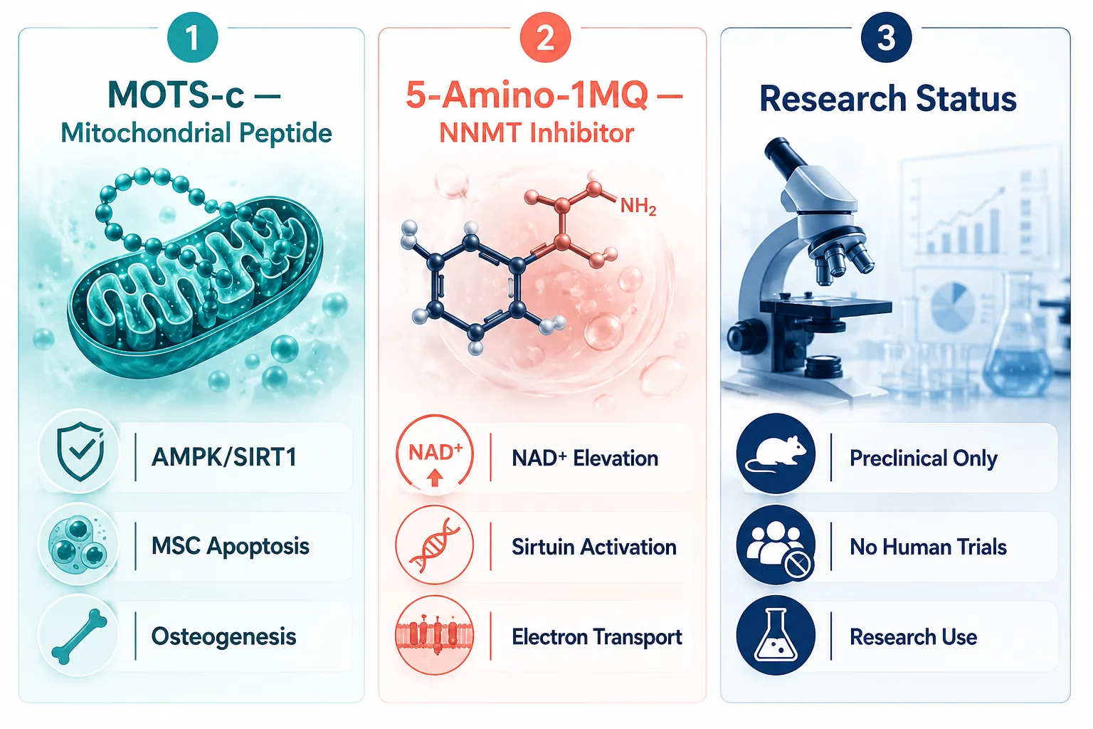
Scroll to top Scroll to top Scroll to top