

Tesamorelin and Ipamorelin Mechanism: How Their Growth-Hormone Signaling Differs in Research Models
{"cover":"Professional landscape format (1536×1024) hero image with bold text overlay: 'Tesamorelin vs Ipamorelin: How Their GH Signaling Differs' in extra large 72pt white bold sans-serif font with dark semi-transparent overlay box, centered upper third. Background shows a dramatic close-up of a pituitary gland cross-section illustration merged with molecular receptor diagrams, deep navy blue and teal color palette, glowing cAMP and calcium signaling pathway nodes rendered in bright white and gold, editorial magazine cover quality, high contrast, scientific research aesthetic.","content":["Detailed landscape format (1536×1024) scientific diagram illustration showing two distinct receptor pathways side by side: left panel depicts GHRH receptor on a pituitary somatotroph cell with cAMP and PKA cascade arrows in cool blue tones, right panel shows GHS-R1a ghrelin receptor activating phospholipase C and intracellular calcium release in warm amber tones, labeled molecular nodes, clean white background with subtle lab texture, research infographic style, high contrast annotations, editorial quality","Landscape format (1536×1024) data visualization showing two overlapping GH pulse waveform graphs on a dark charcoal background: one smooth physiological pulsatile curve in electric blue representing Tesamorelin-driven GH release, one sharp clean spike in gold representing Ipamorelin-induced GH pulse, time axis labeled in hours, amplitude axis labeled in ng/mL, subtle grid lines, cortisol and prolactin flat reference lines in red dashes, preclinical research model aesthetic, clean scientific chart style, editorial quality","Landscape format (1536×1024) overhead view of a modern preclinical research laboratory bench showing two separate experimental setups: left side has molecular model of Tesamorelin peptide chain with trans-3-hexenoic acid modification highlighted in blue, right side shows Ipamorelin pentapeptide structure in gold, surrounded by pharmacokinetic half-life timeline bars, CT scan visceral fat reduction imagery in background, subtle teal and white lab aesthetic, research-grade editorial photography style, high contrast, scientific publication quality"}
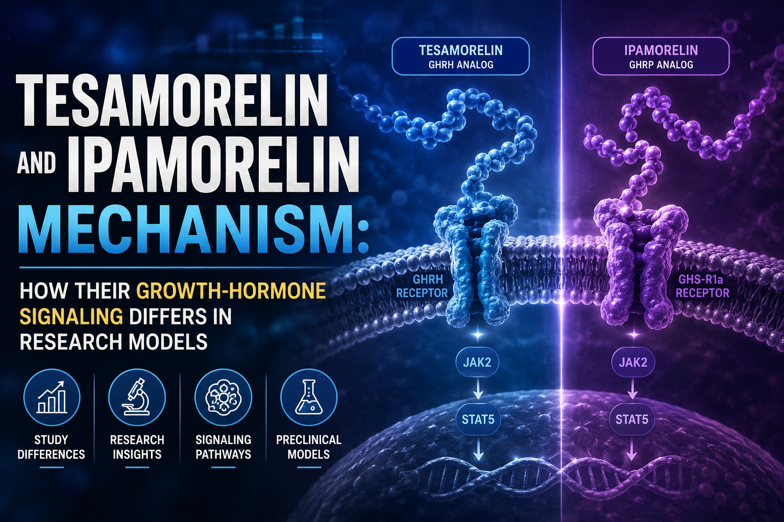
Two peptides can both raise growth hormone levels yet work through entirely separate receptor systems — and that distinction matters enormously when designing preclinical studies. Understanding the Tesamorelin and Ipamorelin mechanism: how their growth-hormone signaling differs in research models is not simply academic. It determines which endpoints are valid, which biomarkers to track, and whether combining the two compounds makes mechanistic sense.
Key Takeaways
- Tesamorelin activates the GHRH receptor via the cAMP/PKA pathway; ipamorelin activates the ghrelin receptor (GHS-R1a) via phospholipase C and intracellular calcium.
- The two pathways are complementary, not redundant, making dual-pathway research designs scientifically justified.
- Tesamorelin preserves physiological GH pulsatility; ipamorelin produces a selective, "clean" GH pulse without elevating cortisol or prolactin.
- Half-life differences (25-40 minutes vs. approximately 2 hours) affect dosing interval choices in animal pharmacokinetic models.
- IGF-1 elevation is a shared downstream endpoint, but the upstream signaling routes remain distinct.
Receptor-Level Differences That Define the Tesamorelin and Ipamorelin Mechanism
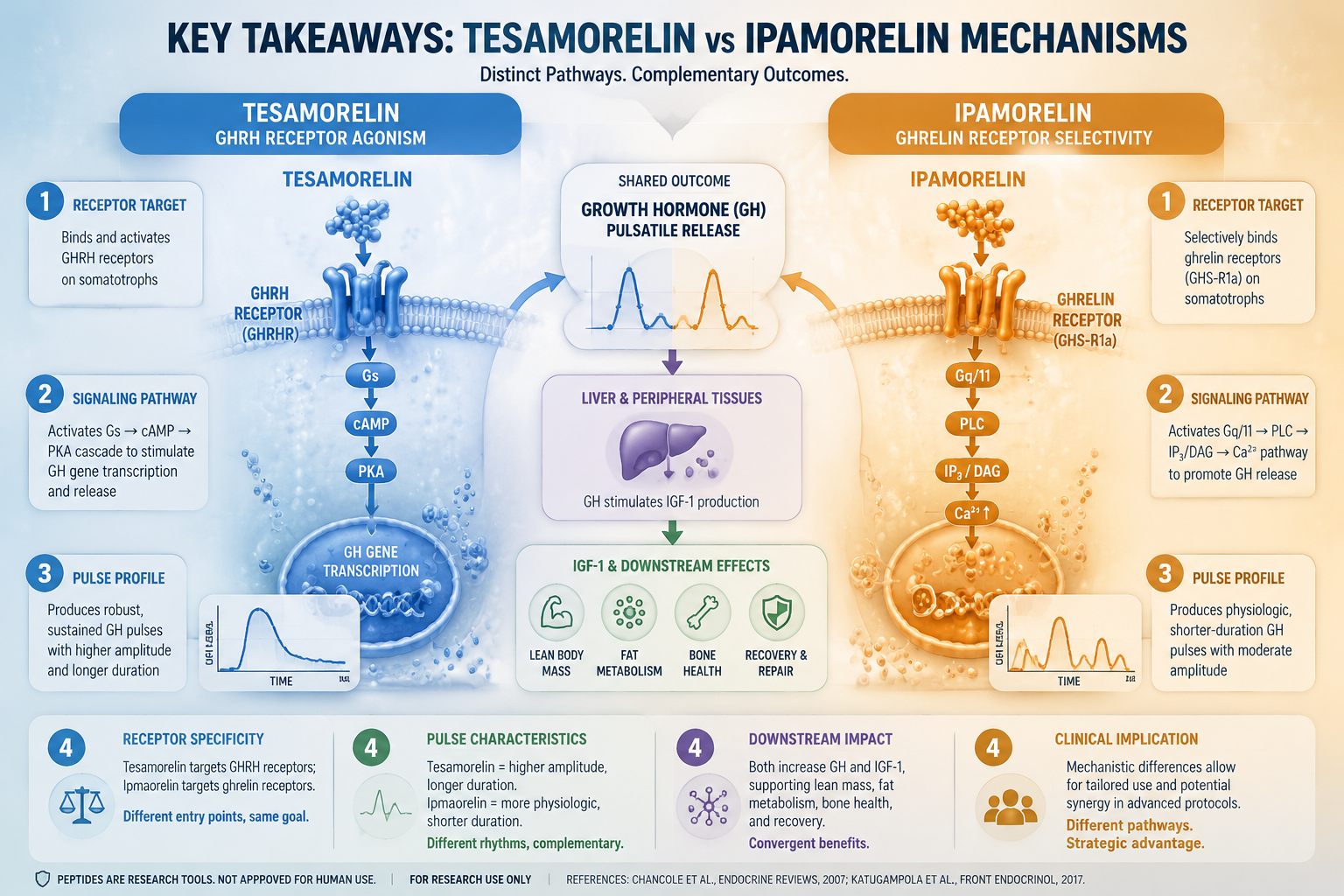
At the receptor level, these two secretagogues operate on separate systems.
Tesamorelin is a synthetic analog of endogenous growth hormone-releasing hormone (GHRH). Its N-terminal modification with trans-3-hexenoic acid protects it from enzymatic degradation, extending its half-life to roughly 25-40 minutes. It binds selectively to the GHRH receptor (GHRHR) on anterior pituitary somatotrophs and activates the cAMP/PKA signaling cascade, which drives GH gene transcription and pulsatile secretion. This mechanism mirrors the body's own GHRH signaling, preserving the natural rhythm of GH release.
Ipamorelin takes a different route entirely. It is a selective agonist of the growth hormone secretagogue receptor type 1a (GHS-R1a) — the same receptor that endogenous ghrelin activates. Rather than cAMP, GHS-R1a engagement triggers phospholipase C (PLC) activation, leading to IP3-mediated calcium release from intracellular stores. This calcium surge is what drives GH secretion in ipamorelin-treated models.
| Feature | Tesamorelin | Ipamorelin |
|---|---|---|
| Target Receptor | GHRHR | GHS-R1a (ghrelin receptor) |
| Signaling Cascade | cAMP / PKA | PLC / intracellular Ca2+ |
| Half-Life | ~25-40 minutes | ~2 hours |
| GH Release Pattern | Pulsatile, physiological | Sharp, selective pulse |
| Cortisol / ACTH Effect | Minimal | Negligible |
For researchers exploring ipamorelin muscle and fat research themes, this receptor distinction is foundational to interpreting results accurately.
GH Pulse Patterns and Downstream IGF-1 Endpoints in Research Models
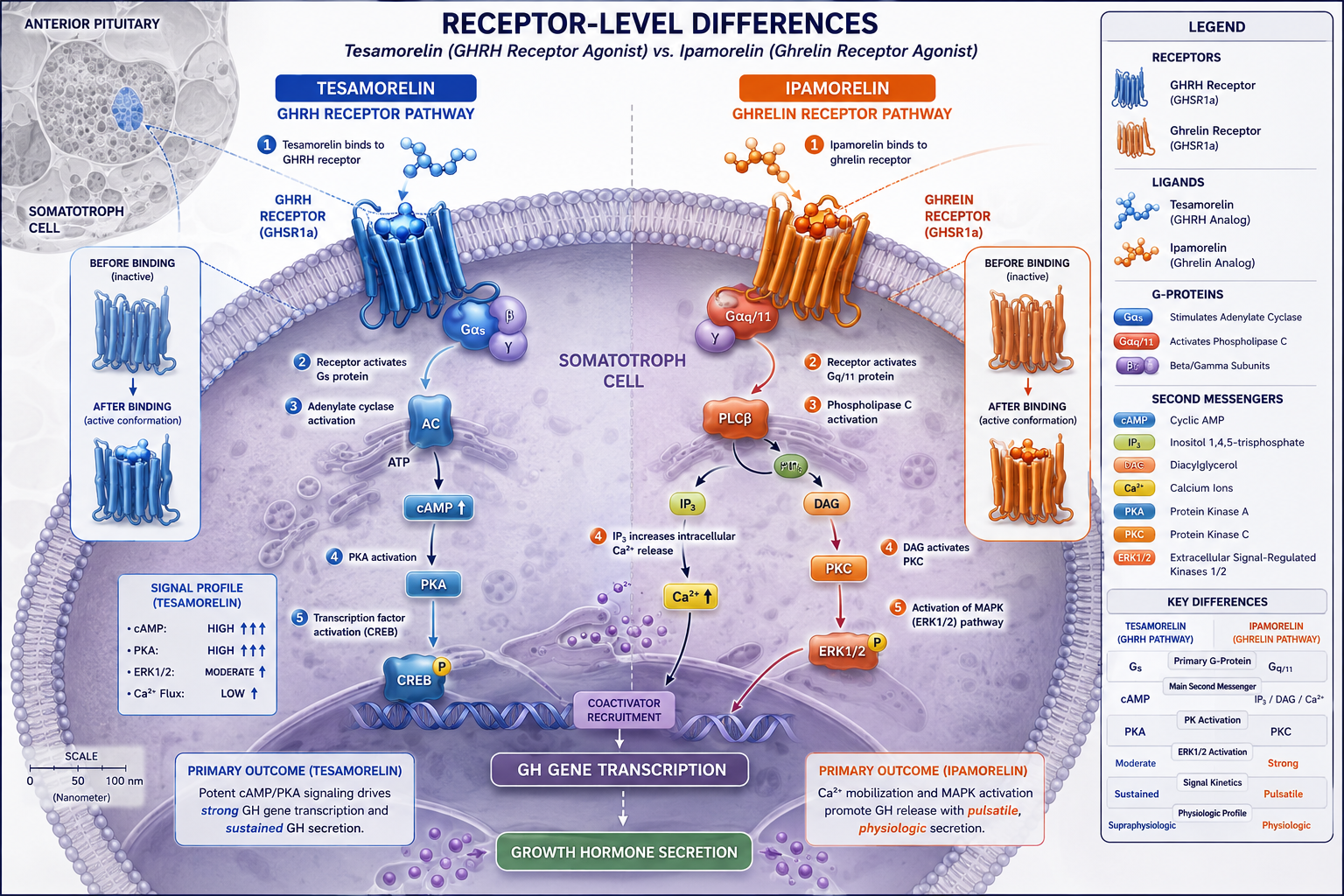
The pattern of GH release produced by each compound is as important as the magnitude.
Tesamorelin's activation of GHRHR amplifies both basal and pulsatile GH secretion, closely replicating the endogenous GHRH-driven rhythm. This physiological pulsatility is considered advantageous in research models where mimicking natural GH dynamics is a priority. Studies examining tesa peptide benefits often highlight this feature as a key differentiator from synthetic GH administration.
Ipamorelin, by contrast, generates what researchers describe as a "clean" GH pulse. Its selectivity for GHS-R1a means it does not significantly elevate cortisol, ACTH, or prolactin — a profile that distinguishes it from earlier GH secretagogues like GHRP-6 or hexarelin. For models where hormonal specificity is critical, this selectivity reduces confounding variables. Detailed analysis of ipamorelin as a GH secretagogue underscores why this selectivity is valued in controlled research settings.
Downstream, both peptides elevate IGF-1, which serves as a practical shared endpoint. Tesamorelin's IGF-1 effects have been documented in Phase 3 clinical trials — including data from HIV-associated lipodystrophy studies showing measurable visceral adipose tissue (VAT) reduction via CT scan. Ipamorelin's IGF-1 elevation has been confirmed in preclinical models, though large-scale clinical quantification remains limited.
"The upstream receptor divergence between these two secretagogues does not prevent a shared downstream outcome — but it does mean the signaling routes, and therefore the research questions, are fundamentally different."
Preclinical Study Design: Applying the Tesamorelin and Ipamorelin Mechanism to Research Endpoints
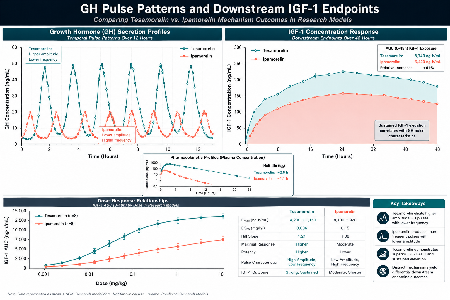
Understanding the Tesamorelin and Ipamorelin mechanism: how their growth-hormone signaling differs in research models has direct implications for study design.
Relevant preclinical endpoints include:
- Serum GH pulse amplitude and frequency (assessed via serial blood sampling)
- Plasma IGF-1 levels at defined intervals post-administration
- Visceral fat mass via imaging or tissue dissection in rodent models
- Cortisol and ACTH levels to confirm ipamorelin's hormonal selectivity
- Muscle protein synthesis markers for anabolic pathway assessment
Because the two pathways are complementary — cAMP/PKA versus PLC/calcium — researchers have proposed dual-pathway designs that combine both compounds. The rationale is that simultaneous GHRHR and GHS-R1a activation may produce synergistic GH release exceeding what either compound achieves alone. Blended formulations explored in Tesamorelin, CJC-1295, and Ipamorelin combination research reflect this mechanistic logic.
Half-life differences also shape dosing interval decisions. Tesamorelin's shorter plasma stability (~25-40 minutes) suggests more frequent administration windows in acute models, while ipamorelin's approximately 2-hour half-life in animal pharmacokinetic studies supports less frequent dosing. Researchers reviewing CJC-1295 and ipamorelin combination dosing will find that pairing compounds with complementary half-lives is a common strategy to sustain GH elevation across a study window.
For broader context on metabolic peptide research, exploring metabolic modulation research lines provides useful comparative frameworks alongside GH secretagogue work.
Conclusion
The mechanistic contrast between tesa and ipamorelin is not a minor technical detail — it is the foundation of any rigorous research design involving these compounds. Tesamorelin drives GH release through GHRHR and cAMP/PKA signaling, preserving physiological pulsatility. Ipamorelin activates GHS-R1a and the PLC/calcium pathway, producing a selective GH pulse without hormonal side effects.
Actionable next steps for researchers:
- Define whether the study requires physiological GH pulsatility (favor tesa) or hormonal selectivity (favor ipamorelin) before choosing a compound.
- Use IGF-1 as a shared downstream biomarker while tracking pathway-specific markers (cAMP vs. intracellular calcium) to confirm receptor engagement.
- Consider dual-pathway designs when the research goal is maximal GH output, accounting for the complementary receptor systems.
- Align dosing intervals with each compound's half-life data from pharmacokinetic models to avoid under- or over-dosing in timed studies.