GHK-Cu Peptide: Collagen Synthesis, Wound Healing & Anti-Aging Research
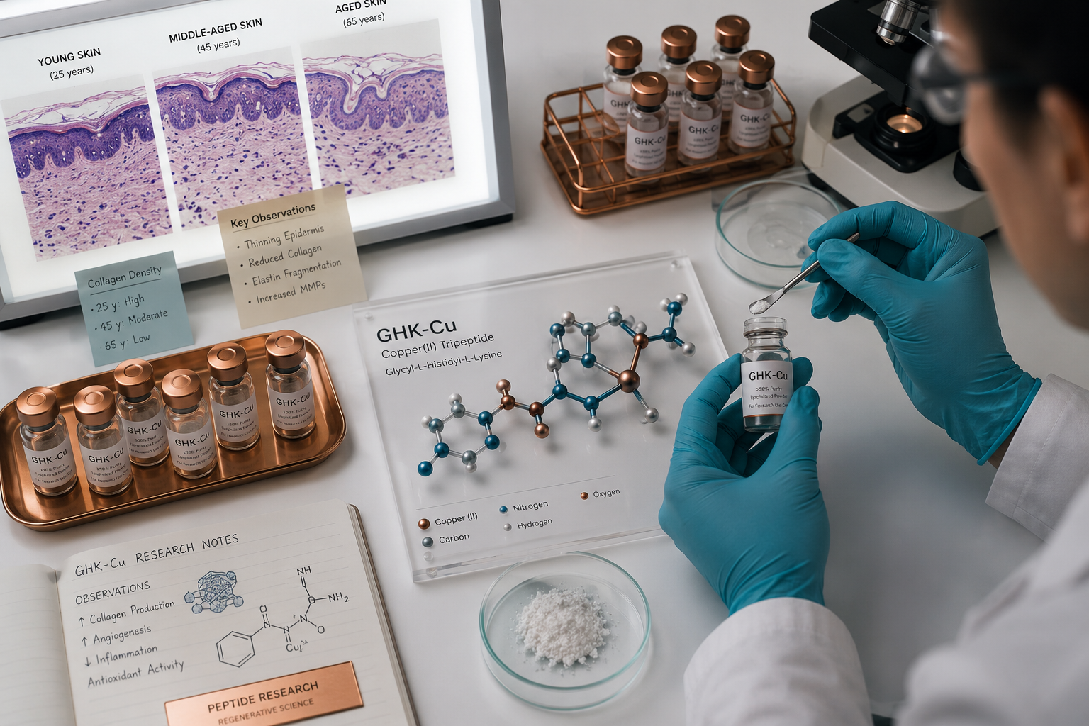
Copper is one of the most biologically active trace metals in the human body, and a tiny three-amino-acid sequence called GHK (glycyl-L-histidyl-L-lysine) has a remarkable ability to bind it. First isolated from human plasma in 1973, GHK-Cu was found to stimulate liver tissue regeneration — a discovery that launched decades of research into its role as a tissue-signaling molecule. Today, GHK-Cu Peptide: Collagen Synthesis, Wound Healing & Anti-Aging Research sits at the intersection of dermatology, wound biology, and longevity science, attracting growing attention from researchers worldwide.
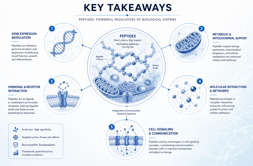
Key Takeaways
- GHK-Cu is a naturally occurring copper-binding tripeptide with documented roles in collagen synthesis and tissue repair.
- Preclinical research shows it activates fibroblasts, upregulates collagen and elastin production, and modulates inflammatory pathways.
- It has demonstrated wound-healing potential in animal models, including accelerated closure and reduced scar formation.
- As of 2026, GHK-Cu remains classified as a cosmetic ingredient and experimental research peptide — no FDA-approved prescription formulation exists.
- Ongoing research explores its anti-aging, antioxidant, and gene-expression-modulating properties.
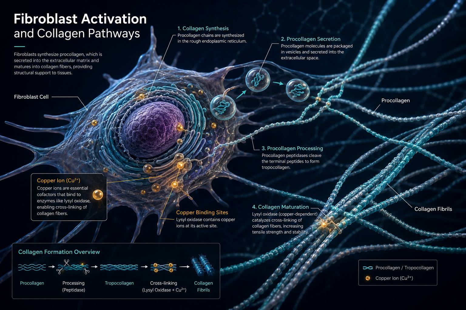
How GHK-Cu Works: Fibroblast Activation and Collagen Pathways
The central mechanism behind GHK-Cu Peptide: Collagen Synthesis, Wound Healing & Anti-Aging Research involves its interaction with fibroblasts — the cells responsible for producing structural proteins in connective tissue.
Key biological actions observed in preclinical studies include:
| Mechanism | Observed Effect |
|---|---|
| Fibroblast stimulation | Increased collagen I and III synthesis |
| Elastin upregulation | Improved tissue elasticity markers |
| MMP modulation | Balanced matrix metalloproteinase activity |
| Antioxidant activity | Reduced oxidative stress markers |
| Gene expression | Activation of over 30 tissue-repair genes |
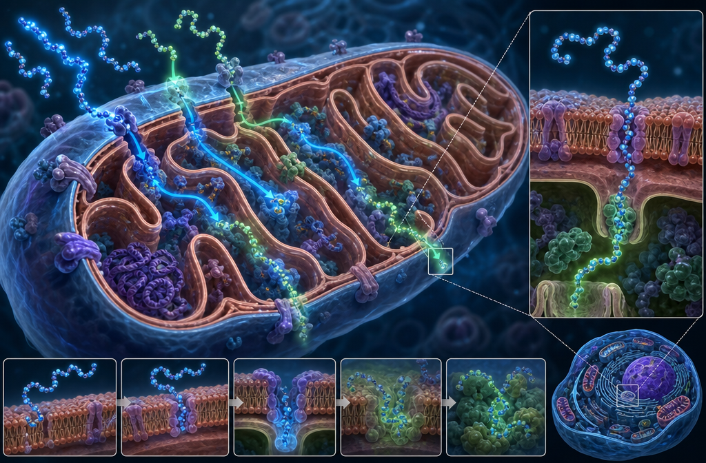
When GHK-Cu binds copper ions, it delivers them directly to enzymes like lysyl oxidase, which cross-links collagen and elastin fibers. This cross-linking is essential for structural integrity in skin, tendons, and vascular tissue.
"GHK-Cu does not simply add collagen — it appears to recalibrate the entire remodeling environment."
Research also shows GHK-Cu modulates transforming growth factor beta (TGF-beta) signaling, which governs both scar formation and normal tissue repair. This dual action — promoting repair while limiting excessive scarring — makes it particularly interesting for wound biology research. For a broader look at how peptides are reshaping tissue science, the latest peptide research updates provide useful context.
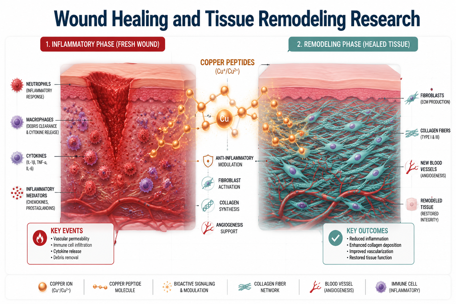
GHK-Cu in Wound Healing and Tissue Remodeling Research
Animal model studies have consistently shown that topical or injected GHK-Cu accelerates wound closure. In rodent excision models, treated wounds demonstrated faster re-epithelialization, denser collagen deposition, and reduced inflammatory cell infiltration compared to controls.
Three wound-healing properties highlighted in preclinical research:
- Angiogenesis support — GHK-Cu promotes the formation of new blood vessels, improving nutrient delivery to healing tissue.
- Nerve outgrowth — Early studies suggest it may support peripheral nerve regeneration at wound sites.
- Anti-inflammatory signaling — It appears to downregulate NF-kB pathways, reducing chronic inflammation that delays healing.
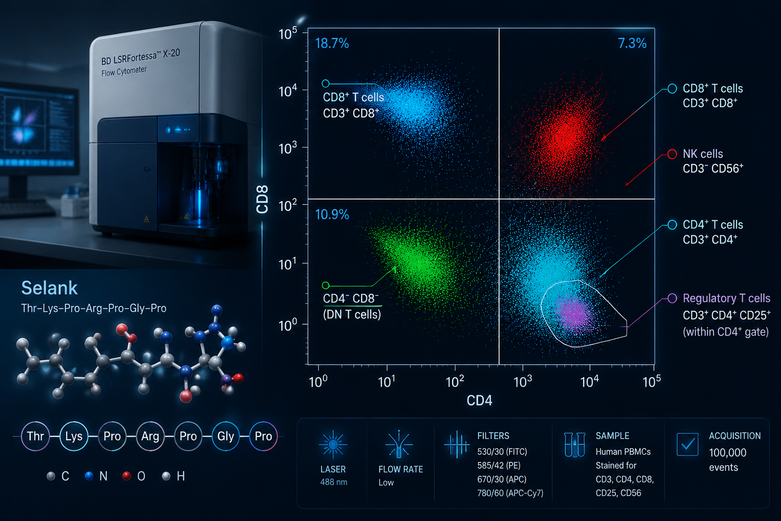
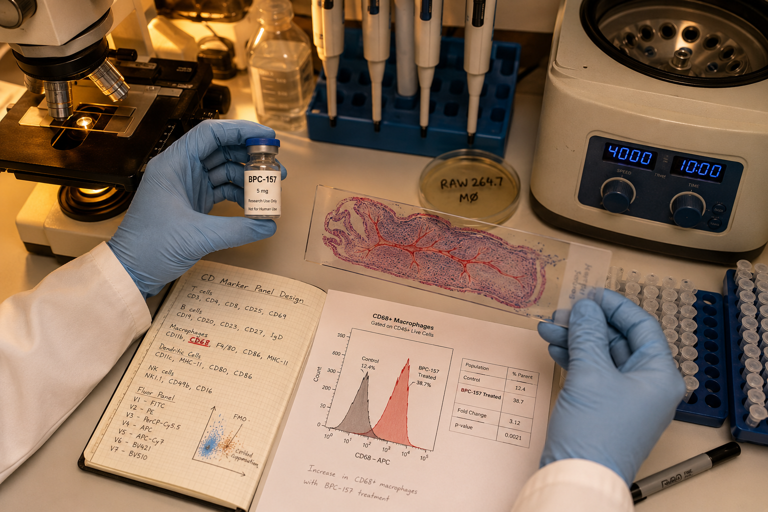
These findings place GHK-Cu alongside other tissue-repair peptides currently under investigation. Researchers interested in comparing repair-focused compounds may also find value in reviewing BPC-157 research themes and TB-500 research, both of which target overlapping tissue remodeling pathways.
The GHK-Cu longevity research overview explores additional preclinical data on systemic aging markers, including its effects on oxidative damage and cellular senescence.
Anti-Aging Research: Gene Expression and Systemic Implications
Beyond skin and wounds, GHK-Cu Peptide: Collagen Synthesis, Wound Healing & Anti-Aging Research has expanded into the field of gene modulation. A landmark analysis found that GHK-Cu reversed the gene expression signature of aged human tissue, activating pathways associated with DNA repair, proteasome function, and mitochondrial activity.
This positions GHK-Cu as more than a topical ingredient. Researchers now classify it as a systemic signaling molecule that may influence:
- Cellular senescence markers
- Oxidative stress response genes
- Tissue regeneration networks across multiple organ systems
The peptide's role in skin aging has been studied in both in vitro and clinical settings. Topical formulations have shown measurable improvements in skin density and fine-line depth in small human trials, though large randomized controlled trials remain limited.
For researchers exploring peptide delivery formats, nasal spray peptide delivery systems and innovative peptide delivery research address how bioavailability affects outcomes for compounds like GHK-Cu. The broader science of peptides in skincare also provides relevant background for understanding topical application research.
Regulatory status in 2026: GHK-Cu is classified as a cosmetic ingredient and research peptide. No FDA-approved prescription formulation exists for any indication — skin, hair, wound, or systemic. NIH-linked sources continue to describe it as experimental, and researchers should distinguish it from approved therapies when designing studies.
Conclusion
GHK-Cu is one of the most studied naturally occurring peptides in tissue biology, with a research profile spanning collagen synthesis, wound repair, antioxidant activity, and gene expression modulation. Its ability to activate fibroblasts, balance matrix remodeling enzymes, and influence aging-related gene signatures makes it a compelling subject for continued preclinical and clinical investigation.
Actionable next steps for researchers:
- Review preclinical wound-healing models to identify gaps where GHK-Cu data could be applied.
- Examine gene expression datasets comparing GHK-Cu-treated versus untreated aged tissue.
- Source research-grade GHK-Cu only from verified, tested suppliers — purity directly affects experimental validity. Reviewing best peptide manufacturer standards is a practical starting point.
- Stay current with evolving regulatory classifications before designing human-subject protocols.
The compound's transition from a plasma-isolated curiosity to a multi-pathway research target reflects the broader maturation of peptide science — and its most significant findings may still be ahead.