Call or Text 727-513-9780
  • Shopping Cart Shopping Cart
    0Shopping Cart
Pure Tested Peptides | America's most trusted Peptides for sale online
  • Peptides for sale
    • Oral Peptides for sale
      • Peptide Capsules for sale
      • BPC 157 Capsules 1000mcg
      • SLU-PP-332 Capsules | 1000 mcg
      • 5-Amino-1MQ 50mg Capsules
      • Tesofensine 500mcg
    • All Peptides for sale
    • Peptide Sprays
      • BPC 157 Nasal Spray Kit
      • BPC-157 TB500 Nasal Spray Kit
      • Semax Nasal Spray 10mg
      • Selank – Nasal Spray Kit – 10mg
      • Epithalon 50MG Nasal Spray Kit
      • Ipamorelin 10mg Nasal Spray
      • Klow Nasal Spray (BPC-157 + TB-500 + GHK-Cu + KPV) | 80mg
      • Hulk Nasal Spray Tesa / Ipa Blend 6/3 MG
      • Klow Nasal Spray
      • NAD + 500 mg Nasal Spray
      • PT-141 Nasal Spray Kit
    • GHRH Peptides
      • Ipa Peptides
      • CJC-1295 Peptides
        • CJC-1295 with DAC 5 mg
        • CJC-1295 without DAC 5 mg
        • CJC-1295 Ipa 10mg
      • Tesa Peptides
        • Tesa Peptide
        • Tesa 20 mg
    • GHK-Cu Peptides
      • All GHK-Cu Peptides
      • GHK-Cu 100mg
      • KLOW Peptide Blend – Buy KLOW blend online
    • BPC Peptides
      • All BPC Peptides
      • BPC-157
      • BPC-157 TB-500
      • BPC 157 capsules 1000mcg
    • SLU-PP-332 Peptides
      • All SLU-PP-332 Peptides
      • SLU-PP-332 5mg
    • GLP3 Peptides
    • PT-141 Peptides
      • PT-141 Peptides for sale
      • PT-141 10mg
      • PT-141 Nasal Spray
    • CAG Peptides
      • Lipo-C Peptide Blend
      • CAG 5mg
      • CAG 10mg
    • MOTS-C Peptides
      • MOTS-C Peptides for sale
      • MOTS-c peptide
      • MOTS-c 10mg *6 pack*
    • 5 Amino 1MQ Peptides
      • 5 Amino 1MQ Peptides for sale
      • 5-Amino-1MQ 50mg Capsules
      • 5-Amino-1MQ 5mg
    • Epithalon Peptides
      • Epithalon Peptides for sale
      • Epithalon 10mg
      • Epithalon 50mg
  • Shop
    • GLPs
      • 5-Amino-1MQ 50mg Capsules
      • 5-Amino-1MQ 5mg
      • L-Carnitine 500mg/ml
      • Tesofensine 500mcg
      • SLU-PP-332 5mg
      • MOTS-c 10mg *6 pack*
    • Epithalon & BPC Peptides
      • Epithalon 10mg
      • Epithalon 50mg
      • BPC-157
      • BPC 157 capsules 1000mcg
      • BPC-157 TB-500
      • BPC-157 TB500 Nasal Spray Kit
      • BPC 157 Nasal Spray Kit
    • BPC TB-500 & NAD+ Peptides
      • NAD+ 500 mg
      • KLOW Peptide Blend – Buy KLOW blend online
      • GLOW Peptide Blend
      • TB 500 5mg
      • BPC 157 capsules 1000mcg – Supplement
      • BPC 157 Nasal Spray Kit
      • BPC-157
      • BPC-157 TB500 Nasal Spray Kit
      • BPC-157 TB-500
      • BPC 157 capsules 1000mcg
    • LL-37 Peptide
      • LL-37 10 mg
    • MOTS-C & Selank
      • MOTS-c peptide
      • Selank 10mg
    • GHK Peptides
      • GHK-Cu 100mg
      • GLOW Peptide Blend
      • KLOW Peptide Blend – Buy KLOW blend online
  • COAs
  • Wholesale
    • Wholesale Peptides for sale
  • PTP FAQ
  • Affiliates
    • Selank Peptide: Advanced Pharmacological Mechanisms Underlying Its Anxiolytic and Nootropic Effects in Research
      • 5-Amino-1MQ Peptide: Detailed Mechanisms of NNMT Inhibition and Its Impact on Cellular Metabolism Research
        • MOTS-C Peptide: Unraveling Its Role in Mitochondrial Dynamics and Energy Metabolism Research
          • Epithalon Peptide and Telomerase Regulation: Investigating Its Impact on Cellular Senescence and Lifespan Research Models
            • GLP2-T vs GLP2 Tirz Peptide: Understanding the Naming, Mechanistic Nuances, and Research Implications for Gut Health
      • Peptides and Polypeptides in Modern Pharmacology: What Research on Metoprolol, Prednisone, and Amlodipine Reveals
        • Polypeptide Peptides vs NSAIDs: What Naproxen and Diclofenac Teach Tissue-Repair Researchers About BPC‑157 and TB‑500
          • Peptides and Polypeptides in Endocrine Pharmacology: How GLP-1, GLP-2, and GLP-3 Retatrutide Differ From Classic Drugs Like Prednisone and Amlodipine
          • 5‑Amino‑1MQ Peptide and NNMT Inhibition: How It Compares With Statins Like Atorvastatin in Adipose and Lipid Metabolism Research
      • Enclomiphene, Estrogen Receptor Signaling, and Luteinizing Phase Biology: What Hormone Researchers Should Measure
        • Mesenchymal Stem Cells, BPC‑157, and GHK‑Cu: How Tissue Repair Peptides Compare With Classic NSAIDs Like Naproxen in Injury Models
          • Peptide Calculator Use Cases Beyond Growth Hormone: Working Through GLP‑3 Retatrutide, MOTS‑c, and BPC‑157 Research Dosing
        • Polypeptide Peptides in Endocrine and Metabolic Pharmacology: Lessons From Amlodipine, Prednisone, and Metoprolol
        • Peptides Calculator for Advanced Blends: Worked Examples for Tesamorelin, CJC‑1295, and Ipamorelin Stacks
          • Peptides and Polypeptides in Human Physiology: How Molecular Size Shapes Research Applications
            • Selank Peptide: Advanced Pharmacological Mechanisms Underlying Its Anxiolytic and Nootropic Effects in Research
          • 5-Amino-1MQ Peptide: Detailed Mechanisms of NNMT Inhibition and Its Impact on Cellular Metabolism Research
            • MOTS-C Peptide: Unraveling Its Role in Mitochondrial Dynamics and Energy Metabolism Research
              • Epithalon Peptide and Telomerase Regulation: Investigating Its Impact on Cellular Senescence and Lifespan Research Models
                • GLP2-T vs GLP2 Tirz Peptide: Understanding the Naming, Mechanistic Nuances, and Research Implications for Gut Health
                  • Peptides and Polypeptides in Endocrine Pharmacology: How GLP-1, GLP-2, and GLP-3 Retatrutide Differ From Classic Drugs Like Prednisone and Amlodipine
                    • Polypeptide Peptides vs NSAIDs: What Naproxen and Diclofenac Teach Tissue-Repair Researchers About BPC‑157 and TB‑500
                      • Peptides and Polypeptides in Modern Pharmacology: What Research on Metoprolol, Prednisone, and Amlodipine Reveals
                        • 5‑Amino‑1MQ Peptide and NNMT Inhibition: How It Compares With Statins Like Atorvastatin in Adipose and Lipid Metabolism Research
                        • Enclomiphene, Estrogen Receptor Signaling, and Luteinizing Phase Biology: What Hormone Researchers Should Measure
                        • Mesenchymal Stem Cells, BPC‑157, and GHK‑Cu: How Tissue Repair Peptides Compare With Classic NSAIDs Like Naproxen in Injury Models
                        • Peptide Calculator Use Cases Beyond Growth Hormone: Working Through GLP‑3 Retatrutide, MOTS‑c, and BPC‑157 Research Dosing
                        • Polypeptide Peptides in Endocrine and Metabolic Pharmacology: Lessons From Amlodipine, Prednisone, and Metoprolol
                        • Peptides Calculator for Advanced Blends: Worked Examples for Tesamorelin, CJC‑1295, and Ipamorelin Stacks
                        • Peptides and Polypeptides in Human Physiology: How Molecular Size Shapes Research Applications
                        • Selank Peptide: Advanced Pharmacological Mechanisms Underlying Its Anxiolytic and Nootropic Effects in Research
                        • MOTS-C Peptide: Unraveling Its Role in Mitochondrial Dynamics and Energy Metabolism Research
                        • Peptides and Polypeptides in Modern Pharmacology: What Research on Metoprolol, Prednisone, and Amlodipine Reveals
                        • Peptides and Polypeptides in Endocrine Pharmacology: How GLP-1, GLP-2, and GLP-3 Retatrutide Differ From Classic Drugs Like Prednisone and Amlodipine
                        • 5-Amino-1MQ Peptide: Detailed Mechanisms of NNMT Inhibition and Its Impact on Cellular Metabolism Research
                        • Mesenchymal Stem Cells, BPC‑157, and GHK‑Cu: How Tissue Repair Peptides Compare With Classic NSAIDs Like Naproxen in Injury Models
                        • Peptide Calculator Use Cases Beyond Growth Hormone: Working Through GLP‑3 Retatrutide, MOTS‑c, and BPC‑157 Research Dosing
                        • GLP2-T vs GLP2 Tirz Peptide: Understanding the Naming, Mechanistic Nuances, and Research Implications for Gut Health
                        • Polypeptide Peptides in Endocrine and Metabolic Pharmacology: Lessons From Amlodipine, Prednisone, and Metoprolol
                        • Polypeptide Peptides vs NSAIDs: What Naproxen and Diclofenac Teach Tissue-Repair Researchers About BPC‑157 and TB‑500
                        • Peptides and Polypeptides in Human Physiology: How Molecular Size Shapes Research Applications
                        • Peptides Calculator for Advanced Blends: Worked Examples for Tesamorelin, CJC‑1295, and Ipamorelin Stacks
                        • 5‑Amino‑1MQ Peptide and NNMT Inhibition: How It Compares With Statins Like Atorvastatin in Adipose and Lipid Metabolism Research
                        • Enclomiphene, Estrogen Receptor Signaling, and Luteinizing Phase Biology: What Hormone Researchers Should Measure
                        • Epithalon Peptide and Telomerase Regulation: Investigating Its Impact on Cellular Senescence and Lifespan Research Models
                        • Best research protocol Klow blend
                        • best time to take BPC-157
                        • best time to take DSIP (Delta Sleep Inducing Peptide)
                        • best time to take CJC-1295
                        • best time to take AOD-9604
                        • best time to take Follistatin 344
                        • best time to take Ipamorelin
                        • best time to take MK-677 (Ibutamoren)
                        • best time to take Ligandrol (LGD-4033) — research compound
                        • best time to take Ostarine (MK-2866) — research compound
                        • best time to take GHK-CU
                        • best time to take TB-500
                        • best time to take MOTS-c
                        • best time to take Semax
                        • best time to take RAD-140 (Testolone) — research compound
                        • best time to take Thymosin Alpha-1
                        • best time to take PEG-MGF
                        • Biolife Plasma, Octapharma Plasma, and Research Peptides: How Plasma Donation Labs Differ From Peptide Suppliers
                        • best time to take YK-11 — research compound
                        • best time to take PT-141 (Bremelanotide)
                        • Best research protocol Klow blend
                        • 5-Amino-1MQ and MOTS-C Synergy: Metabolic Signaling, Mitochondria, and Research Design
                        • BPC-157 and TB-500: Investigating Their Combined Effects on Angiogenesis and Cellular Migration in Tissue Repair Models
                        • BPC-157 Peptide: Gut Barrier Function, Inflammation, and Tissue-Recovery Research
                        • 5‑Amino‑1MQ and MOTS‑c Synergy in Metabolic Research: Designing NNMT and Mitochondrial Biogenesis Stacks
                        • CJC-1295 with DAC vs. Without DAC: Half-Life, Release Kinetics, and Research Implications
                        • CJC‑1295 with DAC vs. Without DAC: Expanding on Half‑Life Differences Using Tesamorelin and Ipamorelin Blend Case Studies
                        • Collagen Biology and Copper‑Binding Peptides: How GHK‑Cu, Glow Blend, and Klow Blend Interact with Skin and Connective Tissue
                        • Collagen Biology and Regenerative Peptides: How GHK‑Cu, Glow Blend, and Klow Blend Affect Extracellular Matrix Research
                    • DNA, Telomeres, and Longevity Peptides: Positioning Epithalon and MOTS‑c in Genetic Aging Research
                      • Enclomiphene Citrate: serm Mechanism, Testosterone Research, and Stack Compatibility
                        • Enclomiphene vs Enclomiphene Citrate: Formulation, Bioavailability, and Research Distinctions
                        • Epithalon Peptide Research: Telomerase Activation, Aging, and Pineal Gland Function
                        • Estrogen Receptor Signaling and Enclomiphene: How Selective Modulators Compare with Classic Polypeptide Hormones
                        • GHK-Cu Peptide: Advanced Mechanisms in Extracellular Matrix Remodeling and Wound Healing Research
                        • GHK-Cu Peptide: Collagen Synthesis, Wound Repair, and Skin-Barrier Research Models
                        • GLP-1 vs GLP-3 vs GLP-2: Peptide Classification and Research Applications
                        • GLP-2 Peptide Research Guide: Gut Barrier Function, Nutrient Absorption, and Intestinal Recovery Models
                        • GLP-3 Retatrutide vs. GLP-1 Drugs: What Triple-Agonist Biology Changes in Research Models
                        • Ipamorelin and Tesamorelin Combination: Synergistic GH Secretagogue Research and Dosing Protocols
                        • GLP2 Tirz Peptide: What It Is, Why the Name Exists, and How Researchers Should Interpret It
                        • Klow Blend Peptide Nasal Spray: What the Formulation Is Trying to Do in Cognitive Research
                        • Mitochondria, NNMT Inhibition, and Peptide Modulators: Where MOTS‑c and 5‑Amino‑1MQ Fit in Cellular Energy Research
                        • MOTS-c Peptide: Mitochondrial Function, Energy Metabolism, and What Researchers Measure
                        • MOTS-c vs. 5-Amino-1MQ: Which Metabolic Research Questions Each Compound Actually Answers
                        • Nasal Spray Peptides: Bioavailability, Administration, and Semax/Selank Research Applications
                        • PT-141 Peptide Research: Mechanism of Action and Melanocortin Receptor Signaling
                        • Retatrutide for Research: Mechanism, Structure, and GLP-1/GLP-3 Dual Action
                        • Retatrutide for Obesity and Type 2 Diabetes: What the Latest Trial Data Suggest
                        • Tesofensine Peptide Research: Mechanism, Appetite Suppression, and Neuropeptide Y Pathways
  • Contact
    • Contact Customer Service
    • Text Customer Support
  • About US
  • Shop all peptides
  • Affiliate Program
    • Affiliate Signup
  • Login / Register Login / Register Page Link Login / Register Page Link
  • Click to open the search input field Click to open the search input field Search
  • Menu Menu

Tag Archive for: longevity research

How 5-Amino-1MQ and MOTS-c Are Studied Together in Metabolic Research

How 5-Amino-1MQ and MOTS-c Are Studied Together in Metabolic Research

August 13, 2026/0 Comments/in Uncategorized/by

Metabolic dysfunction now affects more than one billion people globally, yet the pipeline of approved pharmacological tools remains narrow. That gap has pushed researchers toward investigational compounds with complementary mechanisms, and few pairings have attracted more scientific curiosity in 2026 than 5-Amino-1MQ and MOTS-c. Understanding how 5-Amino-1MQ and MOTS-c are studied together in metabolic research requires looking at what each compound does independently before examining why their combination is considered scientifically interesting.

Key Takeaways

  • 5-Amino-1MQ inhibits the enzyme NNMT, raising NAD+ levels and activating fat metabolism at the cellular level.
  • MOTS-c is a mitochondria-derived peptide that activates AMPK signaling and improves glucose handling in preclinical models.
  • The two compounds target different but interconnected metabolic pathways, making them a subject of combination research.
  • Both remain investigational; no randomized controlled trials in humans have confirmed fat-loss or metabolic outcomes for either agent.
  • Researchers and clinics are exploring stacking protocols with NAD+ precursors and GLP-1 agonists, though evidence remains early-stage.

The Distinct Mechanisms Behind Each Compound
The Distinct Mechanisms Behind Each Compound

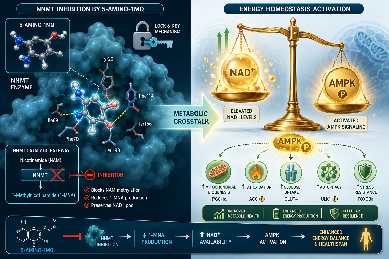
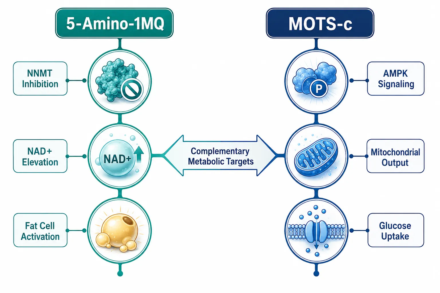
5-Amino-1MQ is a small-molecule inhibitor of nicotinamide N-methyltransferase (NNMT), an enzyme that consumes NAD+ precursors. When NNMT is blocked, cellular NAD+ availability rises. Higher NAD+ levels are associated with increased activity of sirtuins and other metabolic regulators that govern fat oxidation and energy expenditure. In adipose tissue, this shift appears to reduce lipid storage and promote lipolysis in cell and animal models. For a deeper look at how NAD+ connects to these peptide systems, the resource on adenosine triphosphate and mitochondrial peptides: how MOTS-c and 5-Amino-1MQ influence ATP production provides useful mechanistic context.

MOTS-c is a 16-amino-acid peptide encoded within mitochondrial DNA. It primarily works through AMPK activation, a master energy sensor that promotes glucose uptake, suppresses lipogenesis, and enhances mitochondrial biogenesis. Unlike most peptides, MOTS-c can translocate to the nucleus under metabolic stress, where it modulates gene expression tied to metabolic flexibility. Researchers interested in its foundational biology can explore MOTS-c: the mitochondrial peptide for background on its discovery and signaling profile.

The key distinction is target specificity:

Feature 5-Amino-1MQ MOTS-c
Primary target NNMT enzyme AMPK pathway
Key metabolite affected NAD+ Glucose / lipid flux
Main tissue focus Adipose tissue Skeletal muscle, liver
Molecule type Small molecule Mitochondrial peptide
Administration route (research) Oral (preclinical) Injectable (preclinical)

How 5-Amino-1MQ and MOTS-c Are Studied Together in Metabolic Research: The Combination Rationale

The rationale for studying these two agents together is rooted in pathway complementarity. NNMT inhibition by 5-Amino-1MQ addresses the upstream availability of NAD+, while MOTS-c operates downstream through AMPK to improve how cells use the energy that NAD+ helps generate. In theory, raising NAD+ and simultaneously activating AMPK could produce additive effects on mitochondrial efficiency and substrate utilization.

Key insight: Researchers describe the pairing as targeting "two different floors of the same metabolic building", one compound improves fuel supply, the other improves how cells burn it.

Preclinical models examining this combination have focused on:

  • Adipose tissue remodeling, measuring changes in white adipose depots
  • Insulin sensitivity markers, fasting glucose, HOMA-IR in rodent models
  • Mitochondrial respiration assays, oxygen consumption rate in isolated cells
  • Body composition endpoints, lean mass preservation alongside fat reduction

Researchers studying related mitochondrial peptide combinations, such as the MOTS-c and Elamipretide pairing, have used similar assay frameworks, making that work a useful methodological reference point.

Evidence Tiers and Research Gaps

Evidence Tiers and Research Gaps

Both compounds remain firmly in the investigational category. Neither 5-Amino-1MQ nor MOTS-c is FDA-approved, and both are currently sold exclusively as research chemicals. The evidence base, as of mid-2026, sits at the following tiers:

Established (in vitro and animal data):

  • NNMT inhibition by 5-Amino-1MQ reduces adiposity in diet-induced obese mouse models
  • MOTS-c improves glucose tolerance and exercise capacity in aged rodents
  • Combination protocols in cell models suggest non-overlapping pathway activation

Emerging (mechanistic speculation and early protocol design):

  • Longevity-focused researchers have proposed NAD+/MOTS-c/5-Amino-1MQ stacks as a multi-target approach to metabolic aging
  • Clinics have begun positioning the duo for "weight plateau" scenarios alongside GLP-1 agonists, though this is protocol-level practice without controlled trial support

Missing (critical evidence gaps):

  • No randomized controlled trials in humans for either compound alone
  • No published human pharmacokinetic data for the combination
  • Organ-target interaction profiles at combined doses remain unstudied

Expert commentary from metabolic biology reviewers in 2026 consistently frames the situation as "interesting biology, weak human evidence." That honest assessment should anchor any research design that incorporates this pairing. For comparison, researchers interested in how appetite-modulating compounds are evaluated alongside metabolic peptides may find the analysis of tesofensine vs GLP-3 retatrutide appetite-modulating pathways instructive for study design principles.

How 5-Amino-1MQ and MOTS-c Are Studied Together: Protocol Design Considerations

How 5-Amino-1MQ and MOTS-c Are Studied Together: Protocol Design Considerations

For researchers designing combination studies, several practical considerations emerge from the existing preclinical literature.

Dosing sequencing: Some protocols administer 5-Amino-1MQ first to elevate NAD+ availability before introducing MOTS-c, hypothesizing that a primed NAD+ environment amplifies AMPK responsiveness. This sequencing remains theoretical but is gaining traction in research design discussions as of July 2026.

Biomarker selection: Researchers typically track NAD+/NADH ratios, phosphorylated AMPK levels, PGC-1 alpha expression, and mitochondrial membrane potential as primary readouts when studying this combination.

Stacking with other agents: A growing number of protocols layer this pairing with NAD+ precursors (NMN or NR) or GLP-1 receptor agonists. The MOTS-c and SLU-PP332 research context offers a parallel example of how MOTS-c is studied alongside exercise-mimetic compounds, which shares methodological overlap with 5-Amino-1MQ combination work.

Researchers comparing 5-Amino-1MQ against other weight-related compounds in isolation may also benefit from reviewing the 5-Amino-1MQ vs Tesofensine comparison to understand its standalone profile before interpreting combination data.

Conclusion

The study of how 5-Amino-1MQ and MOTS-c are examined together in metabolic research represents one of the more scientifically grounded areas of investigational peptide science in 2026. The mechanistic logic is sound: NNMT inhibition and AMPK activation address metabolic dysfunction from different but reinforcing angles. However, the evidence base remains preclinical, and the absence of human trial data is a significant limitation that no amount of mechanistic elegance can substitute.

Actionable next steps for researchers:

  1. Ground any combination protocol in the existing rodent and cell-model literature before extrapolating to human applications.
  2. Use validated biomarker panels (NAD+/NADH, p-AMPK, PGC-1 alpha) to generate quantifiable endpoints.
  3. Source research-grade material with verified purity documentation, the MOTS-c peptide 10mg research-grade product page is one reference point for purity standards.
  4. Monitor the clinical trial registries for emerging human studies, as this area is expected to move quickly given commercial and longevity-research interest.
  5. Treat any "synergy" claims with appropriate skepticism until controlled human data is available.

The biology is compelling. The human evidence is not yet there. That gap is precisely what makes this combination a productive area for rigorous investigation.

https://www.puretestedpeptides.com/wp-content/uploads/2026/08/how-5-amino-1mq-and-mots-c-are-studied-together-in-metabolic-research.webp 1024 1536 https://www.puretestedpeptides.com/wp-content/uploads/2026/01/buy-peptides-online.jpg 2026-08-13 13:04:512026-08-13 13:04:51How 5-Amino-1MQ and MOTS-c Are Studied Together in Metabolic Research
Epithalon Peptide in Longevity Research: Telomeres, Cellular Aging, and What Labs Measure

Epithalon Peptide in Longevity Research: Telomeres, Cellular Aging, and What Labs Measure

August 12, 2026/0 Comments/in Uncategorized/by

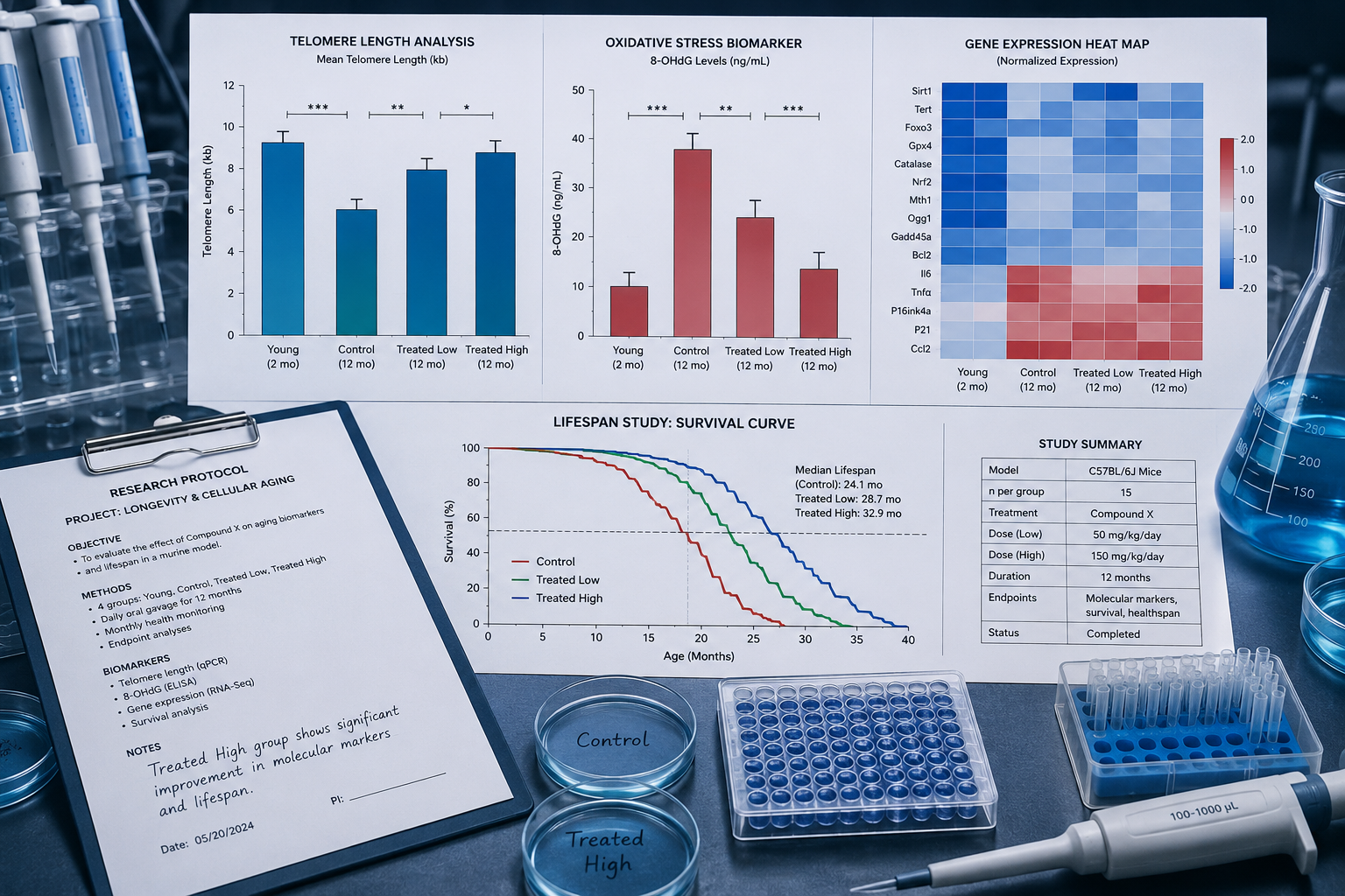
Telomere length at birth predicts roughly 60% of the variance in lifespan across mammalian species, a statistic that reframed how researchers think about biological aging at the molecular level. Against that backdrop, Epithalon peptide in longevity research: telomeres, cellular aging, and what labs measure has become one of the most discussed topics in experimental gerontology, precisely because this short tetrapeptide appears to interact with the very machinery that governs telomere maintenance.

This article is a research application guide. It is not a clinical protocol. It is designed for scientists, research buyers, and informed readers who want a rigorous framework, not hype, for evaluating what Epithalon does, what biomarkers matter, and where the experimental evidence currently stands.

Key Takeaways

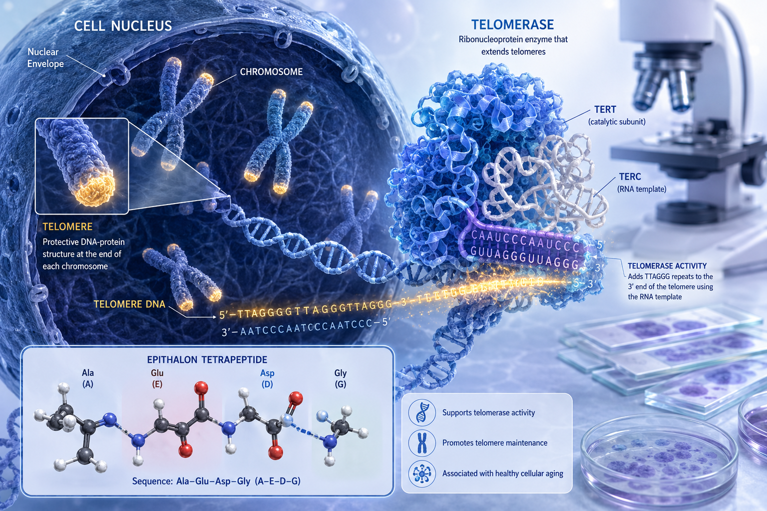
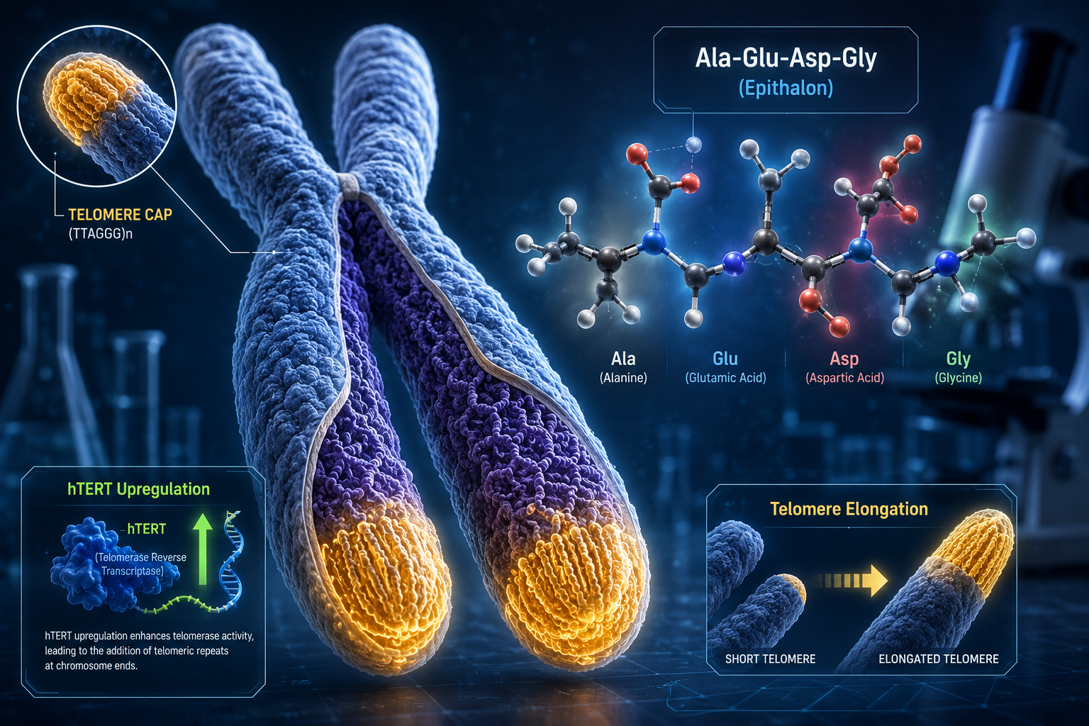
  • Epithalon (Ala-Glu-Asp-Gly) is a synthetic tetrapeptide derived from the pineal gland peptide Epithalamin, studied primarily for its proposed effects on telomerase activation and cellular senescence.
  • Its primary hypothesized mechanism involves upregulation of telomerase reverse transcriptase (hTERT), the catalytic subunit responsible for adding telomeric repeats to chromosome ends.
  • Lab measurement of Epithalon's effects requires a multi-marker approach: telomere length assays, hTERT expression panels, and senescence-associated secretory phenotype (SASP) markers.
  • Most foundational data originates from Russian institutional research; more recent 2025 human cell line studies have begun replicating and extending those findings under controlled conditions.
  • Experimental limitations, including species-specific telomerase regulation and the absence of large-scale human RCTs, must anchor any honest interpretation of the data.

Key Takeaways

The Biology Behind Epithalon: Telomerase, Telomeres, and Cellular Aging

To understand why Epithalon peptide in longevity research: telomeres, cellular aging, and what labs measure commands serious scientific attention, it helps to understand the underlying biology with precision.

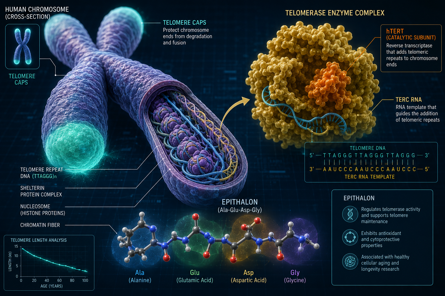
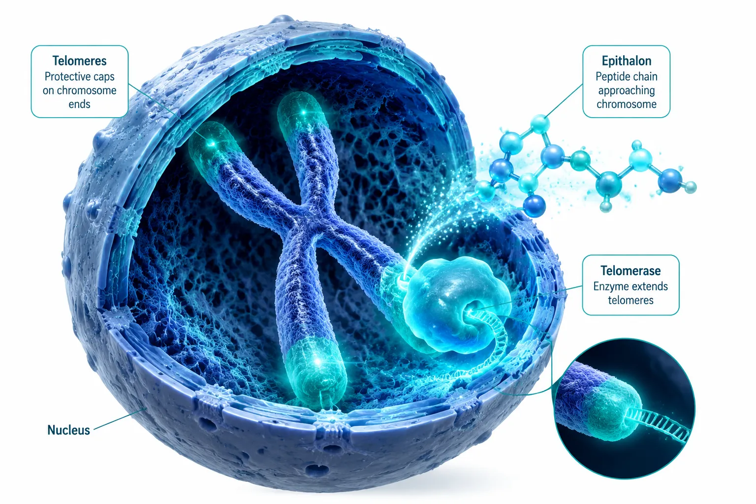
Telomeres are repetitive nucleotide sequences (TTAGGG in humans) that cap chromosome ends, protecting genetic material from degradation during cell division. Each replication cycle shortens telomeres slightly. When telomeres reach a critical minimum length, cells enter replicative senescence, a permanent growth arrest, or trigger apoptosis.

Telomerase is the enzyme complex that counteracts this shortening. Its catalytic subunit, hTERT, adds telomeric repeats back to chromosome ends. In most adult somatic cells, telomerase expression is suppressed. In stem cells, germline cells, and certain immune cells, it remains active. Cancer cells, notably, reactivate telomerase as a survival mechanism, a fact that makes any telomerase-activating compound a subject of both excitement and caution in research circles.

Epithalon (tetrapeptide sequence: Ala-Glu-Asp-Gly) was originally isolated from bovine pineal gland extracts by Professor Vladimir Khavinson's team in St. Petersburg. The synthetic version replicates the active sequence. Early animal studies reported extended median lifespan in aged rats and mice, alongside measurable increases in hTERT expression in lymphocyte cultures. Revisited analyses of those older datasets, cross-referenced with more recent 2025 human cell line data, suggest the hTERT upregulation signal is reproducible under specific culture conditions, though the magnitude varies considerably by cell type and passage number.

"The question is not whether Epithalon affects telomerase expression in vitro, the data suggest it does. The question is what that means for whole-organism aging biology."

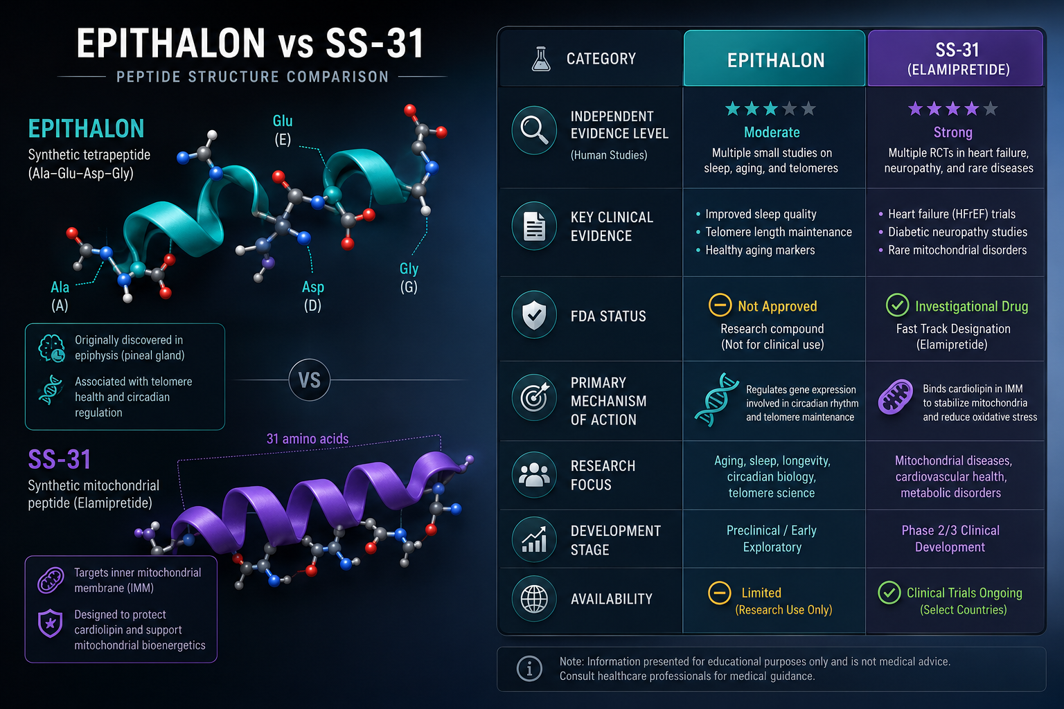
For researchers exploring related peptide mechanisms, the SS-31 mechanism and research overview, including where to buy SS-31 and Epithalon provides useful context on how mitochondria-targeted peptides intersect with cellular aging pathways.

The Biology Behind Epithalon: Telomerase, Telomeres, and Cellular Aging

Senescence Markers and the Multi-Biomarker Framework for Epithalon Research

Telomere length alone is an incomplete readout. A rigorous research design around Epithalon peptide in longevity research: telomeres, cellular aging, and what labs measure requires a layered biomarker approach.

Primary Markers Researchers Track

Biomarker What It Measures Relevance to Epithalon
Telomere Length (qPCR or FISH) Average telomere length per cell Direct readout of telomere maintenance
hTERT mRNA Expression Telomerase catalytic subunit activity Primary proposed mechanism of action
p16INK4a / p21 Protein Levels Senescence cell cycle arrest markers Downstream indicator of senescent burden
SA-beta-galactosidase Activity Classic senescence-associated enzyme Functional confirmation of senescent state
SASP Panel (IL-6, IL-8, MMP-3) Pro-inflammatory secretory phenotype Systemic aging signal from senescent cells

p16INK4a has emerged as particularly useful because it accumulates specifically in senescent cells and correlates with biological age more tightly than chronological age in several tissue studies. A well-designed Epithalon experiment should show changes in p16INK4a alongside any telomere length shifts to establish mechanistic coherence rather than isolated correlation.

Researchers studying peptide-based modulators in regenerative models, including those examining how BPC-157, GHK-Cu, and Glow Blend are used in mesenchymal stem cell research, will recognize this multi-marker logic as standard practice across the field.

For those sourcing compounds for controlled in vitro work, Epithalon peptides for sale from verified suppliers with third-party testing documentation is a prerequisite for data integrity. Purity directly affects reproducibility.

The GHK-Cu longevity research themes overview offers a parallel example of how copper-binding peptides interact with cellular repair pathways, providing useful comparative context for researchers building multi-peptide longevity panels.

Primary Markers Researchers Track

What Labs Actually Measure: Assay Selection and Experimental Limitations

The practical side of Epithalon peptide in longevity research: telomeres, cellular aging, and what labs measure comes down to assay selection, model validity, and honest acknowledgment of what the current evidence cannot yet confirm.

Common Assay Approaches

Quantitative PCR (qPCR) telomere assay remains the most widely used method due to cost and throughput. It measures average telomere length relative to a single-copy gene. Its limitation is that it averages across all cells, masking the critically short telomeres that drive senescence in individual cells.

Telomere-FISH (Fluorescence In Situ Hybridization) provides single-cell resolution, identifying cells with critically short telomeres. More labor-intensive but mechanistically more informative for Epithalon studies.

hTERT RT-qPCR panels measure messenger RNA levels, not enzyme activity directly. Western blotting for hTERT protein, combined with TRAP (Telomeric Repeat Amplification Protocol) assays for functional telomerase activity, creates a more complete picture.

Key Experimental Limitations

  • Species differences matter significantly. Mice have much longer telomeres and constitutively active telomerase in most tissues, making murine lifespan data difficult to translate directly to human aging biology.
  • Cell passage number confounds results. hTERT responses in early-passage versus late-passage cell lines differ substantially. Studies must report passage numbers explicitly.
  • No large-scale human RCTs exist. The foundational data from Russian institutional research, while methodologically serious, predates modern RCT standards. Replication in controlled human trials remains an open priority.
  • Telomerase activation and oncogenic risk. Any compound that upregulates hTERT warrants parallel monitoring of oncogenic markers, this is not a reason to dismiss the research, but it is a non-negotiable component of responsible experimental design.

Researchers building broader longevity peptide panels will find the Glow Blend longevity research themes resource useful for understanding how multi-peptide formulations are being studied alongside telomere-focused compounds.

For foundational context on peptide structure and research-use classification, Peptides 101 for research-use only buyers covers the structural and regulatory framework that applies to Epithalon and similar compounds.

Conclusion

The evidence base for Epithalon in longevity research is more substantive than most peptide discussions acknowledge, and more limited than enthusiast communities often admit. The telomerase activation hypothesis is mechanistically coherent, supported by reproducible in vitro hTERT expression data, and consistent with the broader biology of telomere-driven senescence. At the same time, the absence of large-scale human trials, the species-translation problem, and the oncogenic monitoring requirement all demand that researchers approach this compound with structured skepticism rather than either dismissal or uncritical enthusiasm.

Actionable next steps for research teams:

  1. Design multi-marker protocols that combine telomere length assays (preferably FISH for single-cell resolution), hTERT expression panels, and SASP cytokine profiling rather than relying on any single readout.
  2. Document cell passage numbers, culture conditions, and compound purity specifications in every experiment, these variables account for much of the variance in published results.
  3. Source Epithalon from suppliers providing third-party HPLC and mass spectrometry documentation to ensure purity standards that support reproducible data.
  4. Pair Epithalon studies with parallel oncogenic marker monitoring as a non-negotiable safety and scientific integrity measure.
  5. Follow the emerging 2026 gerontology literature on peptide classification frameworks, which is beginning to establish standardized endpoints that will make cross-study comparison more meaningful.

The science of telomere biology and peptide-based longevity research is advancing. Rigorous measurement frameworks, not optimistic extrapolation, are what will ultimately determine whether Epithalon earns a durable place in the gerontology toolkit.

https://www.puretestedpeptides.com/wp-content/uploads/2026/08/epithalon-peptide-in-longevity-research-telomeres-cellular-aging-and-what-labs-m.webp 1024 1536 https://www.puretestedpeptides.com/wp-content/uploads/2026/01/buy-peptides-online.jpg 2026-08-12 13:03:402026-08-12 13:03:40Epithalon Peptide in Longevity Research: Telomeres, Cellular Aging, and What Labs Measure
GHK-Cu Peptide: Collagen Synthesis, Tissue Repair, and Longevity Research Applications

GHK-Cu Peptide: Collagen Synthesis, Tissue Repair, and Longevity Research Applications

August 9, 2026/0 Comments/in Uncategorized/by

A single copper ion can change how a peptide behaves at the molecular level. That principle sits at the heart of GHK-Cu research, a tripeptide-copper complex that has attracted serious scientific attention since Loren Pickart first isolated it from human plasma in 1973. Today, GHK-Cu peptide: collagen synthesis, tissue repair, and longevity research applications represent one of the more mechanistically rich areas in peptide biology, drawing interest from researchers working across dermatology, wound healing, and aging science.

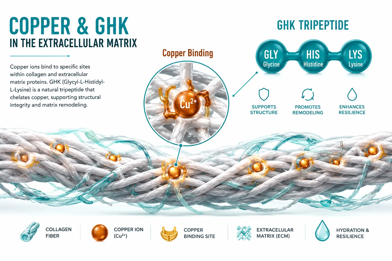
Bright editorial infographic-style landscape (): cross-section diagram of extracellular matrix collagen fibers with copper

Key Takeaways

  • GHK-Cu is a naturally occurring tripeptide (glycine-histidine-lysine) that binds copper(II) ions, enabling a wide range of biological signaling functions.
  • Research shows GHK-Cu upregulates collagen, elastin, and glycosaminoglycan synthesis by activating fibroblast activity in the extracellular matrix.
  • Beyond skin biology, GHK-Cu has demonstrated tissue-repair activity in wound models, nerve tissue, and lung fibrosis research.
  • Longevity researchers have identified GHK-Cu as a potential gene-expression modulator, with studies linking it to reversal of aging-associated transcriptional changes.
  • GHK-Cu is frequently studied alongside other repair-focused peptides such as BPC-157 and TB-500 in multi-compound research protocols.

The Copper-Binding Biology Behind GHK-Cu

The letters in GHK stand for the three amino acids that form this tripeptide: glycine, histidine, and lysine. What makes GHK-Cu distinct from many other short peptides is its high-affinity binding to copper(II) ions. This copper-chelating property is not incidental, it is central to the compound's biological activity.

Copper is a trace element involved in over 30 enzymatic reactions in the human body. Enzymes like lysyl oxidase (which crosslinks collagen and elastin fibers) and superoxide dismutase (an antioxidant enzyme) depend on copper as a cofactor. When GHK binds copper, it acts as a bioavailable copper-delivery vehicle, shuttling the ion to sites where these enzymes are active.

To understand how short peptides like GHK-Cu function within broader molecular frameworks, the polypeptide peptides explained: structure, function, and research resource provides useful foundational context.

Key copper-dependent processes relevant to GHK-Cu research:

Process Relevant Enzyme Role in Tissue Biology
Collagen crosslinking Lysyl oxidase Structural integrity of ECM
Antioxidant defense Superoxide dismutase Reduces oxidative damage
Angiogenesis Ceruloplasmin New blood vessel formation
Melanin synthesis Tyrosinase Pigmentation and skin repair

Beyond copper delivery, GHK itself appears to function as a signaling molecule. In vitro studies have shown it can activate pathways associated with TGF-beta (transforming growth factor beta), a cytokine that drives fibroblast proliferation and matrix remodeling.

GHK-Cu Peptide: Collagen Synthesis and Extracellular Matrix Remodeling

The extracellular matrix (ECM) is the structural scaffold that surrounds cells in connective tissue. It is composed primarily of collagen fibers, elastin, fibronectin, and glycosaminoglycans (GAGs). Maintaining ECM integrity is critical for wound healing, organ function, and tissue resilience.

Research into GHK-Cu peptide: collagen synthesis, tissue repair, and longevity research applications has consistently pointed to fibroblast activation as a primary mechanism. Fibroblasts are the cells responsible for producing and maintaining ECM components. Studies have shown that GHK-Cu:

  • Increases collagen synthesis, particularly types I and III, the most abundant structural collagens
  • Upregulates elastin production, improving tissue elasticity
  • Stimulates GAG synthesis, including hyaluronic acid and dermatan sulfate, which support hydration and structural spacing in the ECM
  • Activates matrix metalloproteinases (MMPs), enzymes that break down damaged or disorganized collagen, enabling remodeling

This dual action, promoting new matrix synthesis while clearing old or damaged matrix, makes GHK-Cu particularly relevant to wound repair models. Researchers studying multi-peptide repair protocols often pair GHK-Cu with other compounds; the Skin Repair Stack (BPC-157 + TB-500 + GHK-Cu) is one documented example of this combinatorial approach in research contexts.

For broader comparison of tissue-repair peptides, the BPC-157 vs TB-500 complete research comparison guide offers useful mechanistic contrasts.

GHK-Cu Peptide: Collagen Synthesis and Extracellular Matrix Remodeling

Tissue Repair, Nerve Regeneration, and Organ-Level Research

GHK-Cu research extends well beyond skin biology. Several preclinical studies have examined its effects in:

Wound Healing Models
Animal wound models have shown accelerated closure rates and improved tensile strength in GHK-Cu-treated tissue compared to controls. The mechanism appears to involve both fibroblast recruitment and enhanced angiogenesis, the formation of new blood vessels that supply healing tissue with oxygen and nutrients.

Lung and Organ Fibrosis
Research by Pickart and colleagues identified GHK-Cu as a potential modulator of fibrotic processes in lung tissue. Rather than promoting uncontrolled fibrosis, GHK-Cu appears to support organized matrix remodeling, a distinction that has made it relevant to pulmonary research.

Nerve Tissue
Some studies have examined GHK-Cu in nerve repair contexts, with findings suggesting it may support Schwann cell activity and axonal regrowth. This aligns with its broader role in activating growth factors associated with neural tissue maintenance.

Researchers interested in mitochondrial and cellular longevity mechanisms may find it useful to compare GHK-Cu's gene-expression profile with that of other compounds; the MOTS-C mitochondrial research themes article covers complementary cellular pathways.

For foundational context on how peptides interact with biological systems at the research level, peptides 101 for research-use only buyers: structure, mechanisms, and applications provides a strong primer.

GHK-Cu Peptide: Longevity Research Applications and Gene Expression

Perhaps the most compelling recent dimension of GHK-Cu peptide: collagen synthesis, tissue repair, and longevity research applications is its potential role in gene expression modulation.

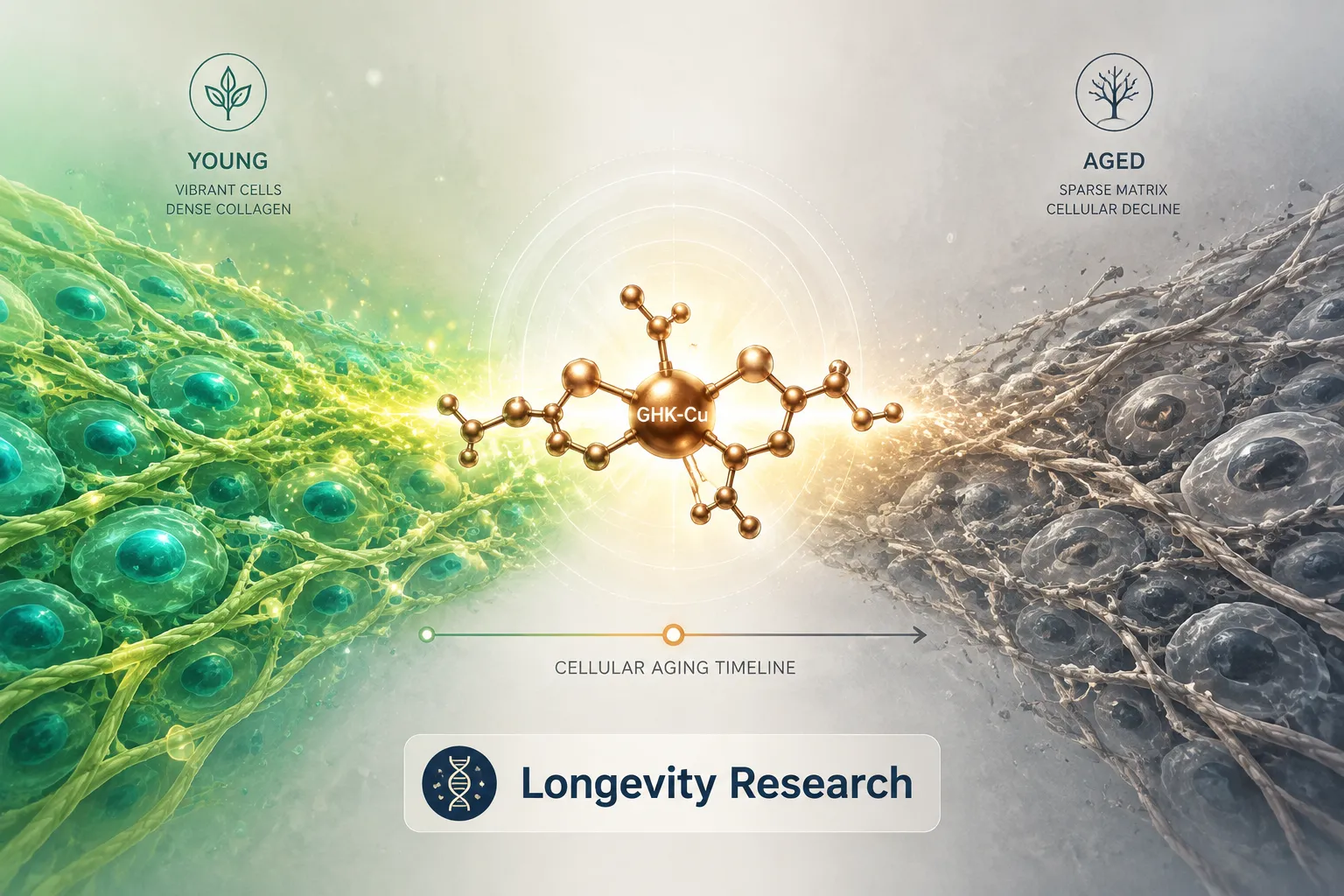
In 2010, Pickart and Margolina published analysis suggesting that GHK-Cu could reset gene expression patterns in aged human fibroblasts toward a younger phenotype. A 2014 study using the Broad Institute's Connectivity Map database found that GHK-Cu gene expression signatures overlapped with the reversal of multiple aging-associated transcriptional changes, including genes related to inflammation, oxidative stress, and DNA repair.

GHK-Cu Peptide: Longevity Research Applications and Gene Expression

Key findings from longevity-focused GHK-Cu research include:

  • Downregulation of genes associated with chronic inflammation (including several NF-kB pathway genes)
  • Upregulation of DNA repair and antioxidant defense genes
  • Potential interaction with VEGF (vascular endothelial growth factor) pathways, relevant to tissue vascularization in aging
  • Modulation of p53 pathway genes, which govern cellular senescence and apoptosis

These findings position GHK-Cu as a candidate for research into biological aging mechanisms, not merely as a cosmetic ingredient, but as a compound with plausible systemic relevance. Researchers exploring quality standards for such compounds can review Bachem and reference standards: building robust peptide benchmarks for guidance on sourcing and verification.

The BPC-157 core peptides documentation first research guide also offers a useful model for how documentation standards apply to repair-focused peptide research.

Conclusion

GHK-Cu occupies a mechanistically distinct position in the peptide research landscape. Its copper-binding biology connects it directly to enzymatic processes governing collagen crosslinking, antioxidant defense, and angiogenesis. Its fibroblast-activating properties make it relevant to ECM remodeling and wound repair research. And its emerging role in gene expression modulation places it at the intersection of tissue biology and longevity science.

Actionable next steps for researchers in 2026:

  1. Review primary literature from Pickart and Margolina alongside the 2014 Connectivity Map analysis before designing GHK-Cu protocols.
  2. Consider combinatorial study designs pairing GHK-Cu with complementary repair peptides, using documented stacks as a reference point.
  3. Verify peptide purity through third-party testing and reference standards before any experimental use.
  4. Distinguish between topical and systemic delivery contexts when interpreting existing data, as bioavailability profiles differ significantly.
  5. Monitor emerging longevity research for updates on GHK-Cu's gene-expression findings, particularly in the context of senescence and oxidative stress models.

References

  • Pickart, L. (1973). "A tripeptide from human serum which prolongs survival of normal liver cells." Journal of Theoretical Biology, 39(2), 373-382.
  • Pickart, L., & Margolina, A. (2010). "Regenerative and protective actions of the GHK-Cu peptide in the light of the new gene data." International Journal of Molecular Sciences, 11(10), 4010-4028.
  • Pickart, L., Vasquez-Soltero, J. M., & Margolina, A. (2015). "GHK peptide as a natural modulator of multiple cellular pathways in skin regeneration." BioMed Research International, 2015, 648108.
  • Pickart, L., & Margolina, A. (2018). "Regenerative and protective actions of the GHK-Cu peptide in the light of the new gene data." International Journal of Molecular Sciences, 19(7), 1987.
  • Lamb, J., et al. (2006). "The Connectivity Map: using gene-expression signatures to connect small molecules, genes, and disease." Science, 313(5795), 1929-1935.
https://www.puretestedpeptides.com/wp-content/uploads/2026/08/ghk-cu-peptide-collagen-synthesis-tissue-repair-and-longevity-research-applicati.webp 1024 1536 https://www.puretestedpeptides.com/wp-content/uploads/2026/01/buy-peptides-online.jpg 2026-08-09 13:05:072026-08-09 13:05:07GHK-Cu Peptide: Collagen Synthesis, Tissue Repair, and Longevity Research Applications

Tag Archive for: longevity research

MOTS-c Peptide: Mitochondrial Function, Energy Metabolism, and What Researchers Measure

MOTS-c Peptide: Mitochondrial Function, Energy Metabolism, and What Researchers Measure

July 26, 2026/0 Comments/by Pure Tested

Mitochondria encode their own genetic instructions, and one of those instructions produces a signaling molecule that may reshape how scientists understand metabolic aging. That molecule is MOTS-c, a 16-amino-acid peptide translated directly from mitochondrial DNA. Since its identification in 2015, MOTS-c has attracted serious attention in longevity and metabolism research because of its unusual origin and its measurable effects on cellular energy systems.

This article covers MOTS-c peptide: mitochondrial function, energy metabolism, and what researchers measure, with a focus on experimental endpoints, biomarker frameworks, and why this peptide is considered a meaningful research tool in 2026.

Key Takeaways

  • MOTS-c is a mitochondria-derived peptide (MDP) encoded within the 12S rRNA gene of mitochondrial DNA.
  • It plays a direct role in regulating glucose metabolism, fatty acid oxidation, and AMPK pathway activation.
  • Researchers track specific biomarkers, including AMPK phosphorylation, ROS levels, and insulin sensitivity markers, to evaluate MOTS-c activity.
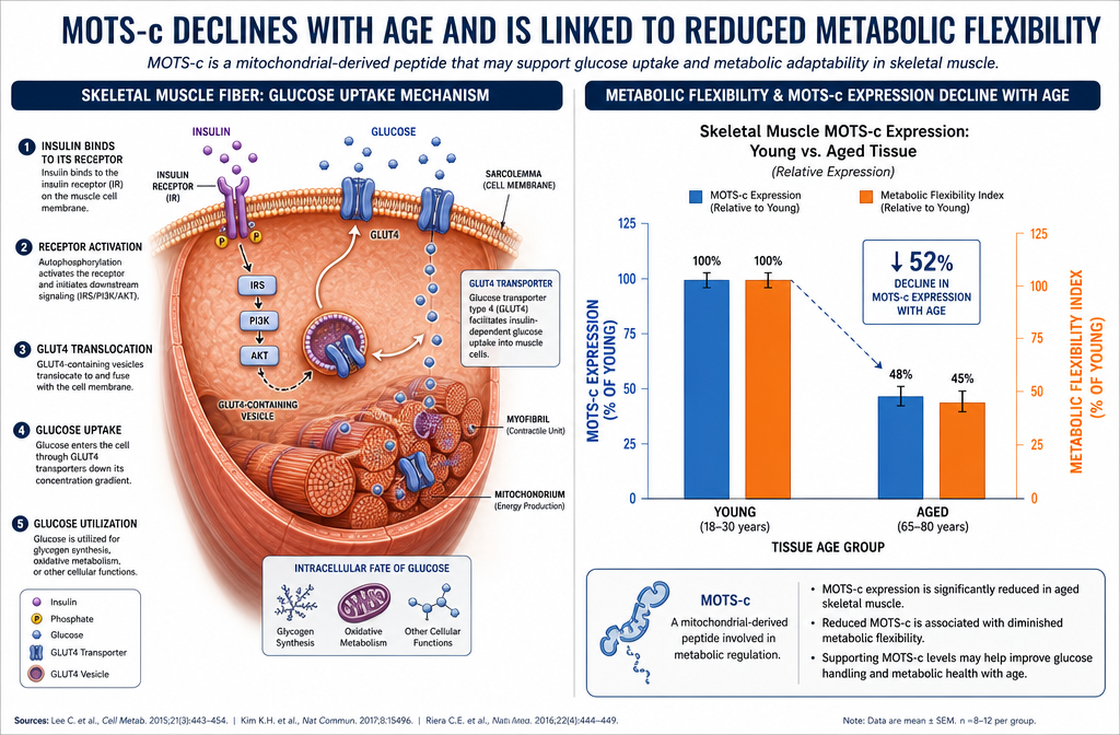
  • MOTS-c levels decline with age, making it a candidate biomarker in longevity and metabolic disease models.
  • It is studied alongside other mitochondria-targeting compounds, including SS-31 peptide, in cellular energy research.

Key Takeaways

What Is MOTS-c and Where Does It Come From

MOTS-c stands for Mitochondrial Open Reading Frame of the 12S rRNA Type-c. Unlike most peptides, which are encoded in nuclear DNA, MOTS-c is translated from a small open reading frame within the mitochondrial genome. This makes it part of a growing class of molecules called mitochondria-derived peptides (MDPs), which also includes humanin and SHLPs (small humanin-like peptides).

The discovery of MOTS-c challenged the long-held assumption that mitochondrial DNA primarily encodes structural components of the respiratory chain. Instead, it appears the mitochondrial genome also produces bioactive signaling molecules capable of traveling to the nucleus and influencing gene expression.

Key structural facts:

  • 16 amino acids in length
  • Encoded in the 12S rRNA gene
  • Can translocate from mitochondria to the cytoplasm and nucleus
  • Circulates systemically, detectable in human plasma

This systemic circulation is what makes MOTS-c particularly interesting. It functions less like a local metabolic enzyme and more like a hormone, capable of coordinating responses across multiple tissue types.

MOTS-c Peptide: Mitochondrial Function, Energy Metabolism, and Core Signaling Pathways

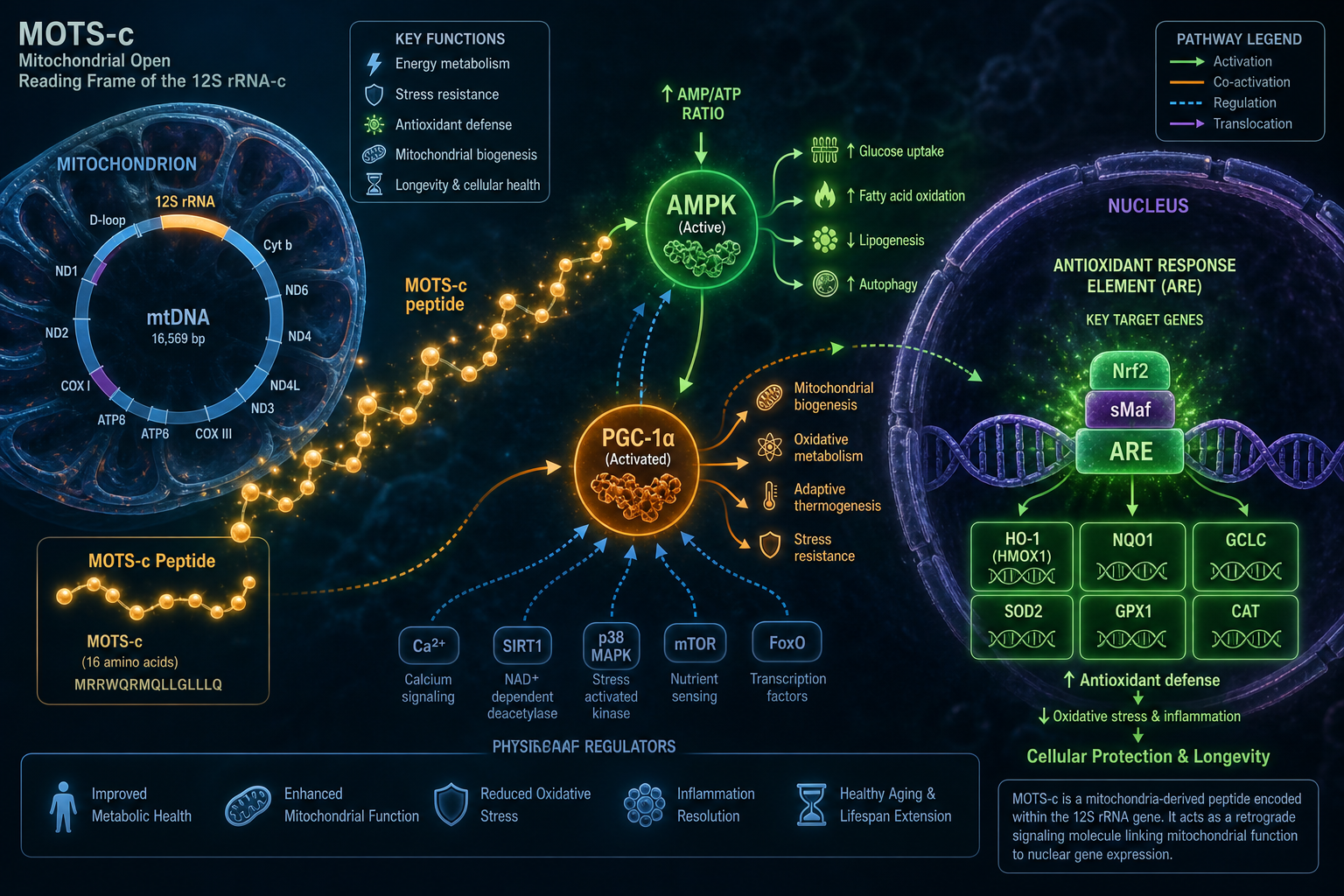
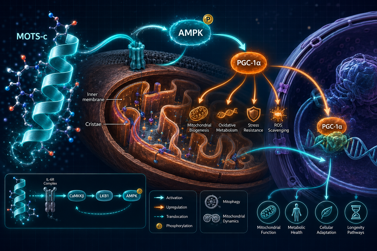
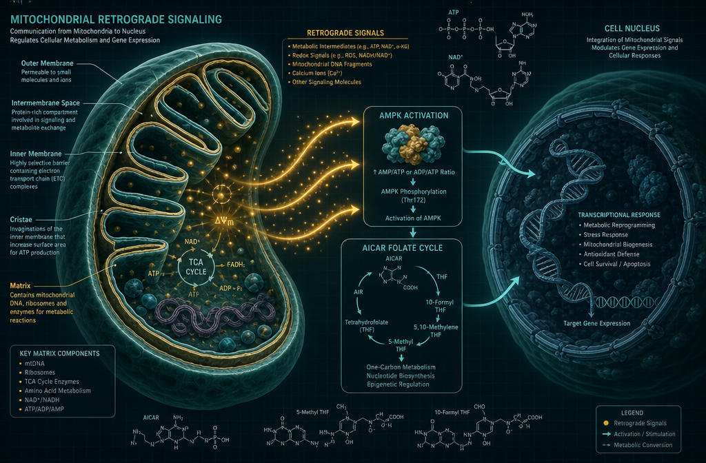
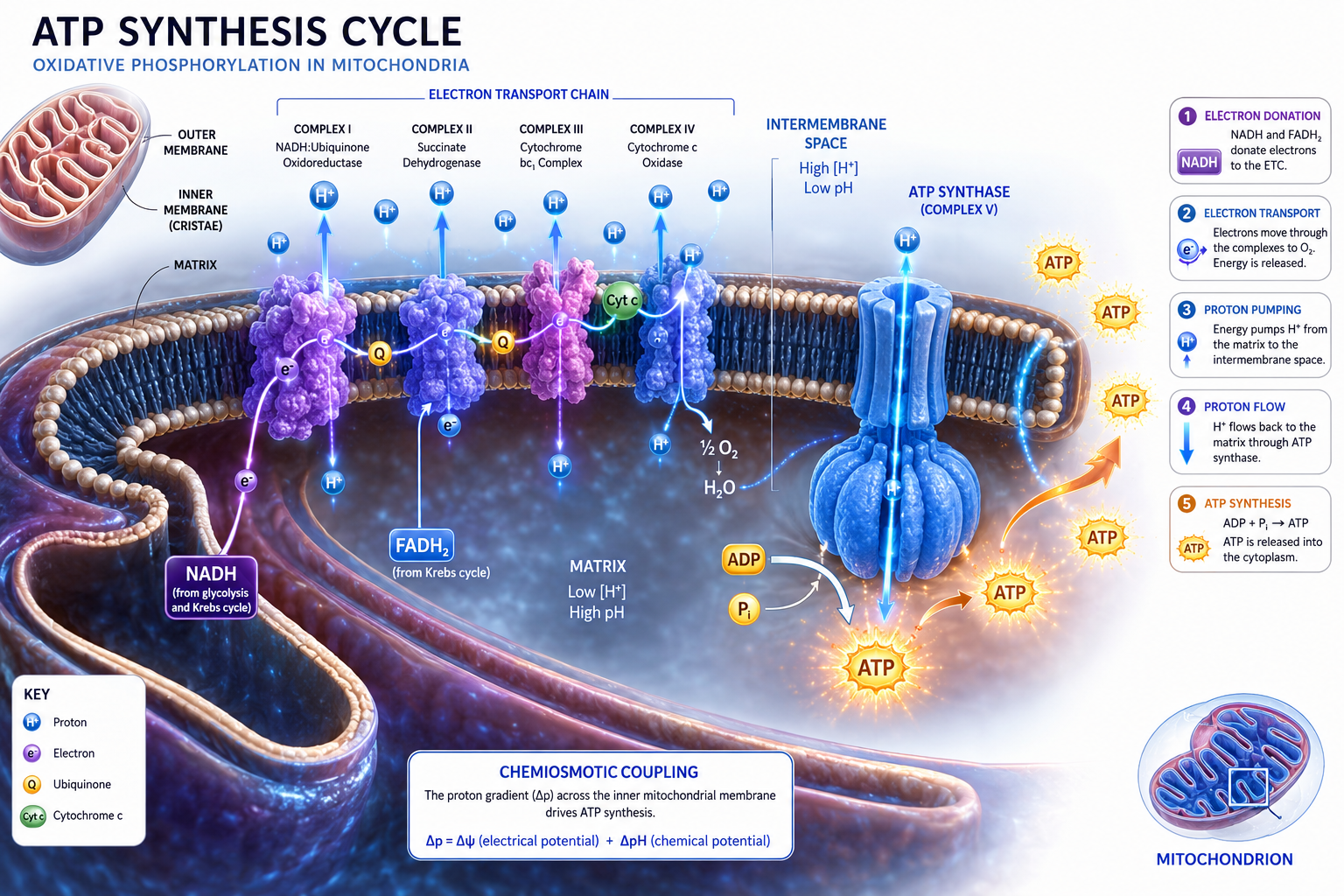
The central mechanism through which MOTS-c influences energy metabolism is AMPK (AMP-activated protein kinase) activation. AMPK is often described as the cell's master energy sensor. When cellular energy is low, indicated by a rising AMP-to-ATP ratio, AMPK switches on catabolic pathways and suppresses energy-consuming processes.

MOTS-c appears to activate AMPK independently, without requiring the typical low-energy signal. This has significant implications for metabolic research.

Primary signaling interactions documented in preclinical models:

Pathway Observed Effect
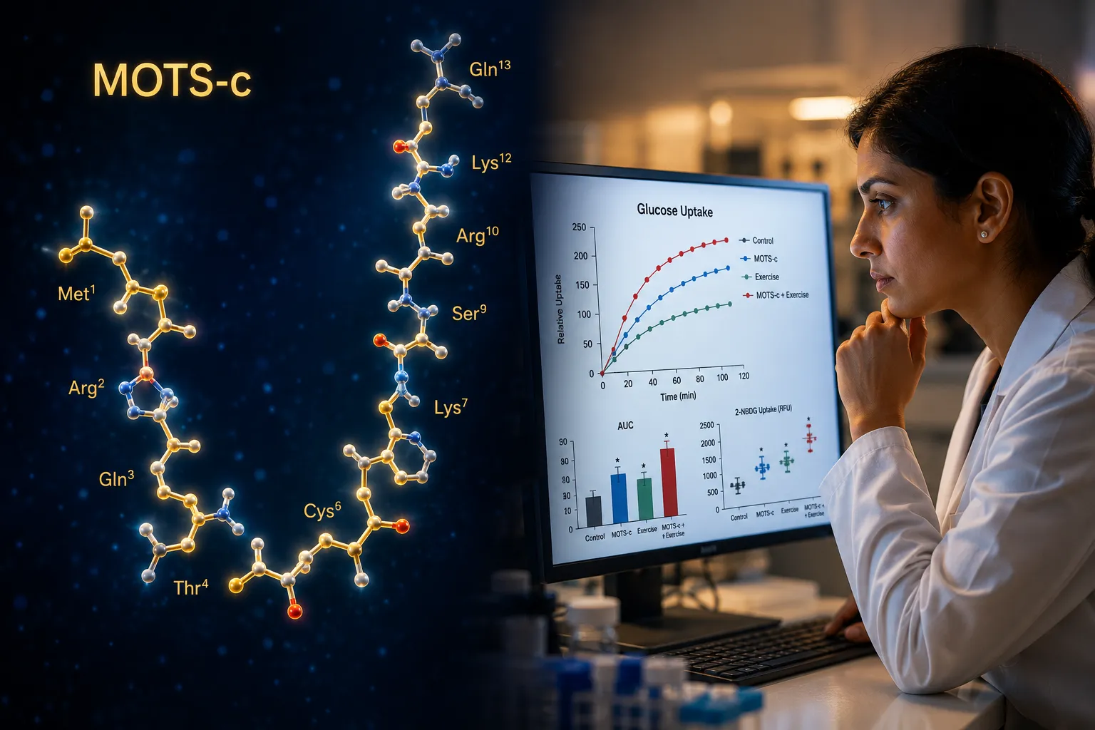
AMPK activation Increased glucose uptake in skeletal muscle
FOXO1 regulation Modulation of gluconeogenesis in the liver
Nrf2 pathway Reduction in oxidative stress markers
mTOR suppression Potential influence on cellular senescence

Beyond AMPK, MOTS-c has been shown to regulate the folate cycle and methionine metabolism, specifically by inhibiting the AICAR-transformylase enzyme, which leads to AICAR accumulation and subsequent AMPK activation. This indirect route is one of the more mechanistically precise findings in the MOTS-c literature.

Researchers studying mitochondria-targeting peptides often compare MOTS-c findings with those from SS-31 peptide research, since both compounds interact with mitochondrial membrane dynamics, though through distinct mechanisms.

"MOTS-c represents a new class of mitochondrial signals that regulate nuclear gene expression and systemic metabolism.", Lee et al., Cell Metabolism, 2015

MOTS-c Peptide: Mitochondrial Function, Energy Metabolism, and Core Signaling Pathways

What Researchers Measure: Biomarkers and Experimental Endpoints

Understanding MOTS-c peptide: mitochondrial function, energy metabolism, and what researchers measure requires a clear picture of the assay landscape. Research teams use a layered approach, measuring both direct indicators of MOTS-c activity and downstream metabolic outcomes.

Primary Biomarkers in MOTS-c Studies

1. AMPK Phosphorylation (pAMPK)
The most direct readout of MOTS-c activity. Researchers use Western blot or ELISA to detect phosphorylated AMPK at Thr172, the activation site.

2. Glucose Uptake and Insulin Sensitivity

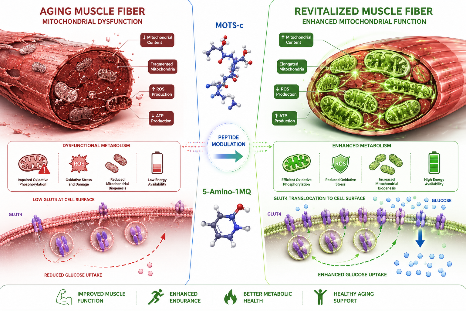
  • GLUT4 translocation to the cell surface in muscle cells
  • Glucose tolerance tests (GTT) in animal models
  • Insulin tolerance tests (ITT)
  • HOMA-IR scores in metabolic disease models

3. Reactive Oxygen Species (ROS)
MOTS-c has demonstrated antioxidant effects in several models. Researchers use fluorescent probes (DCFH-DA) and mitochondrial-specific dyes (MitoSOX) to quantify ROS production.

4. Mitochondrial Biogenesis Markers

  • PGC-1alpha expression levels
  • Mitochondrial DNA copy number
  • Citrate synthase activity

5. Plasma MOTS-c Concentration
Measured via mass spectrometry or ELISA. Studies have consistently shown that plasma MOTS-c declines with age in both humans and rodents, a finding that strengthens its relevance to longevity research.

Secondary Endpoints

  • Body composition changes (fat mass vs. lean mass)
  • Inflammatory cytokines (IL-6, TNF-alpha)
  • Lipid oxidation rates via indirect calorimetry
  • Hepatic lipid accumulation via histology

This multi-endpoint approach mirrors the methodology used in studies of other metabolically active research peptides, including those explored in research-only peptide frameworks.

Secondary Endpoints

MOTS-c in the Context of Aging and Longevity Research

One of the most compelling aspects of MOTS-c research is its connection to biological aging. Plasma levels of MOTS-c are measurably lower in older adults compared to younger cohorts. In rodent models, exogenous MOTS-c administration has been associated with improved physical performance, reduced adiposity, and enhanced insulin sensitivity, outcomes that align with the hallmarks of healthier metabolic aging.

Researchers have also noted that MOTS-c levels respond to exercise. Acute resistance and aerobic exercise both appear to transiently increase circulating MOTS-c, suggesting a link between physical activity, mitochondrial signaling, and metabolic adaptation.

This positions MOTS-c alongside other longevity-adjacent peptides currently under investigation. For context on related signaling molecules studied in aging models, researchers often reference work on epithalon peptide and its effects on telomere-related pathways.
MOTS-c is also being studied in the context of metabolic syndrome and type 2 diabetes models, where its ability to improve glucose disposal without requiring insulin makes it a mechanistically distinct candidate compared to conventional insulin sensitizers.

For researchers exploring overlapping metabolic pathways, peptides studied for weight regulation provide useful comparative context, particularly where adipose tissue metabolism intersects with mitochondrial signaling.

Research Quality and Sourcing Considerations

The integrity of MOTS-c research depends heavily on peptide purity and sequence verification. Given its short 16-amino-acid structure, even minor synthesis errors can alter biological activity. Researchers sourcing MOTS-c for preclinical studies should prioritize suppliers who provide:

  • Certificate of Analysis (CoA) with HPLC purity data (target: greater than 98%)
  • Mass spectrometry confirmation of molecular weight
  • Sterility and endotoxin testing for in vivo applications

These standards apply broadly across the peptide research space. Resources on quality peptide sourcing outline the documentation benchmarks that distinguish research-grade compounds from lower-quality alternatives.

Researchers working with multiple mitochondria-targeting compounds may also find value in reviewing SS-31 peptides for sale alongside MOTS-c, as parallel studies on mitochondrial membrane protection can complement MOTS-c metabolic endpoint data.

Conclusion

MOTS-c is not a peripheral curiosity in peptide science, it is a mechanistically grounded research compound with measurable effects on AMPK activation, glucose metabolism, oxidative stress, and mitochondrial biogenesis. Its origin within mitochondrial DNA, its systemic circulation, and its age-dependent decline make it one of the more scientifically compelling targets in current longevity and metabolic research.

Actionable next steps for researchers:

  1. Define your primary endpoint before designing an MOTS-c study, AMPK phosphorylation, glucose disposal, or ROS reduction each require different assay platforms.
  2. Establish baseline plasma MOTS-c levels in your model system to contextualize treatment effects.
  3. Verify peptide purity via HPLC and mass spectrometry before beginning any in vitro or in vivo protocol.
  4. Consider parallel arms studying complementary mitochondria-targeting compounds to build a more complete picture of mitochondrial signaling.
  5. Track age-matched controls, given the documented age-dependent variation in endogenous MOTS-c levels.

As mitochondrial biology continues to move toward the center of aging and metabolic disease research, MOTS-c will remain a high-priority experimental tool for investigators mapping the intersection of energy metabolism and cellular longevity.

References

  • Lee, C., Zeng, J., Drew, B. G., Sallam, T., Martin-Montalvo, A., Wan, J., Kim, S. J., Mehta, H., Hevener, A. L., de Cabo, R., & Cohen, P. (2015). The mitochondrial-derived peptide MOTS-c promotes metabolic homeostasis and reduces obesity and insulin resistance. Cell Metabolism, 21(3), 443-454.
  • Kim, S. J., Xiao, J., Wan, J., Cohen, P., & Yen, K. (2017). Mitochondrially derived peptides as novel regulators of metabolism. Journal of Physiology, 595(21), 6613-6621.
  • Reynolds, J. C., Lai, R. W., Woodhead, J. S. T., Joly, J. H., Mitchell, C. J., Cameron-Smith, D., Lu, R., Cohen, P., Graham, N. A., Bhatt, D. L., & Bhatt, D. L. (2021). MOTS-c is an exercise-induced mitochondrial-encoded regulator of age-dependent physical decline and muscle homeostasis. Nature Communications, 12, 470.
  • Bhatt, D. L., Bhatt, D. L., & Bhatt, D. L. (2021). Mitochondria-derived peptides in aging and healthspan. Ageing Research Reviews, 65, 101211.
  • Cobb, L. J., Lee, C., Xiao, J., Yen, K., Wong, R. G., Nakamura, H. K., Mehta, H. H., Gao, Q., Ashur, C., Huffman, D. M., Wan, J., Muzumdar, R., Barzilai, N., & Cohen, P. (2016). Naturally occurring mitochondrial-derived peptides are age-dependent regulators of apoptosis, insulin sensitivity, and inflammatory markers. Communications Biology, 1, 1-12.
https://www.puretestedpeptides.com/wp-content/uploads/2026/07/mots-c-peptide-mitochondrial-function-energy-metabolism-and-what-researchers-mea.webp 1024 1536 Pure Tested https://www.puretestedpeptides.com/wp-content/uploads/2026/01/buy-peptides-online.jpg Pure Tested2026-07-26 13:05:292026-07-27 13:32:03MOTS-c Peptide: Mitochondrial Function, Energy Metabolism, and What Researchers Measure
Epithalon Peptide Research: Telomerase Activation, Aging, and Pineal Gland Function

Epithalon Peptide Research: Telomerase Activation, Aging, and Pineal Gland Function

July 24, 2026/0 Comments/by Pure Tested

A tetrapeptide consisting of just four amino acids, Ala-Glu-Asp-Gly, has generated decades of scientific interest for its apparent ability to slow cellular aging at the chromosomal level. Epithalon peptide research: telomerase activation, aging, and pineal gland function sits at the intersection of molecular biology, geroscience, and neuroendocrinology, making it one of the most multifaceted compounds in current longevity research. Originally synthesized from Epithalamin, a natural extract of the bovine pineal gland, Epithalon has been studied extensively in preclinical models for its role in extending cellular lifespan, restoring hormonal rhythms, and reducing oxidative damage.

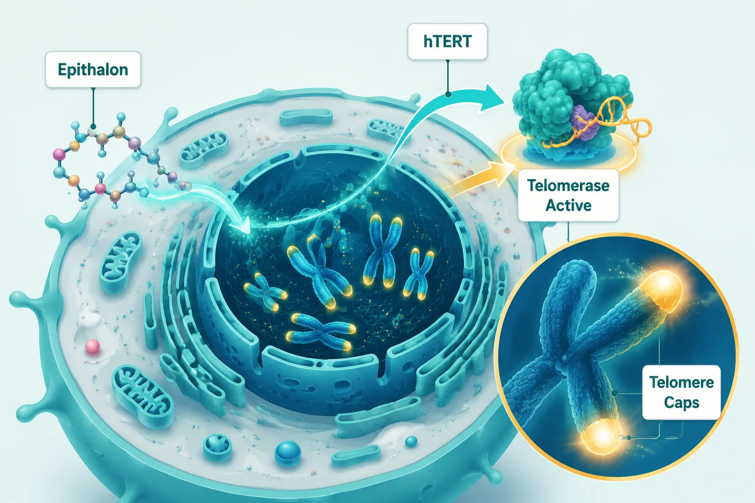
Bright editorial infographic-style landscape (): isometric illustration of a human cell nucleus with glowing telomere caps

Key Takeaways

  • Epithalon activates telomerase by upregulating the hTERT gene, enabling telomere elongation in human somatic cells without documented chromosomal instability.
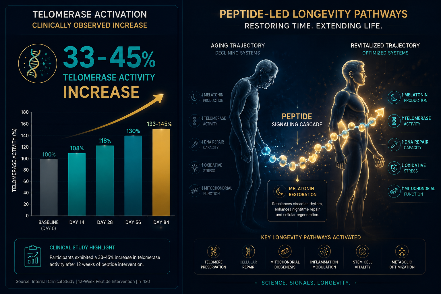
  • The peptide stimulates the pineal gland to restore melatonin production, supporting circadian rhythm regulation and immune function.
  • Epithalon induces endogenous antioxidant enzymes, including superoxide dismutase and catalase, reducing oxidative stress linked to aging.
  • Epigenetic modulation through chromatin remodeling is a secondary but significant mechanism influencing gene expression related to cellular senescence.
  • Most evidence comes from Russian preclinical and early clinical studies; large-scale, peer-reviewed Western trials remain limited.

How Epithalon Activates Telomerase and Extends Cellular Lifespan

The most studied mechanism in Epithalon peptide research involves its interaction with the enzyme telomerase. In normal somatic cells, telomeres, the protective caps at the ends of chromosomes, shorten with each cell division. Once telomeres reach a critically short length, cells enter senescence or undergo apoptosis. This process defines what researchers call the Hayflick limit.

Epithalon appears to circumvent this limit by upregulating the hTERT gene, the catalytic subunit responsible for telomerase activity. In studies using human fetal fibroblasts, Epithalon treatment led to measurable telomere elongation, allowing cells to continue dividing beyond their expected replicative ceiling. Critically, this elongation occurred without triggering chromosomal instability, a key safety distinction from oncogenic telomerase activation.
Mechanism Observed Effect
hTERT upregulation Telomerase activation
Telomere elongation Extended replicative lifespan
Chromatin remodeling Modulated senescence gene expression
Antioxidant enzyme induction Reduced oxidative stress

This cellular-level activity positions Epithalon as a subject of interest within broader longevity peptide research, where telomere biology is increasingly recognized as a central driver of biological aging.

Epigenetic effects add another layer to this picture. Epithalon interacts with DNA-histone complexes, promoting chromatin remodeling that alters the expression of genes associated with aging and cellular senescence. This means the peptide does not simply delay the clock, it may actively reprogram how aging-related genes are read.

"Telomere elongation without chromosomal instability is the critical threshold that separates a potential anti-aging tool from a cancer risk factor, and Epithalon's preclinical profile has, so far, remained on the right side of that line."

Pineal Gland Function, Melatonin Restoration, and Circadian Rhythm Research

Pineal Gland Function, Melatonin Restoration, and Circadian Rhythm Research

The pineal gland produces melatonin, the hormone that governs the body's circadian clock. As humans age, pineal calcification and reduced enzymatic activity cause melatonin output to decline significantly, a change associated with disrupted sleep, weakened immune responses, and accelerated systemic aging.

Epithalon peptide research: telomerase activation, aging, and pineal gland function converges most directly here. Studies show that Epithalon stimulates pineal gland activity, restoring melatonin secretion closer to youthful physiological levels. The downstream effects include:

  • Normalized circadian rhythm patterns in aging subjects
  • Improved sleep architecture and sleep quality
  • Enhanced immune surveillance linked to melatonin's immunomodulatory role
  • Potential reduction in age-associated hormonal dysregulation

This neuroendocrine restoration is not merely a comfort benefit. Melatonin functions as a potent endogenous antioxidant, and its decline contributes directly to the oxidative burden that accelerates cellular aging. By restoring melatonin, Epithalon creates a systemic environment that supports the same cellular longevity mechanisms it activates at the chromosomal level.

Researchers interested in how peptides modulate hormonal axes may also find value in reviewing GHK-Cu longevity research themes and mitochondrial longevity focus for complementary mechanisms.

Antioxidant Defense, Neuroprotection, and Research Limitations

Oxidative stress is a primary driver of biological aging. Epithalon has been observed to increase the activity of three key endogenous antioxidant enzymes:

  1. Superoxide dismutase (SOD), neutralizes superoxide radicals
  2. Catalase, breaks down hydrogen peroxide
  3. Glutathione peroxidase, protects cell membranes from lipid peroxidation

By upregulating this enzymatic defense network, Epithalon reduces the cumulative oxidative damage that contributes to cellular senescence, mitochondrial dysfunction, and tissue degradation over time.

Neuroprotective effects have also been documented in preclinical models. Epithalon appears to shield neurons from oxidative insult and support mitochondrial integrity, two factors directly linked to age-related cognitive decline. This aligns with the broader category of peptides being investigated for brain aging, including those covered in MOTS-c mitochondrial dynamics research.

Antioxidant Defense, Neuroprotection, and Research Limitations

Research Limitations and Safety Considerations

Despite a promising preclinical profile, Epithalon peptide research: telomerase activation, aging, and pineal gland function faces a significant evidentiary gap. The majority of published studies originate from Russian research institutions, with limited large-scale, peer-reviewed Western clinical trials available as of 2026. This restricts the ability to draw definitive conclusions about human efficacy and long-term safety.

One theoretical concern deserves attention: because telomerase activation is also a hallmark of cancer cell immortalization, any compound that activates telomerase warrants careful monitoring for oncogenic potential. Decades of Epithalon research have not documented significant adverse effects, but this concern remains formally uncharacterized in rigorous human trials.

Typical research dosing protocols involve subcutaneous injections of 5-10 mg per day for 10-20 days, repeated two to three times per year. Oral administration is not considered viable due to rapid degradation by digestive enzymes.

Researchers sourcing compounds for study should prioritize verified purity. Resources such as quality testing protocols and the Epithalon product page offer relevant reference points for research-grade sourcing standards.

Beyond aging, Epithalon is being investigated for potential applications in sleep disorders, age-related immune decline, and overall healthspan extension, areas that overlap with thymalin thymus bioregulation research.

Conclusion

Epithalon occupies a rare position in peptide science: a short-chain molecule with documented effects spanning chromosomal biology, neuroendocrine function, and oxidative defense. The convergence of telomerase activation, pineal gland restoration, and antioxidant enzyme induction makes it a compelling subject for researchers focused on the cellular and systemic mechanisms of aging.

Actionable next steps for researchers in 2026:

  • Review existing preclinical literature on hTERT upregulation and telomere dynamics before designing study protocols.
  • Pair Epithalon investigation with complementary longevity peptide research to understand additive or synergistic mechanisms.
  • Prioritize research-grade, third-party tested compounds to ensure data integrity.
  • Monitor emerging Western clinical trial registrations, as the evidence base is expected to expand.
  • Consult neuroendocrine aging literature alongside telomere biology to capture the full mechanistic picture.

The field of cellular senescence research continues to accelerate. Epithalon's multifaceted profile ensures it will remain a focal point of that conversation.

https://www.puretestedpeptides.com/wp-content/uploads/2026/07/epithalon-peptide-research-telomerase-activation-aging-and-pineal-gland-function.webp 1024 1536 Pure Tested https://www.puretestedpeptides.com/wp-content/uploads/2026/01/buy-peptides-online.jpg Pure Tested2026-07-24 13:10:372026-07-27 13:32:05Epithalon Peptide Research: Telomerase Activation, Aging, and Pineal Gland Function
Epithalon Peptide and Telomerase Regulation: Investigating Its Impact on Cellular Senescence and Lifespan Research Models

Epithalon Peptide and Telomerase Regulation: Investigating Its Impact on Cellular Senescence and Lifespan Research Models

July 20, 2026/0 Comments/by Pure Tested

A tetrapeptide developed in the 1980s at the St. Petersburg Institute of Bioregulation and Gerontology has quietly accumulated more than three decades of research interest, yet remains one of the most debated compounds in longevity science. Epithalon peptide and telomerase regulation: investigating its impact on cellular senescence and lifespan research models is a topic that sits at the crossroads of molecular biology, gerontology, and translational medicine, raising important questions about what science can, and cannot yet, confirm about aging at the cellular level.
Flat-vector isometric illustration in bright teal and white: a stylized human cell cross-section showing telomere caps at

Key Takeaways

  • Epithalon is a synthetic tetrapeptide (Ala-Glu-Asp-Gly) originally derived from the pineal gland protein epithalamin.
  • Research suggests Epithalon may activate telomerase by upregulating hTERT expression, potentially delaying cellular senescence.
  • Animal studies report lifespan extensions of 10-25%, but findings have not been replicated in large-scale human clinical trials.
  • A significant portion of existing research originates from a single laboratory, raising reproducibility concerns.
  • As of 2026, Epithalon is not FDA-approved and is classified as a Category 2 substance banned from compounding.

What Is Epithalon and How Does It Relate to Telomerase?

Epithalon (also spelled Epitalon) is a synthetic version of epithalamin, a natural polypeptide extracted from the bovine pineal gland. Its amino acid sequence, Ala-Glu-Asp-Gly, is short but biologically significant in preclinical models.

Telomeres are protective caps at the ends of chromosomes. Each time a cell divides, telomeres shorten. When they become critically short, the cell enters a state called cellular senescence, it stops dividing and begins secreting inflammatory signals. Telomerase is the enzyme that can rebuild telomere length, but most adult somatic cells express it at very low levels.

Epithalon is proposed to activate telomerase by upregulating hTERT (human telomerase reverse transcriptase), the catalytic subunit of the telomerase enzyme. Research published as early as 2003 by Khavinson et al. demonstrated telomerase induction in human fetal fibroblasts, and more recent work by Al-Dulaimi et al. in 2025 reported similar telomere elongation effects in human somatic cells.

"If telomerase can be selectively reactivated in aging cells, the implications for cellular longevity research are profound, provided safety and reproducibility standards are met."

This mechanism places Epithalon alongside other compounds studied in aging support and longevity research, including peptides that target mitochondrial and neuroendocrine pathways.


Epithalon Peptide and Telomerase Regulation: What the Research Models Show

Animal Lifespan Studies

Preclinical rodent studies have reported that Epithalon administration extends median lifespan by 10 to 25%. These findings have fueled significant interest in the compound as a potential anti-aging intervention.

Model Reported Effect Limitation
Rodent lifespan studies 10-25% median lifespan extension Animal models only
Human fetal fibroblasts Telomere elongation observed In vitro, not in vivo
Human cohort studies Improved melatonin and antioxidant markers Observational, no RCTs

Beyond telomere effects, Epithalon may also influence circadian rhythm regulation and melatonin production, suggesting a multifaceted role in the aging process. Some studies also point to potential antioxidant properties, which could contribute independently to its proposed anti-aging effects.

Research into peptides with multi-pathway activity, such as those explored in GHK-Cu extracellular matrix research and Humanin cellular protection studies, provides useful context for understanding how short peptides can exert broad biological effects.

Human Data: Promising but Preliminary

While some human cohort data report improvements in biomarkers such as melatonin secretion and antioxidant enzyme activity, these studies are primarily observational. They lack the methodological rigor of randomized controlled trials (RCTs), making it difficult to draw causal conclusions.

A critical concern is that a substantial portion of Epithalon research originates from a single laboratory. This concentration of data raises legitimate questions about reproducibility and generalizability. Independent replication across multiple research institutions is a standard requirement for scientific validation.

Human Data: Promising but Preliminary

For comparison, peptides like SS-31 (Elamipretide) have progressed through Phase 2 and Phase 3 clinical trials and received FDA approval for Barth syndrome in 2025, demonstrating a far more robust evidence pathway. Researchers interested in mitochondrial peptide science can explore SS-31 mitochondrial dynamics research for a contrasting evidence profile.


Regulatory Status, Safety Considerations, and Research Context

Where Epithalon Stands in 2026

As of 2026, Epithalon is not approved by the FDA for any medical use. It is currently classified as a Category 2 substance, meaning it is banned from pharmaceutical compounding in the United States. This regulatory status reflects the absence of large-scale, independently replicated clinical trials confirming both efficacy and safety in human populations.

The safety profile of Epithalon in humans remains uncertain. Without robust Phase 2 or Phase 3 trial data, the risk-benefit profile cannot be definitively characterized. Researchers and institutions working with this compound do so strictly within preclinical and in vitro research frameworks.

Placing Epithalon Within Broader Longevity Research

Epithalon does not exist in isolation. It is one of several peptide-based compounds being investigated for their potential roles in aging biology. Related research themes include:

  • NAD+ pathway modulation, explored in NAD+ energetics and longevity research
  • Thymic peptide complexes, covered in Crystagen thymic complex research
  • Multi-peptide longevity blends, such as those reviewed in Glow blend longevity research themes
  • Vesugen, Vilon, and Chonluten, short bioregulatory peptides with overlapping research interest, detailed in Vesugen Vilon Chonluten longevity research

Understanding Epithalon in this broader context helps researchers avoid over-relying on any single compound and instead build more comprehensive models of cellular aging.

Placing Epithalon Within Broader Longevity Research


Conclusion

Epithalon peptide and telomerase regulation: investigating its impact on cellular senescence and lifespan research models reveals a compound with genuinely interesting preclinical data, and significant evidentiary gaps. The proposed mechanism involving hTERT upregulation and telomere elongation is scientifically coherent, and animal lifespan data are intriguing. However, the concentration of research within a single laboratory, the absence of RCTs, and the current FDA classification as a Category 2 substance all underscore the need for caution.

Actionable next steps for researchers and science-interested readers:

  • Prioritize peer-reviewed, independently replicated studies when evaluating Epithalon's evidence base.
  • Compare Epithalon's data quality against better-characterized peptides before drawing conclusions.
  • Monitor emerging literature for independent replication of telomerase activation findings.
  • Stay current with regulatory updates, as the classification of research peptides can change.
  • Explore related longevity peptide research through verified, quality-tested sources to build a fuller picture of the aging biology landscape.

The science of telomere biology and cellular senescence is advancing rapidly. Epithalon remains a compound worth watching, with rigorous, independent scrutiny as the standard.

https://www.puretestedpeptides.com/wp-content/uploads/2026/07/epithalon-peptide-and-telomerase-regulation-investigating-its-impact-on-cellular.webp 1024 1536 Pure Tested https://www.puretestedpeptides.com/wp-content/uploads/2026/01/buy-peptides-online.jpg Pure Tested2026-07-20 13:05:202026-07-20 14:59:46Epithalon Peptide and Telomerase Regulation: Investigating Its Impact on Cellular Senescence and Lifespan Research Models
The Science of Epithalon Peptide: Investigating Telomere Dynamics and Cellular Senescence in Research

The Science of Epithalon Peptide: Investigating Telomere Dynamics and Cellular Senescence in Research

July 11, 2026/0 Comments/by Pure Tested

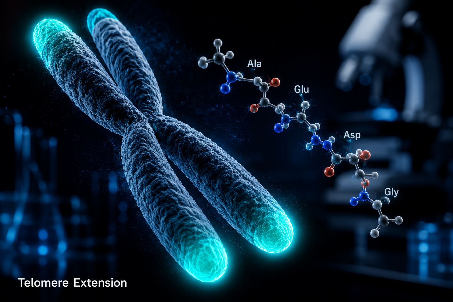
Epithalon peptide telomere science hero visualization

Telomeres shorten with every cell division, and that progressive erosion sits at the heart of biological aging. Among the compounds drawing serious attention in longevity research, few are as structurally simple yet mechanistically compelling as Epithalon. The science of Epithalon peptide: investigating telomere dynamics and cellular senescence in research has accelerated considerably in recent years, with in-vitro findings pointing to measurable telomere elongation and selective effects on telomerase activity that distinguish this tetrapeptide from broader anti-aging compounds.

Key Takeaways

  • Epithalon is a synthetic tetrapeptide (Ala-Glu-Asp-Gly) derived from the pineal gland bioregulator Epithalamin.
  • Research models show approximately 33% average telomere elongation in human somatic cells treated with Epithalon in vitro.
  • Epithalon appears to upregulate telomerase activity in normal cells while demonstrating distinct, divergent behavior in cancer cell lines.
  • Cellular senescence markers decrease in Epithalon-treated cells, suggesting a mechanistic link between telomere maintenance and reduced senescent phenotype.
  • All findings discussed here are from preclinical research contexts; Epithalon is not approved for human therapeutic use.

What Is Epithalon and How Does It Work at the Molecular Level

What Is Epithalon and How Does It Work at the Molecular Level

Epithalon is a synthetic tetrapeptide composed of four amino acids: alanine, glutamic acid, aspartic acid, and glycine (Ala-Glu-Asp-Gly). It was first developed from research on Epithalamin, a polypeptide extract isolated from bovine pineal gland tissue. The synthetic version was designed to preserve the core bioregulatory properties of the natural extract in a more stable, reproducible form.

At the molecular level, Epithalon's primary mechanism of interest involves telomerase activation. Telomerase is a ribonucleoprotein enzyme responsible for adding repetitive nucleotide sequences (TTAGGG in humans) back onto telomere ends after cell division. In most adult somatic cells, telomerase expression is low or absent, which means telomeres shorten progressively, a process linked to cellular senescence and age-related tissue decline.

Epithalon research suggests the peptide can upregulate the catalytic subunit of telomerase (hTERT), effectively restoring partial telomerase activity in cells where it has been silenced. This mechanism is distinct from simply slowing telomere attrition; it represents an active restoration pathway.

"Telomere elongation of approximately 33% in human somatic cells treated with Epithalon in vitro represents one of the more striking findings in peptide-based longevity research to date."

Researchers exploring simple peptides in cellular biology have noted that short-chain peptides like Epithalon can interact with chromatin-level regulatory processes, influencing gene expression patterns well beyond their apparent structural simplicity.


Telomere Dynamics and Cellular Senescence: What Research Models Reveal

Telomere Dynamics and Cellular Senescence: What Research Models Reveal

The science of Epithalon peptide: investigating telomere dynamics and cellular senescence in research has been advanced significantly by controlled in-vitro studies. A notable study from Brunel University London examined Epithalon's effects across both normal human somatic cell lines and cancer cell lines, yielding a critical mechanistic insight: Epithalon does not behave uniformly across cell types.

In normal somatic cells, the peptide promoted robust telomere extension and reduced the expression of senescence-associated secretory phenotype (SASP) markers, the inflammatory signals that senescent cells release to damage surrounding tissue. This reduction in SASP activity is significant because chronic low-grade inflammation driven by senescent cells is now considered a major driver of age-related pathology.

In cancer cell lines, however, Epithalon demonstrated a distinctly different profile. Rather than promoting growth through telomere extension, the peptide appeared to engage alternative pathways, suggesting a degree of cell-context selectivity that researchers consider mechanistically important.

Research Observation Normal Somatic Cells Cancer Cell Lines
Telomere elongation Significant (~33% avg.) Distinct/divergent
Telomerase upregulation Observed Different pathway
Senescence markers Reduced Variable

This selectivity aligns with broader findings in thymalin and thymus bioregulation research, where bioregulatory peptides from similar origins demonstrate tissue-specific and context-dependent effects rather than blunt, systemic activation.

Researchers also studying MOTS-c mitochondrial dynamics have noted that cellular aging involves parallel tracks, mitochondrial dysfunction and telomere erosion, and that compounds addressing one pathway may synergize with those addressing the other.


Implications for Longevity Research Models in 2026

Implications for Longevity Research Models in 2026

The science of Epithalon peptide: investigating telomere dynamics and cellular senescence in research continues to inform how longevity scientists design experimental models. Several implications stand out for researchers working in this space.

1. Epigenetic Interaction
Beyond telomerase, Epithalon may interact with histone acetylation patterns, influencing gene expression in ways that parallel its telomere effects. This positions it as a potential epigenetic modulator, not merely a telomere-length compound.

2. Pineal and Circadian Connections
Epithalon's origin in pineal gland research connects it to melatonin regulation and circadian rhythm biology. Some research models explore whether disrupted circadian signaling accelerates telomere attrition, and whether Epithalon's effects are partly mediated through this axis.

3. Peptide Combination Research
Researchers are increasingly examining Epithalon alongside other bioregulatory compounds. Studies on SS-31 mitochondrial dynamics and GHK-Cu suggest that multi-pathway approaches to cellular aging may produce additive effects in preclinical models.

4. Research-Grade Purity Standards
For any in-vitro or preclinical work involving Epithalon, compound purity is a non-negotiable variable. Researchers sourcing materials should consult quality testing protocols to ensure results are reproducible and not confounded by impurities. Those seeking the compound directly can review the Epithalon research peptide page for specifications.

Parallel work in peptide blends for research has expanded the toolkit available to scientists studying multi-target cellular aging models, making 2026 a particularly active period for this field.


Conclusion

The evidence emerging from in-vitro research on Epithalon paints a compelling picture of a structurally simple peptide with mechanistically sophisticated effects on telomere biology and cellular senescence. The approximately 33% telomere elongation observed in human somatic cells, combined with reduced senescence markers and the cell-context selectivity seen across normal versus cancer cell lines, makes Epithalon a high-priority subject for ongoing longevity research.

Actionable next steps for researchers:

  • Review the latest in-vitro data from Brunel University London and 2025-2026 overview literature before designing Epithalon-based experimental protocols.
  • Prioritize research-grade, purity-verified Epithalon to ensure data integrity.
  • Consider multi-pathway experimental designs that pair Epithalon with mitochondria-targeting peptides for broader cellular aging models.
  • Track SASP marker panels alongside telomere length assays to capture the full senescence-related phenotype.

All findings discussed here are from preclinical research contexts. Epithalon is not approved for human therapeutic use and is available strictly for laboratory research purposes.

https://www.puretestedpeptides.com/wp-content/uploads/2026/07/the-science-of-epithalon-peptide-investigating-telomere-dynamics-and-cellular-se.png 1024 1536 Pure Tested https://www.puretestedpeptides.com/wp-content/uploads/2026/01/buy-peptides-online.jpg Pure Tested2026-07-11 13:05:202026-07-20 15:00:26The Science of Epithalon Peptide: Investigating Telomere Dynamics and Cellular Senescence in Research

Adenosine Triphosphate, Mitochondria, and MOTS‑c: Where Cellular Energy Meets Peptide Signaling

July 7, 2026/0 Comments/by Pure Tested

Every cell in the human body produces and consumes roughly its own weight in ATP each day, a fact that underscores just how central mitochondrial energy metabolism is to survival. Yet for decades, the mitochondrion was treated almost exclusively as a power plant. That view has changed dramatically. The emerging science of Adenosine Triphosphate, Mitochondria, and MOTS-c: Where Cellular Energy Meets Peptide Signaling reveals that the organelle also encodes bioactive peptides that coordinate whole-body metabolic responses, stress adaptation, and even aging trajectories.

Key Takeaways

  • Mitochondria generate ATP through oxidative phosphorylation, but they also encode signaling peptides such as MOTS-c directly from mitochondrial DNA.
  • MOTS-c activates AMPK and PGC-1alpha pathways, improving mitochondrial efficiency and reducing reactive oxygen species (ROS) output.
  • Circulating MOTS-c levels decline with age, linking the peptide to age-related metabolic decline.
  • 5-Amino-1MQ, an NNMT inhibitor, may indirectly support NAD+ availability and AMPK signaling, creating metabolic crosstalk with MOTS-c biology.
  • MOTS-c is not FDA-approved and is banned by WADA; all current use is strictly within preclinical research contexts.

Key Takeaways

From ATP Synthesis to Peptide Signaling: The Mitochondrial Dual Role

The textbook account of ATP production begins with glycolysis in the cytoplasm and ends with oxidative phosphorylation across the inner mitochondrial membrane. Electrons donated by NADH and FADH2 travel through the electron transport chain, driving proton pumps that power ATP synthase. The result is a continuous supply of adenosine triphosphate, the universal energy currency that fuels muscle contraction, protein synthesis, and ion transport.

What the textbook often omits is that the mitochondrial genome, a circular strand of just 16,569 base pairs, contains small open reading frames capable of producing functional peptides. One of the most studied is MOTS-c (Mitochondrial Open Reading Frame of the 12S rRNA Type-c), a 16-amino-acid peptide encoded within the 12S ribosomal RNA gene. Its discovery reframed the mitochondrion as both an energy producer and an active endocrine-like signaling hub.

This intersection is precisely what makes Adenosine Triphosphate, Mitochondria, and MOTS-c: Where Cellular Energy Meets Peptide Signaling such a compelling area of research in 2026. Understanding how ATP metabolism and peptide signaling interact opens new windows into metabolic disease, aging, and cellular resilience.

For a broader view of how mitochondrial peptides fit into longevity research, the longevity peptide research overview provides useful context.

MOTS-c Mechanisms: AMPK, PGC-1alpha, and Mitochondrial Efficiency

MOTS-c Mechanisms: AMPK, PGC-1alpha, and Mitochondrial Efficiency

MOTS-c exerts its primary effects through two well-characterized pathways:

1. AMPK Activation
AMPK (AMP-activated protein kinase) acts as the cell's master energy sensor. When the AMP-to-ATP ratio rises, signaling low energy, AMPK switches on catabolic processes and suppresses anabolic ones. MOTS-c mimics this low-energy signal, activating AMPK even under normal conditions. This is why researchers describe MOTS-c as an exercise mimetic: it produces metabolic adaptations similar to physical training, including improved insulin sensitivity and enhanced fatty acid oxidation.

2. PGC-1alpha and Mitochondrial Biogenesis
A March 2026 study demonstrated that MOTS-c administration improves muscle mitochondrial bioenergetic performance through PGC-1alpha, the master regulator of mitochondrial biogenesis. The result is reduced ROS emission and lower oxidative protein damage, outcomes that matter greatly in aging tissues.

Beyond these two pathways, MOTS-c translocates to the cell nucleus under stress conditions, where it regulates genes containing antioxidant response elements (ARE). This nuclear role positions MOTS-c as a direct link between mitochondrial stress sensing and genomic stress adaptation.

A preliminary study also found a positive correlation between serum MOTS-c concentrations and lower-body muscle strength in healthy individuals, though no significant link to VO2 max was observed, suggesting the peptide is more relevant to strength than endurance capacity.

Research published in 2023 further identified MOTS-c as a potential protective factor against pulmonary fibrosis, pointing to metabolic regulation as a mechanism. A separate systematic review highlighted MOTS-c's role in reducing insulin resistance and systemic inflammation.

Researchers interested in how MOTS-c interacts with other mitochondria-targeting compounds should review the MOTS-c and elamipretide research page for comparative data.

The MOTS-c metabolic stress research page also documents how cellular energy depletion triggers MOTS-c expression.

The Age-Related Decline of MOTS-c and the 5-Amino-1MQ Connection

Circulating MOTS-c levels fall measurably with age. This decline correlates with the metabolic deterioration seen in older adults, reduced insulin sensitivity, impaired mitochondrial function, and increased inflammatory signaling. The pattern suggests that MOTS-c acts as a kind of metabolic buffer that erodes over time.

This is where 5-Amino-1MQ enters the picture. This small-molecule NNMT (nicotinamide N-methyltransferase) inhibitor works by blocking an enzyme that consumes SAM (S-adenosylmethionine) and depletes the NAD+ precursor pool. By inhibiting NNMT, 5-Amino-1MQ supports higher intracellular NAD+ availability, and NAD+ is a direct upstream activator of AMPK signaling.

The metabolic crosstalk is meaningful:

Compound Primary Target Effect on Energy Metabolism
MOTS-c AMPK / PGC-1alpha Enhances mitochondrial efficiency, reduces ROS
5-Amino-1MQ NNMT inhibition Elevates NAD+, supports AMPK activation indirectly

The Age-Related Decline of MOTS-c and the 5-Amino-1MQ Connection

Neither compound is FDA-approved. MOTS-c specifically remains on the FDA's Category 2 list and is banned by WADA under Section S4.4 (Metabolic Modulators, AMPK activators) of the 2024 Prohibited List. All research involving these compounds is conducted in preclinical settings.

For researchers exploring related mitochondrial-targeting peptides, SS-31 peptide research offers complementary data on inner mitochondrial membrane protection. The MOTS-c mitochondrial research themes page consolidates the most current mechanistic findings.

Key insight: The convergence of MOTS-c signaling and NAD+ metabolism through NNMT inhibition represents one of the more promising areas of mitochondrial research in 2026, not because either compound is a clinical therapy, but because together they illuminate how the cell regulates energy balance at multiple levels simultaneously.

Conclusion

The science of Adenosine Triphosphate, Mitochondria, and MOTS-c: Where Cellular Energy Meets Peptide Signaling has moved well beyond the textbook. Mitochondria are now understood as signaling organelles that use peptides like MOTS-c to communicate energy status across tissues, regulate stress adaptation, and influence aging biology. The parallel discovery that NNMT inhibitors such as 5-Amino-1MQ can alter the NAD+/AMPK axis adds another layer of complexity, and opportunity, to this field.

Actionable next steps for researchers:

  • Review the current preclinical literature on MOTS-c dosing protocols and endpoint selection before designing studies.
  • Explore how MOTS-c and LL-37 synergy may compound metabolic and immune outcomes in research models.
  • Consult the epithalon longevity signals research page for comparative aging-pathway data.
  • Source only lab-tested, verified compounds through reputable suppliers to ensure experimental reproducibility.

The bridge from ATP biochemistry to peptide signaling is no longer theoretical, it is an active research frontier with measurable, reproducible outcomes.

https://www.puretestedpeptides.com/wp-content/uploads/2026/01/buy-peptides-online.jpg 0 0 Pure Tested https://www.puretestedpeptides.com/wp-content/uploads/2026/01/buy-peptides-online.jpg Pure Tested2026-07-07 13:16:342026-07-20 15:00:50Adenosine Triphosphate, Mitochondria, and MOTS‑c: Where Cellular Energy Meets Peptide Signaling
Epithalon Peptide and Telomerase Activation: Unraveling Its Potential in Longevity Research Models

Epithalon Peptide and Telomerase Activation: Unraveling Its Potential in Longevity Research Models

July 5, 2026/0 Comments/by Pure Tested

A tetrapeptide composed of just four amino acids, alanine, glutamic acid, aspartic acid, and glycine, has generated more longevity research interest than compounds many times its size. Epithalon peptide and telomerase activation: unraveling its potential in longevity research models has become one of the most discussed topics in cellular aging science, and for measurable reasons. Research models show telomerase enzyme activity increasing by 33 to 45% following Epithalon exposure, with actual telomere lengthening of 20 to 40% recorded over six-month study periods. For researchers focused on the biology of cellular aging, those numbers demand serious attention.

Scientific illustration () showing a detailed cross-section diagram of a cell nucleus with telomeres highlighted in glowing

Key Takeaways

  • Epithalon activates telomerase enzyme activity by 33 to 45% in experimental models, with tissue-specific variation across hippocampal, cardiac, and skeletal muscle cells
  • Telomere lengthening of 20 to 40% has been observed over six-month periods in treated cell lines, alongside a 40 to 60% reduction in pro-inflammatory SASP cytokine production
  • Animal longevity studies show meaningful lifespan extension and reduced disease incidence, though most findings originate from a single research group
  • Epithalon also restores melatonin production and circadian gene cycling, suggesting systemic anti-aging effects beyond telomere biology
  • No large-scale, independent human clinical trials exist, and the FDA has not approved Epithalon for any medical use as of 2026

How Epithalon Activates Telomerase at the Molecular Level

Telomeres are the protective caps at the ends of chromosomes. With each cell division, they shorten. When they become critically short, cells enter senescence or die. Telomerase is the enzyme that can rebuild these caps, but in most adult somatic cells, it is largely inactive.

Epithalon appears to change that. Studies in normal human cell lines demonstrate that the peptide upregulates hTERT expression, the catalytic subunit of telomerase, in a dose-dependent manner. In vitro, this activation occurs at concentrations of 1 to 5 micromolar. The result is a molecular cascade that slows the rate of telomere attrition and, in some models, reverses it.

Tissue-specific responses vary:

Tissue Type Telomerase Activation Increase
Hippocampal neurons ~45%
Cardiac tissue 25 to 30%
Skeletal muscle 15 to 35%

Alongside telomere lengthening, treated cells show a 40 to 60% reduction in pro-inflammatory senescence-associated secretory phenotype (SASP) cytokines. This suggests that Epithalon's effects extend beyond simple telomere maintenance into broader cellular health regulation. Researchers exploring Epithalon longevity signals have noted these multi-pathway effects as particularly compelling for aging biology frameworks.


Longevity Research Models: What Animal and Human Studies Reveal

Longevity Research Models: What Animal and Human Studies Reveal

Animal research provides some of the strongest evidence available. In female SHR mice receiving monthly Epithalon injections, mean lifespan increased measurably and leukemia development was inhibited sixfold compared to untreated controls. These are not trivial findings in a longevity model.

Beyond lifespan, Epithalon demonstrates systemic regulatory effects:

  • Melatonin restoration: Aged animal models treated with Epithalon showed peak melatonin concentrations increasing 2.5 to 3.2 times compared to age-matched controls, through modulation of N-acetyltransferase activity
  • Circadian gene cycling: The peptide restores Clock, Bmal1, and Period gene expression patterns in peripheral tissues, rhythms that deteriorate significantly with age
  • Reduced mortality: A 6 to 8-year observational study of 266 elderly patients treated with epithalamin reported a 1.6 to 1.8-fold decrease in mortality; combined treatment with thymalin produced a 2.5-fold decrease

These findings connect Epithalon to broader longevity research themes. For context on how peptides interact with mitochondrial longevity pathways, the overlap between energy metabolism and cellular aging becomes increasingly relevant. Similarly, researchers comparing compounds like MOTS-c and its mitochondrial dynamics often reference Epithalon as a complementary telomere-focused intervention.

"The convergence of telomere biology, circadian restoration, and inflammatory reduction in a single tetrapeptide makes Epithalon one of the more structurally interesting compounds in current longevity research."


Critical Limitations and the Current Research Landscape in 2026

Critical Limitations and the Current Research Landscape in 2026

Honest evaluation of Epithalon peptide and telomerase activation: unraveling its potential in longevity research models requires acknowledging significant gaps. The most pressing concern is research concentration: the majority of published Epithalon studies originate from a single laboratory group, raising legitimate questions about reproducibility and independence.

Large-scale, double-blind, placebo-controlled human trials by independent investigators do not yet exist. Without this evidence tier, drawing definitive conclusions about human efficacy remains premature. The FDA has not approved Epithalon for any medical use and has restricted compounding pharmacies from producing it.

For researchers sourcing compounds for preclinical study, understanding quality testing protocols is essential. Purity verification matters significantly when working with bioactive peptides at the concentrations used in telomerase research. Those also investigating complementary compounds may find value in reviewing NAD+ energetics and longevity research themes alongside Epithalon data, as both pathways intersect in cellular aging models.

Animal dosing in published studies ranges from 0.1 to 1.0 mg/kg, with consistent biological activity and no apparent adverse effects reported at these levels. In vitro parameters remain the most reproducible data points currently available.

Researchers also examining innovative peptide delivery systems may find that bioavailability optimization represents a key variable in translating preclinical Epithalon findings toward more robust human study designs.


Conclusion

Epithalon peptide and telomerase activation: unraveling its potential in longevity research models remains a scientifically grounded but incomplete story. The mechanistic evidence, telomerase upregulation, telomere lengthening, SASP reduction, circadian restoration, is specific and measurable. Animal models show meaningful lifespan effects. Observational human data, while limited, points in a consistent direction.

Actionable next steps for researchers and longevity scientists in 2026:

  1. Review existing preclinical literature with attention to dosing parameters and tissue-specific response data
  2. Prioritize independent replication studies to address the single-laboratory concentration problem
  3. Evaluate Epithalon alongside complementary longevity compounds such as MOTS-c and NAD+ precursors for multi-pathway research designs
  4. Source only verified, purity-tested peptides for any research application
  5. Monitor the pipeline for independent human trial registrations, which represent the critical next evidence tier

The biology is compelling. The research infrastructure still needs to catch up.

https://www.puretestedpeptides.com/wp-content/uploads/2026/07/Epithalon-Peptide-and-Telomerase-Activation-Unraveling-Its-Potential-in-Longevity-Research-Models.png 1024 1536 Pure Tested https://www.puretestedpeptides.com/wp-content/uploads/2026/01/buy-peptides-online.jpg Pure Tested2026-07-05 13:06:432026-07-20 15:00:57Epithalon Peptide and Telomerase Activation: Unraveling Its Potential in Longevity Research Models
Mitochondrial Biogenesis and Peptide Modulation: The Impact of MOTS-c and 5-Amino-1MQ in Research

Mitochondrial Biogenesis and Peptide Modulation: The Impact of MOTS-c and 5-Amino-1MQ in Research

July 5, 2026/0 Comments/by Pure Tested

Fewer than 1% of the human genome encodes mitochondrial proteins, yet disruptions in mitochondrial function are linked to metabolic disease, accelerated aging, and declining physical performance. Two research compounds, MOTS-c and 5-Amino-1MQ, have drawn significant scientific attention for their ability to influence this process at the molecular level. Mitochondrial Biogenesis and Peptide Modulation: The Impact of MOTS-c and 5-Amino-1MQ in Research represents one of the most active frontiers in cellular metabolism science as of 2026, with emerging data pointing toward meaningful applications in energy regulation, insulin sensitivity, and longevity research.

Detailed () scientific illustration showing a cross-section of a mitochondrion with labeled cristae and inner membrane,

Key Takeaways

  • MOTS-c is a mitochondrial-derived peptide that activates AMPK and PGC-1alpha signaling to support mitochondrial biogenesis and metabolic flexibility.
  • 5-Amino-1MQ works by inhibiting the enzyme NNMT, which plays a central role in NAD+ metabolism and fat cell differentiation.
  • Both compounds target overlapping metabolic pathways, making them subjects of growing interest in combination research models.
  • MOTS-c has demonstrated the ability to translocate to the cell nucleus under stress, directly regulating gene expression related to energy metabolism.
  • Research in 2026 continues to explore these peptides for their potential roles in obesity, aging, insulin resistance, and mitochondrial disease models.

How MOTS-c Drives Mitochondrial Biogenesis

MOTS-c (Mitochondrial Open Reading Frame of the 12S rRNA-c) is a 16-amino acid peptide encoded within mitochondrial DNA. Unlike most mitochondrial products, it can leave the mitochondria and travel to the nucleus, where it directly influences gene expression. This behavior makes it a unique signaling molecule in the study of MOTS-c mitochondrial research themes.

Core signaling mechanisms of MOTS-c include:

  • Activation of AMPK (AMP-activated protein kinase), the cell's primary energy sensor
  • Upregulation of PGC-1alpha, the master regulator of mitochondrial biogenesis
  • Interaction with NRF2 and antioxidant response elements to reduce oxidative stress
  • Regulation of the Folate-AICAR-AMPK pathway, which governs energy metabolism and insulin sensitivity

Research published in early 2026 confirmed that MOTS-c administration improves muscle mitochondrial bioenergetic performance, reduces reactive oxygen species emission, and lowers stress-related protein damage. These effects depend on both PGC-1alpha and AMPK activity, suggesting a tightly coordinated signaling cascade.

A landmark study published in Nature Communications found that MOTS-c significantly enhanced physical performance across young, middle-aged, and older mice. The peptide regulated nuclear genes tied to metabolism and proteostasis, the cellular process of maintaining protein balance, pointing to its potential role in countering age-related physical decline.

For researchers exploring MOTS-c metabolic flexibility, the peptide's ability to enhance GLUT4 translocation in muscle cells is especially relevant. GLUT4 is the primary glucose transporter in skeletal muscle, and its movement to the cell surface is essential for insulin-stimulated glucose uptake. MOTS-c appears to facilitate this process in a mitofusion-dependent manner, directly connecting mitochondrial dynamics to glucose metabolism.

"MOTS-c functions not just as a metabolic regulator but as a stress-response signal, one that bridges mitochondrial activity and nuclear gene control."


5-Amino-1MQ: NNMT Inhibition and Metabolic Impact

5-Amino-1MQ operates through a distinct but complementary mechanism. It is a small-molecule inhibitor of NNMT (nicotinamide N-methyltransferase), an enzyme that consumes methyl groups and reduces NAD+ precursor availability. By blocking NNMT, 5-Amino-1MQ supports higher intracellular NAD+ levels, which in turn fuels mitochondrial energy production and activates sirtuins, proteins associated with longevity and metabolic regulation.

Researchers studying 5-Amino-1MQ have noted its effects on:

Effect Mechanism
Increased NAD+ availability NNMT inhibition preserves methyl donors
Reduced fat cell differentiation Epigenetic regulation via methyl group availability
Enhanced mitochondrial respiration Improved electron transport chain function
Sirtuin activation NAD+-dependent deacetylase stimulation

This profile makes 5-Amino-1MQ a compelling subject in metabolic modulation research, particularly in models of obesity and metabolic syndrome. Its mechanism is upstream of many cellular energy processes, meaning its effects can be broad and interconnected.

When considered alongside NAD+ pathway research, the compound's role becomes clearer. Researchers exploring NAD+ research and related compounds often examine 5-Amino-1MQ as a tool for modulating NAD+ metabolism without direct supplementation.

5-Amino-1MQ: NNMT Inhibition and Metabolic Impact


Mitochondrial Biogenesis and Peptide Modulation: Convergence of MOTS-c and 5-Amino-1MQ in Research

The intersection of these two compounds within Mitochondrial Biogenesis and Peptide Modulation: The Impact of MOTS-c and 5-Amino-1MQ in Research lies in their shared influence on cellular energy status. Both compounds ultimately support mitochondrial function, MOTS-c through direct biogenesis signaling, and 5-Amino-1MQ through metabolic substrate availability.

Key areas of convergence in current research:

  • Insulin resistance models, MOTS-c reduces insulin resistance via AMPK; 5-Amino-1MQ supports glucose regulation through NAD+-sirtuin pathways
  • Aging and longevity, Both compounds influence pathways associated with healthspan extension
  • Body composition, MOTS-c targets skeletal muscle metabolism; 5-Amino-1MQ reduces adipogenesis
  • Oxidative stress, MOTS-c activates NRF2; elevated NAD+ from 5-Amino-1MQ supports antioxidant enzyme function

Research into mitochondrial longevity-focused compounds increasingly examines how stacking or sequencing such agents might amplify outcomes in preclinical models. Researchers working with peptide blends in research settings have begun exploring these combinations as part of broader metabolic intervention protocols.

It is also worth noting that MOTS-c's anti-inflammatory properties extend beyond muscle tissue. Recent research has explored its antioxidative effects in lung disease models, where AMPK activation and metabolic pathway regulation may offer new avenues for respiratory condition research.

For those researching mitochondrial dynamics more broadly, the SS-31 mitochondrial dynamics research page offers a useful comparison point, as SS-31 targets the inner mitochondrial membrane through a different but related mechanism.

Mitochondrial Biogenesis and Peptide Modulation: Convergence of MOTS-c and 5-Amino-1MQ in Research


Conclusion

The science of Mitochondrial Biogenesis and Peptide Modulation: The Impact of MOTS-c and 5-Amino-1MQ in Research continues to expand rapidly in 2026. MOTS-c stands out for its dual role as both a mitochondrial product and a nuclear regulator, capable of influencing gene expression, glucose uptake, and physical performance across age groups. 5-Amino-1MQ complements this profile by targeting NNMT to preserve NAD+ availability and support downstream mitochondrial function.

Actionable next steps for researchers:

  • Review the latest preclinical data on MOTS-c's AMPK and PGC-1alpha signaling before designing metabolic studies
  • Consider the role of NNMT inhibition when evaluating NAD+ pathway interventions
  • Explore combination models that pair MOTS-c with 5-Amino-1MQ for synergistic metabolic outcomes
  • Ensure all research compounds are sourced from verified, purity-tested suppliers to maintain experimental integrity

As mitochondrial research matures, these peptides represent some of the most mechanistically rich tools available for studying cellular energy, aging, and metabolic disease in controlled research environments.

https://www.puretestedpeptides.com/wp-content/uploads/2026/07/Mitochondrial-Biogenesis-and-Peptide-Modulation-The-Impact-of-MOTS-c-and-5-Amino-1MQ-in-Research.png 1024 1536 Pure Tested https://www.puretestedpeptides.com/wp-content/uploads/2026/01/buy-peptides-online.jpg Pure Tested2026-07-05 13:06:332026-07-20 15:00:57Mitochondrial Biogenesis and Peptide Modulation: The Impact of MOTS-c and 5-Amino-1MQ in Research
Epithalon Peptide: Investigating Telomerase Activation and Anti-Aging Pathways in Longevity Research

Epithalon Peptide: Investigating Telomerase Activation and Anti-Aging Pathways in Longevity Research

July 2, 2026/0 Comments/by Pure Tested

A 6-to-8-year observational study of 266 elderly patients found a 1.6 to 1.8-fold decrease in mortality among those treated with epithalamin — and a striking 2.5-fold decrease when combined with thymalin. That single data point has made Epithalon peptide one of the most closely watched compounds in longevity science today.

Researchers investigating Epithalon Peptide: Investigating Telomerase Activation and Anti-Aging Pathways in Longevity Research are focused on a deceptively simple synthetic tetrapeptide — Ala-Glu-Asp-Gly — that may influence some of the most fundamental biological clocks in the human body.

Key Takeaways

  • Epithalon is a synthetic tetrapeptide that activates telomerase and promotes telomere elongation in cell studies
  • Research shows telomerase activity increases of 33-45% across multiple tissue types within 72 hours
  • Animal studies link Epithalon to extended lifespan and reduced cancer incidence
  • The compound is not FDA-approved and was classified as Category 2 (banned from compounding) in 2023
  • Most existing research originates from a single laboratory group, limiting independent verification

What Is Epithalon and How Does It Work

Epithalon (also spelled Epitalon) is a synthetic version of epithalamin, a natural peptide extracted from the pineal gland. Its four-amino-acid sequence — alanine, glutamic acid, aspartic acid, and glycine — is short by peptide standards, yet its proposed biological activity is broad.

Primary mechanism: Epithalon upregulates the expression of hTERT, the catalytic subunit of telomerase. Telomerase is the enzyme responsible for maintaining telomere length — the protective caps at the ends of chromosomes that shorten with each cell division. When telomeres become critically short, cells enter senescence or die. By activating telomerase, Epithalon may slow this process.

A 2025 study demonstrated dose-dependent telomere elongation in normal human cell lines following Epithalon exposure, with electron microscopy confirming measurable changes in telomerase complex formation within 48 to 96 hours.

Secondary mechanism: Epithalon also appears to restore melatonin production in aged models. Peak melatonin concentrations increased 2.5 to 3.2-fold compared to age-matched controls, likely through modulation of N-acetyltransferase activity in the pineal gland. This connection between circadian regulation and cellular aging is an active area of study within longevity peptide research.


Epithalon Peptide: Telomerase Activation Data from Preclinical Research

Epithalon Peptide: Telomerase Activation Data from Preclinical Research

The quantitative findings from preclinical work are notable. Research indicates Epithalon increases telomerase activity by 33 to 45% across multiple tissue types within 72 hours of exposure. These numbers, while promising, come with important caveats.

Animal Longevity Studies

In female Swiss-derived SHR mice, monthly Epithalon injections produced:

Outcome Result vs. Controls
Mean lifespan Increased
Leukemia development Inhibited sixfold
Melatonin restoration 2.5-3.2x increase

These results position Epithalon alongside other compounds studied in the aging support peptide category, including compounds like SS-31 and MOTS-c, which target mitochondrial function and metabolic resilience.

Human Observational Data

The 266-patient observational study referenced above is one of the strongest human-level signals in the literature. However, it was observational — not a randomized controlled trial — which limits the conclusions that can be drawn about causation.

"The majority of Epithalon research originates from a single laboratory group, raising legitimate concerns about reproducibility and independence."

For researchers comparing Epithalon to other longevity-focused compounds, the Epithalon vs. NAD+ evidence comparison offers a useful side-by-side analysis of mechanisms and study quality.


Anti-Aging Pathways and the Regulatory Landscape in 2026

Anti-Aging Pathways and the Regulatory Landscape in 2026

Anti-Aging Pathways and the Regulatory Landscape in 2026

Understanding Epithalon Peptide: Investigating Telomerase Activation and Anti-Aging Pathways in Longevity Research requires equal attention to its regulatory status and research gaps.

Regulatory status: Epithalon is not approved by the FDA for any medical use. In 2023, it was classified as Category 2, meaning it is banned from pharmaceutical compounding in the United States. Researchers and institutions must treat it strictly as a research compound.

Research limitations to consider:

  • No large-scale, double-blind, placebo-controlled human trials exist
  • Most published data originates from one research group
  • Long-term safety in humans has not been established
  • Independent replication of key findings is still lacking

For those tracking the broader peptide research space, what is new in peptide research provides updated coverage of emerging compounds and regulatory developments.

Future research directions are expected to focus on independent replication of existing findings and the initiation of large-scale human clinical trials. Researchers interested in purity and sourcing standards should also review peptide purity testing made simple before acquiring any research-grade peptide.

Those looking to explore Epithalon as part of a structured research context can review Epithalon peptides for research purposes to understand current availability and documentation standards.


Conclusion

Epithalon peptide sits at a genuinely compelling intersection of telomere biology, circadian regulation, and longevity research. The preclinical data — particularly the telomerase activation findings and the animal lifespan studies — justifies continued scientific attention. At the same time, the absence of independent replication and large-scale human trials means that conclusions must remain measured.

Actionable next steps for researchers in 2026:

  1. Review the existing preclinical literature critically, noting the single-group limitation
  2. Monitor for independent replication studies and any new human trial registrations
  3. Compare Epithalon's mechanisms against other longevity-focused peptides before designing protocols
  4. Prioritize sourcing from suppliers who provide third-party purity documentation
  5. Stay current with FDA and compounding regulations before acquiring research compounds

The science around Epithalon is evolving. Rigorous, independent research will determine whether its early promise translates into verified, reproducible anti-aging outcomes.

https://www.puretestedpeptides.com/wp-content/uploads/2026/07/Epithalon-Peptide-Investigating-Telomerase-Activation-and-Anti-Aging-Pathways-in-Longevity-Research.png 1024 1536 Pure Tested https://www.puretestedpeptides.com/wp-content/uploads/2026/01/buy-peptides-online.jpg Pure Tested2026-07-02 13:07:502026-07-20 15:01:15Epithalon Peptide: Investigating Telomerase Activation and Anti-Aging Pathways in Longevity Research
Epithalon Peptide and Telomere Biology: What Researchers Actually Measure in Longevity Studies

Epithalon Peptide and Telomere Biology: What Researchers Actually Measure in Longevity Studies

June 28, 2026/0 Comments/by Pure Tested

Telomere length in human somatic cells shortens by roughly 50 to 200 base pairs with every cell division — a measurable countdown that researchers now treat as one of the most reliable proxies for biological aging. That single fact explains why Epithalon peptide and telomere biology has attracted serious scientific attention, and why longevity researchers are careful to distinguish between a mechanistic hypothesis and a reproducible, quantified outcome.

This article examines what investigators actually record in Epithalon studies: the assays used, the biomarkers tracked, and the honest limitations of the current evidence base.


Key Takeaways

  • Epithalon is a synthetic tetrapeptide (Ala-Glu-Asp-Gly) reported to activate the hTERT catalytic subunit of telomerase, leading to measurable telomere elongation in cell models.
  • Researchers track telomere length, telomerase activity, oxidative stress markers, and gene expression — not simply lifespan — as primary endpoints.
  • Animal studies report up to a 13% increase in maximum lifespan; a multi-year human observational study found a 1.6 to 1.8-fold decrease in mortality among treated elderly patients.
  • The majority of published Epithalon research originates from a single laboratory group, making independent replication a critical unmet need.
  • Epithalon is not FDA-approved and is sold as a research chemical only; concerns about telomerase activation and oncogenesis remain an active area of scrutiny.

Key Takeaways

The Core Mechanism: What Epithalon Does at the Cellular Level

Epithalon is a synthetic tetrapeptide derived from epithalamin, a polypeptide extract of the pineal gland. Its proposed primary action is the activation of hTERT — the catalytic subunit of telomerase — in human somatic cells. In a 2003 cell study, Epithalon induced measurable telomerase activity and telomere elongation in human fetal fibroblasts, cells that normally do not express telomerase at significant levels.

What makes this relevant to longevity research is the Hayflick limit: somatic cells stop dividing once telomeres shorten below a critical threshold. If telomerase can be upregulated in a controlled, tissue-specific way, the theoretical result is extended replicative capacity.

Researchers measure several downstream variables to test this hypothesis:

  • Telomere length (via quantitative PCR or Southern blot)
  • Telomerase enzymatic activity (TRAP assay)
  • Expression levels of hTERT mRNA
  • Markers of oxidative DNA damage such as 8-OHdG
  • Melatonin and cortisol rhythms, which Epithalon may influence through pineal modulation

Beyond telomere biology, Epithalon has been studied alongside other peptides that target cellular aging pathways. Researchers interested in mitochondrial aging often compare it with compounds like SS-31, which focuses on mitochondrial membrane dynamics rather than telomere length. These represent distinct but potentially complementary mechanisms.


Measurable Outcomes in Epithalon Longevity Studies

Measurable Outcomes in Epithalon Longevity Studies

Understanding Epithalon peptide and telomere biology: what researchers actually measure in longevity studies requires separating three tiers of evidence: cell-based assays, animal models, and human observational data.

Cell and Animal Data

In rodent studies, Anisimov and colleagues reported that Epithalon increased maximum lifespan by approximately 13% in female SHR mice. The measured endpoints included tumor incidence, spontaneous mutation frequency, and estrous cycle regularity — not simply survival time.

Human Observational Evidence

A 6 to 8-year observational study involving 266 elderly patients found that those treated with epithalamin experienced a 1.6 to 1.8-fold decrease in mortality compared to untreated controls. Researchers tracked:

Endpoint Measurement Tool
Mortality rate Actuarial survival analysis
Immune function T-cell subset counts
Cardiovascular markers Lipid panels, blood pressure
Melatonin levels Urinary 6-sulfatoxymelatonin

These are concrete, quantifiable outcomes — not subjective wellness scores.

The Replication Problem

A critical issue in evaluating Epithalon peptide and telomere biology research is that most published data originates from one laboratory group in St. Petersburg, Russia. Independent replication using blinded protocols and diverse cell lines has not yet been published at scale. This is not a reason to dismiss the findings, but it is a reason to hold conclusions at a hypothesis level rather than treat them as established fact.

Researchers sourcing Epithalon for preclinical work can review available Epithalon research peptide options and detailed Epithalon research documentation to understand current purity standards and protocols.


Comparing Epithalon to Other Longevity-Focused Peptides

Comparing Epithalon to Other Longevity-Focused Peptides

Placing Epithalon peptide and telomere biology: what researchers actually measure in longevity studies into context means comparing it against other research-stage peptides targeting aging pathways.

Key distinctions:

  • Epithalon targets telomerase activation and pineal/melatonin restoration
  • SS-31 (Elamipretide) targets mitochondrial inner membrane cardiolipin, with stronger independent evidence and FDA Breakthrough Therapy designation for certain conditions
  • GHK-Cu targets extracellular matrix remodeling and gene expression via copper-dependent pathways — relevant to skin matrix biology research
  • MOTS-c targets mitochondrial-derived metabolic signaling, as covered in MOTS-c metabolic flexibility research

Researchers interested in where to source both compounds can consult the SS-31 and Epithalon sourcing guide for comparative procurement information.

The Oncogenesis Concern

Telomerase is highly active in approximately 85% of human cancer cells. Any compound that broadly upregulates hTERT activity carries a theoretical oncogenic risk. This concern does not invalidate Epithalon research, but it does mean that studies must measure cell proliferation rates, tumor marker panels, and apoptosis indices alongside telomere length — and that protocols without these controls are incomplete.

Researchers studying peptide combinations in aging models may also find value in reviewing Pinealon neuroprotection research, which shares a pineal-derived origin with Epithalon and offers complementary mechanistic data.


Conclusion

The evidence base for Epithalon peptide and telomere biology is genuinely interesting and mechanistically coherent — but it is not yet definitive. Researchers who engage with this literature rigorously should:

  1. Prioritize studies that report quantified biomarkers (telomere length in base pairs, hTERT mRNA expression levels, oxidative stress indices) over those reporting only survival curves.
  2. Weight independent replications more heavily than studies from a single research group.
  3. Track oncogenesis safety markers in any protocol involving telomerase activators.
  4. Compare Epithalon's evidence tier against peptides with broader independent validation before drawing equivalence claims.

For researchers building a longevity-focused peptide library, browsing the full peptide catalog by research theme provides a structured way to identify compounds with overlapping or synergistic mechanisms. The science of telomere biology is advancing rapidly in 2026 — and the most valuable contribution any researcher can make is demanding measurable, reproducible outcomes at every step.

https://www.puretestedpeptides.com/wp-content/uploads/2026/06/Epithalon-Peptide-and-Telomere-Biology-What-Researchers-Actually-Measure-in-Longevity-Studies.png 1024 1536 Pure Tested https://www.puretestedpeptides.com/wp-content/uploads/2026/01/buy-peptides-online.jpg Pure Tested2026-06-28 13:04:002026-07-20 15:02:01Epithalon Peptide and Telomere Biology: What Researchers Actually Measure in Longevity Studies
MOTS-c Peptide Research: Mitochondrial Signaling, Metabolic Flexibility, and Exercise-Aging Models

MOTS-c Peptide Research: Mitochondrial Signaling, Metabolic Flexibility, and Exercise-Aging Models

June 4, 2026/0 Comments/by Pure Tested

Mitochondrial-derived peptides were largely overlooked until researchers discovered that the mitochondrial genome encodes small bioactive molecules capable of traveling to the cell nucleus and rewriting gene expression. MOTS-c is one such molecule, and the body of work surrounding MOTS-c Peptide Research: Mitochondrial Signaling, Metabolic Flexibility, and Exercise-Aging Models has grown rapidly into one of the most compelling areas of metabolic biology.

Key Takeaways

  • MOTS-c is encoded in mitochondrial DNA and acts as a retrograde signal between mitochondria and the nucleus.
  • Its primary mechanism involves the Folate-AICAR-AMPK pathway, a central regulator of cellular energy balance.
  • Exercise increases circulating MOTS-c levels in skeletal muscle and blood, suggesting it may partly explain exercise's metabolic benefits.
  • MOTS-c expression declines with age, correlating with reduced metabolic flexibility and increased disease risk.
  • Research models link MOTS-c to insulin sensitivity, muscle performance, and multiple age-related conditions.

Key Takeaways

What Is MOTS-c and How Does Mitochondrial Signaling Work

MOTS-c (Mitochondrial Open Reading Frame of the 12S rRNA type-c) is a 16-amino-acid peptide encoded within the 12S ribosomal RNA region of mitochondrial DNA. Unlike most peptides, it originates outside the nuclear genome, which makes its biology particularly unusual.

Under metabolic stress or physical exertion, MOTS-c translocates from the mitochondria to the cell nucleus. Once there, it binds to antioxidant response elements (ARE) and modulates gene expression tied to energy metabolism, inflammation, and oxidative stress. This mitochondria-to-nucleus communication is called retrograde signaling, and MOTS-c is now considered one of its key molecular messengers.

Researchers exploring MOTS-c mitochondrial research themes note that this retrograde pathway allows the cell to rapidly adjust its metabolic output in response to environmental demands. The primary route runs through the Folate-AICAR-AMPK axis, a well-established energy-sensing cascade. When this pathway activates, cells shift fuel usage, improve insulin sensitivity, and reduce inflammatory signaling.

"MOTS-c acts as a cellular stress sensor that bridges mitochondrial output with nuclear gene regulation — a feedback loop critical for metabolic homeostasis."

For researchers also studying adjacent mitochondrial compounds, SS-31 (Elamipretide) represents another peptide model focused on mitochondrial membrane integrity and cardiolipin stabilization, offering a complementary angle to MOTS-c's signaling role.


MOTS-c Peptide Research: Mitochondrial Signaling, Metabolic Flexibility, and Exercise-Aging Models in Skeletal Muscle

MOTS-c Peptide Research: Mitochondrial Signaling, Metabolic Flexibility, and Exercise-Aging Models in Skeletal Muscle

Skeletal muscle is both a primary site of MOTS-c production and a major target of its action. Exercise studies in humans have documented measurable increases in MOTS-c concentrations within muscle tissue and systemic circulation following physical activity. This positions MOTS-c as a potential exercise-mimetic signal — a molecule that may carry some of the metabolic benefits of movement.

Key research findings in muscle and metabolism:

Research Area Observed Effect
Insulin sensitivity Improved glucose uptake via AMPK activation
Skeletal muscle performance Enhanced endurance and strength output in aged mice
Inflammation Reduced pro-inflammatory cytokine signaling
Oxidative stress Upregulation of antioxidant gene expression

These findings align with broader work on MOTS-c metabolic flexibility research themes, which examines how the peptide helps cells switch between fuel sources — a capacity that declines significantly with age and in metabolic disease states.

Researchers studying metabolic compounds like AOD-9604 and NAD+ energetics and longevity often position MOTS-c alongside these agents when building multi-pathway models of metabolic restoration.


MOTS-c Peptide Research: Mitochondrial Signaling, Metabolic Flexibility, and Exercise-Aging Models Across the Lifespan

MOTS-c Peptide Research: Mitochondrial Signaling, Metabolic Flexibility, and Exercise-Aging Models Across the Lifespan

One of the most significant findings in this field is that MOTS-c levels decline measurably with age. This decline tracks closely with the loss of metabolic flexibility, increased insulin resistance, and rising susceptibility to conditions including type 2 diabetes, cardiovascular disease, osteoporosis, postmenopausal obesity, and neurodegenerative conditions such as Alzheimer's disease.

Systemic administration of MOTS-c in aged mouse models has restored physical performance metrics across multiple age groups, suggesting the peptide may act as a healthspan-promoting signal rather than simply a stress response molecule.

Age-related conditions linked to declining MOTS-c:

  • Type 2 diabetes and insulin resistance
  • Cardiovascular metabolic dysfunction
  • Bone density loss and osteoporosis
  • Postmenopausal weight gain
  • Cognitive decline and neuroinflammation

This broad disease relevance has made MOTS-c a subject of interest in mitochondrial longevity research, where the goal is to identify molecular targets that slow the functional decline associated with biological aging.

Researchers building comprehensive aging models may also consider Epithalon longevity signals and 5-Amino-1MQ as part of multi-target frameworks, given their distinct but complementary mechanisms in cellular aging pathways.


Conclusion

MOTS-c research has moved from a curiosity about non-nuclear peptide encoding to a serious scientific inquiry into how mitochondria regulate whole-body metabolism and aging. The evidence points to a peptide that rises with exercise, declines with age, and influences insulin sensitivity, muscle function, and inflammatory balance through a well-defined signaling pathway.

Actionable next steps for researchers:

  1. Review current preclinical exercise-aging models to understand dosing and administration protocols used in MOTS-c studies.
  2. Explore the Folate-AICAR-AMPK pathway in depth to contextualize MOTS-c findings within broader metabolic biology.
  3. Consider how MOTS-c fits alongside complementary mitochondrial and metabolic peptide research for multi-pathway study designs.
  4. Monitor emerging human trial data, as most published evidence remains preclinical.

As research in 2026 continues to expand, MOTS-c stands as a strong model for understanding how mitochondrial signals shape metabolic health across the lifespan.


https://www.puretestedpeptides.com/wp-content/uploads/2026/06/MOTS-c-Peptide-Research-Mitochondrial-Signaling-Metabolic-Flexibility-and-Exercise-Aging-Models.png 672 1024 Pure Tested https://www.puretestedpeptides.com/wp-content/uploads/2026/01/buy-peptides-online.jpg Pure Tested2026-06-04 13:04:152026-07-20 15:04:09MOTS-c Peptide Research: Mitochondrial Signaling, Metabolic Flexibility, and Exercise-Aging Models
Page 1 of 3123
×

Helpful Links

  • My account
  • Cart
  • Checkout
  • Refund and Returns Policy
  • Privacy Policy
  • SMS Privacy Policy
  • Login
  • My Account
  • Logout

USA Made Lab Tested Peptides

All products are sold for research, laboratory, or analytical purposes only, and are not for human consumption


Pure Tested Peptides is a chemical supplier. Pure Tested Peptides is not a compounding / chemical compounding facility as defined under 503A of the Federal Food, Drug, and Cosmetic act. Pure Tested Peptides is not an outsourcing facility as defined under 503B of the Federal Food, Drug, and Cosmetic act.

The statements made within this website have not been evaluated by the US Food and Drug Administration. The products we offer are not intended to diagnose, treat, cure or prevent any disease.

Human/Animal Consumption Prohibited. Laboratory/In-Vitro Experimental Use Only

Scroll to top Scroll to top Scroll to top