

Roughly 50 million musculoskeletal injuries are treated in the United States each year, yet tendons and ligaments remain notoriously slow to heal, largely because their resident stem cell populations receive weak biochemical signals after damage. That gap has pushed researchers toward a compelling question: can short-chain peptides amplify what mesenchymal stem cells (MSCs) already do naturally? The field of mesenchymal stem cells and peptide-driven tissue repair: comparing BPC-157, TB-500, and GHK-Cu in regeneration studies is now producing some of the most actionable preclinical data in regenerative biology.
Key Takeaways
- MSCs drive repair through migration, differentiation, and paracrine signaling, all three pathways can be modulated by targeted peptides.
- BPC-157 enhances MSC migration and angiogenesis, making it particularly relevant for tendon and ligament models.
- TB-500 (Thymosin Beta-4) promotes actin cytoskeleton remodeling, directly supporting MSC motility and engraftment at injury sites.
- GHK-Cu activates gene expression linked to collagen synthesis and anti-inflammatory signaling in dermal MSC models.
- Peptide purity and validated sourcing are critical variables when interpreting or replicating regeneration study results.
How MSCs Orchestrate Tissue Repair
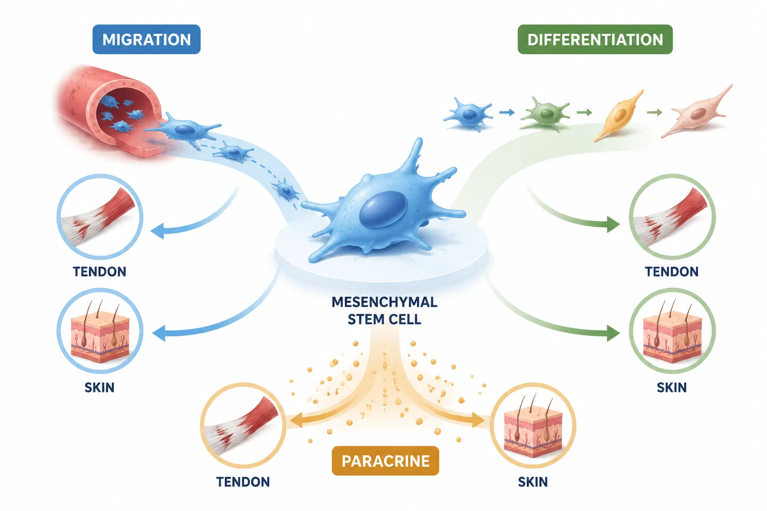
Mesenchymal stem cells are multipotent stromal cells found in bone marrow, adipose tissue, and connective tissue niches. In healthy tissue, they remain largely quiescent. After injury, damage-associated signals recruit MSCs to the wound site, where they contribute through three core mechanisms:
- Migration, chemotactic movement toward injury signals (SDF-1, VEGF, growth factors).
- Differentiation, commitment to tenocyte, fibroblast, or chondrocyte lineages depending on local cues.
- Paracrine signaling, secretion of cytokines, exosomes, and growth factors that modulate inflammation and stimulate resident cells.
Understanding these three pathways is essential for evaluating how peptides interact with MSC biology. For a broader overview of how tissue biology underpins recovery, the recovery and tissue biology overview provides useful foundational context.
Comparing BPC-157, TB-500, and GHK-Cu in Regeneration Studies: MSC-Level Mechanisms
BPC-157: Angiogenesis and MSC Recruitment
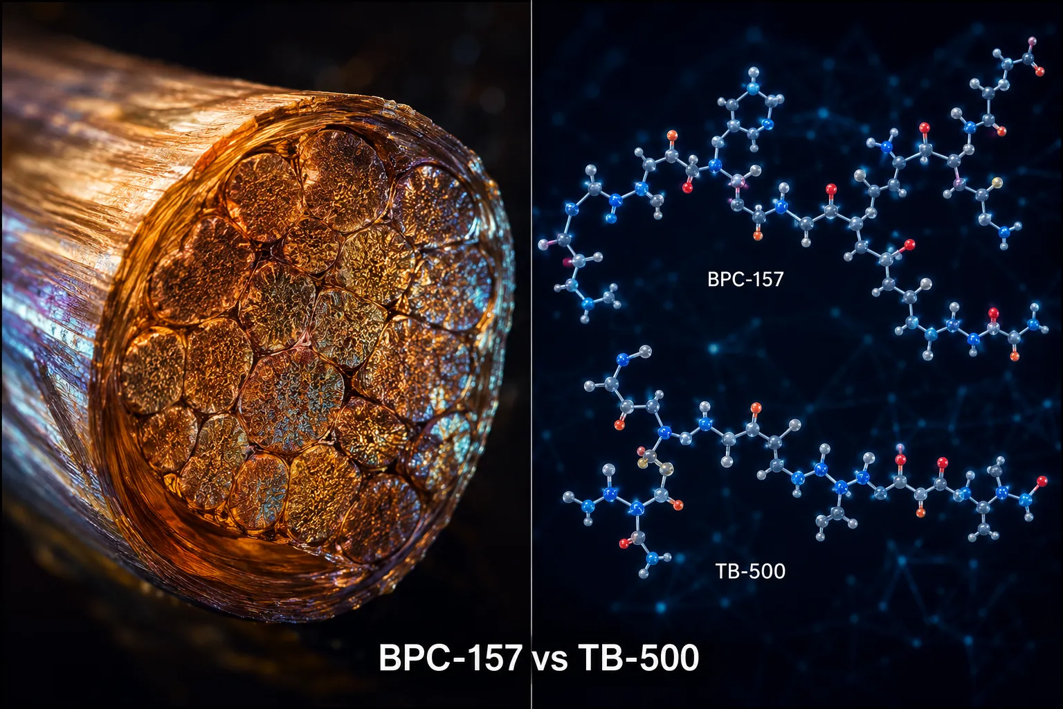
BPC-157 (Body Protection Compound-157) is a 15-amino-acid peptide derived from a gastric protein. In tendon and ligament models, it upregulates VEGF receptor expression and activates the FAK-paxillin pathway, both critical for MSC chemotaxis toward injury zones.
Key findings from preclinical research:
- Accelerated tendon-to-bone healing in rat rotator cuff models
- Increased fibroblast and MSC density at repair sites
- Reduced pro-inflammatory cytokine load (TNF-alpha, IL-6), creating a more permissive environment for MSC engraftment
The BPC-157 research overview and detailed data on BPC-157 nasal and oral delivery formats expand on delivery considerations relevant to tissue-level dosing.
TB-500: Actin Dynamics and MSC Motility
TB-500 is a synthetic analog of Thymosin Beta-4, a 43-amino-acid peptide that sequesters G-actin monomers. Its relevance to MSC biology centers on actin cytoskeleton remodeling, the physical process that allows cells to extend lamellipodia and migrate through extracellular matrix.
"Thymosin Beta-4 does not simply accelerate healing, it changes the cellular architecture that makes directed migration possible."
In muscle and ligament repair models, TB-500 has been shown to:
- Enhance MSC spreading and adhesion on collagen substrates
- Upregulate MMP-2 (matrix metalloproteinase-2), facilitating matrix remodeling
- Promote anti-apoptotic signaling in transplanted MSC populations
Detailed compound data is available on the TB-500 product and research page. Researchers comparing stacking strategies will also find the BPC-157 and TB-500 combination research directly relevant.
GHK-Cu: Gene Activation and Dermal MSC Signaling
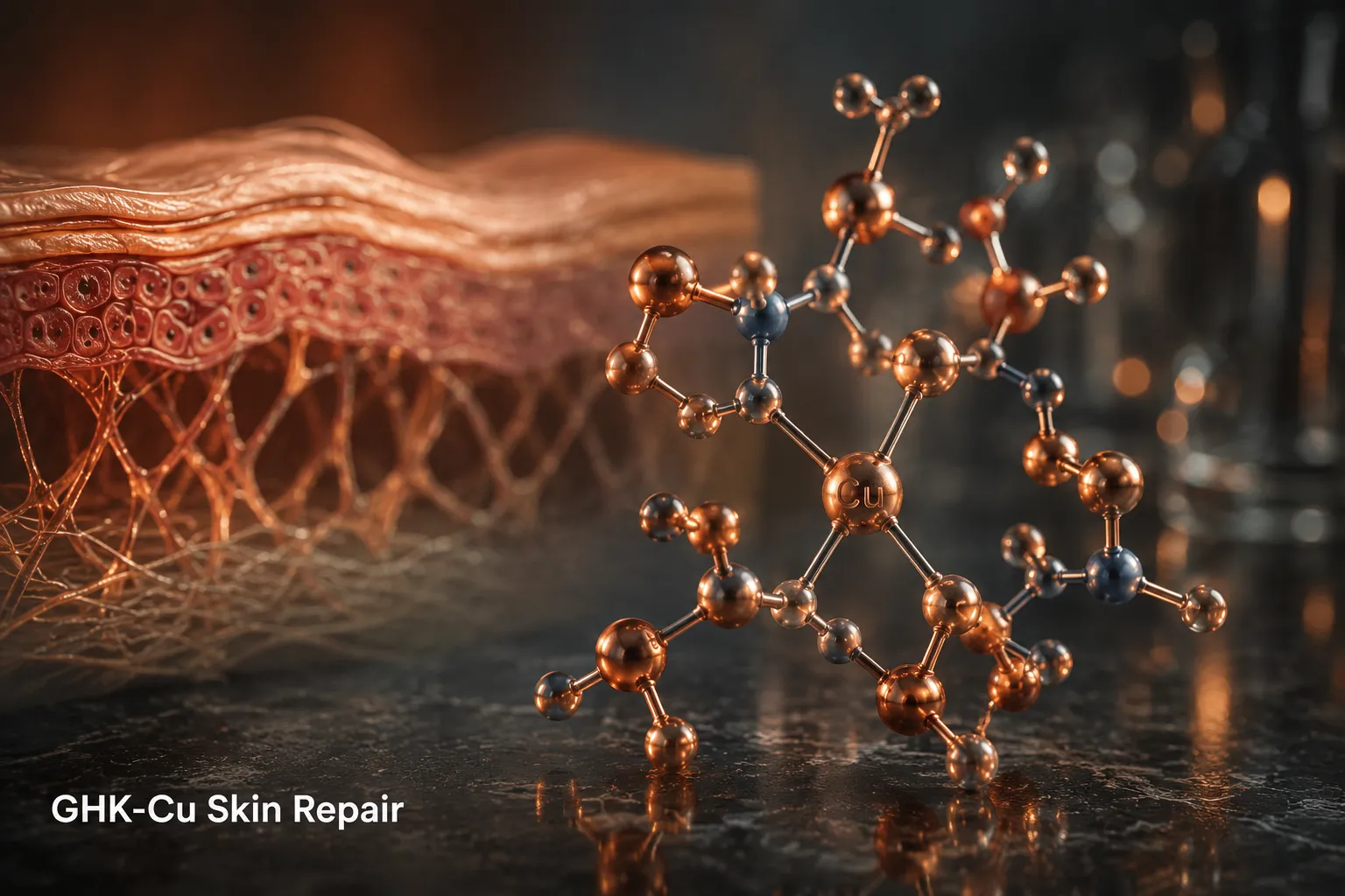
GHK-Cu (glycyl-L-histidyl-L-lysine copper complex) operates through a distinct mechanism. Rather than driving cell motility, it functions primarily as a gene expression modulator, activating over 4,000 human genes in microarray studies, many of them tied to collagen I and III synthesis, anti-inflammatory pathways, and antioxidant defense.
In dermal regeneration models, GHK-Cu:
- Stimulates fibroblast proliferation and MSC-derived collagen deposition
- Downregulates TGF-beta-1 (associated with fibrosis) while upregulating TGF-beta-3 (associated with scarless repair)
- Activates the ubiquitin-proteasome pathway to clear damaged proteins from the extracellular matrix
This makes GHK-Cu particularly valuable in skin and wound-healing contexts, where dermal MSC paracrine output determines scar quality and tissue architecture.
Comparing the Three Peptides: A Functional Summary
| Peptide | Primary MSC Target | Key Tissue Model | Dominant Pathway |
|---|---|---|---|
| BPC-157 | Migration, angiogenesis | Tendon, ligament | VEGF / FAK-paxillin |
| TB-500 | Motility, matrix remodeling | Muscle, ligament | Actin / MMP-2 |
| GHK-Cu | Paracrine gene activation | Dermis, wound healing | TGF-beta / ubiquitin |
These peptides are not interchangeable, they target different nodes of the MSC repair cascade. Researchers exploring broader regenerative peptide categories can also review longevity peptide research for adjacent mechanistic context.
Research Quality and Sourcing Considerations
Reproducibility in MSC and peptide-driven tissue repair studies depends heavily on compound purity. Contaminated or degraded peptides introduce confounding variables that distort migration assays, gene expression data, and histological outcomes. Reference-grade benchmarking, as outlined in resources on Bachem and reference standards for peptide benchmarks, is considered best practice in serious regeneration research.
Researchers sourcing compounds for in vitro or in vivo work should also consult all peptides available for research to evaluate purity specifications before designing studies.
Conclusion
The intersection of mesenchymal stem cells and peptide-driven tissue repair: comparing BPC-157, TB-500, and GHK-Cu in regeneration studies reveals a nuanced picture. Each peptide engages a distinct MSC mechanism, BPC-157 drives recruitment and vascularization, TB-500 enables physical cell migration through matrix remodeling, and GHK-Cu reshapes the paracrine signaling environment at the gene expression level. No single compound covers all three nodes simultaneously.
Actionable next steps for researchers in 2026:
- Design studies that distinguish MSC migration endpoints from differentiation and paracrine outputs to avoid conflating mechanisms.
- Use validated, purity-certified peptide sources to ensure reproducible results across tendon, ligament, and dermal models.
- Consider sequential or combinatorial peptide protocols that address all three MSC repair pathways, informed by the mechanistic distinctions outlined above.
- Cross-reference findings against established tissue biology frameworks before drawing translational conclusions.
The stem cell biology foregrounded here offers a more precise lens than general "healing peptide" narratives, and that precision is exactly what rigorous regeneration research demands.
Leave a Reply
Want to join the discussion?Feel free to contribute!