

Naproxen and Peptide Tissue-Repair Models: COX Inhibition, Inflammation Timing, and Recovery-Endpoint Selection
Prostaglandins do not simply cause pain, they also direct the cellular traffic of tissue repair. When naproxen blocks cyclooxygenase enzymes and suppresses prostaglandin synthesis, it simultaneously alters the signaling environment that peptides like BPC-157 and TB-500 depend on to drive regeneration. Understanding the intersection of Naproxen and Peptide Tissue-Repair Models: COX Inhibition, Inflammation Timing, and Recovery-Endpoint Selection is therefore essential for designing controls and interpreting outcomes in preclinical research.
Key Takeaways
- Naproxen suppresses COX-1 and COX-2, reducing prostaglandin-driven inflammation but also disrupting reparative signaling that tissue-repair peptides rely on.
- The timing of NSAID exposure relative to the inflammatory peak is the single most consequential variable in rodent tendon and muscle repair models.
- Early COX inhibition (days 1-3 post-injury) consistently impairs myosatellite activation and collagen scaffold remodeling in preclinical models.
- BPC-157 and TB-500 experiments require naproxen-treated control arms to isolate peptide-specific effects from background anti-inflammatory noise.
- Recovery-endpoint selection must match the tissue type, repair phase, and peptide mechanism, histological, biochemical, and functional endpoints each capture different dimensions of healing.
How COX Inhibition Reshapes the Prostaglandin Landscape
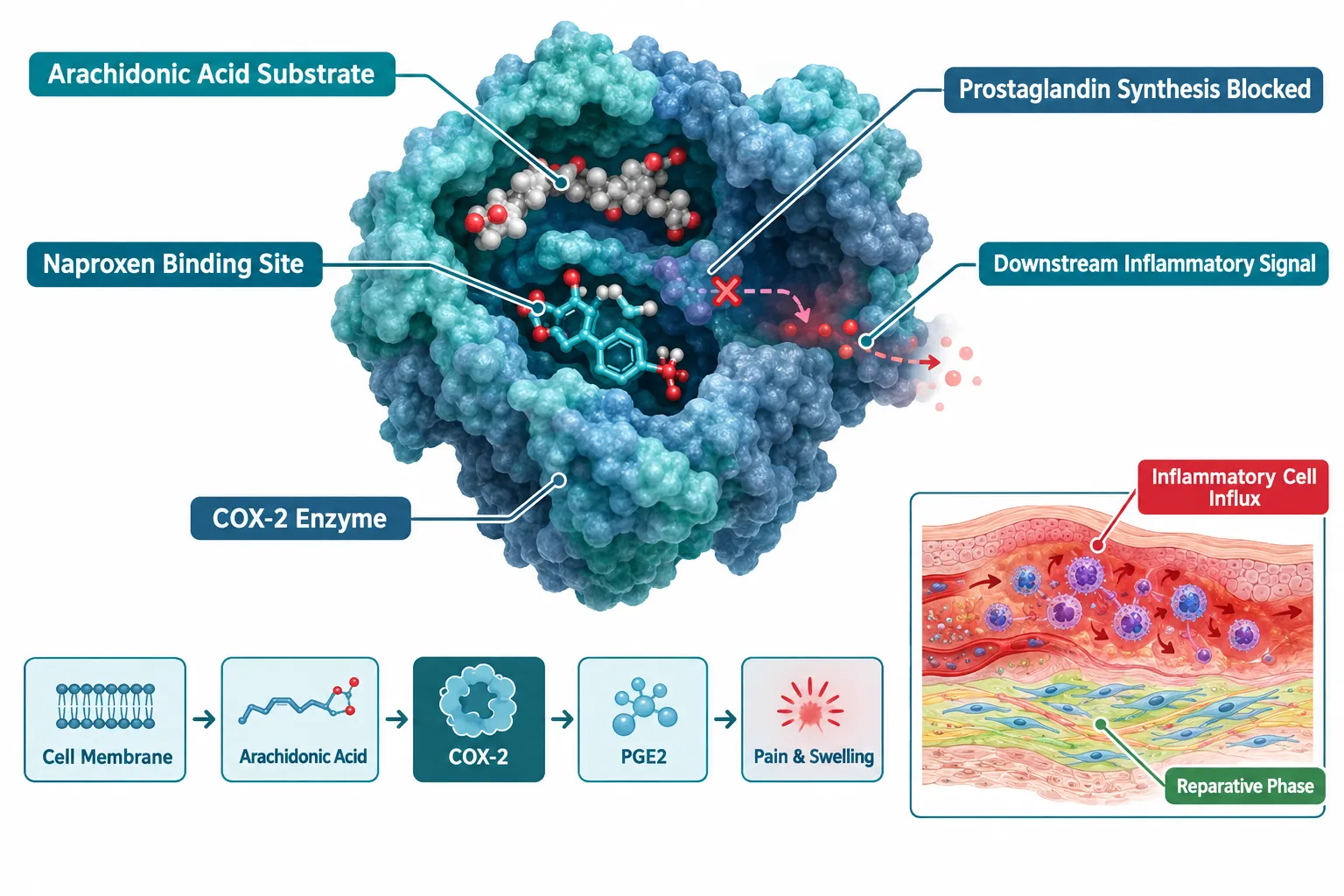
Naproxen is a non-selective NSAID that competitively inhibits both COX-1 and COX-2 isoforms, blocking the conversion of arachidonic acid into prostaglandins, thromboxanes, and prostacyclins. In the context of tissue injury, this matters because prostaglandin E2 (PGE2) is not merely a pain mediator, it recruits macrophages, activates satellite cells in muscle, and upregulates growth factor receptors in tendon fibroblasts.
When naproxen suppresses PGE2 during the first 72 hours after injury, inflammatory cell influx decreases measurably. Neutrophil and macrophage counts in injured tissue drop, which reduces the release of pro-inflammatory cytokines such as IL-6 and TNF-alpha. On the surface, this looks beneficial. In practice, it removes a critical early signal that coordinates the transition from inflammation to proliferative repair.
Researchers working with the NpxFFK naproxen-peptide hydrogel platform have demonstrated that localized COX-2 inhibition can be spatially controlled, offering a useful model for isolating the enzyme's role without systemic suppression. This approach helps distinguish between the anti-inflammatory and anti-regenerative consequences of COX blockade, a distinction that matters enormously when peptide co-treatments are introduced.
For research teams sourcing tissue-repair peptides, the BPC-157 core peptides documentation and first research guide provides a useful framework for understanding how this peptide's angiogenic and collagen-stimulating properties interact with the prostaglandin-depleted environment naproxen creates.
Inflammation Timing: The Critical Variable in Naproxen and Peptide Tissue-Repair Models
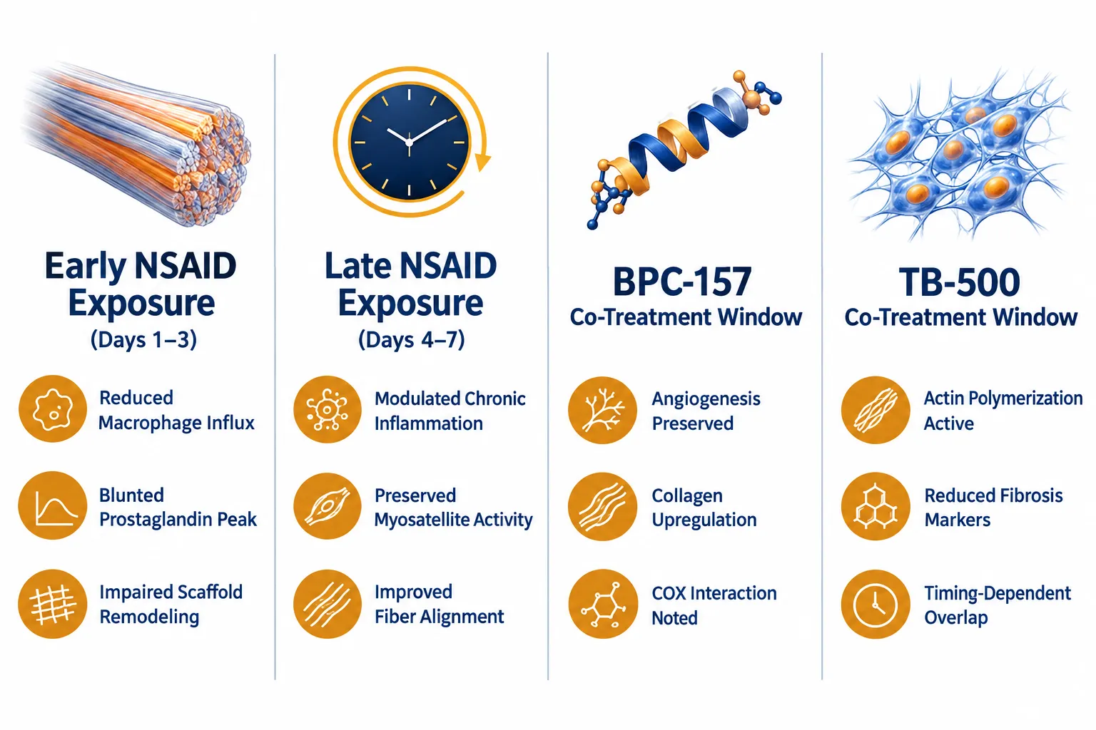
Across rodent Achilles tendon and skeletal muscle repair models, the timing of naproxen administration relative to the inflammatory peak produces dramatically different outcomes. This is the central design challenge in Naproxen and Peptide Tissue-Repair Models: COX Inhibition, Inflammation Timing, and Recovery-Endpoint Selection.
Early exposure (days 1-3): COX inhibition during peak inflammation consistently impairs repair. Myosatellite cell activation is blunted, collagen cross-linking is reduced, and the mechanical strength of healing tendons at 4 weeks is measurably lower than in untreated controls. The reparative macrophage phenotype (M2 polarization) that drives scaffold remodeling is particularly sensitive to early prostaglandin suppression.
Late exposure (days 4-7 onward): Naproxen introduced after the initial inflammatory surge shows a more favorable profile. Chronic, maladaptive inflammation, characterized by persistent neutrophil presence and fibrotic collagen deposition, is reduced without disrupting the early reparative signals that have already been delivered.
"The difference between a reparative and a maladaptive inflammatory response often comes down to a 48-hour window, and naproxen's half-life of 12-17 hours makes dosing schedule a precision variable, not a background detail."
This timing dependency has direct implications for BPC-157 and TB-500 co-treatment experiments. BPC-157 promotes angiogenesis and upregulates growth hormone receptors partly through prostaglandin-dependent pathways. If naproxen is administered simultaneously, the peptide's mechanism of action is partially blocked, confounding outcome data. Researchers should therefore design naproxen control arms with staggered dosing windows rather than concurrent administration.
TB-500, which drives actin polymerization and reduces fibrosis, appears less prostaglandin-dependent in its primary mechanism, but its downstream effects on inflammatory cell migration overlap with COX-2 pathways. The SS-31 peptide benefits literature offers a parallel example of how mitochondrial-targeted peptides navigate the NSAID-altered inflammatory environment, a useful methodological reference.
Chronotherapy data from human postoperative recovery studies adds another layer: NSAIDs administered in the morning show different pharmacodynamic profiles than evening doses, reflecting circadian variation in COX-2 expression. Animal models designed to mirror clinical naproxen use should account for light-cycle-dependent dosing schedules.
Recovery-Endpoint Selection for COX-Inhibited Tissue-Repair Experiments
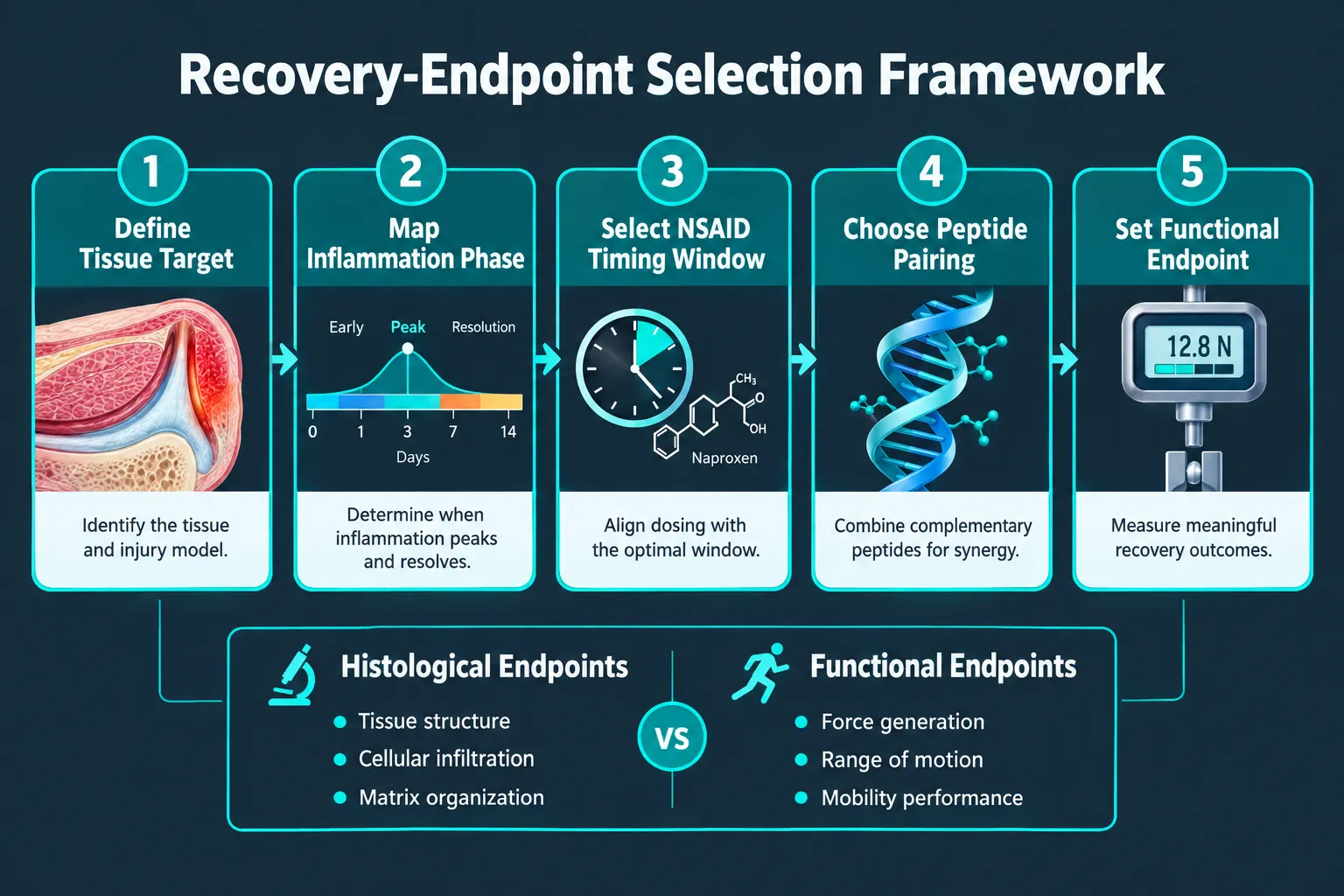
Choosing the wrong endpoint is one of the most common sources of false-negative results in peptide tissue-repair research. Naproxen and Peptide Tissue-Repair Models: COX Inhibition, Inflammation Timing, and Recovery-Endpoint Selection demands a multi-dimensional approach because naproxen affects different aspects of repair at different timepoints.
Recommended endpoint categories:
| Endpoint Type | What It Captures | Best Timepoint |
|---|---|---|
| Histological | Cell composition, collagen architecture | Days 7, 14, 28 |
| Biochemical | PGE2, IL-6, TNF-alpha, hydroxyproline | Days 3, 7, 14 |
| Mechanical | Tensile strength, load-to-failure | Days 21, 42 |
| Functional | Gait analysis, grip strength, swim test | Days 14, 28 |
For BPC-157 experiments, hydroxyproline content (a collagen maturity marker) combined with vascular density staining captures the peptide's dual angiogenic-collagen mechanism. Naproxen-treated controls will show reduced hydroxyproline at day 14, providing a clear baseline against which BPC-157's effect can be measured.
For TB-500 experiments, actin filament density and fibrosis scoring (Masson's trichrome staining) are the most sensitive endpoints. Because TB-500 primarily acts on actin dynamics rather than prostaglandin synthesis, its effects may be partially preserved even in naproxen-treated tissue, making it a useful positive control for separating COX-dependent from COX-independent repair mechanisms.
Pre-emptive COX-2 inhibition studies using celecoxib have shown that wound-healing dynamics are most disrupted when COX blockade precedes injury rather than following it, reinforcing the importance of injury-relative timing in endpoint interpretation.
Researchers sourcing peptides for these models should consult the GHK-Cu peptide purchase and copper peptide research sourcing guide for purity standards applicable to co-treatment protocols, and review research-grade Glow Blend peptide evaluation methods for quality benchmarks in skin and connective-tissue models. For mitochondrial endpoint controls, SS-31 peptide sourcing options provide a validated comparator peptide for oxidative stress endpoints in NSAID-treated tissue.
Conclusion
The relationship between naproxen and peptide tissue-repair models is not adversarial, it is architectural. COX inhibition reshapes the prostaglandin landscape that peptides like BPC-157 and TB-500 navigate, and that reshaping is highly time-dependent. Researchers who treat naproxen as a simple background variable will produce confounded data; those who treat it as a precision timing instrument will generate clean, interpretable results.
Actionable next steps for research design:
- Define the inflammatory phase map for the target tissue before assigning naproxen dosing windows.
- Use staggered rather than concurrent naproxen and peptide administration to isolate mechanism-specific effects.
- Select at least one biochemical endpoint (PGE2 or IL-6), one structural endpoint (collagen content or fiber alignment), and one functional endpoint per experiment.
- Validate peptide purity and lot consistency before initiating multi-arm NSAID co-treatment studies.
- Review chronotherapy data to align dosing schedules with circadian COX-2 expression patterns in the animal model used.
Precision in timing and endpoint selection is what separates publishable data from ambiguous results in this research space.