

Hemoglobin and Hematocrit in Peptide Research: How to Interpret Oxygen-Carrying Capacity and Safety Signals
A hematocrit reading of 54% in a research subject is not just a number, it is a stop signal. Yet many researchers encounter elevated red-cell indices in their bloodwork panels and lack a clear framework for deciding what those values mean, whether they reflect a true physiological change, and what action, if any, is warranted. Understanding hemoglobin and hematocrit in peptide research: how to interpret oxygen-carrying capacity and safety signals is one of the most practical skills any serious investigator can develop.
Key Takeaways
- Hemoglobin measures the protein that carries oxygen; hematocrit measures the percentage of blood volume occupied by red cells, both are essential safety markers in peptide research.
- Baseline values must be established before any research protocol begins, because pre-existing elevations change how subsequent readings are interpreted.
- A hematocrit of 51-52% warrants review; 54% or above typically triggers intervention or protocol cessation.
- Dehydration and red-cell indices like MCV can confound hematocrit readings and must be ruled out before attributing a rise to a peptide compound.
- Monitoring frequency, action thresholds, and management strategies follow a guideline-style algorithm that researchers should apply consistently.
What Hemoglobin and Hematocrit Actually Measure
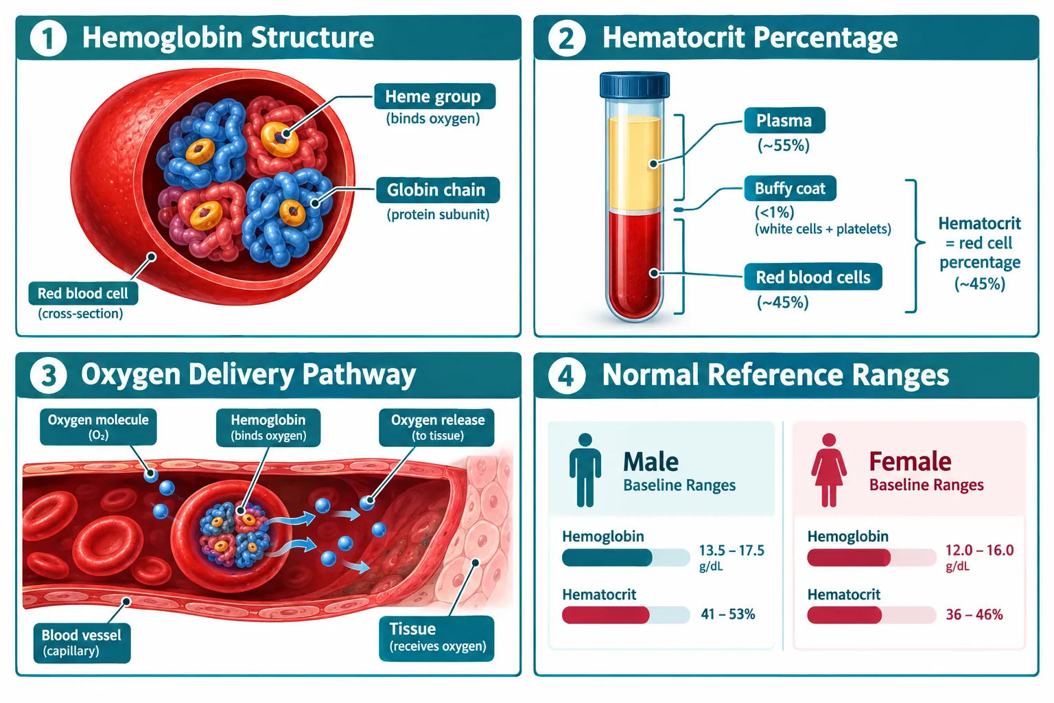
Hemoglobin (Hb) is the iron-containing protein inside red blood cells. Its job is to bind oxygen in the lungs and release it to tissues. It is measured in grams per deciliter (g/dL). Normal ranges sit roughly at 13.5-17.5 g/dL for adult males and 12.0-15.5 g/dL for adult females, though reference intervals vary slightly by laboratory.
Hematocrit (Hct) is a different but related measurement. It expresses the proportion of whole blood that is made up of red blood cells, reported as a percentage. Because hematocrit is a ratio of cells to total blood volume, it can be influenced by both the number of red cells and the volume of plasma. This distinction matters enormously when interpreting results in a research context.
Red-cell indices, including mean corpuscular volume (MCV), mean corpuscular hemoglobin (MCH), and red cell distribution width (RDW), add important context. A high hematocrit paired with a normal MCV and normal RDW looks very different from one paired with microcytic, hypochromic cells, which would suggest iron deficiency rather than true erythrocytosis.
The Difference Between True Erythrocytosis and a Spurious Rise
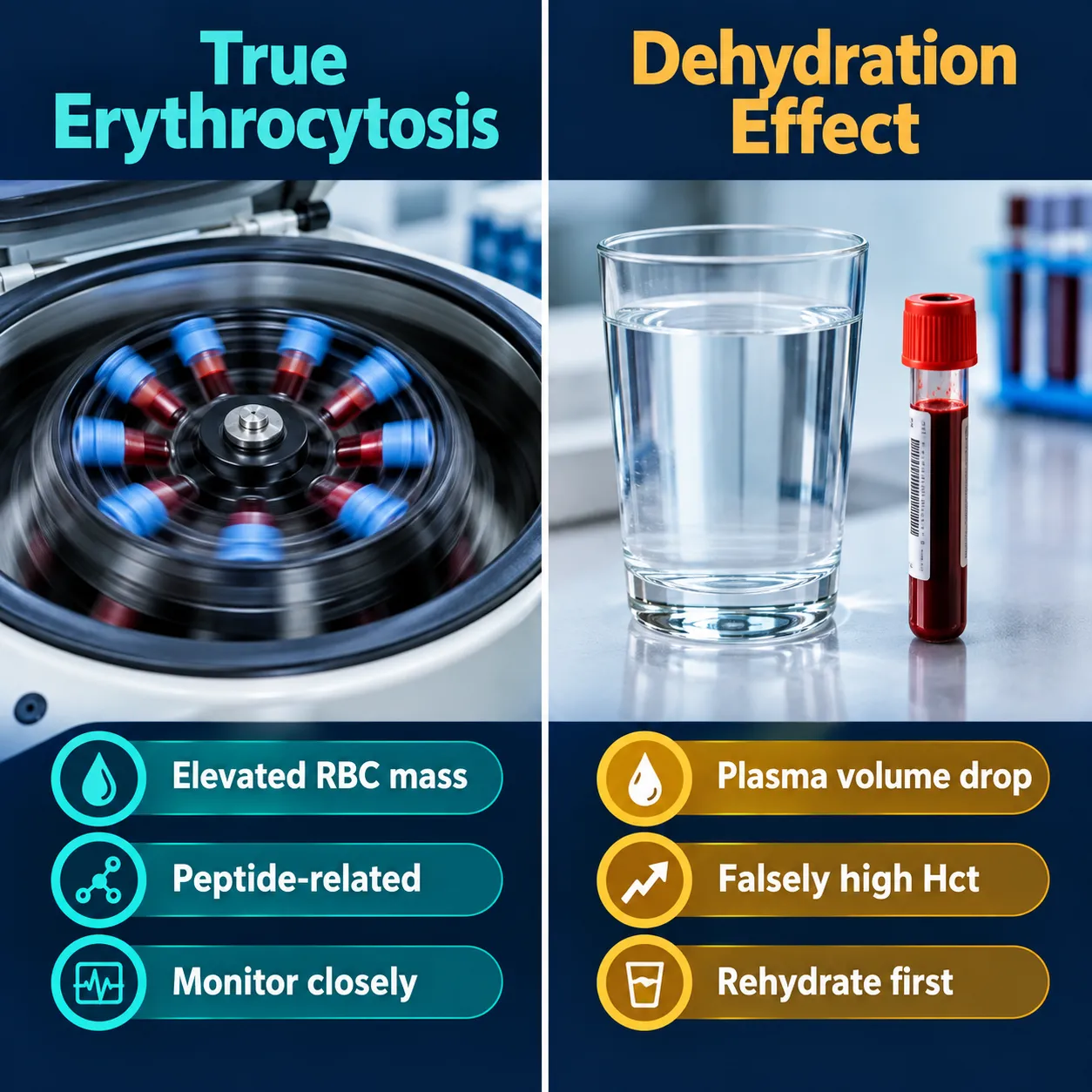
True erythrocytosis means the body is producing more red blood cells, increasing the absolute red cell mass. A spurious rise in hematocrit, by contrast, happens when plasma volume decreases, most commonly through dehydration, without any actual increase in red cell production. A subject who is even mildly dehydrated at the time of blood draw can show a hematocrit several points higher than their true baseline.
Researchers should always confirm adequate hydration before attributing a hematocrit elevation to a peptide compound. When in doubt, repeating the draw after proper rehydration is the most straightforward corrective step.
Monitoring Protocols: Hemoglobin and Hematocrit in Peptide Research
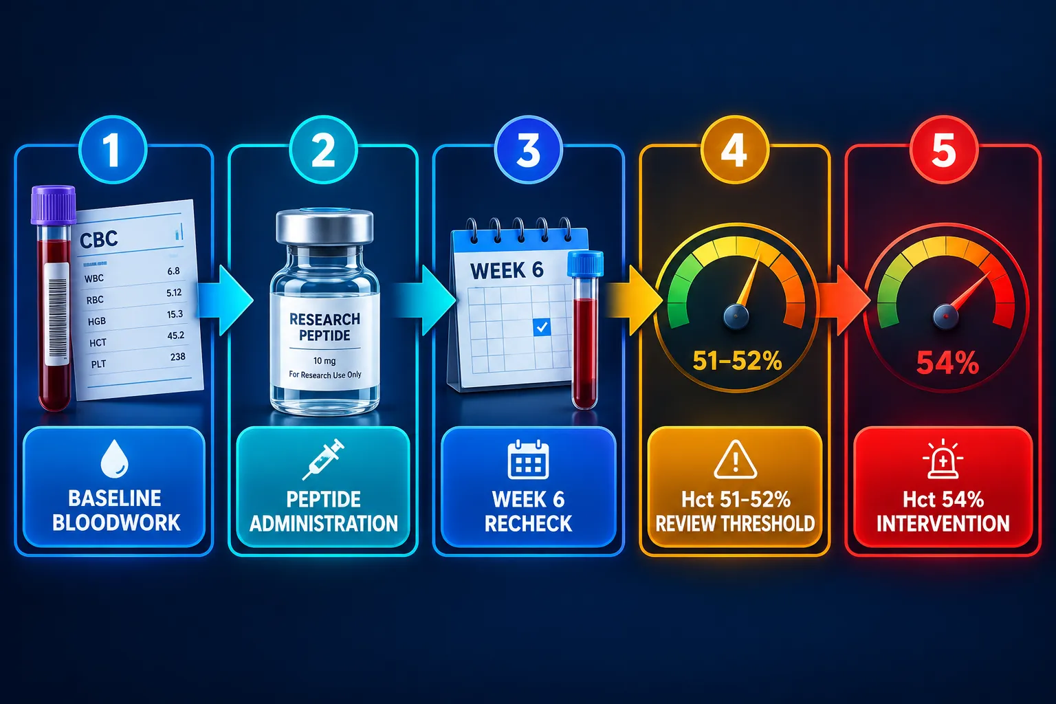
Establishing a monitoring schedule is not optional, it is the foundation of responsible research. The general framework follows a logical sequence:
- Baseline bloodwork before the protocol begins, capturing Hb, Hct, and a full red-cell panel.
- Recheck at 6-8 weeks, which is typically when androgenic or peptide-adjacent compounds begin to exert measurable effects on erythropoiesis.
- Ongoing monitoring every 3-6 months once values are stable.
This schedule mirrors the approach used in testosterone replacement and gender-affirming hormone research, where hematocrit elevation is documented to be several-fold more common than in placebo groups. The same logic applies when investigating compounds that interact with growth hormone axes or metabolic pathways. Researchers exploring options like Tesamorelin peptide benefits or mitochondria-targeted agents such as SS-31 peptides for sale should incorporate hematological monitoring from the outset.
Action Thresholds Every Researcher Should Know
| Hematocrit Level | Recommended Action |
|---|---|
| Below 50% | Continue protocol; monitor per schedule |
| 51-52% | Flag for review; assess hydration and confounders |
| 53% | Increase monitoring frequency; evaluate risk factors |
| 54% or above | Pause protocol; consider intervention or cessation |
These thresholds are not arbitrary. At hematocrit levels above 54%, blood viscosity increases meaningfully, raising the theoretical risk of thromboembolic events. Even in research settings, that risk profile demands a conservative response.
Confounders, Red Flags, and Interpreting Safety Signals
Several factors can confound hematocrit readings and lead to misinterpretation. Researchers using lab tested peptides should be aware of the following:
- Altitude: Living or training at elevation stimulates erythropoietin production naturally, raising baseline Hct independent of any compound.
- Sleep apnea: Chronic intermittent hypoxia drives compensatory erythrocytosis and can be mistaken for a compound-induced effect.
- Polycythemia vera: A rare myeloproliferative condition that elevates red cell mass; it must be ruled out when hematocrit rises without a clear cause.
- Androgens and peptides with androgenic overlap: Compounds like MT-2 research peptides operate through melanocortin receptors, but any compound that indirectly influences testosterone or IGF-1 pathways warrants careful hematological oversight.
When a true elevation is confirmed, management options include dose reduction, increased hydration, therapeutic phlebotomy in severe cases, and in some protocols, temporary cessation. Researchers investigating compounds with systemic metabolic effects, including those in the GLP-3 peptide class or IPA peptides, should document any hematological changes as part of their safety reporting.
It is also worth noting that severe erythrocytosis remains relatively rare when protocols are well-managed. Evidence from gender-affirming testosterone care shows that diligent monitoring catches elevations early, before they reach clinically dangerous levels. The same principle applies to peptide research: early detection through consistent bloodwork is far more effective than reactive management.
Researchers sourcing compounds from a best peptide manufacturer should also confirm purity, since contaminants or misidentified compounds can introduce unexpected physiological effects, including on erythropoiesis.
Conclusion
Interpreting hemoglobin and hematocrit in peptide research: how to interpret oxygen-carrying capacity and safety signals requires more than reading a number off a lab report. It demands understanding what the values represent, what can distort them, and what action each threshold calls for.
Actionable next steps for researchers:
- Establish a full baseline CBC before starting any peptide protocol.
- Rule out dehydration and altitude effects before attributing any Hct rise to the compound under study.
- Apply the 51-52% review threshold and the 54% intervention threshold consistently across all protocols.
- Document red-cell indices alongside Hb and Hct to distinguish true erythrocytosis from nutritional or plasma-volume artifacts.
- Recheck values at 6-8 weeks and every 3-6 months thereafter, adjusting frequency if values trend upward.
Rigorous hematological monitoring is not a bureaucratic formality. It is the mechanism by which researchers protect subject safety and generate data that is actually interpretable. Build it into every protocol from day one.