

GLP-2-T and GLP2 Tirz Peptide Research: How to Design Intestinal Barrier, Organoid, and Epithelial-Repair Assays
Roughly 70% of the body's immune activity is concentrated in the gut lining, yet most peptide research programs treat intestinal barrier function as an afterthought. GLP-2-T and GLP2 Tirz Peptide Research: How to Design Intestinal Barrier, Organoid, and Epithelial-Repair Assays has emerged as a rigorous discipline precisely because the gut epithelium is both a therapeutic target and a sensitive readout system. Designing assays that capture permeability shifts, tight-junction remodeling, and organoid growth requires a structured approach and a clear understanding of what these peptide names actually represent.
Key Takeaways
- GLP-2 drives intestinal growth indirectly through enteric neurons, not directly through epithelial receptors, assay design must account for this.
- "GLP-2-T" and "GLP2 Tirz" are working nomenclature terms, not validated compound names; confirm identity through mass spectrometry and receptor-binding assays before interpreting data.
- Core barrier readouts include transepithelial electrical resistance (TEER), FITC-dextran permeability, and tight-junction protein immunostaining.
- Organoid models using intestinal stem cells (ISCs) and Paneth cells provide the most physiologically relevant epithelial-repair endpoints.
- Purity and sequence verification of research peptides are non-negotiable prerequisites for reproducible results.
Understanding GLP-2 Biology Before Building Assays
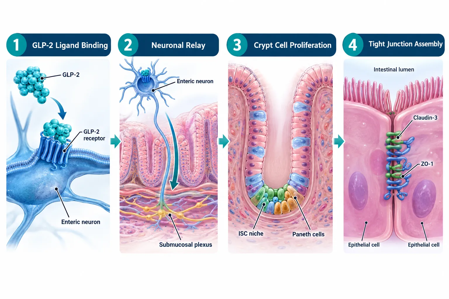
GLP-2 (glucagon-like peptide-2) is a 33-amino-acid hormone secreted by enteroendocrine L-cells in response to luminal nutrients. Its primary role is to stimulate intestinal growth, increase mucosal surface area, and reinforce epithelial barrier integrity. A critical detail that shapes every assay design: the GLP-2 receptor is expressed on enteric neurons and subepithelial myofibroblasts, not on enterocytes themselves. This means GLP-2 acts indirectly, triggering a neuronal relay that ultimately drives crypt cell proliferation and tight-junction assembly.
This neuroepithelial circuit has direct implications for assay selection. Simple monolayer cell cultures lacking a neuronal component will not fully recapitulate GLP-2 signaling. Researchers working on GLP-2-T and GLP2 Tirz concepts must either use co-culture systems or organoid platforms that preserve the cellular architecture of the crypt-villus axis.
Nomenclature clarity matters. The terms "GLP-2-T" and "GLP2 Tirz" circulate in research communities as shorthand for modified GLP-2 analogs or multi-agonist concepts that may incorporate tirzepatide-like dual-receptor targeting. Neither term corresponds to a single, fully characterized compound as of 2026. Before any assay begins, researchers should confirm peptide identity via mass spectrometry, HPLC purity analysis, and receptor-binding confirmation. Explore available GLP peptide research options to understand the range of characterized analogs on the market.
Designing Intestinal Barrier Assays for GLP-2-T and GLP2 Tirz Peptide Research
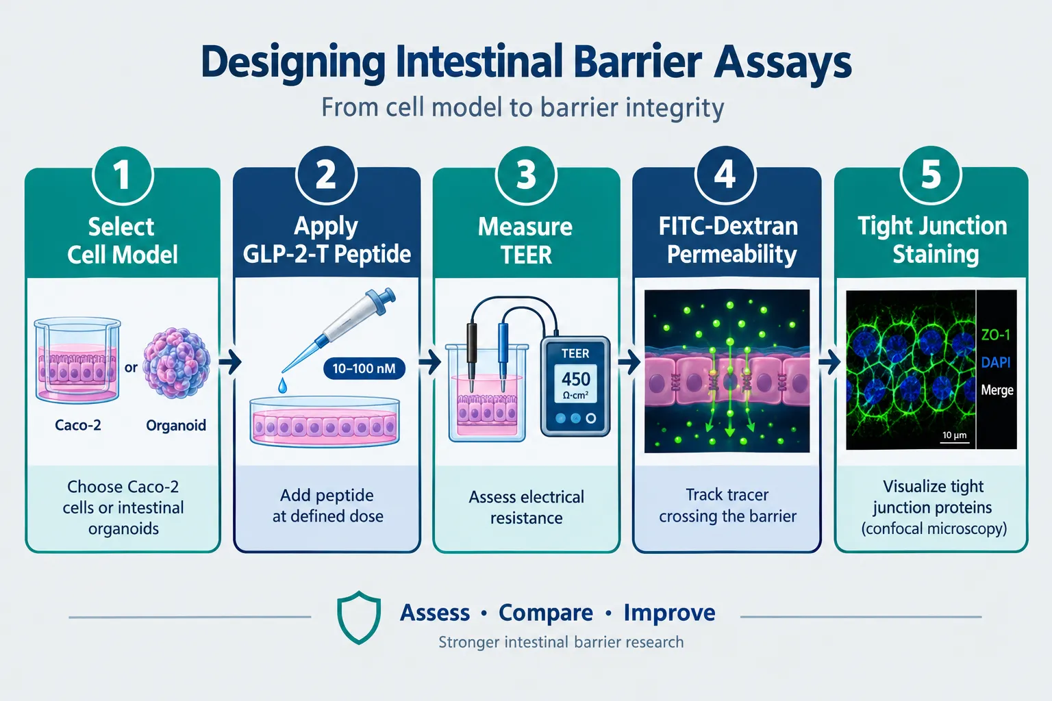
Barrier function assays form the quantitative backbone of GLP-2-T and GLP2 Tirz Peptide Research: How to Design Intestinal Barrier, Organoid, and Epithelial-Repair Assays programs. Two readouts dominate the literature:
Transepithelial Electrical Resistance (TEER)
TEER measures the ionic resistance across a cell monolayer grown on a permeable Transwell insert. Higher resistance values indicate a tighter, more intact barrier. For GLP-2 analog research:
- Seed Caco-2 or T84 cells on 0.4-micron Transwell membranes
- Allow 21 days for full differentiation and tight-junction maturation
- Apply peptide at physiologically relevant concentrations (typically 1-100 nM)
- Record TEER at 24, 48, and 72 hours post-treatment
Key point: A meaningful TEER increase after GLP-2-T treatment suggests tight-junction reinforcement, but must be paired with protein-level confirmation.
FITC-Dextran Permeability
This assay quantifies paracellular leakage by measuring how much fluorescently labeled dextran crosses the monolayer from apical to basolateral compartments. A reduction in FITC-dextran flux after peptide treatment confirms barrier-sealing activity.
Tight-Junction Protein Immunostaining
Immunofluorescence staining for ZO-1, claudin-3, and occludin provides mechanistic evidence. Disrupted tight junctions appear as fragmented or discontinuous staining patterns. Successful GLP-2 analog treatment should restore linear, continuous junction morphology.
Endpoint summary table:
| Assay | Readout | Positive Result |
|---|---|---|
| TEER | Ohms x cm2 | Increased resistance |
| FITC-Dextran | Fluorescence (ng/mL) | Decreased flux |
| ZO-1 Immunostaining | Confocal imaging | Continuous junctions |
| NLRP3 Inflammasome | Western blot / ELISA | Reduced activation |
| LPS Translocation | ELISA (basolateral) | Reduced LPS signal |
For researchers exploring related multi-agonist frameworks, reviewing GLP-3 peptide research compounds can help contextualize receptor-specificity differences when designing controls.
Organoid and Epithelial-Repair Assays in GLP-2-T and GLP2 Tirz Peptide Research
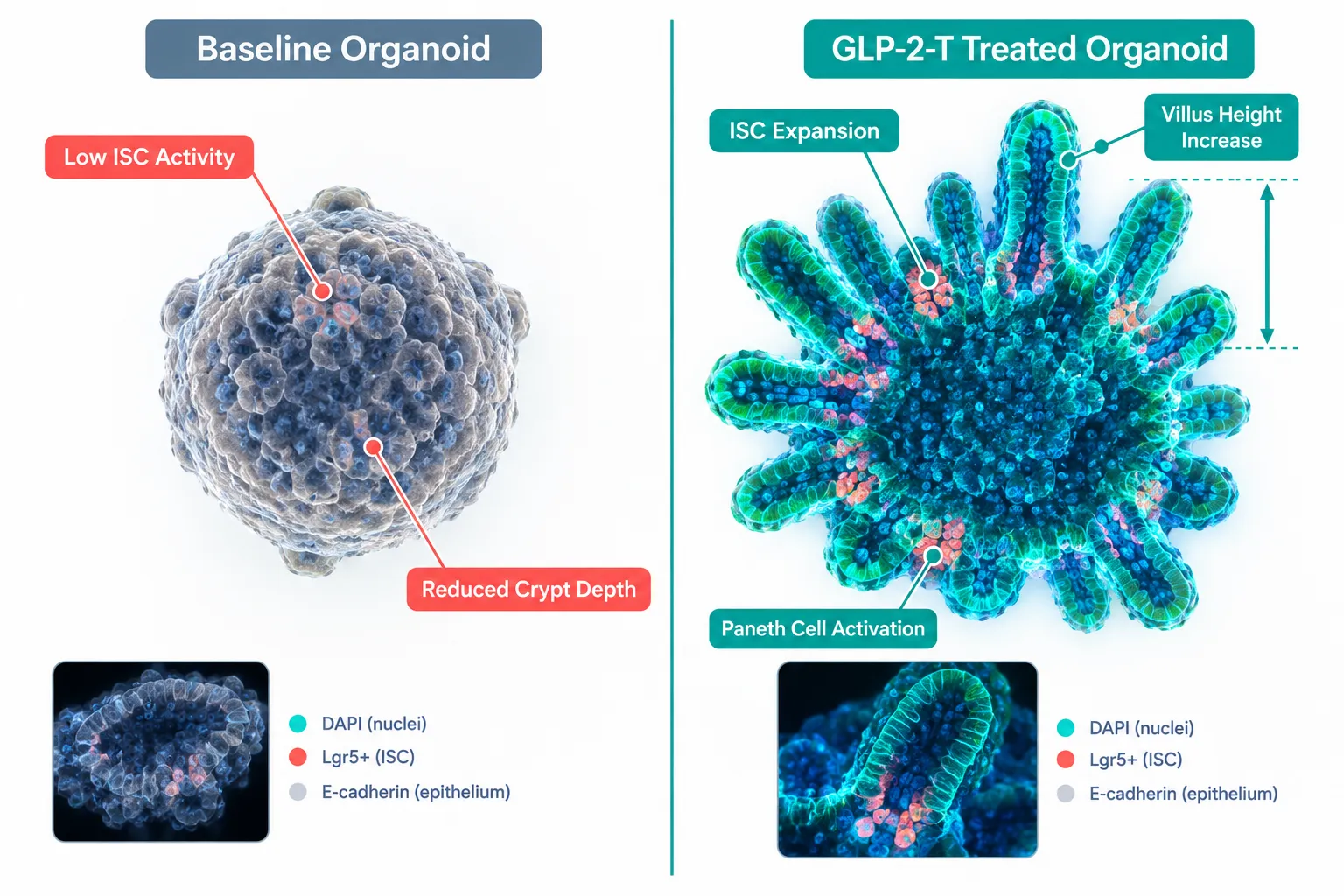
Organoid models represent the most physiologically faithful platform for GLP-2-T and GLP2 Tirz Peptide Research: How to Design Intestinal Barrier, Organoid, and Epithelial-Repair Assays. Intestinal organoids derived from crypts maintain ISC populations, Paneth cells, goblet cells, and enterocytes in a self-organizing three-dimensional structure.
Setting Up the Organoid Assay
- Isolate crypts from murine small intestine or human biopsy tissue using EDTA chelation
- Embed in Matrigel at 37°C and overlay with ENR medium (EGF, Noggin, R-spondin)
- Introduce peptide at day 3 post-seeding, once basal organoids are established
- Quantify growth by measuring organoid surface area, budding frequency, and villus-like domain length at 24-hour intervals
Key Epithelial-Repair Endpoints
- ISC marker expression: Lgr5+ cell frequency by immunofluorescence or flow cytometry
- Paneth cell density: Lysozyme staining as a proxy for niche-support capacity
- Villus height and crypt depth: Morphometric analysis from confocal z-stacks
- Proliferation index: Ki-67 staining in crypt-base columnar cells
GLP-2 analogs consistently increase organoid budding efficiency and villus domain length in preclinical models. Modified analogs with extended half-lives, relevant to the GLP-2-T concept, show amplified effects on crypt proliferation compared to native GLP-2, making organoid size and budding frequency sensitive discriminators between compound variants.
Researchers interested in complementary barrier-repair peptide tools may find value in reviewing SS-31 peptide research for mitochondrial-protection endpoints that can run alongside epithelial-repair assays. Similarly, cagrilinitide peptide research offers a relevant comparator for multi-agonist gut-axis studies.
Disease Model Applications
- IBD models: LPS-challenged organoids or DSS-treated murine intestine provide a relevant inflammatory context for testing GLP-2-T barrier-rescue activity
- Short bowel syndrome (SBS) surrogates: Remnant intestine organoids from partial resection models show blunted adaptation that GLP-2 analogs can rescue, a validated translational endpoint
- GVHD models: Graft-versus-host disease involves severe epithelial damage; GLP-2 receptor agonism has shown protective effects in these systems, making crypt survival the primary endpoint
For those sourcing characterized analogs for these models, GLP-3R peptide options and Reta peptide research compounds represent structurally related tools worth including as comparators.
Conclusion
Rigorous GLP-2-T and GLP2 Tirz Peptide Research: How to Design Intestinal Barrier, Organoid, and Epithelial-Repair Assays programs rest on three pillars: verified peptide identity, mechanistically appropriate cell models, and multi-endpoint readout strategies. The following steps will strengthen any research program:
- Verify compound identity with mass spectrometry and HPLC before any biological assay
- Pair TEER with FITC-dextran and tight-junction immunostaining for a complete barrier profile
- Use organoid models when ISC proliferation and villus morphology are primary endpoints
- Include inflammatory challenge conditions (LPS, DSS) to stress-test barrier-repair claims
- Source research-grade peptides from suppliers with documented purity data, see guidance on evaluating purity for research-grade peptides for a practical framework
As multi-agonist concepts combining GLP-2 activity with tirzepatide-like GIP/GLP-1 receptor engagement continue to develop, the assay toolkit described here will serve as the foundation for evaluating any new analog claiming intestinal barrier or epithelial-repair activity.