

Mitochondria and Research Peptides: What Labs Measure When Studying Cellular Energy Signaling
Fewer than 1% of the roughly 1,500 proteins inside a human mitochondrion have been fully characterized for their role in energy output, yet that small fraction already underpins some of the most active areas in peptide research today. Understanding Mitochondria and Research Peptides: What Labs Measure When Studying Cellular Energy Signaling is no longer a niche concern for biochemists alone. In 2026, it sits at the center of metabolic disease research, rare disease trials, and next-generation therapeutic design.
Key Takeaways
- Mitochondrial function is measured through oxygen consumption rate (OCR) and ATP production assays, most commonly using Seahorse XF technology.
- Research peptides such as SS-31 and MOTS-c target distinct nodes in the mitochondrial energy network and produce measurable, quantifiable effects.
- Labs convert raw OCR data into ATP production rates using established bioenergetic equations, enabling direct comparison across studies.
- Mitochondrial targeting sequences (MTS) are engineered design features that labs validate through membrane potential and proteomics assays.
- Peptide purity and certificate of analysis standards directly affect the reliability of cellular energy signaling data.
Why Mitochondrial Energy Signaling Demands Precise Measurement
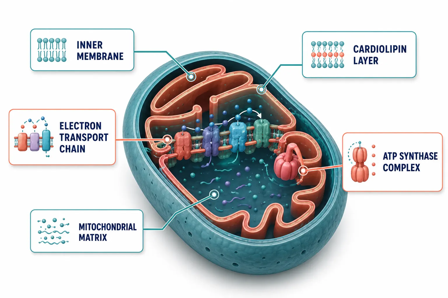
Mitochondria are not static power generators. They are dynamic organelles that constantly shift their output in response to nutrient availability, stress signals, and intercellular communication. When a research peptide enters this environment, it can alter membrane potential, modulate electron transport chain activity, or change the rate at which ATP synthase produces adenosine triphosphate.
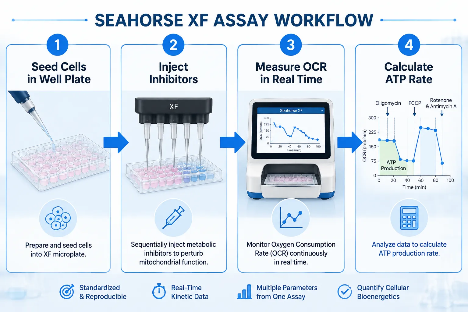
Labs studying cellular energy signaling need quantitative endpoints, not qualitative impressions. The most widely adopted platform for this work is the Seahorse XF Analyzer, which measures oxygen consumption rate (OCR) and extracellular acidification rate (ECAR) in live cells in real time. These two metrics together reveal how a cell balances oxidative phosphorylation against glycolysis, a balance that research peptides can shift in measurable ways.
"The OCR value alone tells you how hard the mitochondria are working. The ATP production rate tells you how efficiently that work translates into usable energy."
Beyond OCR, labs also track:
- Mitochondrial membrane potential (using dyes such as JC-1 or TMRE)
- Reactive oxygen species (ROS) output via fluorescent probes
- NAD+/NADH ratios as indicators of redox balance
- Mitochondrial fusion and fission dynamics through live-cell imaging
A 2025 study examining LRRK2 gene mutations used this exact framework to quantify ATP deficits in patient-derived neurons, demonstrating how OCR-to-ATP conversion math can anchor a disease mechanism to a specific molecular target.
Core Assays in Mitochondria and Research Peptides Studies
The Seahorse XF Mito Stress Test
The standard Seahorse mito stress test injects three compounds in sequence, oligomycin, FCCP, and rotenone/antimycin A, each blocking a different step in the respiratory chain. The resulting OCR curve reveals:
| Parameter | What It Reflects |
|---|---|
| Basal respiration | Baseline mitochondrial workload |
| ATP-linked respiration | OCR directly coupled to ATP synthesis |
| Maximal respiration | Full electron transport chain capacity |
| Spare respiratory capacity | Metabolic reserve under stress |
| Proton leak | Membrane integrity and uncoupling |
Labs convert ATP-linked OCR to an ATP production rate by applying a stoichiometric factor derived from the P/O ratio, roughly 2.73 ATP per oxygen atom consumed during oxidative phosphorylation. This conversion is critical when comparing peptide-treated cells against controls.
Proteomics and Interaction Networks
SS-31 (elamipretide) has become a model compound for understanding how a peptide reshapes the mitochondrial protein interaction landscape. Proteomics studies have mapped SS-31's binding to cardiolipin on the inner mitochondrial membrane, revealing downstream stabilization of cristae architecture and suppression of cytochrome c release. Labs use co-immunoprecipitation and proximity labeling (BioID) to build these interaction networks.
For researchers sourcing this compound, the SS-31 10mg research peptide considerations page outlines purity and handling requirements that directly affect assay reproducibility. Additional context on SS-31 mitochondrial dynamics research is also available for deeper background.
Comparing SS-31 and MOTS-c: Two Distinct Signaling Profiles

In 2026 analyses, researchers have drawn a sharper line between SS-31 and MOTS-c, two peptides that both influence mitochondrial energy output but through fundamentally different mechanisms.
SS-31 acts at the inner mitochondrial membrane. It binds cardiolipin, reduces ROS production, and stabilizes the electron transport chain supercomplexes. Its primary measurable effect is an increase in ATP-linked OCR and a reduction in proton leak, outcomes directly visible in Seahorse assay data. Clinical trials in Barth syndrome and primary mitochondrial myopathy (MMPOWER-3) used functional endpoints such as the six-minute walk test and fatigue scores alongside these bioenergetic markers, though MMPOWER-3 showed limited efficacy improvements over placebo.
MOTS-c, by contrast, is a mitochondria-derived peptide that translocates to the nucleus under metabolic stress. It activates AMPK signaling, upregulates antioxidant gene expression, and shifts cellular metabolism toward glucose utilization. Labs measure its effects through AMPK phosphorylation assays, gene expression panels, and glucose uptake assays rather than pure OCR data.
This distinction matters for experimental design. Researchers exploring metabolic health applications can review the top 5 research peptides for metabolic health to understand how these compounds compare in applied research contexts.
Engineering Mitochondrial Targeting Sequences
Beyond naturally occurring peptides, labs now engineer mitochondrial targeting sequences (MTS), short amphipathic helical peptides that guide attached cargo into the mitochondrial matrix. Key metrics labs validate include:
- Import efficiency (measured by protease protection assays)
- Membrane potential dependence (collapsed by CCCP treatment)
- Submitochondrial localization (outer membrane vs. matrix)
Plant-derived peptides such as roseltide rT1 have served as structural models for MTS design, demonstrating that even non-mammalian sequences can modulate ATP production in cell-free and cell-based systems. Reference standards for these comparisons are discussed in detail in the Bachem and reference standards guide for building robust peptide benchmarks.
Data Quality and Peptide Sourcing in Energy Signaling Research
The reliability of any cellular energy signaling dataset depends on the quality of the peptide used. Impurities in a research-grade compound can independently alter OCR, membrane potential, or ROS output, confounding results in ways that are difficult to detect post hoc.
Labs should require:
- Certificate of Analysis (CoA) with HPLC purity above 98%
- Mass spectrometry confirmation of molecular weight
- Endotoxin testing results for cell-based assays
- Lot-specific documentation for reproducibility across experiments
Researchers can find guidance on evaluating supplier documentation through peptide CoA standards and documentation and peptide supplier comparison resources. For those studying kidney-specific mitochondrial applications, SS-31 kidney health research provides organ-specific context for interpreting bioenergetic data.
Conclusion
The intersection of Mitochondria and Research Peptides: What Labs Measure When Studying Cellular Energy Signaling is defined by rigorous quantitative methods, Seahorse OCR profiling, ATP rate calculations, proteomics interaction mapping, and MTS validation assays. SS-31 and MOTS-c exemplify how two peptides targeting the same organelle can produce entirely different measurable signatures, requiring distinct experimental frameworks.
Actionable next steps for research teams:
- Standardize OCR-to-ATP conversion using published P/O ratios before comparing datasets across labs.
- Pair Seahorse assay data with at least one orthogonal endpoint (membrane potential or ROS) to validate findings.
- Confirm peptide purity via CoA and mass spectrometry before any cell-based energy assay.
- Select peptide compounds based on the specific node of the energy signaling pathway under investigation, membrane-targeted versus nuclear-translocating mechanisms require different readouts.
- Document lot numbers and storage conditions for every experiment to support reproducibility.
As mitochondrial peptide research matures in 2026, the labs that invest in measurement precision will produce the datasets that hold up to scrutiny, and drive the field forward.