

Cellular Energy and Research Peptides: Why ATP Readouts Matter in Mitochondrial Studies
Mitochondria generate roughly 90 percent of the ATP a mammalian cell requires to survive, yet for decades researchers lacked a fast, reliable way to watch that production change in real time. The convergence of cellular energy and research peptides, and specifically the question of why ATP readouts matter in mitochondrial studies, has become one of the most active methodological discussions in preclinical biology in 2026. Understanding the biology behind ATP measurement is essential before interpreting any peptide-related mitochondrial data.
Key Takeaways
- ATP concentration is the most direct proxy for mitochondrial metabolic activity available to researchers today.
- The luciferin-luciferase bioluminescence reaction is the gold-standard method for quantifying cellular ATP in high-throughput formats.
- Compartment-specific luciferase probes now allow researchers to distinguish mitochondrial ATP from cytosolic ATP in living cells.
- Mitochondrial-targeted peptides such as SS-31 and MOTS-c are evaluated partly through ATP-linked bioenergetic endpoints in both preclinical and clinical settings.
- Timing, signal stability, and assay dynamic range are critical variables that determine whether an ATP readout is genuinely quantitative.
Why ATP Is the Right Proxy for Mitochondrial Activity
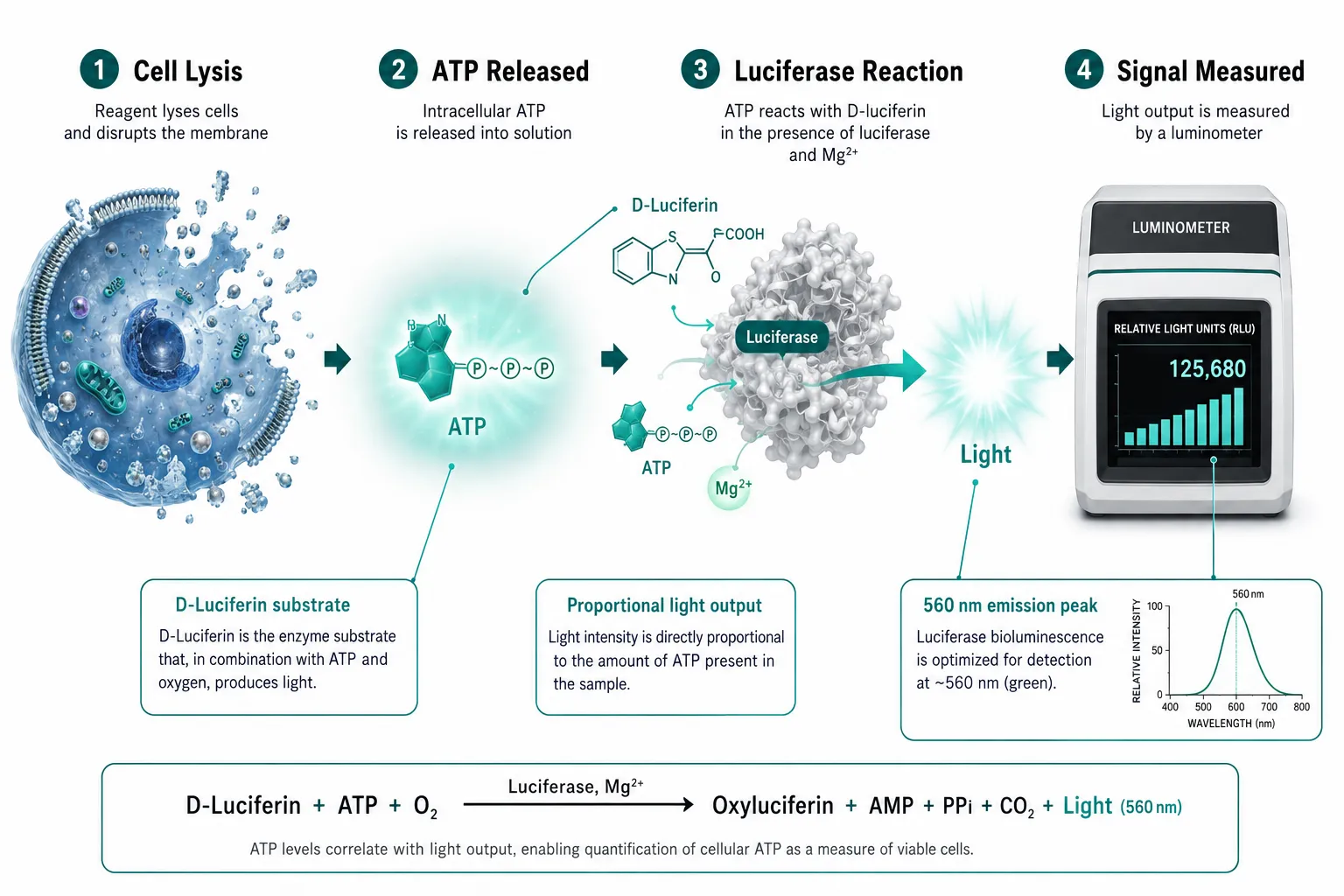
Adenosine triphosphate is not simply a fuel molecule, it is a real-time indicator of how well the entire oxidative phosphorylation chain is functioning. When mitochondria are stressed, damaged, or pharmacologically targeted, ATP output drops before most other measurable parameters shift. That sensitivity is exactly why intracellular ATP measurement is now established as a primary proxy for mitochondrial activity in research settings.
The dominant detection method is the luciferin-luciferase bioluminescence assay. Firefly luciferase catalyzes a reaction between D-luciferin and ATP, producing light. Because luminescence intensity is directly proportional to ATP concentration when ATP is the limiting reagent, the assay delivers a quantitative signal without requiring radioactive tracers or complex instrumentation. Most current protocols use a single working reagent that simultaneously lyses cells and generates luminescence, with measurements taken within one minute to prevent signal drift.
A standard workflow looks like this:
- Add equal volumes of sample and working solution to a 96-well plate.
- Incubate at 25 degrees Celsius for a fixed period (commonly 10 minutes per validated protocols).
- Read luminescence immediately to capture peak signal before kinetic decay.
Timing matters. Even a two-minute delay after adding the reaction mixture can introduce measurable error. Researchers building mitochondrial assay panels must treat the ATP readout as a time-sensitive endpoint, not a stable colorimetric measurement.
One additional consideration is dynamic range. When cell density is high or mitochondrial activity is elevated, the luminescent signal can saturate. Adjusting substrate volume or diluting lysate before adding the luciferase reagent is standard practice to keep measurements within the linear range of the assay.
Compartment-Specific Measurement and the Role of Cellular Energy and Research Peptides in Mitochondrial Studies
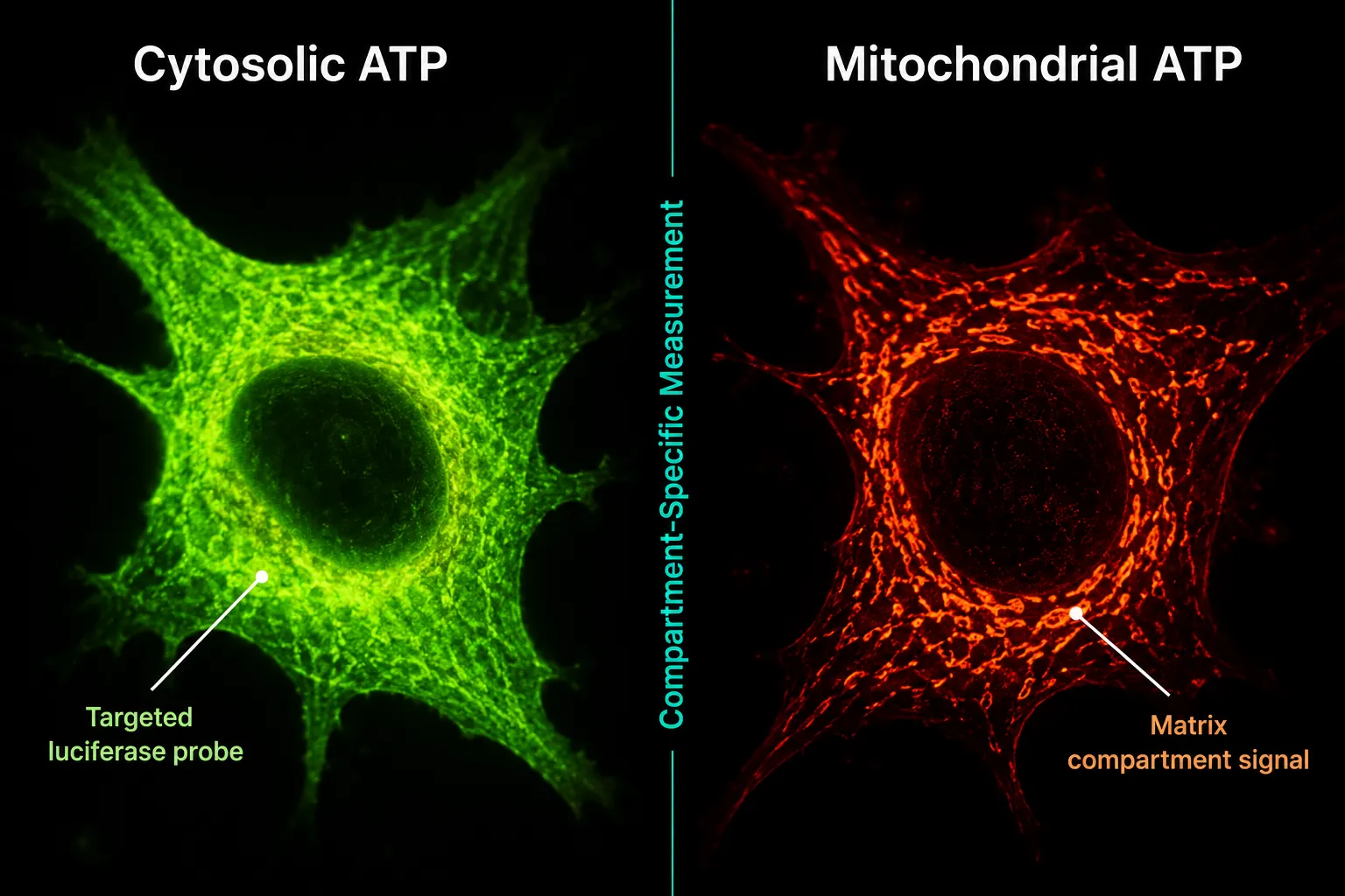
A whole-cell ATP readout captures the sum of all nucleotide pools, cytosolic, mitochondrial, and pericellular. For many screening applications that aggregate signal is sufficient. But when the research question is specifically about how a peptide alters mitochondrial energy production, a whole-cell number can obscure the answer.
Targeted luciferase chimeras solve this problem. By fusing a luciferase gene to a mitochondrial matrix-targeting sequence, researchers can direct the reporter protein to a specific subcellular compartment. Luminescence from that probe reflects only the ATP pool in that location. The same strategy works for the cytosol and pericellular space, enabling simultaneous multi-compartment profiling across multi-day experiments.
This level of resolution matters for evaluating SS-31 mitochondrial dynamics because the peptide's proposed mechanism involves direct interaction with cardiolipin in the inner mitochondrial membrane. A whole-cell ATP assay might show a modest aggregate increase, while a matrix-targeted probe could reveal a substantially larger improvement confined to the mitochondrial compartment, a distinction with real mechanistic significance.
Beyond single-nucleotide assays, dual-detection platforms now allow simultaneous quantitation of both GTP and ATP from the same sample well. This matters because GTP is a direct product of the TCA cycle succinyl-CoA synthetase reaction, making it an independent indicator of mitochondrial metabolic flux. Combining GTP and ATP readouts in a single luminescent assay provides a more complete picture of cellular energy metabolism than either measurement alone.
For researchers mapping metabolic pathway dependency, ATP assays also help distinguish how much a cell relies on glycolysis versus oxidative phosphorylation. By selectively inhibiting one pathway and measuring the ATP response, investigators can characterize a cell line's bioenergetic phenotype, information that is directly relevant when screening peptide candidates for metabolic effects. Readers exploring that intersection may find the top 5 research peptides for metabolic health guide a useful companion resource.
Translating ATP Readouts to Peptide Research: Clinical and Preclinical Implications
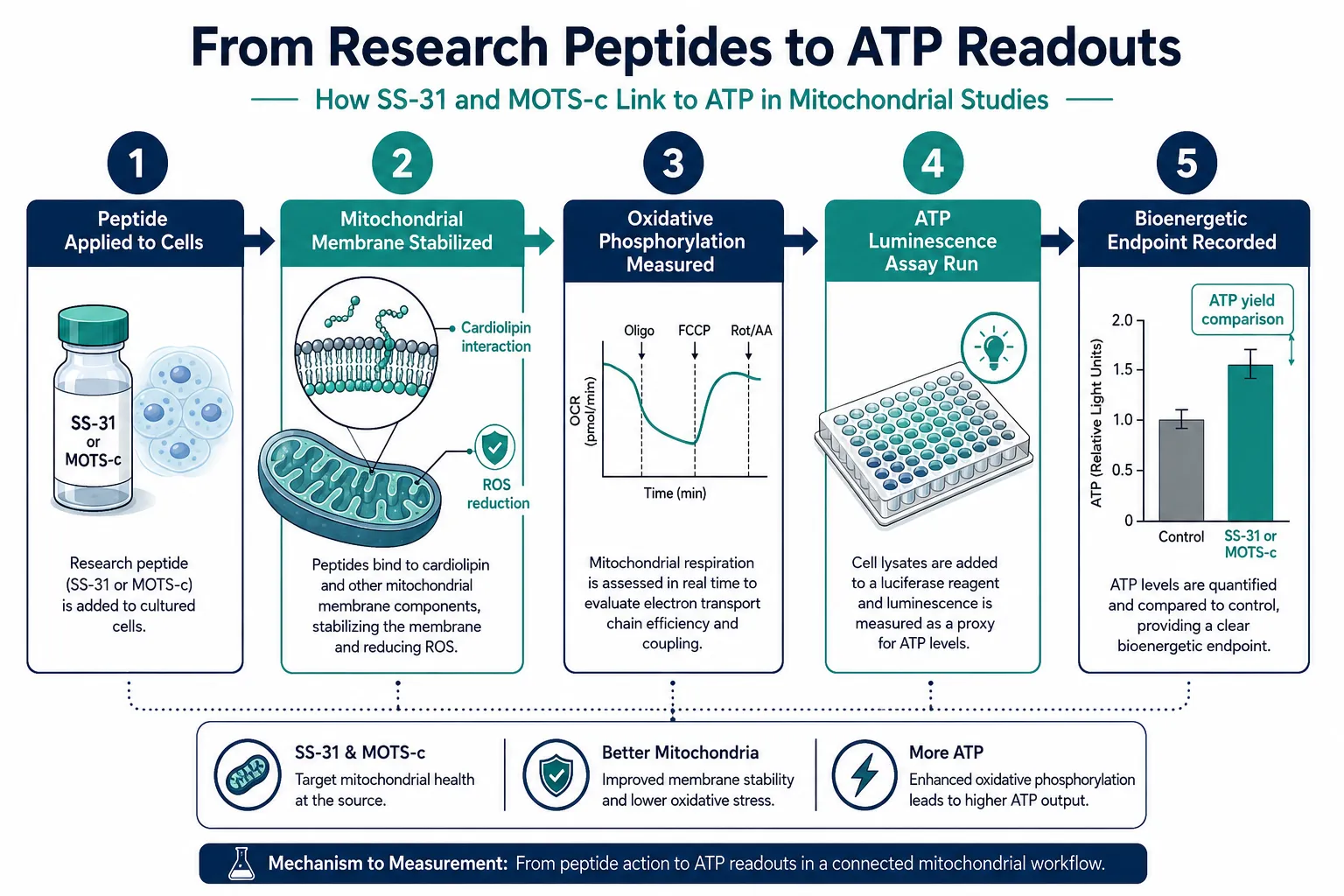
The question of why ATP readouts matter in mitochondrial studies becomes most concrete when examining how mitochondrial-targeted peptides are actually evaluated in research programs. Two peptides illustrate the point clearly.
Elamipretide (SS-31) is a small, cell-permeable tetrapeptide that associates with cardiolipin in the inner mitochondrial membrane. Preclinical data consistently show that it improves mitochondrial respiration, reduces reactive oxygen species, and enhances ATP production. In clinical heart failure trials, even when primary endpoints such as infarct size reduction were not met, improvements in mitochondrial function and reductions in cardiac injury biomarkers were observed. This pattern suggests that ATP-linked bioenergetic measures may be more sensitive indicators of therapeutic effect than some anatomical endpoints. Two ongoing Phase 3 trials, ReNEW and ReGAIN, are expected to report data in 2026, and bioenergetic endpoints will be central to interpreting those results. Researchers can review the SS-31 mechanism and research overview for additional background on its mitochondrial targets.
MOTS-c is a mitochondria-encoded peptide that has entered human trials, with Phase 1 completion projected for mid-2026 and Phase 2 initiation anticipated later in the year. Mitochondrial respiratory capacity is a planned endpoint, and ATP and respiration measures are expected to quantify metabolic responses. The SS-31 mitochondrial research themes resource provides relevant context on how mitochondrial endpoints are structured across similar peptide programs.
The broader implication is that ATP assays are transitioning from purely preclinical screening tools to clinically meaningful biomarkers. As late-2026 trial data accumulate, consistent improvements in ATP-linked markers alongside clinical outcomes would further validate ATP readouts as surrogate endpoints for mitochondrial peptide therapies.
For research teams sourcing compounds for these studies, working with lab-tested peptides ensures that purity data are available to separate compound-related effects from assay artifacts, a non-trivial concern when ATP luminescence is the primary readout.
| Peptide | Primary Mitochondrial Target | ATP-Related Endpoint | Trial Stage (2026) |
|---|---|---|---|
| Elamipretide (SS-31) | Cardiolipin / inner membrane | ATP production, ROS reduction | Phase 3 (ReNEW, ReGAIN) |
| MOTS-c | Mitochondrial genome / AMPK | Respiratory capacity, insulin sensitivity | Phase 1 completion / Phase 2 initiation |
Conclusion
ATP concentration is not a peripheral metric in mitochondrial research, it is the most direct, quantifiable signal of whether the organelle is doing its job. The luciferin-luciferase platform has made high-throughput ATP measurement practical, but reliable data require strict attention to timing, dynamic range, and compartment specificity. As the fields of cellular energy and research peptides converge more tightly, ATP readouts are becoming the common language between bench assays and clinical endpoints.
Actionable next steps for research teams:
- Validate assay timing protocols before comparing treatment groups; even small delays introduce quantitative error.
- Consider compartment-targeted luciferase probes when the research question is specifically about mitochondrial (not total cellular) ATP.
- Pair ATP assays with GTP or oxygen consumption measurements to capture a fuller bioenergetic profile.
- When evaluating mitochondrial peptides such as SS-31, design studies to capture ATP-linked secondary endpoints alongside primary anatomical or functional measures.
- Source compounds from verified suppliers and review the SS-31 kidney health research literature to understand how ATP endpoints have been applied across different tissue models.
The 2026 clinical readouts from ongoing mitochondrial peptide trials will test whether ATP-based bioenergetic markers can carry the weight of surrogate endpoints. The methodology to support that claim is already in place.