

How to Read Hemoglobin, Hematocrit, and MCHC Results in Peptide Research Studies
A single out-of-range CBC value can derail an entire peptide research protocol, yet most of those flags are artifacts, not true biological signals. Knowing how to read hemoglobin, hematocrit, and MCHC results in peptide research studies is therefore one of the most practical skills a researcher can develop. These three indices are tightly interconnected, and misreading one in isolation leads to faulty conclusions about safety and efficacy.
Key Takeaways
- Hemoglobin (Hb), hematocrit (Hct), and MCHC are mathematically linked; changes in one almost always shift the others.
- MCHC is primarily a quality-control flag, a value above 36 g/dL often signals a laboratory artifact rather than a true physiological change.
- Hct thresholds (typically above 54% in males or 48% in females) serve as intervention triggers in peptide research monitoring protocols.
- No single index should be interpreted in isolation; always cross-reference with MCV, RDW, and reticulocyte count.
- Monitoring cadence matters: baseline, four-week, and twelve-week CBC panels are the current consensus practice for most peptide study designs.
Understanding the Three Core Red-Cell Indices
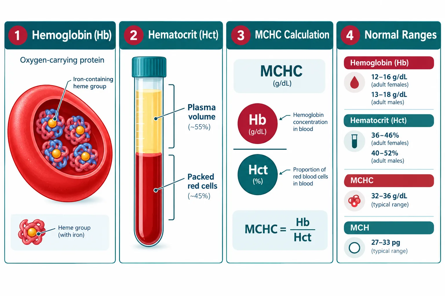
Hemoglobin (Hb) is the iron-containing protein inside red blood cells that carries oxygen. It is measured in grams per deciliter (g/dL). Standard reference ranges are approximately 13.5-17.5 g/dL for adult males and 12.0-15.5 g/dL for adult females, though laboratory-specific ranges vary slightly.
Hematocrit (Hct) expresses the proportion of whole blood occupied by red blood cells, reported as a percentage. Normal values run roughly 41-53% for males and 36-46% for females. Because Hct is essentially three times the Hb value in healthy subjects, a large discrepancy between the two is itself a diagnostic clue.
MCHC (Mean Corpuscular Hemoglobin Concentration) measures the average concentration of hemoglobin within a given volume of packed red cells. It is calculated as:
MCHC (g/dL) = (Hb / Hct) x 100
Normal MCHC falls between 32 and 36 g/dL. Because hemoglobin physically cannot exceed its solubility limit inside a red cell, a result above 36-37 g/dL almost always indicates a pre-analytical error, lipemia, hemolysis, or a cold agglutinin, rather than a genuine finding.
Why These Three Indices Are Inseparable
The mathematical relationship among Hb, Hct, and MCHC means they must be read as a set. If Hb rises due to hemoconcentration during a peptide protocol, Hct will rise proportionally and MCHC will remain stable. If MCHC spikes while Hb and Hct stay flat, the sample is almost certainly compromised.
A Practical Workflow for Reading CBC Results in Peptide Studies
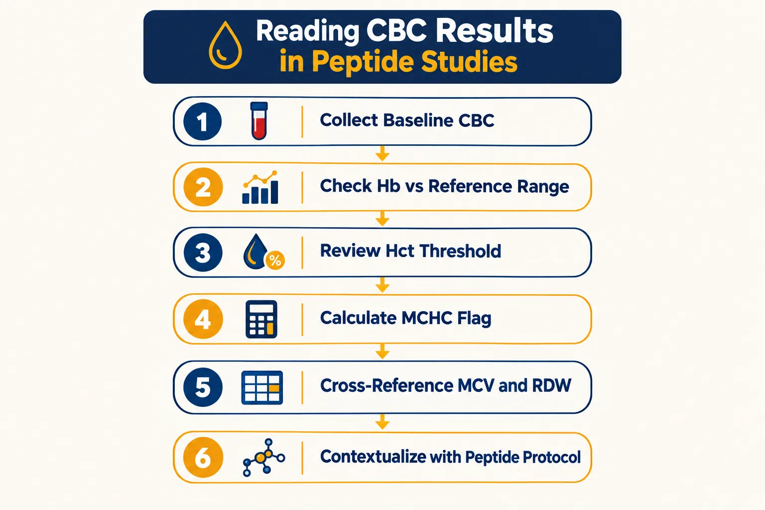
Researchers working with compounds such as SS-31 peptide or Ipamorelin peptide for sale formulations routinely collect complete blood counts (CBCs) to monitor hematological safety. The following six-step workflow reflects 2026 consensus practice.
Step 1, Establish a true baseline. Collect a CBC before any compound is introduced. A single pre-study value is insufficient; two baseline draws separated by at least one week reduce intra-individual variability noise.
Step 2, Evaluate Hb against sex-specific reference ranges. Flag values more than 1.5 g/dL above the upper reference limit. In peptide studies involving erythropoietic compounds, rising Hb is the earliest signal of red-cell mass expansion.
Step 3, Apply Hct thresholds as intervention triggers. Current guidance treats Hct above 54% (males) or 48% (females) as a threshold requiring protocol pause and clinical review. These numbers align with cardiovascular risk data from polycythemia research.
Step 4, Use MCHC as a quality-control gate, not a primary endpoint. Before interpreting any MCHC shift, confirm sample integrity. An MCHC above 36.5 g/dL should trigger a repeat draw before any protocol decision is made.
Step 5, Cross-reference MCV and RDW. Mean corpuscular volume (MCV) and red-cell distribution width (RDW) add context. A rising Hb with normal MCV and low RDW suggests true erythrocytosis. A rising Hb with low MCV and high RDW points toward iron-deficiency compensation, a very different finding.
Step 6, Contextualize within the peptide protocol timeline. Some compounds, including those explored in SS-31 mitochondrial research, affect cellular energy metabolism in ways that may indirectly influence reticulocyte production. Timing the CBC draw relative to dosing schedule matters.
Monitoring Cadence
| Timepoint | Recommended Panel |
|---|---|
| Baseline (pre-study) | Full CBC with differential, iron studies |
| Week 4 | CBC with differential |
| Week 12 | Full CBC, iron studies, reticulocyte count |
| Any symptom event | Immediate CBC plus MCHC quality check |
Avoiding Overinterpretation: MCHC as a Quality-Control Flag
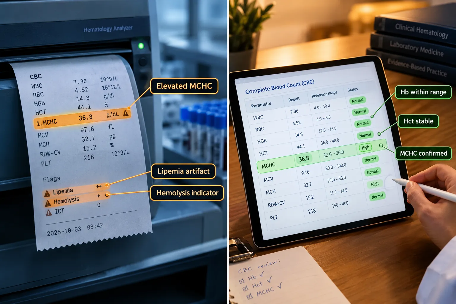
The most common error in reading hemoglobin, hematocrit, and MCHC results in peptide research studies is treating every flagged value as a true biological event. MCHC is the index most vulnerable to this mistake.
Common pre-analytical artifacts that falsely elevate MCHC:
- Lipemia (high triglycerides interfere with spectrophotometric Hb measurement)
- In-vitro hemolysis from rough sample handling
- Cold agglutinins causing red-cell clumping
- Delayed sample processing beyond four hours
When sourcing compounds from a best peptide supplier for structured trials, researchers should also document sample collection procedures as rigorously as they document dosing. A poorly handled blood draw produces data that looks like a hematological crisis but is simply a laboratory error.
Low MCHC (hypochromia, below 32 g/dL) is more likely to be genuine. It points toward iron deficiency, thalassemia trait, or chronic disease anemia, all of which can confound peptide study endpoints and must be resolved before attributing changes to the compound under investigation.
Researchers exploring compounds such as those detailed in SS-31 mechanism and research should note that mitochondrial-targeted peptides may influence oxidative stress markers that interact with red-cell membrane integrity, making clean sample handling even more critical.
The Integration Principle
No single CBC index is a standalone decision-maker. Hb confirms oxygen-carrying capacity. Hct confirms red-cell mass. MCHC confirms data quality. Together, they form a three-point verification system. Researchers working with wholesale peptides across multi-subject trials should build this three-point check into their standard operating procedures.
Conclusion
Reading hemoglobin, hematocrit, and MCHC results in peptide research studies requires a systematic, index-by-index approach rather than a reflexive response to any single out-of-range flag. The practical next steps are clear:
- Set sex-specific baselines before any protocol begins, using two draws to reduce variability.
- Apply the Hct threshold rule (54%/48%) as the primary intervention trigger, not MCHC alone.
- Treat elevated MCHC as a quality-control alert first, repeat the draw before drawing any biological conclusion.
- Always cross-reference MCV, RDW, and reticulocyte count to distinguish true erythrocytosis from iron-deficiency compensation.
- Document sample handling procedures with the same rigor applied to dosing logs.
Researchers who build this workflow into their CBC review process will produce cleaner data, avoid false protocol pauses, and generate findings that hold up to scrutiny. As peptide research continues to mature in 2026, standardized hematological monitoring will increasingly separate rigorous studies from anecdotal reports.