

Epithalon Research: Comparing Telomerase, DNA-Damage, and Senescence Assays Across Cell Models
Telomerase activity, telomere length, gamma-H2AX foci, p16/p21 expression, and replicative senescence are five distinct biological readouts, yet researchers studying Epithalon frequently treat them as interchangeable proxies for the same outcome. They are not. Each measures a different node in the aging cascade, and conflating them produces misleading conclusions. A rigorous look at Epithalon research: comparing telomerase, DNA-damage, and senescence assays across cell models reveals exactly where the evidence is strong, where it is thin, and what future studies must address.
Key Takeaways
- Epithalon consistently activates telomerase (TRAP assay, hTERT mRNA) in normal human fibroblast and epithelial models, with fold-changes of roughly 2- to 12-fold depending on dose and cell type.
- In malignant cell lines, Epithalon extends telomeres primarily through the alternative lengthening of telomeres (ALT) pathway, not by further boosting telomerase catalytic activity, making TRAP-only assay designs inadequate for cancer models.
- DNA-damage endpoints (gamma-H2AX, chromosomal aberration counts) show benefit in mouse oocyte and senescence-accelerated animal models but remain largely unmeasured in human somatic cell studies.
- Senescence markers such as SA-beta-galactosidase staining and p16/p21 protein expression are rarely co-reported with telomerase data, leaving a critical gap in mechanistic understanding.
- As of 2026, all robust Epithalon data remain preclinical; no completed human trial has systematically measured telomere or senescence outcomes under controlled conditions.
Why Assay Choice Defines What Epithalon Research Can Conclude
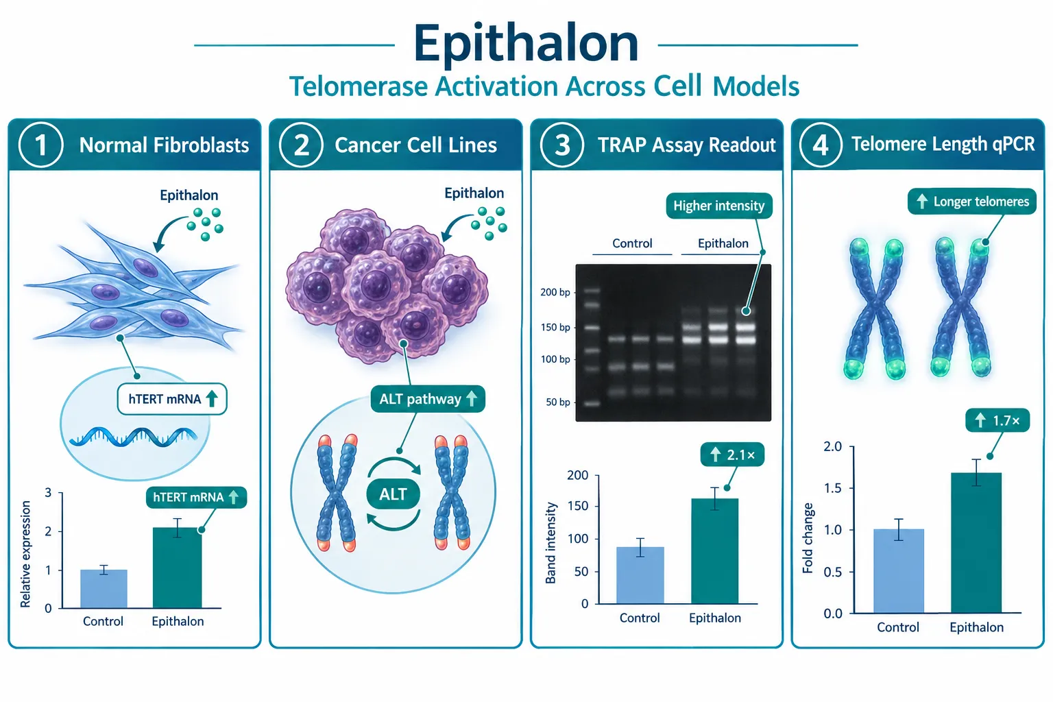
The tetrapeptide Epithalon (Ala-Glu-Asp-Gly) was first characterized in pineal gland extracts and has since become one of the most studied peptides in cellular aging research. The bulk of Epithalon research: comparing telomerase, DNA-damage, and senescence assays across cell models shows that most published work clusters around one assay type, the telomeric repeat amplification protocol (TRAP), while other endpoints receive far less systematic attention.
Why this matters: TRAP measures the catalytic activity of the telomerase enzyme complex. It does not measure telomere length, DNA strand-break burden, or whether a cell has entered stable growth arrest. A peptide could theoretically raise TRAP signal while leaving telomere length unchanged, or could reduce DNA damage without touching telomerase at all. Treating a TRAP result as evidence of "anti-aging" broadly defined is a category error.
Telomerase Assays: The Strongest Evidence Base
A 2025 Biogerontology study conducted at Brunel University London provided the most rigorous independent data to date. In normal human fibroblast and epithelial cells, Epithalon dose-dependently increased hTERT mRNA by up to approximately 12-fold at 1 microgram per milliliter in one cell line, with corresponding increases in TRAP-measured telomerase activity and telomere lengthening confirmed by both qPCR and immunofluorescence. A correction published later in 2025 clarified figure labeling issues but explicitly upheld all main quantitative conclusions, reinforcing confidence in the core findings.
Earlier work in human somatic fibroblasts reported that Epithalon extended average telomere length by approximately 33.3% and pushed proliferative lifespan from roughly 34 passages to more than 44, well beyond the classical Hayflick limit. Independent laboratory replication in fibroblast, endothelial, and mesenchymal stem cell cultures has produced roughly 2.0- to 3.2-fold TRAP activation, closely matching published values and suggesting reasonable technical reproducibility across labs.
Key point: Fetal lung fibroblast cultures exposed to Epithalon at concentrations between 20 nM and 2 micromolar showed approximately 2- to 3-fold TRAP increases versus untreated controls. These telomerase changes connect mechanistically to replicative senescence thresholds via the ATM/ATR-p53-p21 DNA-damage response pathway, but that connection is inferred from pathway biology, not from co-measured senescence markers in the same experiments.
"A TRAP result tells you the enzyme is more active. It does not tell you whether the cell has escaped senescence, repaired its DNA breaks, or changed its inflammatory secretome."
DNA-Damage Assays: A Narrower but Informative Evidence Window
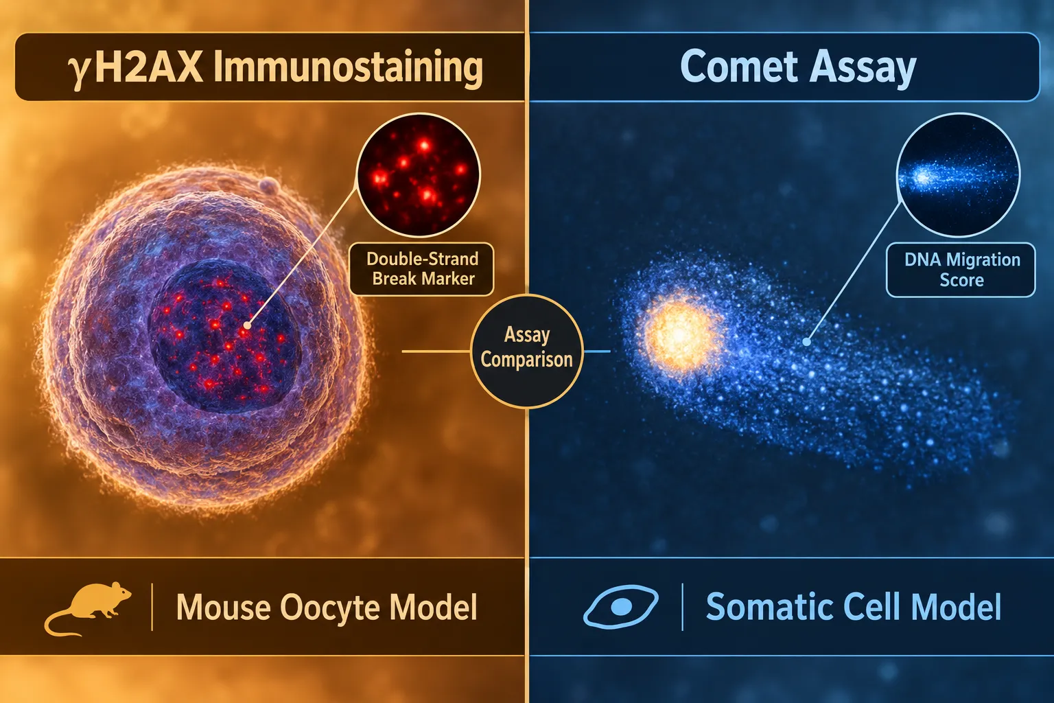
DNA-damage readouts in Epithalon research are concentrated in two model systems: mouse oocytes undergoing in-vitro post-ovulatory aging, and senescence-accelerated mouse strains. Neither maps directly onto human somatic cell telomere biology, which is an important limitation.
In the oocyte model, adding Epithalon during aging reduced gamma-H2AX foci, a validated marker of double-strand DNA breaks, lowered apoptosis rates, and improved spindle and chromosome morphology. These findings are meaningful because they show the peptide can act on DNA-integrity endpoints independently of a pure telomerase mechanism. Gamma-H2AX immunostaining and apoptosis assays are well-validated, quantitative, and reproducible, making this a technically sound data set even if the cell type is specialized.
In senescence-accelerated mouse models, Epithalon treatment reduced the incidence of chromosome aberrations by approximately 17.9 to 30.1 percent compared to age-matched controls. Cytogenetic assays measuring chromosomal instability are a legitimate DNA-damage endpoint, and this magnitude of effect is biologically meaningful.
What is missing: The comet assay (single-cell gel electrophoresis), 8-OHdG oxidative DNA-damage quantification, and 53BP1 foci analysis have not been systematically reported in Epithalon studies. These assays would allow researchers to distinguish between double-strand break repair, base-excision repair, and oxidative damage pathways, distinctions that matter for understanding mechanism.
| Assay | Reported in Epithalon Studies | Cell/Animal Model Used |
|---|---|---|
| TRAP (telomerase activity) | Yes, extensively | Human fibroblasts, epithelial cells, endothelial cells |
| hTERT mRNA (qPCR) | Yes | Human fibroblasts, epithelial cells |
| Telomere length (qPCR/IF) | Yes | Human fibroblasts, breast cancer lines |
| gamma-H2AX foci | Yes, limited | Mouse oocytes |
| Chromosomal aberration count | Yes, limited | Senescence-accelerated mice |
| SA-beta-galactosidase staining | Rarely reported | Not systematically published |
| p16/p21 protein expression | Rarely reported | Not systematically published |
| Comet assay | Not reported | — |
Senescence Markers and the Gap That Most Studies Leave Open
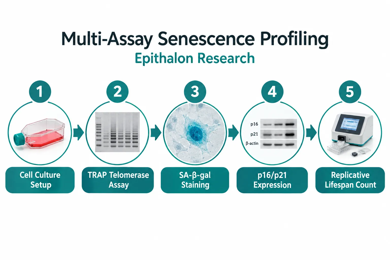
Cellular senescence is not simply "short telomeres." A cell enters stable senescence when multiple reinforcing signals converge: telomere dysfunction triggers ATM/ATR kinases, which stabilize p53, which induces p21 (CDKN1A), which blocks CDK2 and halts the cell cycle. Separately, p16 (CDKN2A) inhibits CDK4/6 independently of telomere length. SA-beta-galactosidase activity at pH 6.0 is a widely used cytochemical marker of this arrested state. None of these endpoints are routinely co-reported in published Epithalon studies alongside TRAP data.
This is the central methodological gap in Epithalon research: comparing telomerase, DNA-damage, and senescence assays across cell models. A study that shows telomerase activation without measuring p16, p21, SA-beta-gal, or long-term growth arrest cannot confirm that telomerase activation translated into delayed senescence. The mechanistic chain is plausible, and the proliferative lifespan data from fibroblast passage studies supports it indirectly, but direct senescence marker co-measurement is largely absent.
Researchers and labs sourcing lab-tested peptides for cell-based work should design experiments that pair TRAP assays with at least SA-beta-gal staining and p21 immunoblotting in the same culture wells. This design would allow direct correlation between telomerase activation magnitude and senescence entry delay, producing far more interpretable data than single-endpoint studies.
For context on how complementary peptides with mitochondrial and oxidative-stress mechanisms are studied alongside telomere-focused compounds, the SS-31 mechanism and research overview provides a useful parallel. Similarly, SS-31 kidney health research demonstrates how multi-endpoint organ-level studies are structured when a single mechanism does not capture the full biological picture.
Cancer Cell Models Require ALT Marker Inclusion
The 2025 Biogerontology study identified a critical model-specific finding: in breast cancer cell lines, Epithalon extended telomeres primarily through the alternative lengthening of telomeres (ALT) pathway rather than by further increasing telomerase catalytic activity. This means that a TRAP-only assay design in malignant models will systematically underestimate, or entirely miss, the telomere-lengthening effect. ALT activity should be confirmed with C-circle assay, APB (ALT-associated PML bodies) immunofluorescence, or telomere sister chromatid exchange (T-SCE) analysis whenever cancer-derived lines are used.
For researchers exploring peptide purity standards relevant to cell-based assay reliability, the GHK-Cu peptide purchase and copper peptide research sourcing guide and the research-grade Glow Blend peptide evaluation guide both address how impurity profiles can confound cell-model outcomes, a concern equally relevant to Epithalon studies.
Conclusion
The evidence base for Epithalon is more coherent than critics suggest and more limited than proponents often claim. Telomerase activation in normal human fibroblasts and epithelial cells is the best-supported finding, with independent replication and a plausible mechanistic pathway. DNA-damage reduction is supported in specialized models but not yet in human somatic cell systems. Senescence marker data is the weakest link across the entire literature.
Actionable next steps for researchers:
- Design multi-endpoint experiments. Pair every TRAP assay with SA-beta-gal staining, p21 immunoblot, and a telomere-length measurement in the same culture condition.
- Add ALT markers in any cancer-derived model. C-circle assay or APB immunofluorescence should accompany TRAP when using malignant lines.
- Include DNA-damage co-readouts in human somatic models. Gamma-H2AX and 53BP1 foci analysis would bridge the gap between the oocyte/mouse data and human cell findings.
- Prioritize normal stem cell and immune cell models. Mesenchymal stem cells and T-lymphocytes are biologically relevant to systemic aging and remain understudied in this context.
- Use verified, high-purity research-grade material. Assay reproducibility depends on peptide integrity; impurity-driven artifacts are a known confounder in low-concentration cell-culture work.
As of 2026, no human clinical trial has systematically measured telomere length or telomerase activity before and after Epithalon under controlled conditions. Until that data exists, Epithalon remains a mechanistic research tool of genuine scientific interest, not an established therapeutic. The path from promising preclinical signal to validated intervention runs directly through the multi-assay, multi-model experimental designs described above.