

GLP-3 Retatrutide vs GLP-2-T: Choosing Metabolic, Intestinal, and Organoid Readouts for Comparative Research
Retatrutide produced mean weight reductions exceeding 24% over 48 weeks in phase 2 trials, a figure that no single-receptor incretin therapy had previously achieved. That single data point signals why researchers face a genuine decision problem when designing comparative studies: retatrutide operates through a fundamentally different biological model than GLP-2-type agents, and conflating their readout frameworks produces unreliable conclusions.
This article builds a practical decision framework for GLP-3 Retatrutide vs GLP-2-T: Choosing Metabolic, Intestinal, and Organoid Readouts for Comparative Research. The goal is to separate retatrutide's multi-receptor systemic model from GLP-2-T's intestinal-repair context, then map each to the endpoints, assay systems, and interpretation boundaries that fit them best.
Key Takeaways
- Retatrutide simultaneously activates GLP-1R, GIPR, and GCGR, making systemic metabolic endpoints the primary readout domain.
- GLP-2-type agents act principally on intestinal GLP-2 receptors; their readouts center on mucosal structure, barrier function, and organoid proliferation.
- Mixing readout frameworks across these two agent classes introduces mechanistic confounds that weaken comparative data.
- Receptor expression confirmation is a non-negotiable first step before running any parallel assay.
- Organoid platforms can serve both arms only when receptor expression and endpoint domains are kept strictly separate.
Understanding the Mechanistic Divide
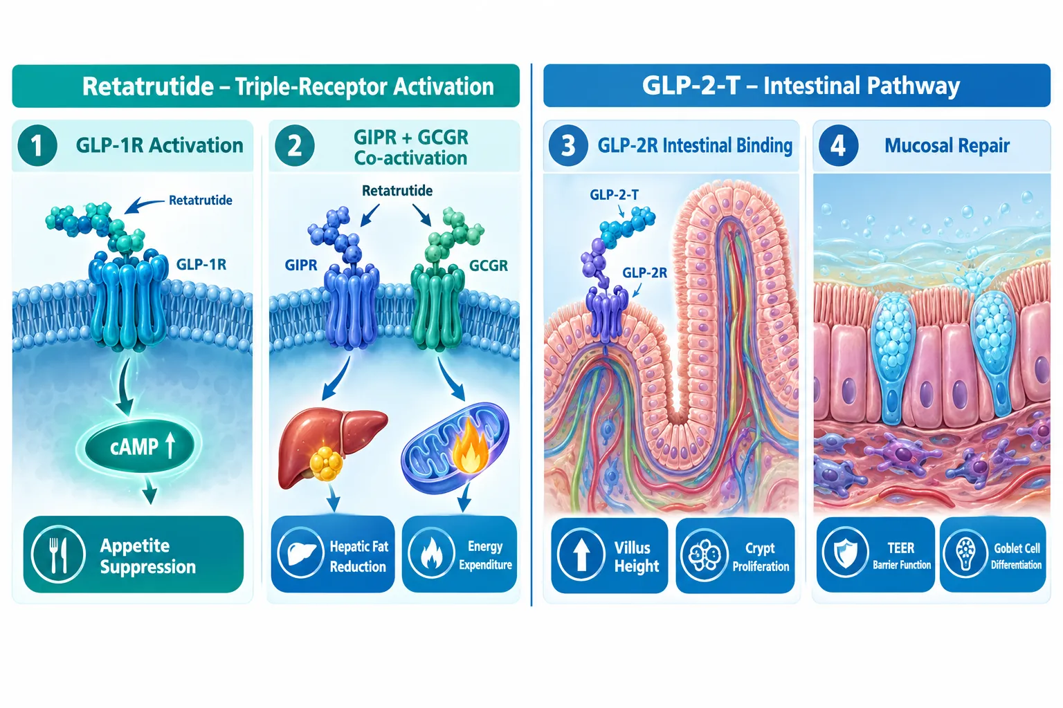
The core challenge in GLP-3 Retatrutide vs GLP-2-T comparative research is that these two agent classes do not share a primary tissue target. Retatrutide (LY3437943) is the first GLP-1/GIP/glucagon triple agonist to complete phase 2 trials in both obesity and type 2 diabetes. It engages three distinct receptors, GLP-1R, GIPR, and GCGR, through a single peptide conjugated to a fatty diacid moiety. The downstream effects span appetite suppression, hepatic fat reduction, and systemic energy expenditure.
GLP-2-type agents (GLP-2-T), by contrast, bind the GLP-2 receptor expressed predominantly on intestinal subepithelial myofibroblasts and enteroendocrine cells. Their documented effects are gut-trophic: increased villus height, crypt cell proliferation, improved mucosal barrier integrity, and enhanced nutrient absorption. These are the readouts that define clinical use cases such as short bowel syndrome and intestinal failure.
The mechanistic divide is not a matter of degree, it is a matter of tissue domain. Retatrutide's phase 2 MASLD trial reinforced this by demonstrating meaningful reductions in liver fat measured by MRI-proton density fat fraction (MRI-PDFF), alongside improvements in hepatic enzymes and non-invasive fibrosis markers. None of those endpoints are relevant to GLP-2-T's primary mechanism.
"Choosing the wrong readout domain does not just weaken a study, it actively misrepresents the biology of both agents."
Researchers looking to source well-characterized material for this work can review options through the Buy Retatrutide Peptide page or explore the broader GLP-3 Retatrutide buy online USA resource for purity and specification details.
Building the Readout Decision Framework
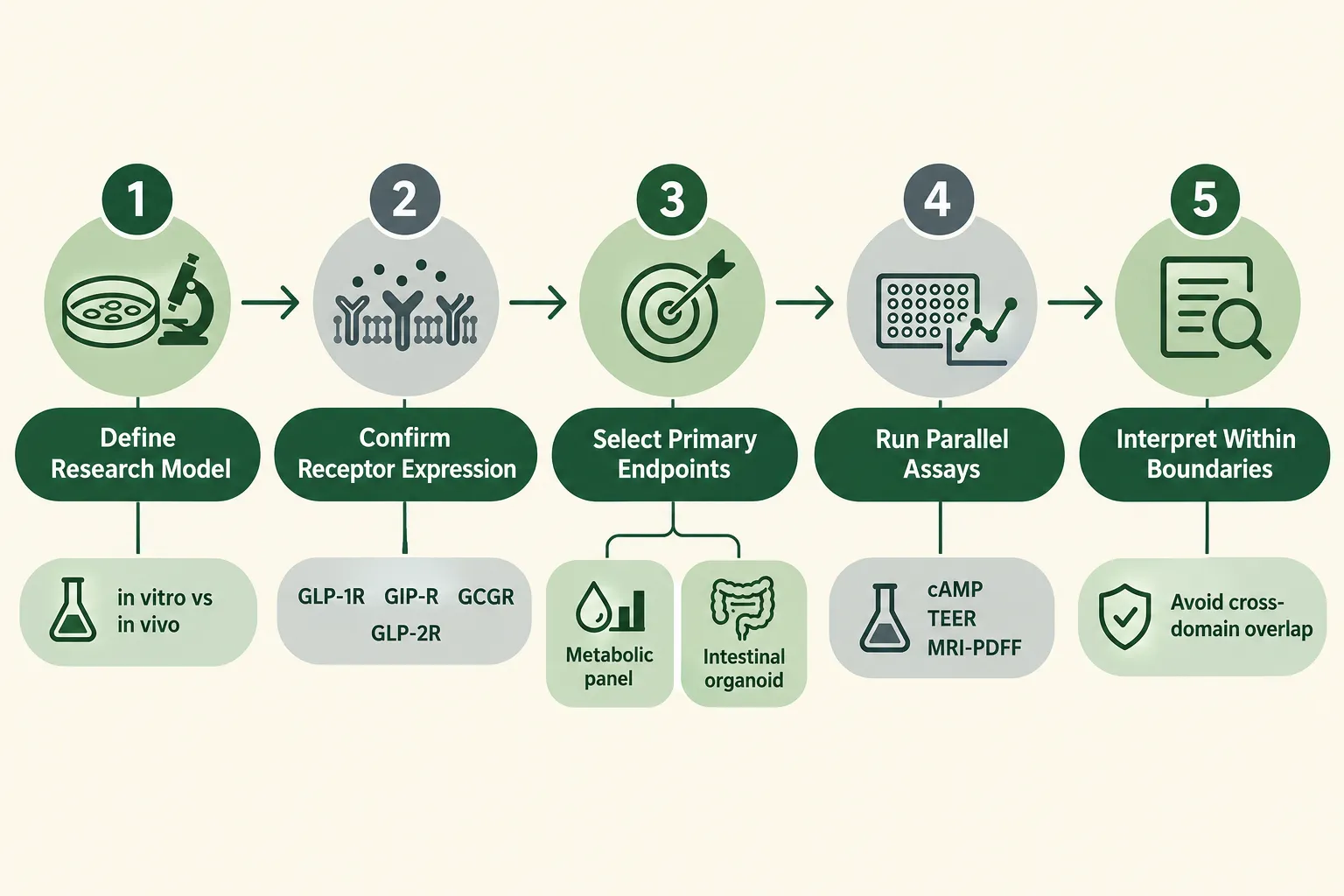
A structured five-step decision process reduces the risk of endpoint misalignment when designing GLP-3 Retatrutide vs GLP-2-T: Choosing Metabolic, Intestinal, and Organoid Readouts for Comparative Research studies.
Step 1: Define the Research Model
Choose whether the study is in vitro, ex vivo, or in vivo before selecting any endpoint. Retatrutide's receptor profile requires models that co-express GLP-1R, GIPR, and GCGR, hepatocyte lines, adipocyte cultures, pancreatic islet preparations, or diet-induced obesity rodent models. GLP-2-T studies require intestinal epithelial models: primary organoids derived from small intestinal crypts, Caco-2 monolayers, or murine intestinal preparations.
Step 2: Confirm Receptor Expression
This step is non-negotiable. Structural work published in 2024 clarified that retatrutide engages all three receptors with defined binding modes and biased signaling profiles. Before running any assay, confirm receptor expression using qPCR or immunofluorescence. A GLP-2-T study run in a model lacking adequate GLP-2R expression produces noise, not data.
Step 3: Select Primary Endpoints by Agent Class
| Endpoint Category | Retatrutide (GLP-3-like) | GLP-2-T |
|---|---|---|
| Primary metabolic | Body weight, HbA1c, fasting glucose, lipids | Not applicable as primary |
| Hepatic | MRI-PDFF, ALT/AST, fibrosis markers | Minimal relevance |
| Intestinal structure | Secondary / safety monitoring | Villus height, crypt depth, cell proliferation |
| Barrier function | GI tolerability (adverse event tracking) | TEER, tight junction proteins (ZO-1, claudin) |
| Receptor signaling | cAMP, beta-arrestin, downstream kinase activity | cAMP via GLP-2R, PI3K/Akt pathway |
| Organoid readouts | Hepatic organoid fat accumulation, glucose uptake | Enterocyte and goblet cell differentiation, proliferation index |
Step 4: Run Parallel Assays With Separated Domains
Phase 2 safety data for retatrutide identified dose-dependent gastrointestinal adverse events, nausea and diarrhea, alongside signals for lean mass changes and modest heart rate increases. These findings make body composition and heart rate monitoring essential safety-related readouts for the retatrutide arm. They are not shared endpoints with GLP-2-T, which does not produce systemic energy expenditure effects. Running parallel assays only works when each arm's endpoint set remains internally consistent.
Step 5: Interpret Within Defined Boundaries
No published 2025-2026 literature presents a formal GLP-3 vs GLP-2-T comparative schema. The framework described here is constructed from separate evidence streams. Researchers must therefore resist interpreting GLP-2-T intestinal organoid results as informative about systemic metabolic efficacy, and vice versa.
For additional context on related peptide research methodologies, the SLUPP332 oral versus injectable research resource offers useful parallel considerations on route-of-administration variables that affect readout selection.
Organoid Platforms: Where the Two Frameworks Can Intersect
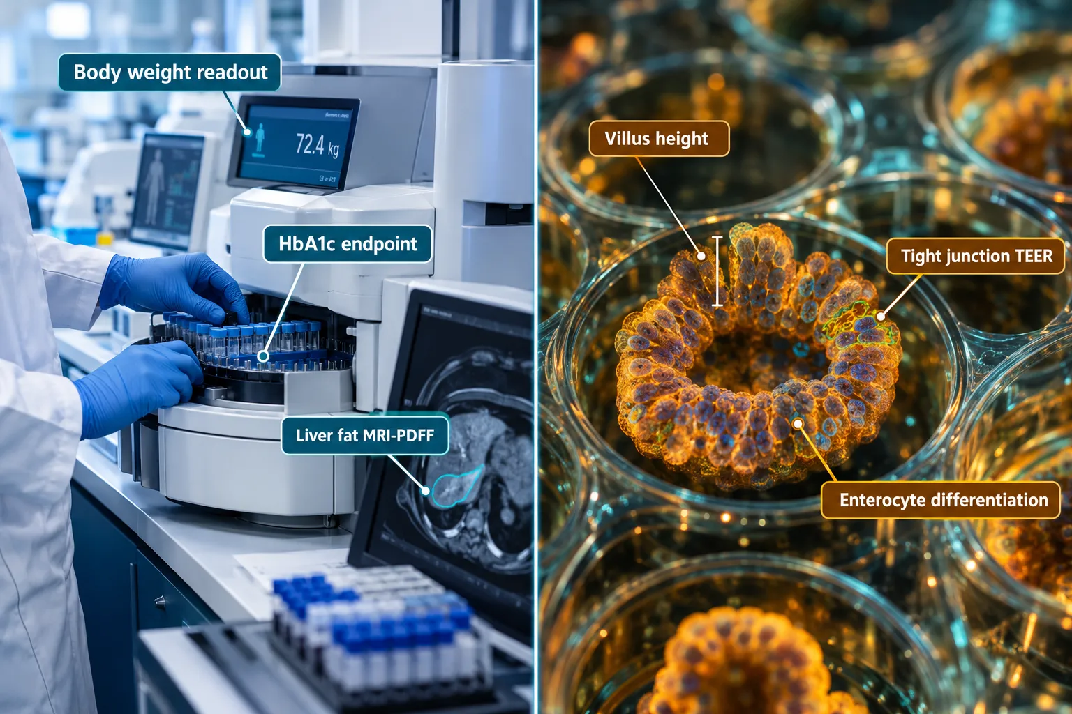
Organoid technology is the one research context where GLP-3 Retatrutide vs GLP-2-T: Choosing Metabolic, Intestinal, and Organoid Readouts for Comparative Research can share a platform, but only with strict compartmentalization.
For retatrutide, hepatic organoids derived from primary hepatocytes or iPSC-derived liver cells provide a controlled system for measuring fat accumulation (lipid droplet quantification), glucose output, and receptor-level signaling (cAMP production, beta-arrestin recruitment). These models mirror the liver-fat and glycemic endpoints validated in the MASLD phase 2a trial.
For GLP-2-T, intestinal organoids derived from small intestinal crypts are the gold-standard platform. Key readouts include:
- Organoid surface area expansion as a proxy for villus-like growth
- Goblet cell and enterocyte differentiation ratios
- Tight junction protein expression (ZO-1, occludin, claudin-3)
- Transepithelial electrical resistance (TEER) as a barrier integrity measure
- Nutrient transporter expression (SGLT1, GLUT2)
The intersection point is co-culture or multi-organ chip systems that link hepatic and intestinal compartments. In that setting, retatrutide's hepatic effects and GLP-2-T's intestinal effects can be observed in the same platform without conflating their primary readout domains. This remains a speculative but scientifically grounded direction for 2026 research programs.
Researchers sourcing material for organoid-based work should verify purity documentation carefully. The GLP3 RETA CAG 10mg for sale and GLP3 RETA CAG 10mg GA5 product pages provide specification details relevant to in vitro dosing decisions. For broader sourcing considerations, the GLP-3 Retatrutide buy online USA cheap resource addresses cost-quality tradeoffs that matter in multi-arm organoid studies.
Conclusion
The central lesson from aligning retatrutide's clinical trial evidence with GLP-2-T's intestinal biology is straightforward: these agents require separate readout frameworks, not a shared one. Retatrutide's primary endpoints are systemic, body weight, HbA1c, liver fat, lipids, and cardiometabolic markers. GLP-2-T's primary endpoints are structural and functional at the intestinal level, villus morphology, barrier integrity, and organoid proliferation.
Actionable next steps for research teams in 2026:
- Audit existing assay panels and remove any endpoints that do not align with the receptor biology of the agent being tested.
- Confirm GLP-1R, GIPR, and GCGR co-expression in every model used for retatrutide; confirm GLP-2R expression in every model used for GLP-2-T.
- Use MRI-PDFF, ALT/AST, and body composition as the metabolic arm's anchor endpoints.
- Use TEER, tight junction protein quantification, and organoid proliferation index as the intestinal arm's anchor endpoints.
- Consider multi-organ chip platforms only after single-domain validation is complete.
Maintaining this discipline protects the interpretive value of comparative data and positions research outputs for stronger translational relevance as triple-agonist therapies move through phase 3 development.