

Meloxicam and Peptide Research: A Practical Guide to COX Inhibition, Pain Scores, and Tissue-Repair Confounding
A single oral dose of meloxicam can suppress prostaglandin E2 levels by roughly 40% within eight hours, a biochemical shift significant enough to reshape the inflammatory environment that repair-focused peptides like BPC-157 and TB-500 are specifically designed to modulate. That overlap is the central problem this guide addresses.
This practical guide to meloxicam and peptide research covers COX inhibition, pain score interpretation, and tissue-repair confounding so that researchers can design cleaner studies and interpret existing data with far greater accuracy.
Key Takeaways
- Meloxicam preferentially inhibits COX-2 but meaningfully suppresses COX-1 as well, altering the same prostanoid signals that tissue-repair peptides target.
- Concurrent meloxicam use can reduce pain scores independently of any peptide effect, inflating apparent analgesic efficacy in research endpoints.
- Timing matters critically: late-phase COX-2 inhibition (days 11-20 post-injury) demonstrably impairs tendon load tolerance and may mask peptide-driven remodeling gains.
- Bone, tendon, and muscle repair models are all susceptible to meloxicam-induced confounding; each tissue type carries distinct mechanistic risks.
- Practical mitigation strategies, including analgesic stratification, topical-only NSAID policies, and covariate adjustment, can protect the integrity of peptide trial endpoints.
How COX Inhibition Reshapes the Peptide Research Baseline
Understanding the COX pathway is not optional background knowledge in peptide research, it is the foundation of every pain and repair endpoint used in translational models.
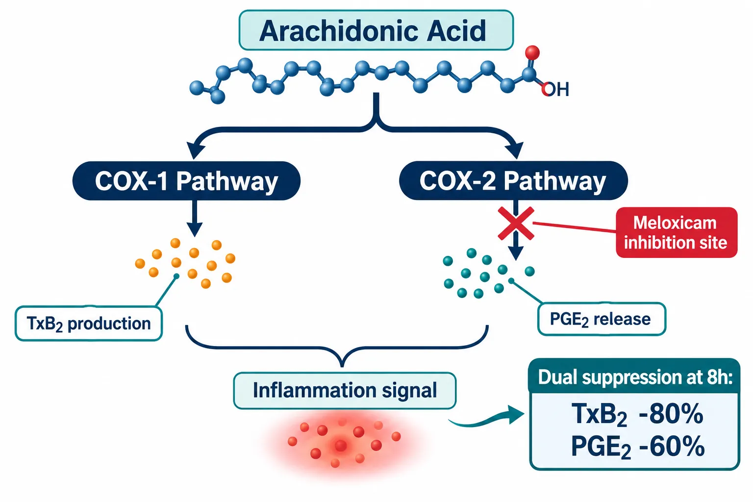
Meloxicam blocks cyclooxygenase enzymes that convert arachidonic acid into prostaglandins and thromboxanes. It has a preference for COX-2 but is not a selective COX-2 inhibitor in the strict pharmacological sense. Pharmacodynamic data from animal models confirm that a standard single dose reduces thromboxane B2 (a COX-1 product) to approximately 80% of baseline and prostaglandin E2 (a COX-2 product) to roughly 60% of baseline, both measured at the eight-hour mark. That dual suppression is consequential because prostaglandins E2 and I2 are not simply pain mediators; they are active regulators of angiogenesis, stem cell recruitment, and matrix remodeling.
Why this directly conflicts with peptide mechanisms:
- BPC-157 is documented in preclinical models to upregulate growth factor expression partly through prostanoid-sensitive signaling cascades. When meloxicam flattens the PGE2 signal, the upstream environment BPC-157 operates within is fundamentally altered. For a thorough breakdown of that mechanism, see the BPC-157 core peptides documentation and first research guide.
- TB-500 (thymosin beta-4) promotes actin polymerization and cell migration in a milieu shaped partly by prostanoid gradients.
- Peptides targeting mitochondrial integrity, such as those reviewed in SS-31 mitochondrial research, operate in an oxidative stress environment that COX-2 activity helps define.
Researchers who fail to account for background NSAID use are, in effect, running peptide trials on a biochemically altered baseline.
Tissue-Repair Confounding Across Bone, Tendon, and Muscle
The confounding risk is not uniform across tissue types. Each repair model carries a distinct mechanistic vulnerability tied to COX-2 biology.
Bone Repair
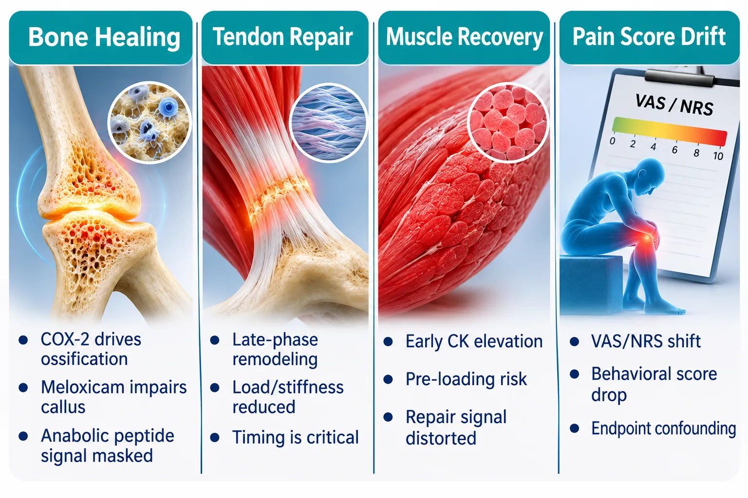
Endochondral ossification, the primary mechanism of fracture healing, depends on COX-2 activity during the cartilaginous callus phase. In rat fracture models, ten days of post-fracture meloxicam significantly impaired healing compared to controls. In vitro work confirms reduced bone nodule formation. For trials testing bone-anabolic peptides or growth factors, concurrent meloxicam use can suppress the very biological machinery the peptide is meant to stimulate, producing false-negative or attenuated efficacy signals.
Tendon and Soft Tissue
A well-characterized rotator cuff repair model found that meloxicam administered during the late proliferative and remodeling phase (days 11 through 20 post-surgery) significantly reduced both maximal load and stiffness at three weeks. Earlier administration was less detrimental. This timing dependency is critical: a peptide trial assessing connective tissue mechanical endpoints must specify exactly when NSAID co-administration occurred, or the biomechanical data are uninterpretable. Broader systematic reviews on soft-tissue healing echo this concern, noting that COX-2-selective agents can reduce tendon and ligament failure loads in controlled models.
Muscle Recovery
An eccentric exercise damage model found that early meloxicam administration, given before the damaging exercise bout, produced higher post-exercise creatine kinase (CK) levels than dosing 48 hours after damage. This suggests that premature COX-2 blockade can amplify the magnitude of muscle injury rather than limit it. For peptide studies targeting muscle repair or hypertrophy, pre-loading subjects with meloxicam before the damage stimulus could substantially inflate CK signals, making a peptide's subsequent effect appear smaller by comparison.
Key point: The mechanism of harm differs by tissue type, but the shared theme is consistent, meloxicam alters the inflammatory signaling environment that repair peptides depend on, in ways that can either mask efficacy or distort biomarker readings.
Pain Scores, Analgesic Confounding, and Practical Protocol Design
Meloxicam's analgesic efficacy is robust. In veterinary trials, meloxicam-treated subjects produced significantly lower pain scores than comparators at days 15, 30, and 60. In behavioral animal models, treated subjects show markedly reduced pain scores versus saline controls within 60 to 180 minutes of dosing. That reliability is exactly what makes it a confounding threat in peptide research.
If a trial uses a visual analogue scale (VAS), numeric rating scale (NRS), or behavioral pain score as a primary endpoint, an uncontrolled meloxicam co-administration can reduce scores by 25% to 60% independently of any peptide effect. Non-NSAID analgesic comparators, such as cetylated fatty acid formulations, have demonstrated VAS reductions of 45% to 64% over eight weeks, confirming that analgesic alternatives exist and can be deployed to avoid COX-2-mediated pain endpoint contamination.
Practical mitigation steps for peptide protocol designers:
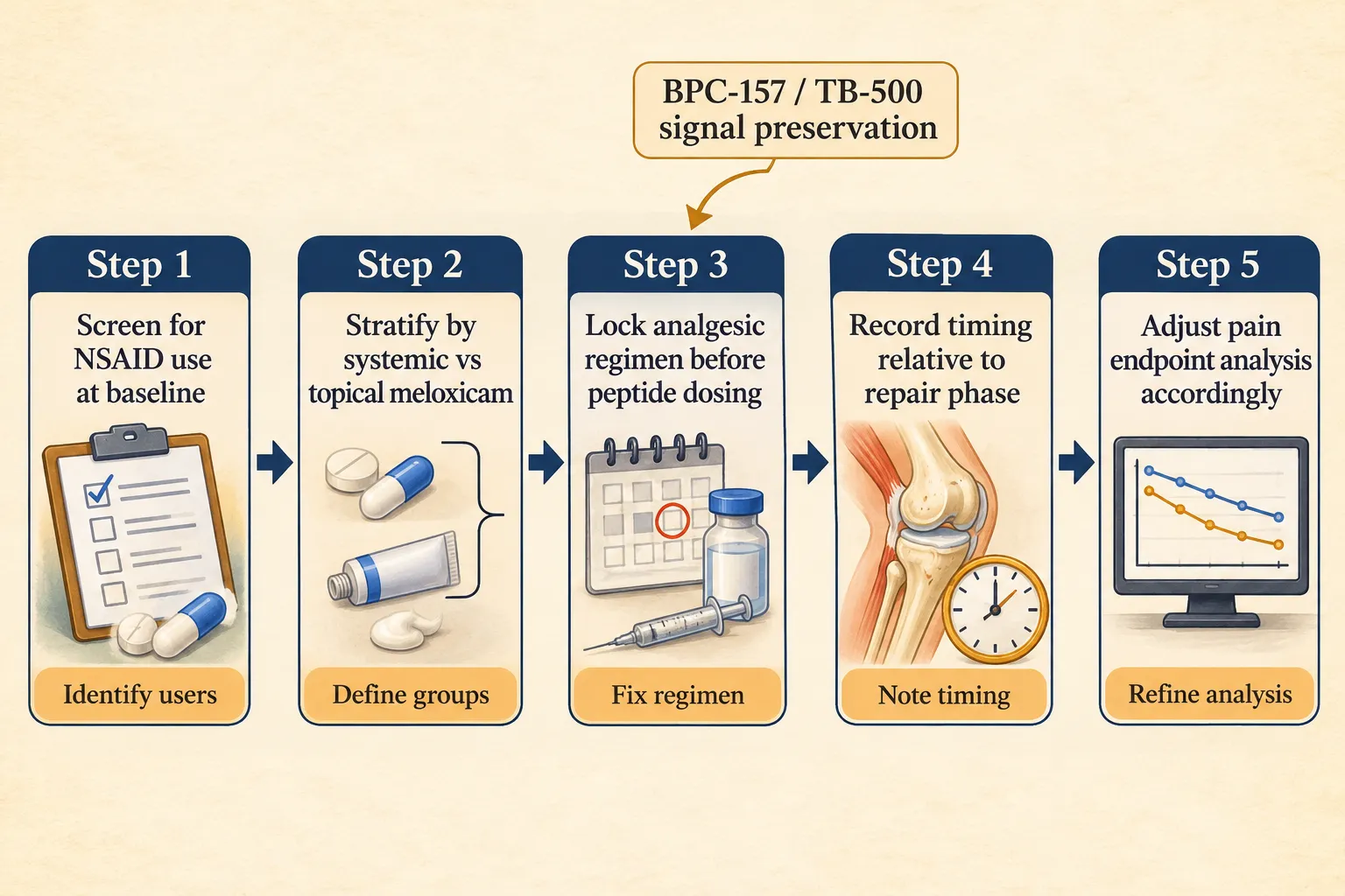
- Screen all subjects for NSAID use at baseline and during the washout period.
- Stratify enrolled subjects by systemic versus topical meloxicam exposure before randomization.
- Lock the analgesic rescue regimen before peptide dosing begins, do not allow ad hoc NSAID initiation during the trial.
- Document the timing of any meloxicam doses relative to the injury or repair phase.
- Pre-specify meloxicam use as a covariate in the statistical analysis plan.
Topical meloxicam formulations, including film-forming sprays and emulgels, deliver meaningful local COX-2 suppression with potentially reduced systemic exposure. However, even localized COX-2 inhibition can alter joint tissue remodeling endpoints in regenerative research, so topical use should be documented and stratified rather than dismissed as inconsequential.
For researchers sourcing compounds for controlled studies, using lab-tested peptides with verified purity documentation is essential when analgesic confounders are already adding variability. Guidance on building robust peptide benchmarks with reference standards is equally relevant to this quality-control conversation.
Clinical practice guidance increasingly recommends topical NSAIDs and multimodal analgesia, combining acetaminophen with topical agents, to minimize systemic COX-2 inhibition while still managing pain. Aligning peptide trial analgesic protocols with these frameworks is now viewed as best practice. Researchers exploring metabolic or mitochondrial peptides in conjunction with pain management should also review the SS-31 mechanism and research context for a parallel example of how co-interventions interact with endpoint selection.
For newer investigational peptides where the interaction with COX biology has not yet been characterized, supplier-level comparisons and quality documentation become the first line of defense. Resources like peptide supplier comparisons help establish the purity and identity baseline needed before any confounding-variable analysis is meaningful.
Conclusion
Meloxicam is a highly effective analgesic and anti-inflammatory agent, and that effectiveness is precisely what makes it a methodological hazard in peptide and tissue-repair research. Its dual COX-1 and COX-2 suppression reshapes the prostanoid environment that BPC-157, TB-500, and similar compounds are designed to work within. It independently reduces pain scores, potentially inflating apparent peptide analgesic efficacy. And in bone, tendon, and muscle repair models, its timing relative to the injury phase can attenuate the mechanical and cellular outcomes a trial is trying to attribute to the peptide.
Actionable next steps for 2026 and beyond:
- Treat systemic meloxicam as a primary covariate in any tissue-repair peptide trial, not a nuisance variable.
- Adopt topical-only analgesic policies during critical repair windows (especially days 10-20 post-injury) when COX-2 activity is mechanistically essential.
- Use validated non-NSAID analgesic comparators when pain endpoints are primary outcomes.
- Document analgesic timing with the same rigor applied to peptide dosing schedules.
- Review study designs against updated systematic evidence on NSAID and soft-tissue interactions, as the human trial literature continues to grow.
Cleaner analgesic protocols do not just protect data integrity, they make the signal from investigational peptides far more interpretable, reproducible, and ultimately useful.