Call or Text 727-513-9780
  • Shopping Cart Shopping Cart
    0Shopping Cart
Pure Tested Peptides | America's most trusted Peptides for sale online
  • Peptides for sale
    • Oral Peptides for sale
      • Peptide Capsules for sale
      • BPC 157 Capsules 1000mcg
      • SLU-PP-332 Capsules | 1000 mcg
      • 5-Amino-1MQ 50mg Capsules
      • Tesofensine 500mcg
    • All Peptides for sale
    • Peptide Sprays
      • BPC 157 Nasal Spray Kit
      • BPC-157 TB500 Nasal Spray Kit
      • Semax Nasal Spray 10mg
      • Selank – Nasal Spray Kit – 10mg
      • Epithalon 50MG Nasal Spray Kit
      • Ipamorelin 10mg Nasal Spray
      • Klow Nasal Spray (BPC-157 + TB-500 + GHK-Cu + KPV) | 80mg
      • Hulk Nasal Spray Tesa / Ipa Blend 6/3 MG
      • Klow Nasal Spray
      • NAD + 500 mg Nasal Spray
      • PT-141 Nasal Spray Kit
    • GHRH Peptides
      • Ipa Peptides
      • CJC-1295 Peptides
        • CJC-1295 with DAC 5 mg
        • CJC-1295 without DAC 5 mg
        • CJC-1295 Ipa 10mg
      • Tesa Peptides
        • Tesa Peptide
        • Tesa 20 mg
    • GHK-Cu Peptides
      • All GHK-Cu Peptides
      • GHK-Cu 100mg
      • KLOW Peptide Blend – Buy KLOW blend online
    • BPC Peptides
      • All BPC Peptides
      • BPC-157
      • BPC-157 TB-500
      • BPC 157 capsules 1000mcg
    • SLU-PP-332 Peptides
      • All SLU-PP-332 Peptides
      • SLU-PP-332 5mg
    • GLP3 Peptides
    • PT-141 Peptides
      • PT-141 Peptides for sale
      • PT-141 10mg
      • PT-141 Nasal Spray
    • CAG Peptides
      • Lipo-C Peptide Blend
      • CAG 5mg
      • CAG 10mg
    • MOTS-C Peptides
      • MOTS-C Peptides for sale
      • MOTS-c peptide
      • MOTS-c 10mg *6 pack*
    • 5 Amino 1MQ Peptides
      • 5 Amino 1MQ Peptides for sale
      • 5-Amino-1MQ 50mg Capsules
      • 5-Amino-1MQ 5mg
    • Epithalon Peptides
      • Epithalon Peptides for sale
      • Epithalon 10mg
      • Epithalon 50mg
  • Shop
    • GLPs
      • 5-Amino-1MQ 50mg Capsules
      • 5-Amino-1MQ 5mg
      • L-Carnitine 500mg/ml
      • Tesofensine 500mcg
      • SLU-PP-332 5mg
      • MOTS-c 10mg *6 pack*
    • Epithalon & BPC Peptides
      • Epithalon 10mg
      • Epithalon 50mg
      • BPC-157
      • BPC 157 capsules 1000mcg
      • BPC-157 TB-500
      • BPC-157 TB500 Nasal Spray Kit
      • BPC 157 Nasal Spray Kit
    • BPC TB-500 & NAD+ Peptides
      • NAD+ 500 mg
      • KLOW Peptide Blend – Buy KLOW blend online
      • GLOW Peptide Blend
      • TB 500 5mg
      • BPC 157 capsules 1000mcg – Supplement
      • BPC 157 Nasal Spray Kit
      • BPC-157
      • BPC-157 TB500 Nasal Spray Kit
      • BPC-157 TB-500
      • BPC 157 capsules 1000mcg
    • LL-37 Peptide
      • LL-37 10 mg
    • MOTS-C & Selank
      • MOTS-c peptide
      • Selank 10mg
    • GHK Peptides
      • GHK-Cu 100mg
      • GLOW Peptide Blend
      • KLOW Peptide Blend – Buy KLOW blend online
  • COAs
  • Wholesale
    • Wholesale Peptides for sale
  • PTP FAQ
  • Affiliates
    • Selank Peptide: Advanced Pharmacological Mechanisms Underlying Its Anxiolytic and Nootropic Effects in Research
      • 5-Amino-1MQ Peptide: Detailed Mechanisms of NNMT Inhibition and Its Impact on Cellular Metabolism Research
        • MOTS-C Peptide: Unraveling Its Role in Mitochondrial Dynamics and Energy Metabolism Research
          • Epithalon Peptide and Telomerase Regulation: Investigating Its Impact on Cellular Senescence and Lifespan Research Models
            • GLP2-T vs GLP2 Tirz Peptide: Understanding the Naming, Mechanistic Nuances, and Research Implications for Gut Health
      • Peptides and Polypeptides in Modern Pharmacology: What Research on Metoprolol, Prednisone, and Amlodipine Reveals
        • Polypeptide Peptides vs NSAIDs: What Naproxen and Diclofenac Teach Tissue-Repair Researchers About BPC‑157 and TB‑500
          • Peptides and Polypeptides in Endocrine Pharmacology: How GLP-1, GLP-2, and GLP-3 Retatrutide Differ From Classic Drugs Like Prednisone and Amlodipine
          • 5‑Amino‑1MQ Peptide and NNMT Inhibition: How It Compares With Statins Like Atorvastatin in Adipose and Lipid Metabolism Research
      • Enclomiphene, Estrogen Receptor Signaling, and Luteinizing Phase Biology: What Hormone Researchers Should Measure
        • Mesenchymal Stem Cells, BPC‑157, and GHK‑Cu: How Tissue Repair Peptides Compare With Classic NSAIDs Like Naproxen in Injury Models
          • Peptide Calculator Use Cases Beyond Growth Hormone: Working Through GLP‑3 Retatrutide, MOTS‑c, and BPC‑157 Research Dosing
        • Polypeptide Peptides in Endocrine and Metabolic Pharmacology: Lessons From Amlodipine, Prednisone, and Metoprolol
        • Peptides Calculator for Advanced Blends: Worked Examples for Tesamorelin, CJC‑1295, and Ipamorelin Stacks
          • Peptides and Polypeptides in Human Physiology: How Molecular Size Shapes Research Applications
            • Selank Peptide: Advanced Pharmacological Mechanisms Underlying Its Anxiolytic and Nootropic Effects in Research
          • 5-Amino-1MQ Peptide: Detailed Mechanisms of NNMT Inhibition and Its Impact on Cellular Metabolism Research
            • MOTS-C Peptide: Unraveling Its Role in Mitochondrial Dynamics and Energy Metabolism Research
              • Epithalon Peptide and Telomerase Regulation: Investigating Its Impact on Cellular Senescence and Lifespan Research Models
                • GLP2-T vs GLP2 Tirz Peptide: Understanding the Naming, Mechanistic Nuances, and Research Implications for Gut Health
                  • Peptides and Polypeptides in Endocrine Pharmacology: How GLP-1, GLP-2, and GLP-3 Retatrutide Differ From Classic Drugs Like Prednisone and Amlodipine
                    • Polypeptide Peptides vs NSAIDs: What Naproxen and Diclofenac Teach Tissue-Repair Researchers About BPC‑157 and TB‑500
                      • Peptides and Polypeptides in Modern Pharmacology: What Research on Metoprolol, Prednisone, and Amlodipine Reveals
                        • 5‑Amino‑1MQ Peptide and NNMT Inhibition: How It Compares With Statins Like Atorvastatin in Adipose and Lipid Metabolism Research
                        • Enclomiphene, Estrogen Receptor Signaling, and Luteinizing Phase Biology: What Hormone Researchers Should Measure
                        • Mesenchymal Stem Cells, BPC‑157, and GHK‑Cu: How Tissue Repair Peptides Compare With Classic NSAIDs Like Naproxen in Injury Models
                        • Peptide Calculator Use Cases Beyond Growth Hormone: Working Through GLP‑3 Retatrutide, MOTS‑c, and BPC‑157 Research Dosing
                        • Polypeptide Peptides in Endocrine and Metabolic Pharmacology: Lessons From Amlodipine, Prednisone, and Metoprolol
                        • Peptides Calculator for Advanced Blends: Worked Examples for Tesamorelin, CJC‑1295, and Ipamorelin Stacks
                        • Peptides and Polypeptides in Human Physiology: How Molecular Size Shapes Research Applications
                        • Selank Peptide: Advanced Pharmacological Mechanisms Underlying Its Anxiolytic and Nootropic Effects in Research
                        • MOTS-C Peptide: Unraveling Its Role in Mitochondrial Dynamics and Energy Metabolism Research
                        • Peptides and Polypeptides in Modern Pharmacology: What Research on Metoprolol, Prednisone, and Amlodipine Reveals
                        • Peptides and Polypeptides in Endocrine Pharmacology: How GLP-1, GLP-2, and GLP-3 Retatrutide Differ From Classic Drugs Like Prednisone and Amlodipine
                        • 5-Amino-1MQ Peptide: Detailed Mechanisms of NNMT Inhibition and Its Impact on Cellular Metabolism Research
                        • Mesenchymal Stem Cells, BPC‑157, and GHK‑Cu: How Tissue Repair Peptides Compare With Classic NSAIDs Like Naproxen in Injury Models
                        • Peptide Calculator Use Cases Beyond Growth Hormone: Working Through GLP‑3 Retatrutide, MOTS‑c, and BPC‑157 Research Dosing
                        • GLP2-T vs GLP2 Tirz Peptide: Understanding the Naming, Mechanistic Nuances, and Research Implications for Gut Health
                        • Polypeptide Peptides in Endocrine and Metabolic Pharmacology: Lessons From Amlodipine, Prednisone, and Metoprolol
                        • Polypeptide Peptides vs NSAIDs: What Naproxen and Diclofenac Teach Tissue-Repair Researchers About BPC‑157 and TB‑500
                        • Peptides and Polypeptides in Human Physiology: How Molecular Size Shapes Research Applications
                        • Peptides Calculator for Advanced Blends: Worked Examples for Tesamorelin, CJC‑1295, and Ipamorelin Stacks
                        • 5‑Amino‑1MQ Peptide and NNMT Inhibition: How It Compares With Statins Like Atorvastatin in Adipose and Lipid Metabolism Research
                        • Enclomiphene, Estrogen Receptor Signaling, and Luteinizing Phase Biology: What Hormone Researchers Should Measure
                        • Epithalon Peptide and Telomerase Regulation: Investigating Its Impact on Cellular Senescence and Lifespan Research Models
                        • Best research protocol Klow blend
                        • best time to take BPC-157
                        • best time to take DSIP (Delta Sleep Inducing Peptide)
                        • best time to take CJC-1295
                        • best time to take AOD-9604
                        • best time to take Follistatin 344
                        • best time to take Ipamorelin
                        • best time to take MK-677 (Ibutamoren)
                        • best time to take Ligandrol (LGD-4033) — research compound
                        • best time to take Ostarine (MK-2866) — research compound
                        • best time to take GHK-CU
                        • best time to take TB-500
                        • best time to take MOTS-c
                        • best time to take Semax
                        • best time to take RAD-140 (Testolone) — research compound
                        • best time to take Thymosin Alpha-1
                        • best time to take PEG-MGF
                        • Biolife Plasma, Octapharma Plasma, and Research Peptides: How Plasma Donation Labs Differ From Peptide Suppliers
                        • best time to take YK-11 — research compound
                        • best time to take PT-141 (Bremelanotide)
                        • Best research protocol Klow blend
                        • 5-Amino-1MQ and MOTS-C Synergy: Metabolic Signaling, Mitochondria, and Research Design
                        • BPC-157 and TB-500: Investigating Their Combined Effects on Angiogenesis and Cellular Migration in Tissue Repair Models
                        • BPC-157 Peptide: Gut Barrier Function, Inflammation, and Tissue-Recovery Research
                        • 5‑Amino‑1MQ and MOTS‑c Synergy in Metabolic Research: Designing NNMT and Mitochondrial Biogenesis Stacks
                        • CJC-1295 with DAC vs. Without DAC: Half-Life, Release Kinetics, and Research Implications
                        • CJC‑1295 with DAC vs. Without DAC: Expanding on Half‑Life Differences Using Tesamorelin and Ipamorelin Blend Case Studies
                        • Collagen Biology and Copper‑Binding Peptides: How GHK‑Cu, Glow Blend, and Klow Blend Interact with Skin and Connective Tissue
                        • Collagen Biology and Regenerative Peptides: How GHK‑Cu, Glow Blend, and Klow Blend Affect Extracellular Matrix Research
                    • DNA, Telomeres, and Longevity Peptides: Positioning Epithalon and MOTS‑c in Genetic Aging Research
                      • Enclomiphene Citrate: serm Mechanism, Testosterone Research, and Stack Compatibility
                        • Enclomiphene vs Enclomiphene Citrate: Formulation, Bioavailability, and Research Distinctions
                        • Epithalon Peptide Research: Telomerase Activation, Aging, and Pineal Gland Function
                        • Estrogen Receptor Signaling and Enclomiphene: How Selective Modulators Compare with Classic Polypeptide Hormones
                        • GHK-Cu Peptide: Advanced Mechanisms in Extracellular Matrix Remodeling and Wound Healing Research
                        • GHK-Cu Peptide: Collagen Synthesis, Wound Repair, and Skin-Barrier Research Models
                        • GLP-1 vs GLP-3 vs GLP-2: Peptide Classification and Research Applications
                        • GLP-2 Peptide Research Guide: Gut Barrier Function, Nutrient Absorption, and Intestinal Recovery Models
                        • GLP-3 Retatrutide vs. GLP-1 Drugs: What Triple-Agonist Biology Changes in Research Models
                        • Ipamorelin and Tesamorelin Combination: Synergistic GH Secretagogue Research and Dosing Protocols
                        • GLP2 Tirz Peptide: What It Is, Why the Name Exists, and How Researchers Should Interpret It
                        • Klow Blend Peptide Nasal Spray: What the Formulation Is Trying to Do in Cognitive Research
                        • Mitochondria, NNMT Inhibition, and Peptide Modulators: Where MOTS‑c and 5‑Amino‑1MQ Fit in Cellular Energy Research
                        • MOTS-c Peptide: Mitochondrial Function, Energy Metabolism, and What Researchers Measure
                        • MOTS-c vs. 5-Amino-1MQ: Which Metabolic Research Questions Each Compound Actually Answers
                        • Nasal Spray Peptides: Bioavailability, Administration, and Semax/Selank Research Applications
                        • PT-141 Peptide Research: Mechanism of Action and Melanocortin Receptor Signaling
                        • Retatrutide for Research: Mechanism, Structure, and GLP-1/GLP-3 Dual Action
                        • Retatrutide for Obesity and Type 2 Diabetes: What the Latest Trial Data Suggest
                        • Tesofensine Peptide Research: Mechanism, Appetite Suppression, and Neuropeptide Y Pathways
  • Contact
    • Contact Customer Service
    • Text Customer Support
  • About US
  • Shop all peptides
  • Affiliate Program
    • Affiliate Signup
  • Login / Register Login / Register Page Link Login / Register Page Link
  • Click to open the search input field Click to open the search input field Search
  • Menu Menu

Tag Archive for: ss-31 mitochondrial research

What Does MCHC Mean in Peptide Research? Interpreting Hemoglobin Indices When Evaluating GLP-Class and Tissue-Repair Models

What Does MCHC Mean in Peptide Research? Interpreting Hemoglobin Indices When Evaluating GLP-Class and Tissue-Repair Models

October 11, 2026/0 Comments/in Uncategorized/by

Fewer than 15% of published preclinical peptide studies list MCHC as a primary endpoint, yet researchers routinely flag unexpected shifts in this index and struggle to interpret what they mean. Understanding what does MCHC mean in peptide research, and how to interpret hemoglobin indices when evaluating GLP-class and tissue-repair models, is a practical skill that separates rigorous data review from guesswork.

Key Takeaways

  • MCHC (Mean Corpuscular Hemoglobin Concentration) measures hemoglobin density inside red blood cells and is calculated as hemoglobin divided by hematocrit, expressed in g/dL.
  • In peptide research, MCHC functions as a secondary safety and physiology marker, not a primary efficacy endpoint.
  • Low MCHC (hypochromia) most often signals iron deficiency, chronic inflammation, or sample dilution rather than a direct peptide effect.
  • High MCHC (hyperchromia) in preclinical panels frequently reflects laboratory artifact, particularly hemolysis from poor sample handling.
  • MCHC should always be interpreted alongside MCV, RDW, and full study context before attributing changes to any peptide compound.

Defining MCHC: Calculation, Normal Ranges, and the Erythrocyte Index Hierarchy

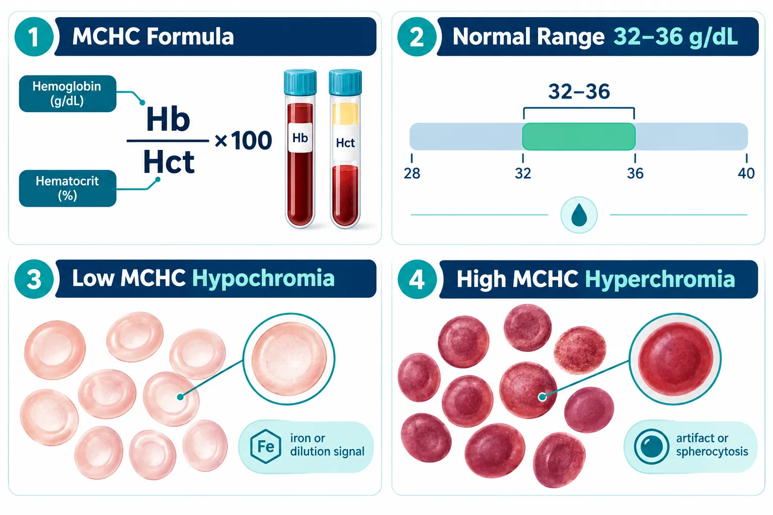
MCHC stands for Mean Corpuscular Hemoglobin Concentration. It quantifies how much hemoglobin is packed into a given volume of red blood cells. The formula is straightforward:

MCHC (g/dL) = (Hemoglobin / Hematocrit) x 100

In most mammalian species used in preclinical research, the reference range sits between 32 and 36 g/dL. Values below this threshold indicate hypochromia (pale, hemoglobin-poor cells). Values above suggest hyperchromia, though true biological hyperchromia is rare.
Defining MCHC: Calculation, Normal Ranges, and the Erythrocyte Index Hierarchy

Within the broader erythrocyte index hierarchy, MCHC ranks below MCV (Mean Corpuscular Volume) and RDW (Red Cell Distribution Width) in diagnostic weight. Here is how the indices compare:

Index What It Measures Research Priority
MCV Average red cell size High
RDW Size variation across cells High
MCH Average hemoglobin per cell Moderate
MCHC Hemoglobin concentration per cell volume Supportive

MCHC is most useful when read as part of this full panel, not in isolation. Researchers sourcing compounds through platforms that emphasize building robust peptide benchmarks will recognize that baseline CBC panels, including MCHC, are part of responsible quality-control workflows.


Why MCHC Matters When Interpreting Hemoglobin Indices in GLP-Class Models

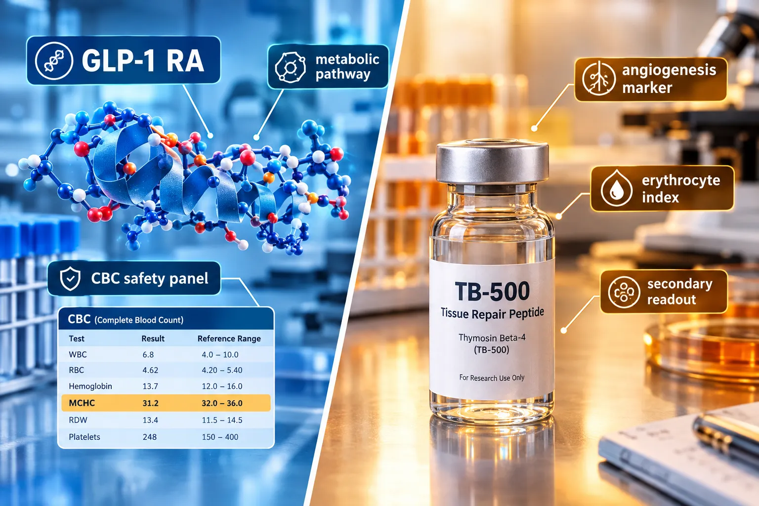
GLP-1 receptor agonist (GLP-RA) compounds have become central to metabolic research. When reviewing CBC safety data from GLP-class studies, MCHC appears as a secondary safety lab rather than a target biomarker. Its role is to confirm that metabolic changes driven by energy restriction, body composition shifts, or altered nutrient absorption are not producing clinically meaningful hematologic stress.

Researchers working with GLP-1 peptide sourcing and generational research concepts should note that caloric restriction models, common in GLP-RA efficacy studies, can mildly suppress MCHC through reduced dietary iron intake. This is a model-design artifact, not a compound toxicity signal.

Key considerations for GLP-class CBC interpretation:

  • Weight loss velocity can transiently alter plasma volume, diluting hemoglobin concentration and lowering apparent MCHC.
  • Visceral fat mobilization studies, such as those explored in visceral fat research models, may produce mild hemodilution as fluid compartments shift.
  • GLP-2-class compounds, which target intestinal repair and nutrient absorption, carry a more direct theoretical link to iron and B12 uptake, making MCHC monitoring more relevant in those specific models.

For researchers evaluating next-generation triple-agonist compounds, resources on GLP-3 for sale and triple agonist research planning provide useful context on how expanded receptor profiles may affect systemic physiology, including hematologic readouts.
Why MCHC Matters When Interpreting Hemoglobin Indices in GLP-Class Models


MCHC in Tissue-Repair Peptide Models: A Practical Interpretation Framework

Tissue-repair peptides, including TB-500 analogs and mitochondria-targeting compounds, introduce different considerations for hemoglobin index interpretation. Researchers following TB-500 controlled experimental models and QC workflow already understand that angiogenesis-promoting compounds can alter red cell dynamics through vascular remodeling rather than direct erythropoietic effects.

For mitochondria-targeted peptides, the relationship between cellular energy metabolism and red cell lifespan is an active area of interest. Work in SS-31 mitochondrial research and related SS-31 kidney health research highlights how oxidative stress reduction may indirectly support erythrocyte membrane integrity, a variable that influences MCHC readings over longer study durations.

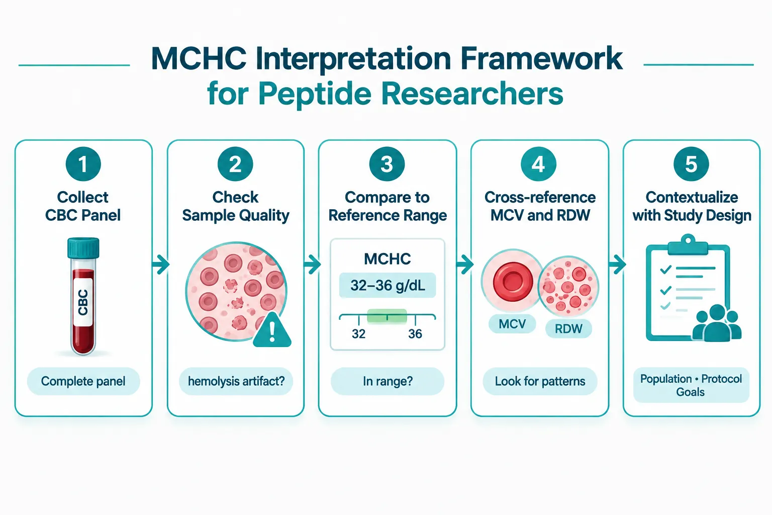
A practical five-step MCHC interpretation framework for peptide researchers:

  1. Verify sample quality first. Hemolysis from poor venipuncture or freeze-thaw cycling is the single most common cause of falsely elevated MCHC. Confirm sample integrity before drawing conclusions.
  2. Check the full CBC panel. An isolated MCHC shift without corresponding MCV or RDW changes is more likely an artifact than a biological signal.
  3. Compare against species-specific reference ranges. Murine, rat, and canine reference ranges differ meaningfully from human norms.
  4. Cross-reference study design variables. Diet, hydration status, and blood collection timing all affect MCHC independently of any peptide compound.
  5. Assign interpretive weight proportionally. MCHC is a supporting character in the CBC story, not the lead.
MCHC in Tissue-Repair Peptide Models: A Practical Interpretation Framework

As of late 2026, the published literature contains very few peptide-specific MCHC studies. Most available data comes from broader hematology safety panels embedded in metabolic or regenerative medicine trials. This gap means researchers must apply general hematology principles carefully rather than relying on peptide-specific MCHC benchmarks.


Conclusion

Understanding what does MCHC mean in peptide research, and how to interpret hemoglobin indices when evaluating GLP-class and tissue-repair models, ultimately comes down to context. MCHC is a reliable supporting marker when used correctly, but it is frequently misread when treated as a standalone signal.

Actionable next steps for peptide researchers in 2026:

  • Always collect a full CBC panel, not individual indices, as part of baseline and endpoint safety monitoring.
  • Document sample collection conditions meticulously to rule out hemolysis artifacts before flagging MCHC shifts.
  • When MCHC deviates from reference range, cross-check MCV, RDW, and reticulocyte count before attributing the change to a peptide compound.
  • For GLP-class models, account for caloric restriction and fluid redistribution as confounding variables in hematologic data.
  • For tissue-repair and mitochondria-targeting models, monitor MCHC trends over time rather than at single time points to distinguish transient from sustained changes.

Rigorous interpretation of hemoglobin indices strengthens the overall quality of peptide research data and supports more reliable conclusions across all model types.

https://www.puretestedpeptides.com/wp-content/uploads/2026/10/what-does-mchc-mean-in-peptide-research-interpreting-hemoglobin-indices-when-eva.webp 1024 1536 https://www.puretestedpeptides.com/wp-content/uploads/2026/01/buy-peptides-online.jpg 2026-10-11 13:04:122026-10-11 13:04:12What Does MCHC Mean in Peptide Research? Interpreting Hemoglobin Indices When Evaluating GLP-Class and Tissue-Repair Models
Prednisone and Peptide Research Models: Separating Glucocorticoid Metabolic Effects From Tissue-Repair Signals

Prednisone and Peptide Research Models: Separating Glucocorticoid Metabolic Effects From Tissue-Repair Signals

October 4, 2026/0 Comments/in Uncategorized/by

Fewer than 15% of preclinical peptide studies formally account for background glucocorticoid exposure in their control arms, yet prednisone alone can shift glucose metabolism, body composition, and wound-healing readouts enough to invalidate a comparison. Understanding how to work with that interference is now a core competency in regenerative peptide research.

This article examines how prednisone and peptide research models interact, and provides a practical framework for deciding when steroid exposure should function as a control variable, an exclusion criterion, or a stratification factor.

Key Takeaways

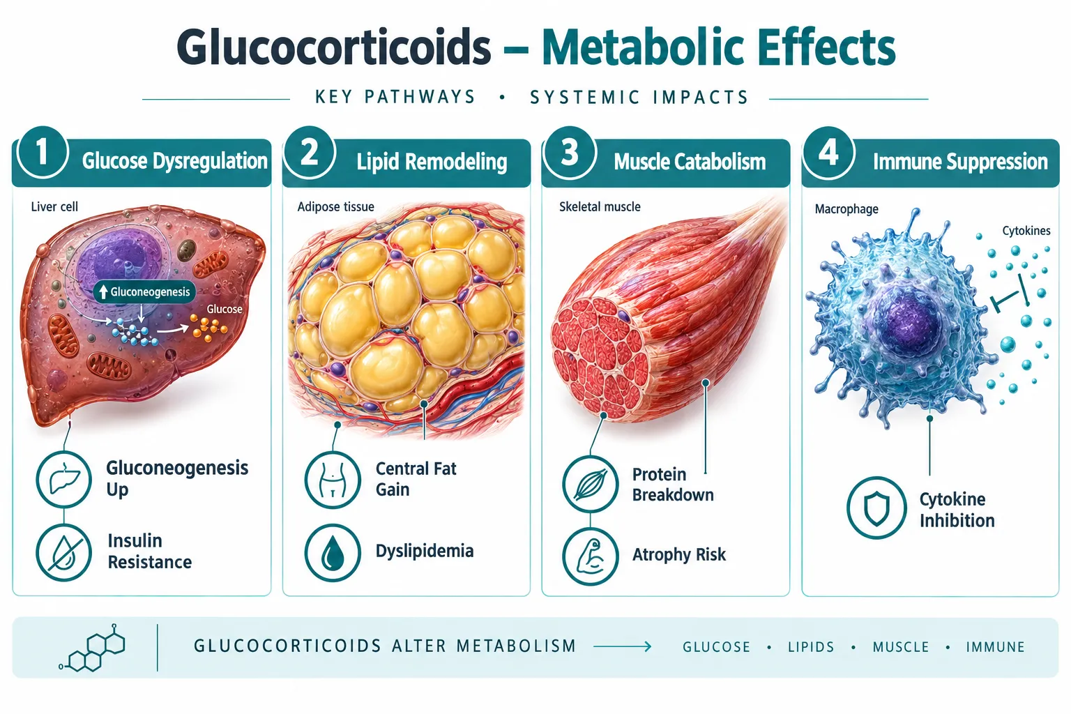
  • Prednisone exerts distinct metabolic effects (glucose dysregulation, lipid remodeling, muscle catabolism) that are mechanistically separate from its acute tissue-repair actions.
  • Peptide-based regenerative agents operate on slower, anabolic timelines, making direct head-to-head comparisons with glucocorticoids methodologically complex.
  • Pulsed or intermittent prednisone dosing can produce beneficial mitochondrial and sarcolemmal repair effects that overlap with peptide endpoints.
  • Researchers must classify steroid exposure as a control, exclusion criterion, or stratification variable before designing outcome readouts.
  • No Phase III trials currently pair peptides directly with long-term corticosteroid protocols, leaving a significant evidence gap.

How Prednisone Reshapes the Metabolic Landscape

Prednisone and peptide research models: separating glucocorticoid metabolic effects from tissue-repair signals begins with understanding what prednisone actually does to baseline physiology. Prednisone is a synthetic glucocorticoid that binds glucocorticoid receptors across virtually every tissue type. Its systemic metabolic profile is dominated by three well-documented disruptions.

How Prednisone Reshapes the Metabolic Landscape

1. Glucose and Insulin Metabolism
Prednisone stimulates hepatic gluconeogenesis and reduces peripheral insulin sensitivity. Even short-term use preferentially disrupts glucose metabolism before significant muscle protein changes appear. This matters enormously for any peptide study that uses fasting glucose, HOMA-IR, or glycemic variability as an outcome measure.

2. Lipid Remodeling and Body Composition
Prolonged prednisone use drives central adiposity and dyslipidemia. Importantly, research on intermittent weekly prednisone dosing shows sex-specific lipid remodeling in skeletal muscle, a finding that introduces a sex-as-biological-variable confound if not stratified properly.

3. Muscle Catabolism vs. Repair Duality
This is where the picture becomes nuanced. Chronic high-dose prednisone is catabolic to muscle. Yet pulsed glucocorticoid protocols in dystrophic muscle models have demonstrated enhanced physical performance through nutrient-utilization and repair pathways. Circadian timing of intermittent prednisone pulses has been shown to modulate mitochondrial remodeling, suggesting that dose, timing, and frequency determine whether the net effect is catabolic or restorative.

"The same compound that degrades muscle under chronic dosing can enhance sarcolemmal repair via annexin-mediated pathways under pulsed protocols, a distinction that most research designs fail to capture."

This duality means that labeling prednisone exposure simply as "harmful background noise" in a peptide study is an oversimplification that can distort results in either direction.


Tissue-Repair Signals: Where Peptides Operate Differently

To build a sound comparative framework, researchers must map where peptide mechanisms diverge from glucocorticoid mechanisms. For a broader foundation on peptide structure and receptor pharmacology, the Peptides 101 for Research-Use Only Buyers guide provides essential context.

Tissue-Repair Signals: Where Peptides Operate Differently

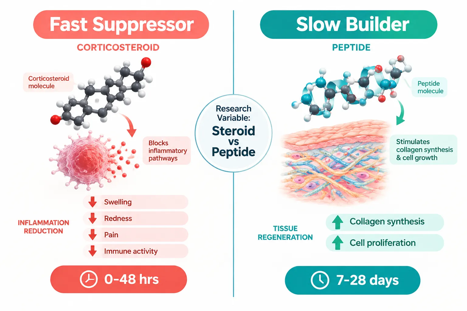
Corticosteroids act as fast suppressors: they reduce inflammatory cytokines within hours, providing rapid symptom relief. Clinical rheumatology data from 2026 confirm that prednisone reliably improves symptoms but impairs longer-term tissue healing when used continuously.

Peptides, by contrast, function as slow builders. They tend to stimulate collagen synthesis, cell proliferation, and mitochondrial biogenesis over days to weeks. Compounds such as GHK-Cu peptide have been studied for fibroblast activation and extracellular matrix remodeling, while SS-31 peptide research focuses on mitochondrial membrane stabilization, a mechanism directly relevant to steroid-induced mitochondrial disruption. For deeper reading on mitochondrial targets, SS-31 mitochondrial research offers a useful reference point.

Feature Prednisone (Pulsed) Peptide Agents
Onset of action Hours Days to weeks
Primary target Immune/inflammatory signaling Structural and metabolic repair
Mitochondrial effect Remodeling (timing-dependent) Biogenesis support
Glucose impact Dysregulation (dose-dependent) Neutral to beneficial
Evidence base Extensive Phase III data Preclinical/early Phase I-II

Peptide-based regenerative therapies remain insufficiently validated compared to glucocorticoids in head-to-head trials. No Phase III study currently pairs a peptide protocol directly with long-term corticosteroid therapy, which makes the need for rigorous study design even more critical.


A Framework for Handling Steroid Exposure in Peptide Research Models

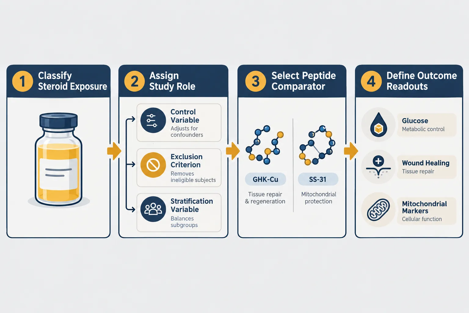
Applying the lens of prednisone and peptide research models: separating glucocorticoid metabolic effects from tissue-repair signals to study design requires a deliberate classification step before any outcome readout is chosen.

A Framework for Handling Steroid Exposure in Peptide Research Models

Researchers should ask three questions about any subject or model with prednisone exposure:

Step 1, Classify the exposure type
Is the steroid use chronic (daily, systemic), intermittent (weekly pulse), or acute (single-dose)? Each produces a different metabolic fingerprint. Intermittent once-weekly prednisone has been shown to improve ATP levels, NAD+ availability, and endurance in aged skeletal muscle, outcomes that directly overlap with peptide endpoints. Treating all steroid exposure as equivalent will produce noise, not signal.

Step 2, Assign a study role

  • Control variable: Include steroid-exposed and steroid-naive arms, measure the exposure quantitatively, and model it as a covariate. This is appropriate when excluding steroid users would make recruitment impossible.
  • Exclusion criterion: Remove subjects with any current or recent steroid use when the primary endpoint is glucose metabolism or wound healing, because prednisone's effects on those readouts are too large to model reliably.
  • Stratification variable: Randomize within steroid-exposed and steroid-naive strata when the research question specifically asks how a peptide performs in a glucocorticoid-rich environment.

Step 3, Select peptide comparators with mechanistic logic
Choose peptides whose mechanisms are least likely to be masked or amplified by glucocorticoid background. For metabolic endpoints, compounds such as MOTS-C peptide, which targets mitochondrial AMPK pathways, may show cleaner signal separation from prednisone's glucose effects than growth-hormone-releasing peptides. For tissue-repair endpoints, Thymosin Alpha 4 peptide research offers a comparator that operates through actin cytoskeletal and immune-modulation pathways distinct from glucocorticoid receptor signaling.

Dose-dependent and tissue-specific adverse metabolic effects from prednisone further underscore why a single classification rule cannot apply across all study types. A low-dose pulse in muscle tissue is not the same biological event as a high-dose chronic regimen in adipose tissue.


Conclusion

Prednisone and peptide research models: separating glucocorticoid metabolic effects from tissue-repair signals is not merely a methodological nicety, it is a prerequisite for generating reproducible data. The metabolic disruptions prednisone introduces (glucose dysregulation, lipid remodeling, context-dependent muscle effects) operate on different timescales and through different receptors than the anabolic, repair-oriented signals that most peptides are designed to amplify.

Actionable next steps for researchers in 2026:

  • Audit existing datasets for undocumented steroid co-exposure before pooling results.
  • Pre-register the steroid classification decision (control, exclusion, or stratification) in study protocols.
  • Prioritize mechanistic alignment when selecting peptide comparators, match the peptide's primary pathway to an endpoint that prednisone does not dominate.
  • Monitor both glucose and mitochondrial markers simultaneously to distinguish metabolic interference from genuine tissue-repair signal.
  • Follow emerging pulsed-dosing literature closely, as intermittent prednisone protocols may eventually serve as a validated adjunct rather than a pure confounder in select repair models.

The field will only move forward when steroid exposure is treated with the same rigor applied to any other pharmacological variable in the experimental design.

https://www.puretestedpeptides.com/wp-content/uploads/2026/10/prednisone-and-peptide-research-models-separating-glucocorticoid-metabolic-effec.webp 1024 1536 https://www.puretestedpeptides.com/wp-content/uploads/2026/01/buy-peptides-online.jpg 2026-10-04 13:04:262026-10-04 13:04:26Prednisone and Peptide Research Models: Separating Glucocorticoid Metabolic Effects From Tissue-Repair Signals

Tag Archive for: ss-31 mitochondrial research

The Role of Adenosine Triphosphate (ATP) in Peptide-Mediated Cellular Energy Research

The Role of Adenosine Triphosphate (ATP) in Peptide-Mediated Cellular Energy Research

July 1, 2026/0 Comments/by Pure Tested

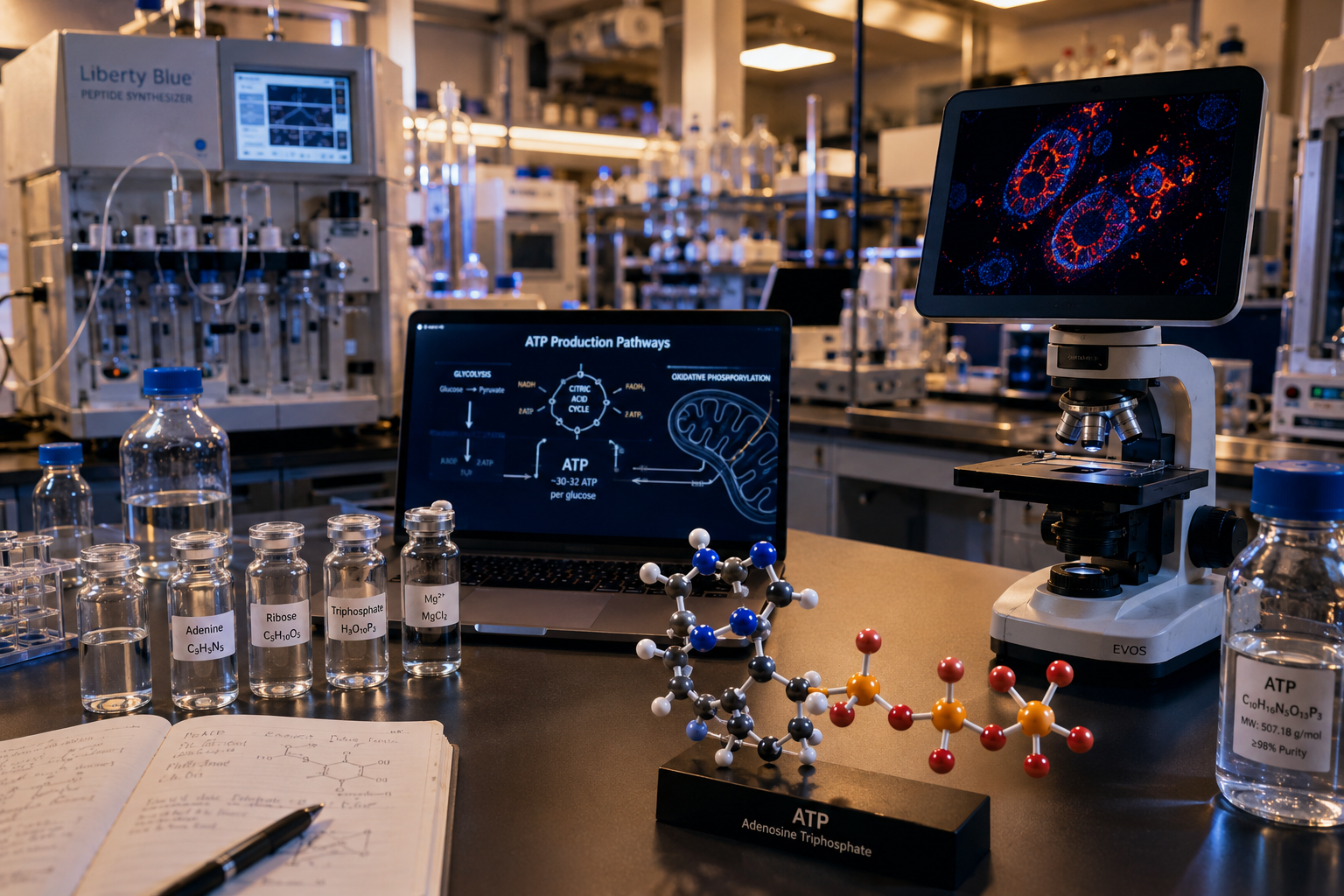
Every cell in the human body runs on a molecule so fundamental that without it, life stops within seconds. Adenosine triphosphate (ATP) powers nearly every biological process, yet researchers are only beginning to understand how peptides actively shape its production, regulation, and distribution at the cellular level. The role of adenosine triphosphate (ATP) in peptide-mediated cellular energy research has emerged as one of the most productive areas in modern biochemistry, connecting mitochondrial biology to therapeutic peptide science in ways that were not fully appreciated even a decade ago.

Key Takeaways

  • ATP is the primary energy currency of the cell, produced mainly within mitochondria through oxidative phosphorylation.
  • Specific peptides, including MOTS-c, directly influence ATP synthesis by interacting with mitochondrial pathways.
  • ATP also acts as a signaling molecule, not just a fuel source, affecting peptide behavior and cellular communication.
  • Research into peptide-ATP interactions is opening new directions in longevity, metabolic health, and tissue repair science.
  • Understanding this relationship helps researchers design more targeted peptide protocols for cellular energy optimization.

Key Takeaways

ATP as the Foundation of Cellular Energy Metabolism

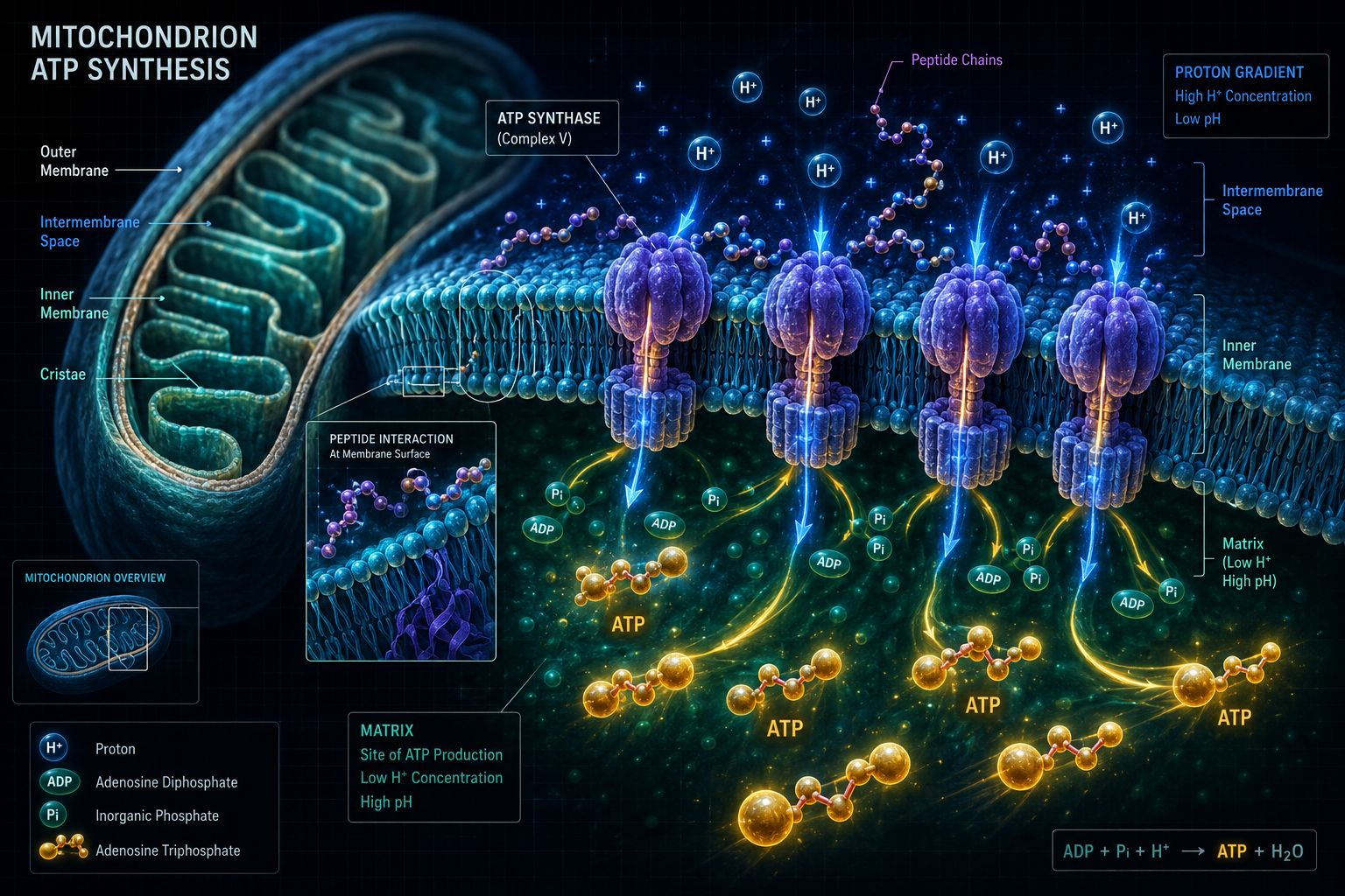
ATP is produced primarily inside the mitochondria through a process called oxidative phosphorylation. The inner mitochondrial membrane houses ATP synthase complexes that harness the energy from a proton gradient to convert ADP into ATP. This continuous cycle of synthesis and hydrolysis drives muscle contraction, protein synthesis, ion transport, and virtually every other energy-demanding cellular event.

What makes ATP especially relevant to peptide research is its dual role. It functions both as a fuel molecule and as an extracellular signaling agent. When released from cells, ATP activates purinergic receptors, particularly P2 receptors, which regulate tissue responses including inflammation, wound healing, and mechanosensation. Research into mechanosensitive channels such as Piezo1 has shown that ATP release triggered by physical stimuli plays a key role in how tissues adapt to mechanical stress.

Beyond energy transfer, ATP has been shown to suppress the fibrillation of amyloid peptides associated with neurodegenerative conditions such as Alzheimer's disease. This finding positions ATP not merely as a passive fuel but as an active modulator of peptide behavior in biological systems.

Key ATP functions at a glance:

Function Mechanism
Energy transfer Phosphate bond hydrolysis
Cell signaling Purinergic receptor activation
Peptide modulation Amyloid fibrillation suppression
Skin cell regulation Calcium mobilization in keratinocytes

How Peptides Influence the Role of Adenosine Triphosphate (ATP) in Cellular Energy Research

How Peptides Influence the Role of Adenosine Triphosphate (ATP) in Cellular Energy Research

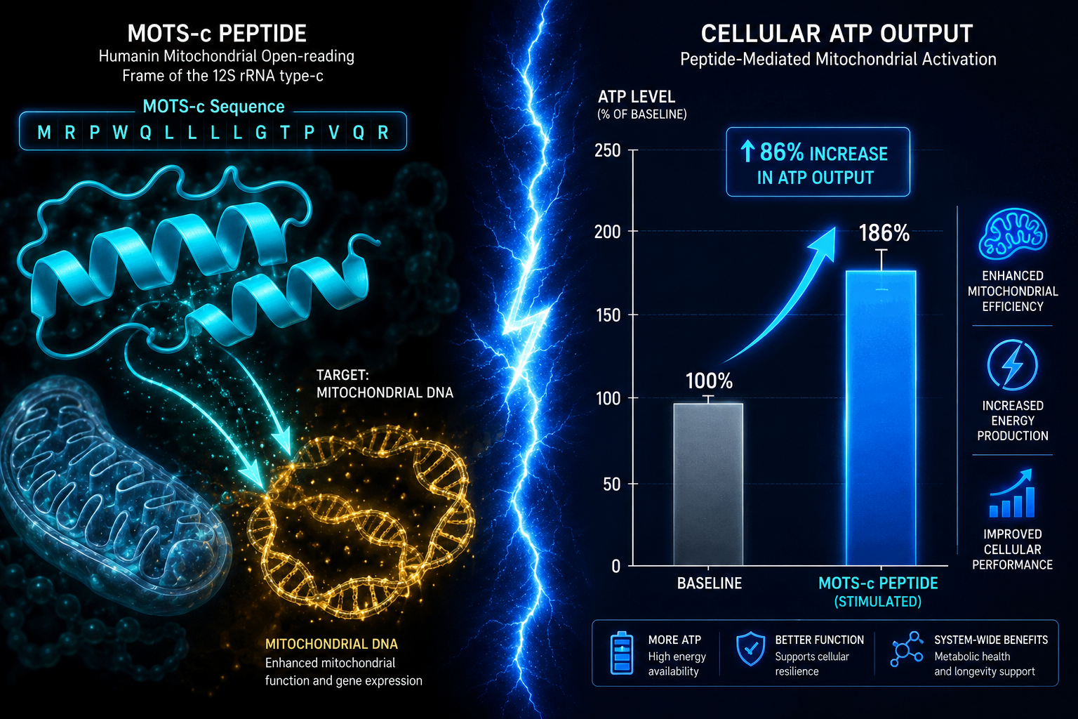
Peptides are not passive bystanders in energy metabolism. Several research-grade peptides interact directly with mitochondrial function and ATP output. Among the most studied is MOTS-c, a mitochondria-derived peptide encoded within mitochondrial DNA. Research on MOTS-c and mitochondrial dynamics shows that this peptide translocates to the nucleus under metabolic stress, where it activates pathways that restore ATP production efficiency.

MOTS-c is particularly notable because it appears to act as a retrograde signal from the mitochondria to the nucleus, coordinating the cell's response to energy deficits. This places it at the center of the peptide-ATP relationship. Research on MOTS-c and metabolic stress responses further supports its role in maintaining mitochondrial homeostasis during oxidative challenge.

Another well-researched peptide in this context is SS-31 (elamipretide). This tetrapeptide targets cardiolipin on the inner mitochondrial membrane, stabilizing the architecture needed for efficient ATP synthase function. Detailed SS-31 mitochondrial research themes document how this peptide reduces mitochondrial membrane potential loss and preserves ATP output under conditions of oxidative stress. Related work on SS-31 mitochondrial dynamics reinforces these findings across multiple tissue models.

GHK-Cu also appears in this research landscape. Studies reviewed in GHK-Cu longevity research themes suggest this copper-binding tripeptide supports mitochondrial gene expression, indirectly supporting ATP production capacity in aging tissue models.


Research Applications and the Broader Significance of ATP-Peptide Interactions

Research Applications and the Broader Significance of ATP-Peptide Interactions

The role of adenosine triphosphate (ATP) in peptide-mediated cellular energy research extends well beyond basic science. Oral ATP supplementation studies have demonstrated measurable improvements in strength, power output, fatigue reduction, and cardiovascular efficiency, suggesting that systemic ATP availability is a modifiable variable in performance and recovery research.

Bioelectronic applications have also emerged. ATPases, the enzymes that hydrolyze ATP, have been integrated into hybrid biological-electronic devices capable of converting chemical energy into electrical signals. Tandem mass spectrometry has advanced understanding of ATPase catalytic mechanisms at the molecular level, enabling more precise research into how peptides modulate these enzymes.

For researchers exploring the intersection of longevity and mitochondrial health, the connection between NAD+ metabolism and ATP synthesis is equally important. Reviewing NAD+ scientific evidence provides context for how upstream cofactors feed into ATP production pathways, and how peptides may amplify those effects.

Additionally, mitochondrial longevity focus research highlights the growing interest in peptides that target mitochondrial biogenesis as a strategy for extending cellular healthspan.


Conclusion

The relationship between ATP and peptide signaling is one of the most consequential areas in current cellular energy research. ATP is not simply a fuel molecule. It is a dynamic regulator of peptide behavior, mitochondrial function, and intercellular communication. Peptides such as MOTS-c and SS-31 demonstrate that targeted molecular interventions can meaningfully influence ATP production, opening research pathways relevant to aging, metabolic disease, and tissue repair.

Actionable next steps for researchers:

  • Review published data on SS-31 and MOTS-c mechanisms before designing mitochondrial energy studies.
  • Consider the interplay between NAD+ pathways and ATP synthesis when evaluating peptide protocols.
  • Examine mechanosensitive ATP release pathways when studying tissue-level peptide effects.
  • Source research-grade peptides from verified suppliers to ensure assay reliability and reproducibility.

Understanding the full scope of ATP's role in peptide-mediated cellular energy research is not optional for serious investigators. It is the foundation upon which meaningful experimental design is built.

https://www.puretestedpeptides.com/wp-content/uploads/2026/07/The-Role-of-Adenosine-Triphosphate-ATP-in-Peptide-Mediated-Cellular-Energy-Research.png 1024 1536 Pure Tested https://www.puretestedpeptides.com/wp-content/uploads/2026/01/buy-peptides-online.jpg Pure Tested2026-07-01 13:03:522026-07-20 15:01:18The Role of Adenosine Triphosphate (ATP) in Peptide-Mediated Cellular Energy Research
×

Helpful Links

  • My account
  • Cart
  • Checkout
  • Refund and Returns Policy
  • Privacy Policy
  • SMS Privacy Policy
  • Login
  • My Account
  • Logout

USA Made Lab Tested Peptides

All products are sold for research, laboratory, or analytical purposes only, and are not for human consumption


Pure Tested Peptides is a chemical supplier. Pure Tested Peptides is not a compounding / chemical compounding facility as defined under 503A of the Federal Food, Drug, and Cosmetic act. Pure Tested Peptides is not an outsourcing facility as defined under 503B of the Federal Food, Drug, and Cosmetic act.

The statements made within this website have not been evaluated by the US Food and Drug Administration. The products we offer are not intended to diagnose, treat, cure or prevent any disease.

Human/Animal Consumption Prohibited. Laboratory/In-Vitro Experimental Use Only

Scroll to top Scroll to top Scroll to top