

Prednisone, Methylprednisolone, and Dexamethasone: Steroid Controls for Metabolic and Tissue-Repair Peptide Models
Roughly one in four hospitalized patients in 2026 receives a systemic glucocorticoid at some point during their stay, yet these same drugs are used deliberately as controlled stressors in peptide research to provoke metabolic and inflammatory states that mirror real disease. Understanding how prednisone, methylprednisolone, and dexamethasone function as steroid controls for metabolic and tissue-repair peptide models is not optional for rigorous experimental design; it is foundational to producing results that translate beyond the bench.
Key Takeaways
- Prednisone, methylprednisolone, and dexamethasone differ in potency, half-life, and receptor kinetics, making steroid selection critical in peptide control arms.
- Their dose-equivalency relationship, roughly 5 mg prednisone equals 4 mg methylprednisolone equals 0.75 mg dexamethasone, must be matched carefully across experimental groups.
- Glucocorticoids disturb glucose handling, insulin sensitivity, adipokine signaling, and HPA axis function, each of which can confound peptide readouts.
- Dexamethasone is the most widely used positive control for anti-inflammatory peptide benchmarking in both in vivo and clinical settings.
- Adverse-event profiles, including infection risk, edema, and impaired tissue repair, must be accounted for when interpreting peptide efficacy data against these controls.
How Glucocorticoids Function as Metabolic Messengers and Confounders
Glucocorticoids are described in current mechanistic literature as "pivotal metabolic messengers" that reshape transcriptional programs governing glucose and lipid metabolism. When researchers introduce prednisone, methylprednisolone, or dexamethasone into a peptide experiment, they are not adding a neutral background agent. Each drug activates the glucocorticoid receptor (GR), triggering cascades that drive hepatic gluconeogenesis, reduce peripheral glucose uptake, impair beta-cell function, and disturb hypothalamic signaling related to appetite and energy balance.
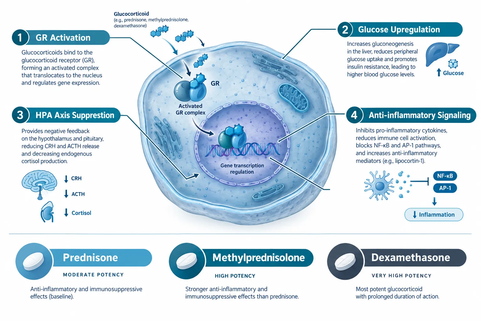
These disruptions translate directly into confounders. A peptide targeting insulin sensitivity, appetite regulation, or fat oxidation, such as those explored in GLP-1 peptide research, will produce very different results in a glucocorticoid-treated animal versus a naive one. Unless the steroid control arm is precisely defined, dose-matched, and pharmacokinetically characterized, effect-size estimates become unreliable.
Key metabolic confounders introduced by glucocorticoid controls:
| Variable | Effect |
|---|---|
| Glucose handling | Elevated fasting glucose, impaired insulin signaling |
| Adipokine profile | Reduced adiponectin, altered leptin sensitivity |
| HPA axis | Suppression of endogenous cortisol feedback |
| Immune tone | Reduced cytokine production, altered macrophage polarization |
| Edema and fluid retention | Altered sodium handling, tissue fluid balance |
For metabolic peptide researchers exploring tools like tesa peptide benefits or MOTS-C peptide, these steroid-induced metabolic shifts represent the very conditions the peptide may be designed to counter, making glucocorticoids both indispensable tools and sources of potential bias.
Prednisone, Methylprednisolone, and Dexamethasone: Steroid Controls for Metabolic and Tissue-Repair Peptide Models, Dose Equivalency and Pharmacokinetic Design
Choosing the wrong steroid, or the wrong dose, can invalidate an entire peptide experiment. Systems pharmacology modeling work has developed detailed eight-parameter GR activation models for all three drugs, with full human pharmacokinetic curves for both intravenous and oral routes. These models confirm that while the three steroids share a common receptor target, their binding kinetics, half-lives, and exposure profiles diverge substantially.
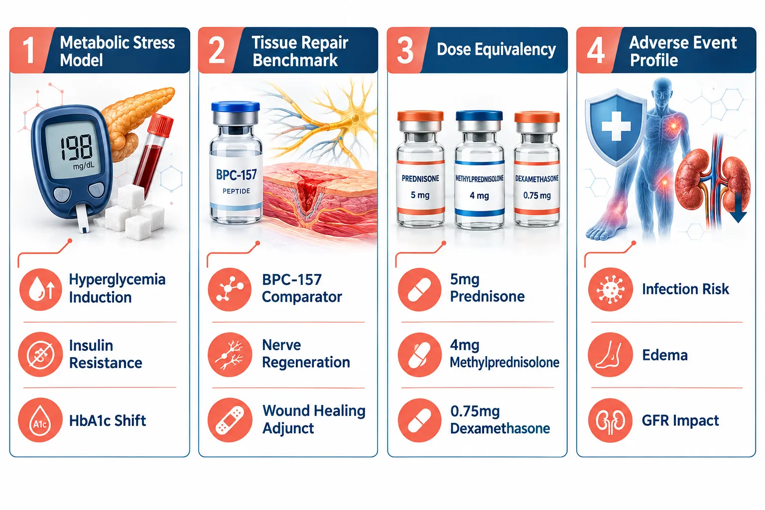
Potency equivalency (anti-inflammatory basis):
- 5 mg prednisone, moderate potency, short half-life, requires hepatic conversion to prednisolone
- 4 mg methylprednisolone, slightly higher potency, minimal mineralocorticoid activity
- 0.75 mg dexamethasone, highest relative potency, longest effective duration, no mineralocorticoid activity
These figures matter enormously when selecting a control arm. An experiment pairing a GLP-1 class peptide, such as those available as GLP-3 peptides, against a methylprednisolone-induced hyperglycemia model needs to document the exposure metric (AUC, Cmax) used for steroid dosing, not just the nominal dose.
Real-world clinical evidence reinforces how severe the metabolic challenge can be. High-dose methylprednisolone has precipitated severe hyperglycemia in patients, with baseline HbA1c values exceeding 12%, requiring multi-drug management and producing meaningful weight loss only after months of intervention. This type of acute metabolic disruption represents the stress-test condition that metabolic peptide models aim to rescue, making it one of the most clinically valid control paradigms available.
Research note: When using dexamethasone as a metabolic control, its longer receptor occupancy can mask early peptide-driven recovery signals. Timing of peptide administration relative to the steroid dosing window must be pre-specified in the protocol.
Applying Steroid Controls in Tissue-Repair Peptide Research
The role of prednisone, methylprednisolone, and dexamethasone as steroid controls for metabolic and tissue-repair peptide models extends well beyond glucose. In tissue-repair contexts, dexamethasone has been co-administered alongside regenerative peptides, including BPC-157, TB-500, and ARA-290, in nerve injury protocols, functioning simultaneously as an anti-inflammatory adjunct and a clinical anchor against which experimental peptide effects are contextualized.

This dual role is important to understand. In anti-inflammatory peptide benchmarking, novel short-chain compounds are routinely tested against dexamethasone-induced suppression of ear swelling in murine models, with efficacy described as "comparable to dexamethasone." That phrasing signals that dexamethasone is not just a co-treatment, it is the reference standard.
For researchers sourcing compounds for these protocols, reviewing peptide supplier comparisons and ensuring lab-tested peptides are used alongside characterized steroid controls is essential for reproducibility. The GHK-Cu peptide sourcing guide offers a practical example of how sourcing rigor connects to experimental credibility.
Tissue-repair confounders to account for in steroid-controlled models:
- Wound healing impairment: Glucocorticoids suppress collagen synthesis and keratinocyte proliferation, which can artifactually depress peptide wound-healing readouts.
- Vascularization suppression: Anti-angiogenic effects of dexamethasone may mask pro-vascular peptide actions.
- Immune modulation: Steroid-driven cytokine suppression alters the inflammatory milieu in which tissue-repair peptides operate.
- Edema reduction: Glucocorticoid-driven edema reduction can overlap with, or obscure, peptide-driven fluid normalization effects.
Evidence from IgA nephropathy real-world data is instructive: systemic glucocorticoid use was associated with higher infection rates, hospitalizations, and no clear glomerular filtration benefit compared with non-steroid cohorts. This adverse-event burden underscores why peptide-based or steroid-sparing strategies represent a meaningful research direction, and why the steroid control arm must be treated as an active, risk-carrying intervention, not a passive baseline.
Conclusion
Prednisone, methylprednisolone, and dexamethasone are not interchangeable background agents in peptide research. Each brings a distinct pharmacokinetic fingerprint, a specific metabolic disruption profile, and a well-documented adverse-event burden that can confound, amplify, or mask peptide-driven effects across glucose, appetite, inflammatory signaling, immune suppression, edema, and wound repair.
Actionable next steps for researchers:
- Pre-specify steroid selection and dose using validated equivalency ratios and exposure metrics (AUC, Cmax), not nominal dose alone.
- Document the timing gap between steroid administration and peptide intervention, GR occupancy duration varies significantly across the three drugs.
- Account for metabolic confounders (glucose, adipokines, HPA axis) as co-primary readouts, not secondary observations.
- Use dexamethasone as the positive control benchmark for anti-inflammatory peptide efficacy, and match dose to the specific inflammatory model used.
- Treat the steroid control arm as an active pharmacological condition with its own risk profile, design and interpret accordingly.
Researchers who build these considerations into their protocols from the outset will produce peptide-model data that is both scientifically defensible and clinically meaningful.