Call or Text 727-513-9780
  • Shopping Cart Shopping Cart
    0Shopping Cart
Pure Tested Peptides | America's most trusted Peptides for sale online
  • Peptides for sale
    • Oral Peptides for sale
      • Peptide Capsules for sale
      • BPC 157 Capsules 1000mcg
      • SLU-PP-332 Capsules | 1000 mcg
      • 5-Amino-1MQ 50mg Capsules
      • Tesofensine 500mcg
    • All Peptides for sale
    • Peptide Sprays
      • BPC 157 Nasal Spray Kit
      • BPC-157 TB500 Nasal Spray Kit
      • Semax Nasal Spray 10mg
      • Selank – Nasal Spray Kit – 10mg
      • Epithalon 50MG Nasal Spray Kit
      • Ipamorelin 10mg Nasal Spray
      • Klow Nasal Spray (BPC-157 + TB-500 + GHK-Cu + KPV) | 80mg
      • Hulk Nasal Spray Tesa / Ipa Blend 6/3 MG
      • Klow Nasal Spray
      • NAD + 500 mg Nasal Spray
      • PT-141 Nasal Spray Kit
    • GHRH Peptides
      • Ipa Peptides
      • CJC-1295 Peptides
        • CJC-1295 with DAC 5 mg
        • CJC-1295 without DAC 5 mg
        • CJC-1295 Ipa 10mg
      • Tesa Peptides
        • Tesa Peptide
        • Tesa 20 mg
    • GHK-Cu Peptides
      • All GHK-Cu Peptides
      • GHK-Cu 100mg
      • KLOW Peptide Blend – Buy KLOW blend online
    • BPC Peptides
      • All BPC Peptides
      • BPC-157
      • BPC-157 TB-500
      • BPC 157 capsules 1000mcg
    • SLU-PP-332 Peptides
      • All SLU-PP-332 Peptides
      • SLU-PP-332 5mg
    • GLP3 Peptides
    • PT-141 Peptides
      • PT-141 Peptides for sale
      • PT-141 10mg
      • PT-141 Nasal Spray
    • CAG Peptides
      • Lipo-C Peptide Blend
      • CAG 5mg
      • CAG 10mg
    • MOTS-C Peptides
      • MOTS-C Peptides for sale
      • MOTS-c peptide
      • MOTS-c 10mg *6 pack*
    • 5 Amino 1MQ Peptides
      • 5 Amino 1MQ Peptides for sale
      • 5-Amino-1MQ 50mg Capsules
      • 5-Amino-1MQ 5mg
    • Epithalon Peptides
      • Epithalon Peptides for sale
      • Epithalon 10mg
      • Epithalon 50mg
  • Shop
    • GLPs
      • 5-Amino-1MQ 50mg Capsules
      • 5-Amino-1MQ 5mg
      • L-Carnitine 500mg/ml
      • Tesofensine 500mcg
      • SLU-PP-332 5mg
      • MOTS-c 10mg *6 pack*
    • Epithalon & BPC Peptides
      • Epithalon 10mg
      • Epithalon 50mg
      • BPC-157
      • BPC 157 capsules 1000mcg
      • BPC-157 TB-500
      • BPC-157 TB500 Nasal Spray Kit
      • BPC 157 Nasal Spray Kit
    • BPC TB-500 & NAD+ Peptides
      • NAD+ 500 mg
      • KLOW Peptide Blend – Buy KLOW blend online
      • GLOW Peptide Blend
      • TB 500 5mg
      • BPC 157 capsules 1000mcg – Supplement
      • BPC 157 Nasal Spray Kit
      • BPC-157
      • BPC-157 TB500 Nasal Spray Kit
      • BPC-157 TB-500
      • BPC 157 capsules 1000mcg
    • LL-37 Peptide
      • LL-37 10 mg
    • MOTS-C & Selank
      • MOTS-c peptide
      • Selank 10mg
    • GHK Peptides
      • GHK-Cu 100mg
      • GLOW Peptide Blend
      • KLOW Peptide Blend – Buy KLOW blend online
  • COAs
  • Wholesale
    • Wholesale Peptides for sale
  • PTP FAQ
  • Affiliates
    • Selank Peptide: Advanced Pharmacological Mechanisms Underlying Its Anxiolytic and Nootropic Effects in Research
      • 5-Amino-1MQ Peptide: Detailed Mechanisms of NNMT Inhibition and Its Impact on Cellular Metabolism Research
        • MOTS-C Peptide: Unraveling Its Role in Mitochondrial Dynamics and Energy Metabolism Research
          • Epithalon Peptide and Telomerase Regulation: Investigating Its Impact on Cellular Senescence and Lifespan Research Models
            • GLP2-T vs GLP2 Tirz Peptide: Understanding the Naming, Mechanistic Nuances, and Research Implications for Gut Health
      • Peptides and Polypeptides in Modern Pharmacology: What Research on Metoprolol, Prednisone, and Amlodipine Reveals
        • Polypeptide Peptides vs NSAIDs: What Naproxen and Diclofenac Teach Tissue-Repair Researchers About BPC‑157 and TB‑500
          • Peptides and Polypeptides in Endocrine Pharmacology: How GLP-1, GLP-2, and GLP-3 Retatrutide Differ From Classic Drugs Like Prednisone and Amlodipine
          • 5‑Amino‑1MQ Peptide and NNMT Inhibition: How It Compares With Statins Like Atorvastatin in Adipose and Lipid Metabolism Research
      • Enclomiphene, Estrogen Receptor Signaling, and Luteinizing Phase Biology: What Hormone Researchers Should Measure
        • Mesenchymal Stem Cells, BPC‑157, and GHK‑Cu: How Tissue Repair Peptides Compare With Classic NSAIDs Like Naproxen in Injury Models
          • Peptide Calculator Use Cases Beyond Growth Hormone: Working Through GLP‑3 Retatrutide, MOTS‑c, and BPC‑157 Research Dosing
        • Polypeptide Peptides in Endocrine and Metabolic Pharmacology: Lessons From Amlodipine, Prednisone, and Metoprolol
        • Peptides Calculator for Advanced Blends: Worked Examples for Tesamorelin, CJC‑1295, and Ipamorelin Stacks
          • Peptides and Polypeptides in Human Physiology: How Molecular Size Shapes Research Applications
            • Selank Peptide: Advanced Pharmacological Mechanisms Underlying Its Anxiolytic and Nootropic Effects in Research
          • 5-Amino-1MQ Peptide: Detailed Mechanisms of NNMT Inhibition and Its Impact on Cellular Metabolism Research
            • MOTS-C Peptide: Unraveling Its Role in Mitochondrial Dynamics and Energy Metabolism Research
              • Epithalon Peptide and Telomerase Regulation: Investigating Its Impact on Cellular Senescence and Lifespan Research Models
                • GLP2-T vs GLP2 Tirz Peptide: Understanding the Naming, Mechanistic Nuances, and Research Implications for Gut Health
                  • Peptides and Polypeptides in Endocrine Pharmacology: How GLP-1, GLP-2, and GLP-3 Retatrutide Differ From Classic Drugs Like Prednisone and Amlodipine
                    • Polypeptide Peptides vs NSAIDs: What Naproxen and Diclofenac Teach Tissue-Repair Researchers About BPC‑157 and TB‑500
                      • Peptides and Polypeptides in Modern Pharmacology: What Research on Metoprolol, Prednisone, and Amlodipine Reveals
                        • 5‑Amino‑1MQ Peptide and NNMT Inhibition: How It Compares With Statins Like Atorvastatin in Adipose and Lipid Metabolism Research
                        • Enclomiphene, Estrogen Receptor Signaling, and Luteinizing Phase Biology: What Hormone Researchers Should Measure
                        • Mesenchymal Stem Cells, BPC‑157, and GHK‑Cu: How Tissue Repair Peptides Compare With Classic NSAIDs Like Naproxen in Injury Models
                        • Peptide Calculator Use Cases Beyond Growth Hormone: Working Through GLP‑3 Retatrutide, MOTS‑c, and BPC‑157 Research Dosing
                        • Polypeptide Peptides in Endocrine and Metabolic Pharmacology: Lessons From Amlodipine, Prednisone, and Metoprolol
                        • Peptides Calculator for Advanced Blends: Worked Examples for Tesamorelin, CJC‑1295, and Ipamorelin Stacks
                        • Peptides and Polypeptides in Human Physiology: How Molecular Size Shapes Research Applications
                        • Selank Peptide: Advanced Pharmacological Mechanisms Underlying Its Anxiolytic and Nootropic Effects in Research
                        • MOTS-C Peptide: Unraveling Its Role in Mitochondrial Dynamics and Energy Metabolism Research
                        • Peptides and Polypeptides in Modern Pharmacology: What Research on Metoprolol, Prednisone, and Amlodipine Reveals
                        • Peptides and Polypeptides in Endocrine Pharmacology: How GLP-1, GLP-2, and GLP-3 Retatrutide Differ From Classic Drugs Like Prednisone and Amlodipine
                        • 5-Amino-1MQ Peptide: Detailed Mechanisms of NNMT Inhibition and Its Impact on Cellular Metabolism Research
                        • Mesenchymal Stem Cells, BPC‑157, and GHK‑Cu: How Tissue Repair Peptides Compare With Classic NSAIDs Like Naproxen in Injury Models
                        • Peptide Calculator Use Cases Beyond Growth Hormone: Working Through GLP‑3 Retatrutide, MOTS‑c, and BPC‑157 Research Dosing
                        • GLP2-T vs GLP2 Tirz Peptide: Understanding the Naming, Mechanistic Nuances, and Research Implications for Gut Health
                        • Polypeptide Peptides in Endocrine and Metabolic Pharmacology: Lessons From Amlodipine, Prednisone, and Metoprolol
                        • Polypeptide Peptides vs NSAIDs: What Naproxen and Diclofenac Teach Tissue-Repair Researchers About BPC‑157 and TB‑500
                        • Peptides and Polypeptides in Human Physiology: How Molecular Size Shapes Research Applications
                        • Peptides Calculator for Advanced Blends: Worked Examples for Tesamorelin, CJC‑1295, and Ipamorelin Stacks
                        • 5‑Amino‑1MQ Peptide and NNMT Inhibition: How It Compares With Statins Like Atorvastatin in Adipose and Lipid Metabolism Research
                        • Enclomiphene, Estrogen Receptor Signaling, and Luteinizing Phase Biology: What Hormone Researchers Should Measure
                        • Epithalon Peptide and Telomerase Regulation: Investigating Its Impact on Cellular Senescence and Lifespan Research Models
                        • Best research protocol Klow blend
                        • best time to take BPC-157
                        • best time to take DSIP (Delta Sleep Inducing Peptide)
                        • best time to take CJC-1295
                        • best time to take AOD-9604
                        • best time to take Follistatin 344
                        • best time to take Ipamorelin
                        • best time to take MK-677 (Ibutamoren)
                        • best time to take Ligandrol (LGD-4033) — research compound
                        • best time to take Ostarine (MK-2866) — research compound
                        • best time to take GHK-CU
                        • best time to take TB-500
                        • best time to take MOTS-c
                        • best time to take Semax
                        • best time to take RAD-140 (Testolone) — research compound
                        • best time to take Thymosin Alpha-1
                        • best time to take PEG-MGF
                        • Biolife Plasma, Octapharma Plasma, and Research Peptides: How Plasma Donation Labs Differ From Peptide Suppliers
                        • best time to take YK-11 — research compound
                        • best time to take PT-141 (Bremelanotide)
                        • Best research protocol Klow blend
                        • 5-Amino-1MQ and MOTS-C Synergy: Metabolic Signaling, Mitochondria, and Research Design
                        • BPC-157 and TB-500: Investigating Their Combined Effects on Angiogenesis and Cellular Migration in Tissue Repair Models
                        • BPC-157 Peptide: Gut Barrier Function, Inflammation, and Tissue-Recovery Research
                        • 5‑Amino‑1MQ and MOTS‑c Synergy in Metabolic Research: Designing NNMT and Mitochondrial Biogenesis Stacks
                        • CJC-1295 with DAC vs. Without DAC: Half-Life, Release Kinetics, and Research Implications
                        • CJC‑1295 with DAC vs. Without DAC: Expanding on Half‑Life Differences Using Tesamorelin and Ipamorelin Blend Case Studies
                        • Collagen Biology and Copper‑Binding Peptides: How GHK‑Cu, Glow Blend, and Klow Blend Interact with Skin and Connective Tissue
                        • Collagen Biology and Regenerative Peptides: How GHK‑Cu, Glow Blend, and Klow Blend Affect Extracellular Matrix Research
                    • DNA, Telomeres, and Longevity Peptides: Positioning Epithalon and MOTS‑c in Genetic Aging Research
                      • Enclomiphene Citrate: serm Mechanism, Testosterone Research, and Stack Compatibility
                        • Enclomiphene vs Enclomiphene Citrate: Formulation, Bioavailability, and Research Distinctions
                        • Epithalon Peptide Research: Telomerase Activation, Aging, and Pineal Gland Function
                        • Estrogen Receptor Signaling and Enclomiphene: How Selective Modulators Compare with Classic Polypeptide Hormones
                        • GHK-Cu Peptide: Advanced Mechanisms in Extracellular Matrix Remodeling and Wound Healing Research
                        • GHK-Cu Peptide: Collagen Synthesis, Wound Repair, and Skin-Barrier Research Models
                        • GLP-1 vs GLP-3 vs GLP-2: Peptide Classification and Research Applications
                        • GLP-2 Peptide Research Guide: Gut Barrier Function, Nutrient Absorption, and Intestinal Recovery Models
                        • GLP-3 Retatrutide vs. GLP-1 Drugs: What Triple-Agonist Biology Changes in Research Models
                        • Ipamorelin and Tesamorelin Combination: Synergistic GH Secretagogue Research and Dosing Protocols
                        • GLP2 Tirz Peptide: What It Is, Why the Name Exists, and How Researchers Should Interpret It
                        • Klow Blend Peptide Nasal Spray: What the Formulation Is Trying to Do in Cognitive Research
                        • Mitochondria, NNMT Inhibition, and Peptide Modulators: Where MOTS‑c and 5‑Amino‑1MQ Fit in Cellular Energy Research
                        • MOTS-c Peptide: Mitochondrial Function, Energy Metabolism, and What Researchers Measure
                        • MOTS-c vs. 5-Amino-1MQ: Which Metabolic Research Questions Each Compound Actually Answers
                        • Nasal Spray Peptides: Bioavailability, Administration, and Semax/Selank Research Applications
                        • PT-141 Peptide Research: Mechanism of Action and Melanocortin Receptor Signaling
                        • Retatrutide for Research: Mechanism, Structure, and GLP-1/GLP-3 Dual Action
                        • Retatrutide for Obesity and Type 2 Diabetes: What the Latest Trial Data Suggest
                        • Tesofensine Peptide Research: Mechanism, Appetite Suppression, and Neuropeptide Y Pathways
  • Contact
    • Contact Customer Service
    • Text Customer Support
  • About US
  • Shop all peptides
  • Affiliate Program
    • Affiliate Signup
  • Login / Register Login / Register Page Link Login / Register Page Link
  • Click to open the search input field Click to open the search input field Search
  • Menu Menu

Tag Archive for: telomerase activity

Epithalon in Cellular-Aging Models: Separating Telomerase Activity, Telomere Length, DNA Damage, and Senescence

Epithalon in Cellular-Aging Models: Separating Telomerase Activity, Telomere Length, DNA Damage, and Senescence

October 14, 2026/0 Comments/in Uncategorized/by

Fewer than 1% of peptides studied in aging research generate data across four distinct cellular endpoints simultaneously, yet Epithalon, a synthetic tetrapeptide derived from the pineal gland protein epithalamin, has accumulated a body of evidence touching telomerase activation, telomere elongation, DNA damage reduction, and senescence delay. The challenge for researchers in 2026 is not whether Epithalon affects these pathways, but how to cleanly separate them. Conflating a short-term telomerase signal with genuine telomere lengthening, or mistaking reduced oxidative damage for a true senescence delay, leads to inflated claims and flawed experimental design. This article presents a structured, longitudinal assay framework for studying Epithalon in cellular-aging models: separating telomerase activity, telomere length, DNA damage, and senescence with methodological rigor.

Key Takeaways

  • Telomerase activation and telomere lengthening are distinct, time-separated events that require different assays and measurement windows.
  • Epithalon produces approximately 30-35% telomere extension in cell models, but this effect takes weeks to manifest after initial telomerase signaling.
  • DNA damage reduction appears to operate through a mechanism partially independent of telomere dynamics.
  • Senescence delay, measured by SA-beta-galactosidase activity and p21 expression, represents a downstream phenotypic outcome rather than a primary mechanism.
  • As of 2026, Epithalon holds no regulatory approval; all findings remain in the preclinical and early exploratory human-data stage.

The Four Endpoints Are Not the Same Thing

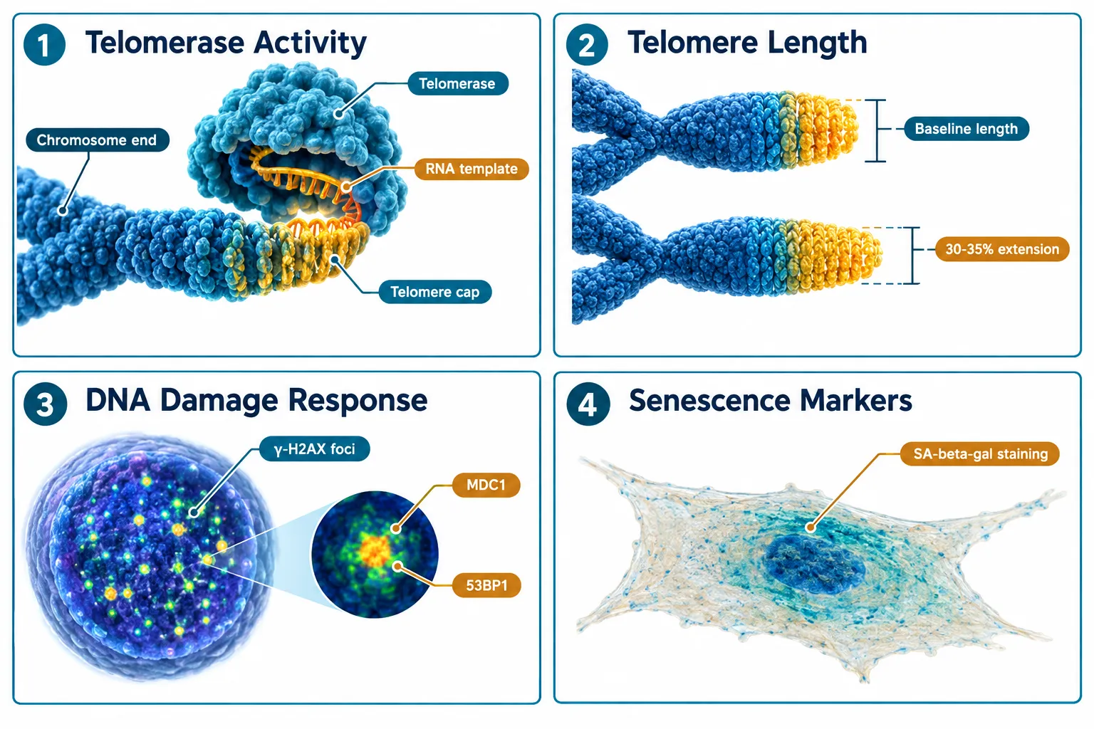
A common error in Epithalon literature is treating telomerase activity as a proxy for telomere length. These are related but temporally and mechanistically distinct. Telomerase is an enzyme; its activity can spike within 24 to 72 hours of peptide exposure. Telomere length, by contrast, reflects cumulative replication history and changes over days to weeks, not hours.
The Four Endpoints Are Not the Same Thing

Similarly, DNA damage response, quantified through gamma-H2AX foci counts or comet assay tail moment, reflects genomic stress that may or may not be telomere-driven. Senescence, the terminal arrest of proliferating cells, is a downstream phenotype shaped by all three upstream signals combined. Researchers studying SS-31 mechanism and research alongside Epithalon have noted similar endpoint-separation challenges in mitochondrial peptide work, underscoring that this is a broader methodological issue in peptide aging research.

The four endpoints and their primary assays:

Endpoint Primary Assay Measurement Window
Telomerase activity TRAP assay, RT-qPCR for hTERT 24-72 hours
Telomere length Q-FISH, Southern blot TRF 2-6 weeks
DNA damage Comet assay, gamma-H2AX foci 48 hours to 2 weeks
Senescence SA-beta-gal, p21/p16 expression 3-8 weeks

Building a Longitudinal Assay Plan for Epithalon in Cellular-Aging Models

The most defensible experimental design treats each endpoint as a separate dependent variable with its own measurement schedule. Collapsing them into a single time point produces noise, not signal.
Building a Longitudinal Assay Plan for Epithalon in Cellular-Aging Models

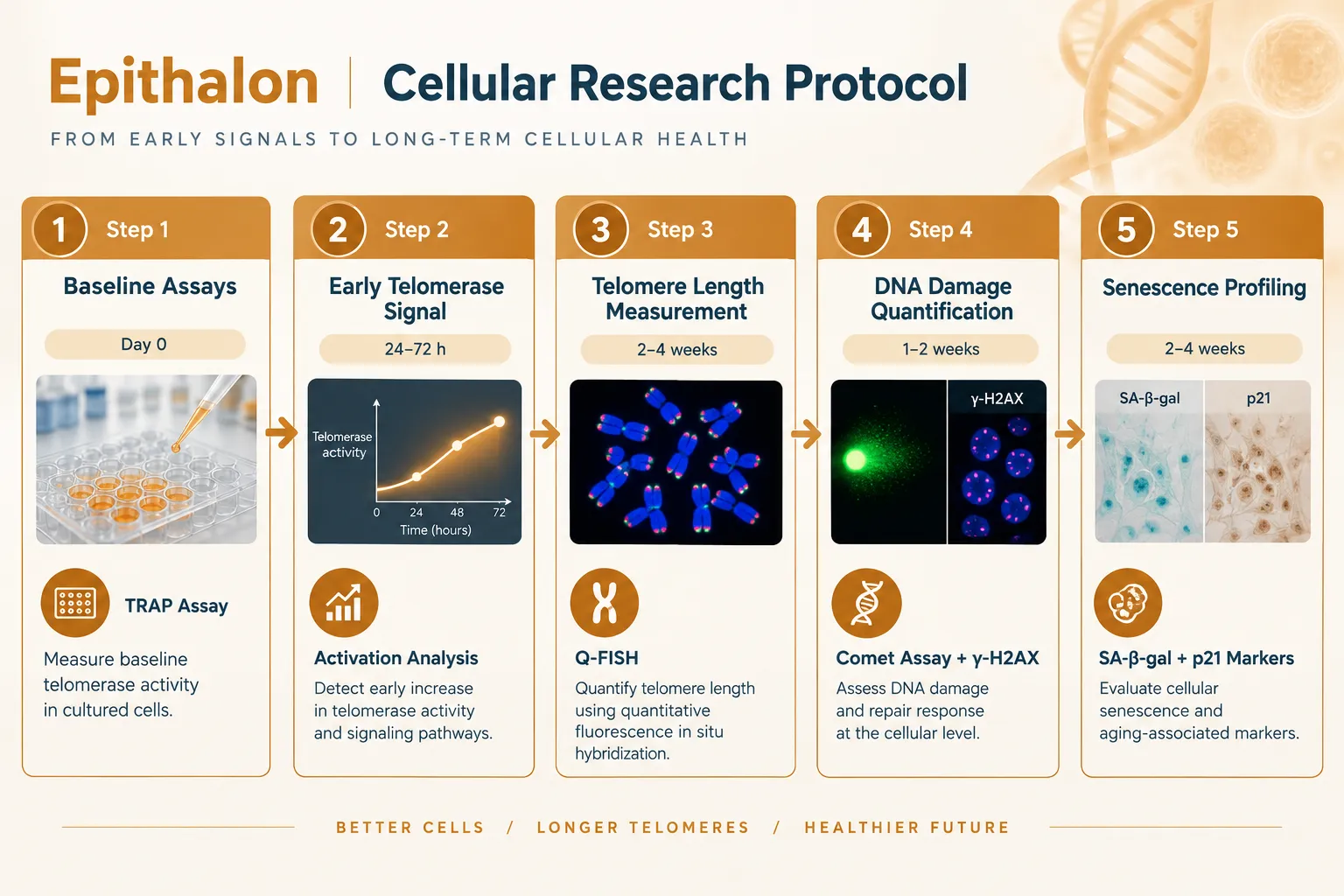
A five-stage longitudinal framework:

Stage 1, Baseline (Day 0). Establish pre-treatment values for all four endpoints in the same cell population. Human diploid fibroblasts (IMR-90) and WI-38 lines are well-validated for this purpose. Record passage number, population doubling level, and baseline SA-beta-galactosidase positivity.

Stage 2, Early telomerase signal (24-72 hours). Apply TRAP assay and hTERT mRNA quantification. Magnitude of activation in published cell models has ranged from modest to substantial depending on cell type and peptide concentration. This window captures the enzymatic signal but says nothing about whether telomeres have actually lengthened.

Stage 3, Telomere length measurement (Weeks 2-4). Use quantitative fluorescence in situ hybridization (Q-FISH) or terminal restriction fragment (TRF) analysis. Independent replication work from multiple groups has reported approximately 30-35% telomere extension relative to untreated controls at this stage. Critically, this measurement must be taken at a population doubling interval, not a fixed calendar interval, to control for proliferation rate differences.

Stage 4, DNA damage quantification (Days 3-14). Run parallel comet assays and gamma-H2AX immunofluorescence. Evidence from multiple cell-model studies suggests that Epithalon reduces markers of oxidative DNA damage even in cells where telomere lengthening is modest, pointing to a partially independent mechanism, possibly involving antioxidant enzyme upregulation rather than telomere-mediated genomic stability.

Stage 5, Senescence profiling (Weeks 4-8). Quantify SA-beta-galactosidase-positive cells, p21 and p16 protein expression, and secretory phenotype markers (IL-6, IL-8 as SASP indicators). Extended replicative lifespan, the number of population doublings before crisis, is the gold-standard functional readout. Studies have reported meaningful delays in replicative senescence onset in Epithalon-treated cultures.

Key insight: Running all five stages in parallel, rather than sequentially, allows researchers to build a causal map rather than a simple correlation between peptide exposure and aging phenotype.

For researchers designing controlled experimental models, the quality-control workflow considerations discussed in TB-500 controlled experimental models and QC workflow offer transferable lessons on blinding, passage controls, and contamination prevention.


Evidence Across Model Systems: What the Data Actually Show

Separating Epithalon in cellular-aging models from broader claims requires looking at each model type individually.
Evidence Across Model Systems: What the Data Actually Show

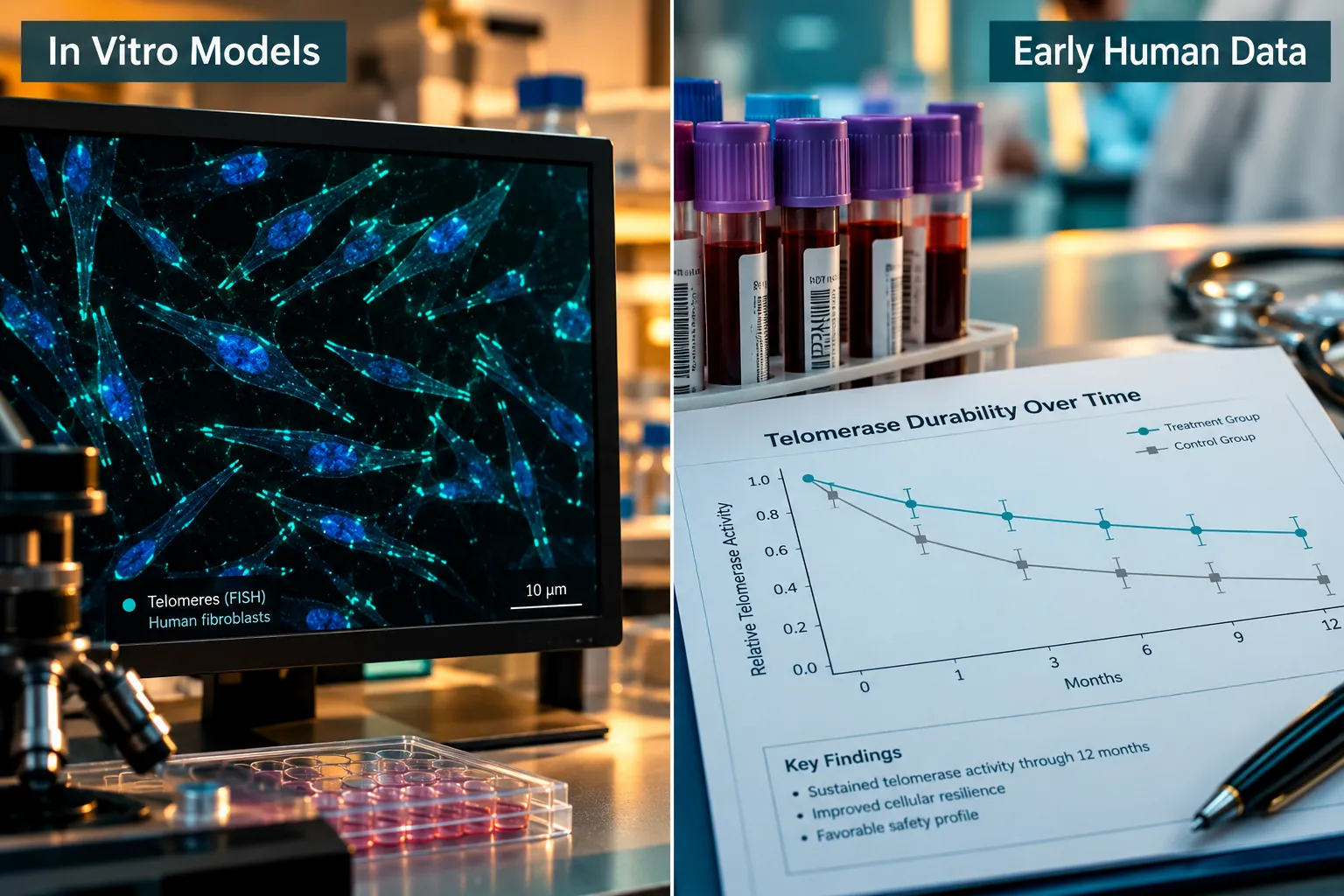
In vitro cell models provide the most mechanistically detailed data. Telomerase activation is consistently observed. Telomere extension at the 30-35% range has been replicated by independent groups including work published from Brunel University and affiliated researchers in 2025-2026. DNA damage markers decrease. Senescence is delayed. The effect size varies by cell type, with fetal lung fibroblasts showing stronger responses than primary skin fibroblasts.

Animal aging models extend findings to whole-organism endpoints. Rodent studies have reported increased mean and maximum lifespan, reduced lipid peroxidation markers, and improved immune function in aged animals. These outcomes are consistent with the cellular data but introduce confounders, diet, housing stress, and genetic background, that make direct mechanistic attribution difficult.

Reproductive models represent a newer frontier. A 2025 study using bovine cumulus-oocyte complexes found that Epithalon treatment improved oocyte competence and reduced oxidative stress markers, suggesting that telomere-protective effects extend to non-somatic cell types with high replicative demand.

Early human data remain limited. Peripheral blood mononuclear cell studies have detected telomerase activity increases following Epithalon administration, with some evidence of durability extending weeks beyond the treatment period. These findings are preliminary and have not been replicated in adequately powered trials.

Researchers interested in how peptide sourcing quality affects experimental reproducibility can find relevant context in resources on where to buy SS-31 and Epithalon online and the importance of purity verification for research-grade compounds. Related mitochondrial aging research on SS-31 mitochondrial dynamics provides useful comparative context for understanding oxidative-stress endpoints.


Safety, Cancer Risk, and the Hype-Evidence Gap

No discussion of Epithalon in cellular-aging models: separating telomerase activity, telomere length, DNA damage, and senescence is complete without addressing the cancer risk question. Telomerase activation is a double-edged signal. While it protects normal somatic cells from replicative senescence, it is also a hallmark of most cancer cells. Preclinical data on Epithalon have not shown tumor-promoting activity in standard carcinogenicity assays, and some studies report anti-tumor effects in aged animals. However, long-term safety data in humans are absent.

The regulatory position as of 2026 is clear: Epithalon has no approved therapeutic indication in any major jurisdiction. It is available as a research compound, and its use in human subjects outside formal clinical trial protocols raises significant ethical and safety concerns.

The expert consensus in late 2026 is cautiously interested but appropriately skeptical. The mechanistic data are real and replicable. The translational gap, from cell culture to clinically meaningful human outcomes, remains wide. Researchers and commentators who collapse this gap prematurely do a disservice to a genuinely interesting area of science.


Conclusion

The value of studying Epithalon in cellular-aging models lies precisely in the discipline of keeping its four major endpoints, telomerase activity, telomere length, DNA damage, and senescence, analytically separate. A longitudinal assay plan that stages measurements across the appropriate biological windows, uses validated assays for each endpoint, and controls for proliferation rate and passage number will produce data that actually advances understanding. The 30-35% telomere extension finding is worth replicating rigorously. The DNA damage reduction signal deserves mechanistic dissection independent of telomere biology. The senescence delay data need larger, better-controlled models before translational claims are warranted.

Actionable next steps for researchers:

  • Design experiments with a minimum five-stage longitudinal structure, not a single endpoint snapshot.
  • Always measure telomerase activity and telomere length in separate assay windows, never use one as a proxy for the other.
  • Include DNA damage endpoints (comet assay, gamma-H2AX) as independent variables, not secondary observations.
  • Source research-grade Epithalon with documented purity certificates and replicate findings across at least two cell lines before drawing mechanistic conclusions.
  • Track the evolving regulatory and safety literature; the field is moving quickly in 2026 and responsible research requires staying current.
https://www.puretestedpeptides.com/wp-content/uploads/2026/10/epithalon-in-cellular-aging-models-separating-telomerase-activity-telomere-lengt.webp 1024 1536 https://www.puretestedpeptides.com/wp-content/uploads/2026/01/buy-peptides-online.jpg 2026-10-14 13:04:142026-10-14 13:04:14Epithalon in Cellular-Aging Models: Separating Telomerase Activity, Telomere Length, DNA Damage, and Senescence

Epithalon and Telomere Research: Designing Longitudinal Experiments That Separate Telomere Length From Senescence

October 5, 2026/0 Comments/in Uncategorized/by

Cover Image

Telomere length and cellular senescence are often treated as synonyms in aging research, but they are not the same thing, and conflating them produces flawed experiments. A 2025 study published in Biogerontology by Al-Dulaimi and colleagues demonstrated that the synthetic tetrapeptide epitalon (Ala-Glu-Asp-Gly) extends telomeres in four distinct human cell lines through two separate mechanisms, yet the study measured no downstream senescence endpoints. That gap is precisely where the field of Epithalon and Telomere Research: Designing Longitudinal Experiments That Separate Telomere Length From Senescence becomes both urgent and scientifically rich.

Key Takeaways

  • Epitalon extends telomeres in normal human cells primarily by upregulating hTERT and telomerase activity, and in certain cancer lines via the ALT pathway.
  • Telomere length and cellular senescence are related but distinct biological variables that require separate measurement in well-designed experiments.
  • Current evidence for epitalon is confined to in vitro and animal models; no well-controlled longitudinal human trials exist as of 2026.
  • Robust longitudinal study designs must track telomere metrics, telomerase activity, DNA-damage markers, and senescence-associated phenotypes in parallel arms over multiple passages.
  • Epitalon's greatest current value is as a probe compound for dissecting telomere biology in experimental systems, not as a clinically validated anti-aging therapy.

What Epitalon Does at the Molecular Level

Epithalon peptide telomere activation mechanism diagram

Epitalon is a four-amino-acid peptide originally derived from pineal gland research. Its molecular action is unusually well-characterized for a short peptide. In normal human fibroblasts and mammary epithelial cells, treatment at 0.2-1 μg/ml for four days produces dose-dependent telomere extension. The mechanism centers on hTERT, the catalytic subunit of telomerase. At 1 μg/ml, hTERT mRNA levels rose approximately 12-fold in one cell line and about 5-fold at 0.5 μg/ml in another, accompanied by measurable increases in telomerase enzyme activity.

In breast cancer cell lines (21NT and BT474), the picture shifts. Epitalon still extends telomeres, but the dominant mechanism is the Alternative Lengthening of Telomeres (ALT) pathway rather than canonical telomerase. This distinction matters enormously for experimental design: a peptide that engages two different telomere-maintenance mechanisms depending on cellular context cannot be treated as a single-mechanism probe without controlling for cell type.

For researchers exploring the broader landscape of peptide mechanisms, the Unlocking The Benefits Epithalon Peptide For Sale resource provides useful background on epitalon's molecular profile.

Key insight: Telomere extension via ALT in cancer lines raises legitimate safety questions about using telomere-activating agents in any setting where neoplastic cells may be present.


Why Telomere Length Is Not the Same as Senescence

Telomere length versus senescence marker split-screen comparison

The Hayflick limit describes how normal somatic cells stop dividing after a fixed number of replications. Short telomeres are one trigger for this arrest, but they are not the only one. Senescence can also be induced by oxidative stress, DNA double-strand breaks, oncogene activation, and mitochondrial dysfunction, none of which require telomere shortening.

This is why Epithalon and Telomere Research: Designing Longitudinal Experiments That Separate Telomere Length From Senescence demands a multi-marker framework. An experiment that measures only telomere length after epitalon treatment cannot determine whether:

  • Cells with longer telomeres are functionally younger
  • Senescence-associated secretory phenotype (SASP) factors have decreased
  • p16-INK4a expression has been suppressed
  • SA-beta-galactosidase (SA-β-gal) staining is reduced

The 2025 Biogerontology work confirmed telomere elongation but did not report these downstream senescence endpoints. A 2026 review on therapeutic peptides in gerontology reinforced this gap, noting that epitalon data remain confined to in vitro and animal models with no clinical evidence of systemic anti-senescence effects in humans.

For context on how mitochondrial dysfunction feeds into cellular aging independently of telomeres, the SS31 Mitochondrial Dynamics research overview is instructive.


Designing Longitudinal Experiments That Separate Telomere Length From Senescence

Longitudinal experiment design step-by-step flow for epitalon research

Rigorous longitudinal experiments in this space require parallel measurement arms and extended passage tracking. The table below outlines a practical framework:

Variable Category Specific Markers Measurement Method
Telomere Length Mean TRF length, telomere FISH Southern blot, q-FISH
Telomerase Activity hTERT mRNA, TRAP assay RT-qPCR, telomeric repeat amplification
DNA Damage gamma-H2AX foci, 53BP1 Immunofluorescence
Senescence Phenotype p16-INK4a, SA-β-gal Western blot, cytochemical staining
SASP Profile IL-6, IL-8, MMP-3 ELISA, multiplex cytokine panel
Functional Capacity Proliferation rate, differentiation BrdU incorporation, lineage assays

Three design principles stand out:

  1. Parallel arms with non-telomeric stressors. One arm receives epitalon or hTERT overexpression to extend telomeres. A second arm applies oxidative stress or mitochondrial inhibitors at matched passage numbers. Comparing outcomes isolates telomere-dependent from telomere-independent senescence pathways.

  2. Repeated sampling over extended passages. Short-term studies (3-4 days) capture telomere length changes but miss passage-level senescence dynamics. Sampling at passages 5, 10, 20, and 30 reveals whether early telomere gains translate into delayed senescence entry.

  3. Cell-type stratification. Because epitalon activates ALT in cancer lines and telomerase in normal cells, experiments must not pool these populations. Each cell type needs its own arm with matched controls.

The first independent replication of epitalon's telomere-extending effects outside the original Khavinson group, the Brunel University London work by Al-Dulaimi, Thomas, Matta, and Roberts, strengthens confidence in the core finding while also underscoring how narrow the current evidence base remains.

Researchers interested in how other peptides fit into aging-support research can explore the Aging Support category for comparative context. Those examining mitochondrial co-interventions may also find the SS-31 Kidney Health Research overview relevant, given the overlap between mitochondrial stress and senescence signaling.


The Translational Gap and What It Means for Researchers

As of 2026, there are no published, well-controlled longitudinal human trials measuring both telomere length and comprehensive senescence endpoints, frailty scores, organ function, senescent-cell biomarkers, over multi-year periods in epitalon-treated subjects. Multiple evidence summaries published in 2025-2026 agree on this point explicitly: while epitalon consistently increases telomerase activity and telomere length in cultured human cells, there is currently no strong clinical evidence that it lengthens telomeres in living humans or reverses biological aging.

A 2026 methodological commentary also warned that common anti-aging peptide protocols extrapolate epitalon doses from cell-culture experiments without appropriate scaling. The core evidence base comprises the 2025 four-cell-line study and earlier Khavinson-group work, a foundation that does not yet support clinical dosing recommendations.

For researchers comparing peptide mechanisms across different biological systems, the Polypeptide Peptides in Cardiometabolic Models article illustrates how preclinical-to-clinical translation challenges appear across peptide research broadly.


Conclusion

Epithalon and Telomere Research: Designing Longitudinal Experiments That Separate Telomere Length From Senescence is not merely a methodological concern, it is the central scientific question that will determine whether this peptide has genuine geroprotective value or remains a fascinating in vitro tool.

Actionable next steps for researchers:

  • Build multi-marker panels that capture telomere length, telomerase activity, DNA-damage response, and senescence phenotypes simultaneously in every experiment.
  • Include parallel arms with non-telomeric stressors to isolate telomere-dependent aging pathways.
  • Stratify by cell type to account for epitalon's context-dependent mechanism switching between telomerase and ALT.
  • Design passage-level longitudinal sampling rather than single-timepoint snapshots.
  • Resist translating in vitro dosing data to human protocols without appropriate pharmacokinetic scaling.

Epitalon's ability to extend telomeres without gene-therapy vectors makes it a uniquely tractable probe compound. Realizing that potential requires experiments built to answer the right question: not just whether telomeres get longer, but whether longer telomeres actually delay the senescence phenotype, and under what conditions.

https://www.puretestedpeptides.com/wp-content/uploads/2026/01/buy-peptides-online.jpg 0 0 https://www.puretestedpeptides.com/wp-content/uploads/2026/01/buy-peptides-online.jpg 2026-10-05 13:18:052026-10-05 13:18:05Epithalon and Telomere Research: Designing Longitudinal Experiments That Separate Telomere Length From Senescence
Epithalon, Telomerase, and Senescence Assays: Choosing qPCR, Telomere-Length, and Senescence-Associated Readouts

Epithalon, Telomerase, and Senescence Assays: Choosing qPCR, Telomere-Length, and Senescence-Associated Readouts

September 27, 2026/0 Comments/in Uncategorized/by

,

Professional () hero image with 'Epithalon, Telomerase, and Senescence' in crisp white modern on a deep navy

Fewer than 5% of published peptide-aging studies correctly distinguish between telomerase activity, average telomere length, and telomere distribution, yet each endpoint tells a fundamentally different biological story. When studying Epithalon, Telomerase, and Senescence Assays: Choosing qPCR, Telomere-Length, and Senescence-Associated Readouts is not a matter of preference; it is a matter of endpoint validity. Selecting the wrong assay can produce data that looks compelling but answers the wrong question entirely.

Key Takeaways

  • Telomerase activity, average telomere length, and telomere distribution are distinct endpoints that require different assay platforms.
  • Epithalon's documented effects on telomerase are primarily from in vitro cell-culture models; direct human extrapolation remains premature as of 2026.
  • qPCR-based telomere length measurement is cost-effective but reports only a relative average; Flow-FISH and TRF provide distribution and absolute sizing.
  • Senescence-associated readouts (SA-beta-gal, p21, p16, SASP panels) must complement telomere assays to confirm biological aging phenotypes.
  • Assay choice should be driven by the specific research question, not by convenience or cost alone.

Understanding What Each Endpoint Actually Measures

Understanding What Each Endpoint Actually Measures

Before selecting any platform, researchers must clarify which biological event they want to capture. The four major endpoints in Epithalon, Telomerase, and Senescence Assays: Choosing qPCR, Telomere-Length, and Senescence-Associated Readouts work are not interchangeable.

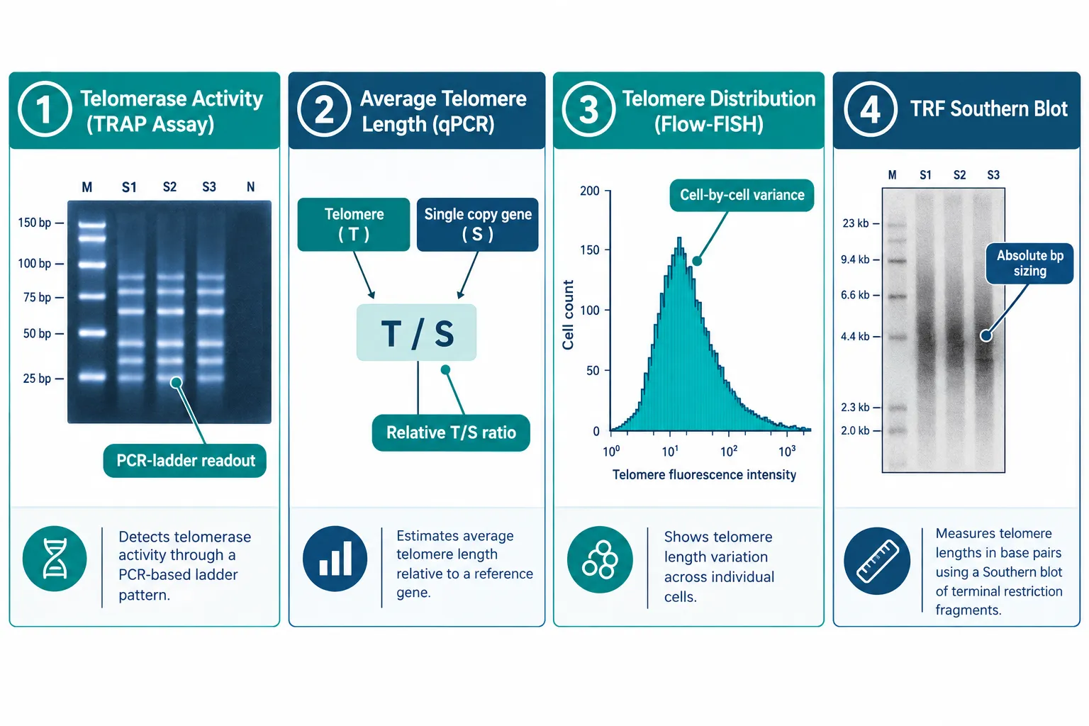
Telomerase Activity measures the enzymatic capacity of the telomerase complex to extend telomere repeats. The gold-standard method is the Telomeric Repeat Amplification Protocol (TRAP assay), which generates a PCR ladder pattern scored by gel densitometry or fluorescence. A positive TRAP result confirms the enzyme is functional, it says nothing about whether telomeres have actually lengthened.

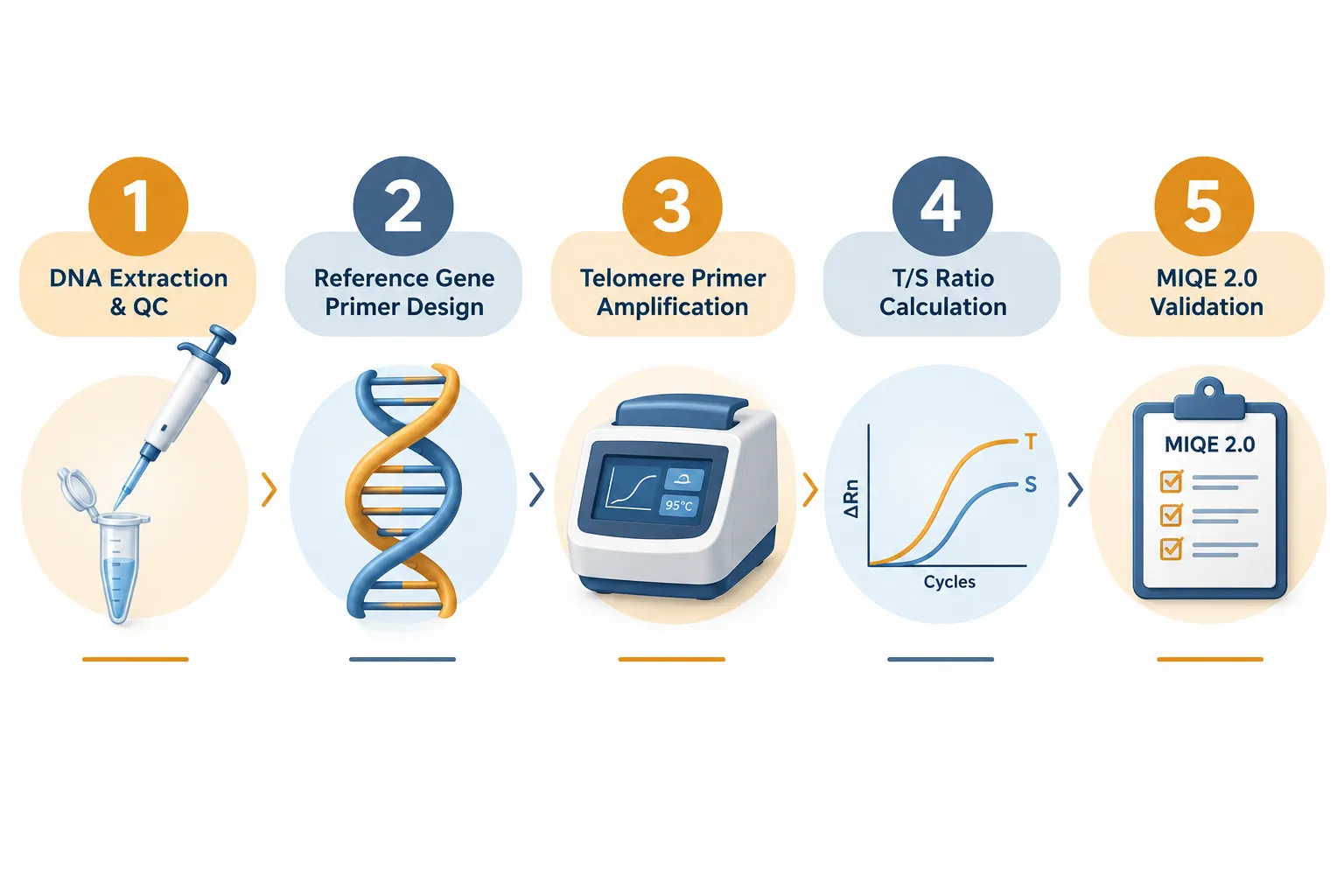
Average Telomere Length captures the mean repeat-count across all chromosomes in a cell population. Quantitative PCR (qPCR) reports this as a T/S ratio, telomere signal normalized to a single-copy reference gene. This is the most scalable method but collapses the entire distribution into one number.

Telomere Length Distribution reveals cell-to-cell and chromosome-to-chromosome variance. Flow-FISH (fluorescence in situ hybridization combined with flow cytometry) and Q-FISH on metaphase spreads provide this granular view. Critically, a cell population with a normal average T/S ratio can still harbor a subset of critically short telomeres driving senescence, information invisible to qPCR alone.

Terminal Restriction Fragment (TRF) Southern Blot remains the reference method for absolute base-pair sizing. It is low-throughput and requires large DNA inputs, but it anchors other assays to a concrete molecular ruler.

Assay Endpoint Throughput Key Limitation
TRAP Telomerase activity Medium Does not measure length
qPCR (T/S ratio) Average telomere length High Relative, not absolute
Flow-FISH Length distribution Medium Requires fresh cells
TRF Southern Blot Absolute length (bp) Low High DNA input needed
Q-FISH Chromosome-specific length Low Labor-intensive

Epithalon's Documented Effects and Assay-Selection Implications

Epithalon's Documented Effects and Assay-Selection Implications

Epithalon (Ala-Glu-Asp-Gly) is a synthetic tetrapeptide studied for its capacity to upregulate telomerase expression in human cell lines. Independent work from Brunel University and subsequent 2026 corrections to earlier datasets confirm that Epithalon can increase TERT (telomerase reverse transcriptase) mRNA levels and TRAP-positive activity in cultured cells. Bovine oocyte maturation studies published in 2025 further demonstrated telomerase activation in a reproductive biology context.

However, a 2026 systematic review concluded that the strongest evidence remains confined to cell culture. Dosing parameters used in vitro do not translate directly to in vivo concentrations, and no peer-reviewed human clinical trial has yet validated the telomere-lengthening claims circulating in popular media.

What this means for assay design:

  • If the goal is to confirm Epithalon's mechanism, a TRAP assay is the correct first-line tool, it directly measures the enzymatic output the peptide is claimed to stimulate.
  • If the goal is to detect downstream telomere elongation, qPCR T/S ratio is appropriate for screening, but positive findings must be confirmed by TRF or Flow-FISH before any mechanistic claim is made.
  • Cell-line studies should report passage number, serum conditions, and peptide concentration with full MIQE 2.0 compliance to allow cross-study comparison.

Researchers interested in related peptides that interact with cellular repair pathways, such as those exploring GHK-Cu peptide for sale options or wound healing peptide research, face similar endpoint-selection challenges when designing aging-biology experiments.

"Telomerase activation and telomere elongation are sequential, not synonymous. Reporting one without the other is an incomplete experiment."


Senescence-Associated Readouts: Completing the Picture

Senescence-Associated Readouts: Completing the Picture

Telomere data alone cannot confirm cellular senescence or its reversal. Senescence-Associated Secretory Phenotype (SASP) markers and classical senescence biomarkers must be layered in to validate any aging-biology claim.

Core senescence readouts to pair with telomere assays:

  • SA-beta-galactosidase (SA-beta-gal): Histochemical staining at pH 6.0; the most widely used senescence marker. Simple but not exclusive to senescence.
  • p21 (CDKN1A) and p16 (CDKN2A): Cyclin-dependent kinase inhibitors upregulated in senescent cells. Measured by Western blot or RT-qPCR. p16 is particularly stable and less influenced by acute stress responses.
  • SASP cytokine panels: IL-6, IL-8, MMP-3, and GDF-15 are frequently elevated. Multiplex ELISA or Luminex platforms allow simultaneous quantification.
  • Gamma-H2AX foci: Marks DNA double-strand breaks, including those at dysfunctional telomeres. Immunofluorescence co-localization with telomere probes (TIF assay) directly links telomere dysfunction to DNA damage signaling.

Combining these with telomere and telomerase readouts creates a multi-dimensional senescence profile. For example, a cell population showing elevated TRAP activity, stable T/S ratio, reduced p16, and decreased SA-beta-gal staining would provide much stronger evidence of senescence reversal than any single marker alone.

Researchers working with mitochondria-targeted peptides such as SS-31 Elamipretide or studying synergy of LL-37 and SS-31 in aging models should apply the same multi-endpoint logic when designing senescence assay panels.

For those sourcing research-grade compounds, verified suppliers offering NAD 500 and related longevity compounds alongside peptides provide a useful reference point for quality benchmarks.


Practical Assay-Selection Framework

Choosing between platforms for Epithalon, Telomerase, and Senescence Assays: Choosing qPCR, Telomere-Length, and Senescence-Associated Readouts projects should follow a tiered logic:

  1. Define the primary question first. Is the study asking whether Epithalon activates telomerase (use TRAP), changes average length (use qPCR), or alters the proportion of senescent cells (use SA-beta-gal + p16)?
  2. Apply MIQE 2.0 standards for all qPCR work. The Telomere Research Network's 2026 absolute telomere length protocol provides an improved step-by-step method that reduces inter-laboratory variability significantly.
  3. Use at least two orthogonal assays. No single platform is sufficient for publication-quality senescence claims.
  4. Report raw Ct values and efficiency data. T/S ratios without amplification efficiency correction are not reproducible across laboratories.
  5. Acknowledge the translational gap explicitly. In vitro Epithalon findings cannot be applied to human dosing without pharmacokinetic bridging studies.

Conclusion

Rigorous research into Epithalon, Telomerase, and Senescence Assays: Choosing qPCR, Telomere-Length, and Senescence-Associated Readouts demands that investigators treat each endpoint as a distinct biological question. TRAP assays confirm enzymatic activity; qPCR T/S ratios screen for average length changes; Flow-FISH and TRF resolve distribution and absolute sizing; and SA-beta-gal, p16, and SASP panels confirm the senescence phenotype.

Actionable next steps for researchers in 2026:

  • Audit existing Epithalon datasets for MIQE 2.0 compliance before drawing mechanistic conclusions.
  • Add at least one senescence marker (p16 or SA-beta-gal) to any telomere-length study to contextualize findings biologically.
  • Pre-register assay selection criteria before data collection to prevent endpoint switching.
  • Treat all in vitro Epithalon findings as hypothesis-generating, not clinically prescriptive, until controlled human studies are available.

The field will advance faster when endpoint selection is treated as a scientific decision, not an afterthought.

https://www.puretestedpeptides.com/wp-content/uploads/2026/09/epithalon-telomerase-and-senescence-assays-choosing-qpcr-telomere-length-and-sen.webp 1024 1536 https://www.puretestedpeptides.com/wp-content/uploads/2026/01/buy-peptides-online.jpg 2026-09-27 13:03:452026-09-27 13:03:45Epithalon, Telomerase, and Senescence Assays: Choosing qPCR, Telomere-Length, and Senescence-Associated Readouts
×

Helpful Links

  • My account
  • Cart
  • Checkout
  • Refund and Returns Policy
  • Privacy Policy
  • SMS Privacy Policy
  • Login
  • My Account
  • Logout

USA Made Lab Tested Peptides

All products are sold for research, laboratory, or analytical purposes only, and are not for human consumption


Pure Tested Peptides is a chemical supplier. Pure Tested Peptides is not a compounding / chemical compounding facility as defined under 503A of the Federal Food, Drug, and Cosmetic act. Pure Tested Peptides is not an outsourcing facility as defined under 503B of the Federal Food, Drug, and Cosmetic act.

The statements made within this website have not been evaluated by the US Food and Drug Administration. The products we offer are not intended to diagnose, treat, cure or prevent any disease.

Human/Animal Consumption Prohibited. Laboratory/In-Vitro Experimental Use Only

Scroll to top Scroll to top Scroll to top