Meloxicam and BPC-157 Research Models: Separating COX Inhibition From Barrier, Angiogenesis, and Repair Signals
Selective COX-2 inhibition can reduce myofiber cross-sectional area by up to 33% in the first three weeks after muscle injury, a finding that forces researchers to ask a sharper question: when an NSAID quiets inflammation, what else does it silence? That question is precisely where Meloxicam and BPC-157 Research Models: Separating COX Inhibition From Barrier, Angiogenesis, and Repair Signals becomes a productive experimental framework rather than a simple drug-versus-peptide comparison.
Key Takeaways
- Meloxicam is a preferential COX-2 inhibitor that suppresses prostaglandin E2-driven inflammatory cascades but may simultaneously blunt early regenerative signaling in muscle, bone, and tendon models.
- BPC-157 activates FAK-paxillin, PI3K/Akt, VEGF, and eNOS pathways, functioning as a "signal-upregulating" repair agent rather than an anti-inflammatory compound.
- Gastrointestinal barrier models show that BPC-157 can restore mucosal integrity even when NSAID-driven COX inhibition has already disrupted prostaglandin synthesis.
- BPC-157's angiogenic effects are context-dependent: pro-angiogenic in wound and musculoskeletal models, anti-angiogenic in pathological settings such as liver cirrhosis.
- All BPC-157 data remain preclinical as of 2026; no peer-reviewed controlled human trials exist, making rigorous experimental design essential.
How Meloxicam Defines the COX-Inhibition Arm of These Research Models
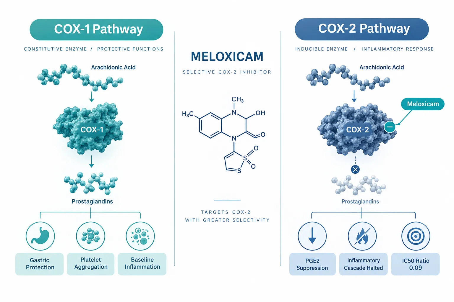
Meloxicam occupies a specific mechanistic lane. Its COX-2:COX-1 IC50 ratio sits at approximately 0.09 in human whole-blood assays, meaning COX-2 is inhibited at concentrations far below those needed to meaningfully affect COX-1. At therapeutic doses, this translates to strong suppression of prostaglandin E2 and related COX-2-dependent mediators that drive the classical redness, heat, and swelling of acute tissue injury.
That selectivity is valuable for isolating variables in a research model. When meloxicam is used as the COX-inhibition comparator, investigators can attribute downstream changes in inflammatory cytokines, edema volume, or pain behavior specifically to the prostaglandin axis rather than to broader cyclooxygenase disruption.
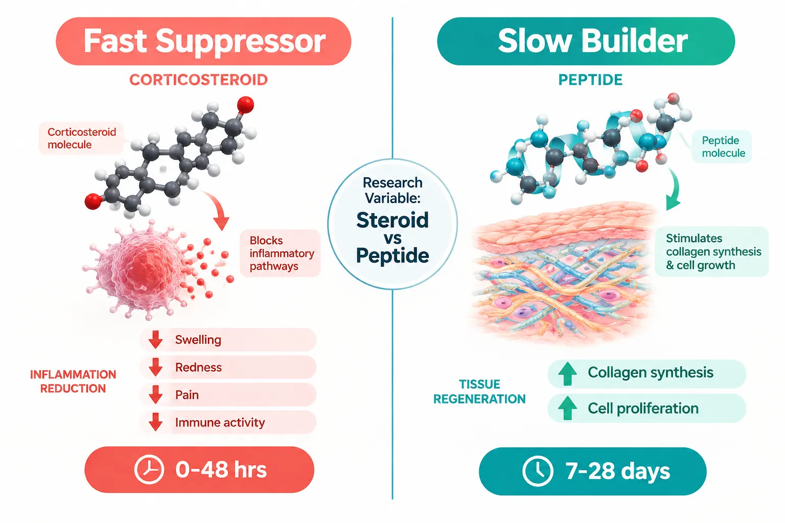
The trade-off, however, is regenerative. Prostaglandins are not only inflammatory mediators, they also coordinate satellite cell activation, osteoblast recruitment, and tendon fibroblast proliferation. Models that apply meloxicam-class COX-2 inhibition during the first weeks of healing consistently show reduced regenerative output alongside reduced inflammation. This creates a measurable "repair deficit" that serves as a useful baseline against which a repair-promoting agent can be tested.
| Meloxicam Mechanism | Research Utility | Observed Limitation |
|---|---|---|
| Preferential COX-2 blockade | Isolates prostaglandin axis | Blunts satellite cell and fibroblast signaling |
| PGE2 suppression | Reduces inflammatory noise in assays | May delay bone and tendon callus formation |
| Low COX-1 interference | Preserves gastric baseline prostaglandins | Does not address vascular remodeling readouts |
BPC-157 as the Repair and Barrier Comparator in Meloxicam and BPC-157 Research Models
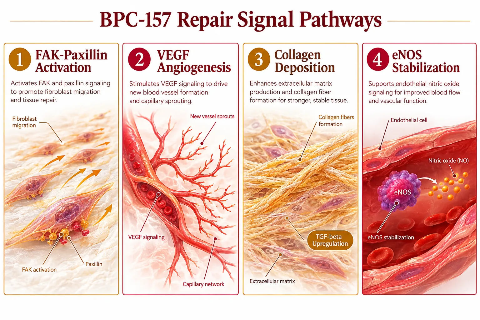
BPC-157 is a stable synthetic pentadecapeptide derived from a gastric protein sequence. Its mechanistic profile is essentially the inverse of meloxicam's: rather than suppressing a signaling cascade, it amplifies several repair programs simultaneously.
Core pathways activated by BPC-157 include:
- FAK-paxillin signaling, which accelerates fibroblast migration and attachment
- PI3K/Akt, which promotes cell survival and proliferation
- VEGF and VEGFR2, which drive new capillary formation
- TGF-beta, which upregulates collagen synthesis and extracellular matrix remodeling
- eNOS stabilization, which maintains endothelial nitric oxide output and microvascular tone
More than 150 preclinical studies, predominantly rodent models, document pro-healing effects across muscle, tendon, ligament, bone, and gastrointestinal tissue. Researchers exploring wound healing mechanisms will recognize BPC-157 as a benchmark agent for angiogenesis and collagen-deposition assays.
Crucially, BPC-157 has also been shown to reduce COX-2 expression, myeloperoxidase activity, and pro-inflammatory cytokines such as IL-6 and TNF-alpha in gastrointestinal inflammation models. This partial overlap with NSAID-like outcomes occurs through modulation of inflammatory signaling rather than direct enzyme inhibition, a distinction that matters enormously when designing assays intended to separate mechanisms.
"Meloxicam turns down prostaglandin-mediated inflammatory cascades; BPC-157 turns up repair programs in fibroblasts, endothelial cells, and extracellular matrix. These are complementary but non-equivalent actions."
Separating COX Inhibition From Barrier, Angiogenesis, and Repair Signals: Key Experimental Readouts
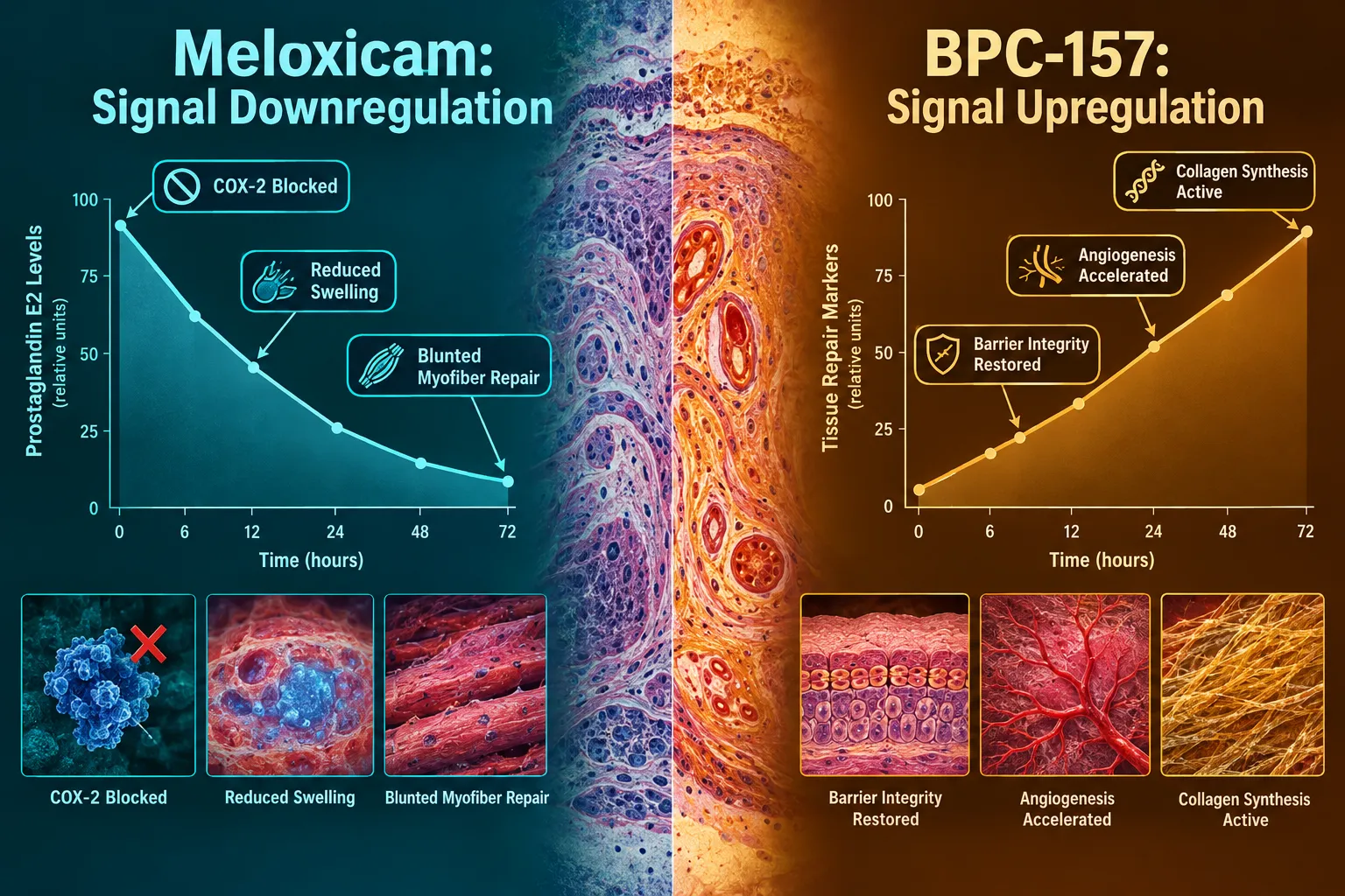
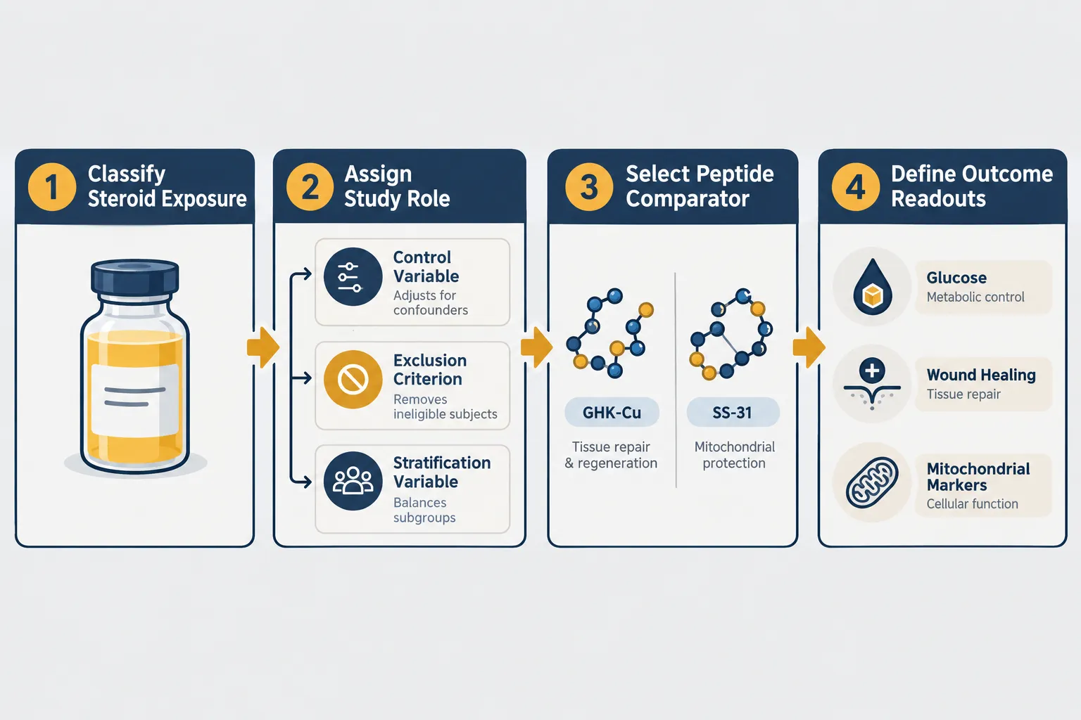
The most informative use of Meloxicam and BPC-157 Research Models: Separating COX Inhibition From Barrier, Angiogenesis, and Repair Signals lies in assigning distinct readout categories to each agent.
Timing windows matter. Meloxicam's effects are most pronounced during the acute inflammatory phase (days 1-5 post-injury). BPC-157's angiogenic and barrier effects accumulate over days 3-21, making sequential or overlapping dosing protocols necessary to capture both mechanisms without conflation.
Vascular readouts require specific assays. BPC-157 drives angiogenesis through VEGFR2-PI3K-Akt-eNOS and Src-caveolin-1-eNOS pathways. A 2026 study identified FBXO22-dependent stabilization of the transcription factor BACH1 as an additional angiogenic driver. Meloxicam does not meaningfully engage these pathways, so CD31 staining, microvessel density counts, and Doppler perfusion measures will reflect BPC-157 activity without NSAID interference.
Barrier integrity assays are particularly diagnostic. In gastrointestinal models using indomethacin- or aspirin-induced ulceration, co-administration of BPC-157 reduces mucosal hemorrhage, lowers ulcer index scores, and helps maintain local prostaglandin levels despite upstream COX inhibition. This demonstrates that barrier repair can proceed through peptide-driven pathways even when prostaglandin synthesis is suppressed, a clean mechanistic separation.
Context-dependent angiogenesis is a critical nuance. In wound and musculoskeletal models, BPC-157 is pro-angiogenic. In liver cirrhosis and portal hypertension models, it suppresses pathological angiogenesis and normalizes NO signaling. Researchers must define the vascular pathology context before interpreting angiogenesis data.
For investigators comparing peptide repair mechanisms, related mitochondrial and cellular energy research, such as SS-31 mitochondrial research themes, offers useful parallel frameworks for understanding how peptides modulate tissue recovery at the cellular level. Similarly, copper peptide research documented in the GHK-Cu copper peptide research sourcing guide provides additional context for collagen and angiogenesis assay design.
Recommended assay matrix for separating mechanisms:
- PGE2 ELISA, meloxicam-sensitive, BPC-157-independent
- Microvessel density (CD31 IHC), BPC-157-sensitive, meloxicam-independent
- Transepithelial electrical resistance (TEER), barrier integrity, BPC-157 primary driver
- Myofiber cross-sectional area (H&E morphometry), detects NSAID-induced regenerative deficit
- Collagen content (Sirius Red staining), BPC-157 upregulated, not NSAID-dependent
- eNOS phosphorylation (Western blot), BPC-157 pathway marker
Researchers sourcing peptide compounds for these protocols should consult resources such as the PT-141 peptide research context QA and controls guide for quality and control benchmarks applicable across peptide research settings.
A 2026 Cambridge-affiliated review on BPC-157's role in tissue repair and pain management underscores that its pleiotropic effects, spanning angiogenesis, collagen synthesis, fibroblast activity, and nitric oxide modulation, could carry off-target risks if translated to humans. Rigorous dose-finding, toxicity screening, and long-term safety trials remain prerequisites before any clinical application. All current conclusions are strictly preclinical.
Conclusion
Meloxicam and BPC-157 Research Models: Separating COX Inhibition From Barrier, Angiogenesis, and Repair Signals is most valuable when investigators move beyond a binary NSAID-versus-peptide framing and instead assign each agent to specific mechanistic readouts at defined time windows. Meloxicam reliably controls the prostaglandin axis and creates a measurable regenerative deficit that quantifies the cost of COX-2 suppression. BPC-157 provides a parallel track of vascular, barrier, and matrix repair signals that operate largely independently of COX enzyme activity.
Actionable next steps for research teams:
- Define the injury phase (acute vs. proliferative) and select assay timing accordingly before combining agents in a single protocol.
- Use PGE2 ELISA as the meloxicam-specific readout and microvessel density plus TEER as BPC-157-specific readouts to prevent cross-mechanism attribution errors.
- Specify the vascular pathology context when interpreting angiogenesis data, given BPC-157's demonstrated context-dependent pro- and anti-angiogenic behavior.
- Treat all BPC-157 findings as preclinical until controlled human trial data are available; design protocols with appropriate safety and toxicity checkpoints.
- Consult established sourcing and quality-control frameworks, including resources on GHK-Cu for sale and related peptide compound standards, to ensure reagent integrity across experimental runs.
Separating these mechanisms with precision is not merely an academic exercise. It is the foundation on which any future translational work in tissue repair, barrier biology, and vascular remodeling will need to stand.