Call or Text 727-513-9780
  • Shopping Cart Shopping Cart
    0Shopping Cart
Pure Tested Peptides | America's most trusted Peptides for sale online
  • Peptides for sale
    • Oral Peptides for sale
      • Peptide Capsules for sale
      • BPC 157 Capsules 1000mcg
      • SLU-PP-332 Capsules | 1000 mcg
      • 5-Amino-1MQ 50mg Capsules
      • Tesofensine 500mcg
    • All Peptides for sale
    • Peptide Sprays
      • BPC 157 Nasal Spray Kit
      • BPC-157 TB500 Nasal Spray Kit
      • Semax Nasal Spray 10mg
      • Selank – Nasal Spray Kit – 10mg
      • Epithalon 50MG Nasal Spray Kit
      • Ipamorelin 10mg Nasal Spray
      • Klow Nasal Spray (BPC-157 + TB-500 + GHK-Cu + KPV) | 80mg
      • Hulk Nasal Spray Tesa / Ipa Blend 6/3 MG
      • Klow Nasal Spray
      • NAD + 500 mg Nasal Spray
      • PT-141 Nasal Spray Kit
    • GHRH Peptides
      • Ipa Peptides
      • CJC-1295 Peptides
        • CJC-1295 with DAC 5 mg
        • CJC-1295 without DAC 5 mg
        • CJC-1295 Ipa 10mg
      • Tesa Peptides
        • Tesa Peptide
        • Tesa 20 mg
    • GHK-Cu Peptides
      • All GHK-Cu Peptides
      • GHK-Cu 100mg
      • KLOW Peptide Blend – Buy KLOW blend online
    • BPC Peptides
      • All BPC Peptides
      • BPC-157
      • BPC-157 TB-500
      • BPC 157 capsules 1000mcg
    • SLU-PP-332 Peptides
      • All SLU-PP-332 Peptides
      • SLU-PP-332 5mg
    • GLP3 Peptides
    • PT-141 Peptides
      • PT-141 Peptides for sale
      • PT-141 10mg
      • PT-141 Nasal Spray
    • CAG Peptides
      • Lipo-C Peptide Blend
      • CAG 5mg
      • CAG 10mg
    • MOTS-C Peptides
      • MOTS-C Peptides for sale
      • MOTS-c peptide
      • MOTS-c 10mg *6 pack*
    • 5 Amino 1MQ Peptides
      • 5 Amino 1MQ Peptides for sale
      • 5-Amino-1MQ 50mg Capsules
      • 5-Amino-1MQ 5mg
    • Epithalon Peptides
      • Epithalon Peptides for sale
      • Epithalon 10mg
      • Epithalon 50mg
  • Shop
    • GLPs
      • 5-Amino-1MQ 50mg Capsules
      • 5-Amino-1MQ 5mg
      • L-Carnitine 500mg/ml
      • Tesofensine 500mcg
      • SLU-PP-332 5mg
      • MOTS-c 10mg *6 pack*
    • Epithalon & BPC Peptides
      • Epithalon 10mg
      • Epithalon 50mg
      • BPC-157
      • BPC 157 capsules 1000mcg
      • BPC-157 TB-500
      • BPC-157 TB500 Nasal Spray Kit
      • BPC 157 Nasal Spray Kit
    • BPC TB-500 & NAD+ Peptides
      • NAD+ 500 mg
      • KLOW Peptide Blend – Buy KLOW blend online
      • GLOW Peptide Blend
      • TB 500 5mg
      • BPC 157 capsules 1000mcg – Supplement
      • BPC 157 Nasal Spray Kit
      • BPC-157
      • BPC-157 TB500 Nasal Spray Kit
      • BPC-157 TB-500
      • BPC 157 capsules 1000mcg
    • LL-37 Peptide
      • LL-37 10 mg
    • MOTS-C & Selank
      • MOTS-c peptide
      • Selank 10mg
    • GHK Peptides
      • GHK-Cu 100mg
      • GLOW Peptide Blend
      • KLOW Peptide Blend – Buy KLOW blend online
  • COAs
  • Wholesale
    • Wholesale Peptides for sale
  • PTP FAQ
  • Affiliates
    • Selank Peptide: Advanced Pharmacological Mechanisms Underlying Its Anxiolytic and Nootropic Effects in Research
      • 5-Amino-1MQ Peptide: Detailed Mechanisms of NNMT Inhibition and Its Impact on Cellular Metabolism Research
        • MOTS-C Peptide: Unraveling Its Role in Mitochondrial Dynamics and Energy Metabolism Research
          • Epithalon Peptide and Telomerase Regulation: Investigating Its Impact on Cellular Senescence and Lifespan Research Models
            • GLP2-T vs GLP2 Tirz Peptide: Understanding the Naming, Mechanistic Nuances, and Research Implications for Gut Health
      • Peptides and Polypeptides in Modern Pharmacology: What Research on Metoprolol, Prednisone, and Amlodipine Reveals
        • Polypeptide Peptides vs NSAIDs: What Naproxen and Diclofenac Teach Tissue-Repair Researchers About BPC‑157 and TB‑500
          • Peptides and Polypeptides in Endocrine Pharmacology: How GLP-1, GLP-2, and GLP-3 Retatrutide Differ From Classic Drugs Like Prednisone and Amlodipine
          • 5‑Amino‑1MQ Peptide and NNMT Inhibition: How It Compares With Statins Like Atorvastatin in Adipose and Lipid Metabolism Research
      • Enclomiphene, Estrogen Receptor Signaling, and Luteinizing Phase Biology: What Hormone Researchers Should Measure
        • Mesenchymal Stem Cells, BPC‑157, and GHK‑Cu: How Tissue Repair Peptides Compare With Classic NSAIDs Like Naproxen in Injury Models
          • Peptide Calculator Use Cases Beyond Growth Hormone: Working Through GLP‑3 Retatrutide, MOTS‑c, and BPC‑157 Research Dosing
        • Polypeptide Peptides in Endocrine and Metabolic Pharmacology: Lessons From Amlodipine, Prednisone, and Metoprolol
        • Peptides Calculator for Advanced Blends: Worked Examples for Tesamorelin, CJC‑1295, and Ipamorelin Stacks
          • Peptides and Polypeptides in Human Physiology: How Molecular Size Shapes Research Applications
            • Selank Peptide: Advanced Pharmacological Mechanisms Underlying Its Anxiolytic and Nootropic Effects in Research
          • 5-Amino-1MQ Peptide: Detailed Mechanisms of NNMT Inhibition and Its Impact on Cellular Metabolism Research
            • MOTS-C Peptide: Unraveling Its Role in Mitochondrial Dynamics and Energy Metabolism Research
              • Epithalon Peptide and Telomerase Regulation: Investigating Its Impact on Cellular Senescence and Lifespan Research Models
                • GLP2-T vs GLP2 Tirz Peptide: Understanding the Naming, Mechanistic Nuances, and Research Implications for Gut Health
                  • Peptides and Polypeptides in Endocrine Pharmacology: How GLP-1, GLP-2, and GLP-3 Retatrutide Differ From Classic Drugs Like Prednisone and Amlodipine
                    • Polypeptide Peptides vs NSAIDs: What Naproxen and Diclofenac Teach Tissue-Repair Researchers About BPC‑157 and TB‑500
                      • Peptides and Polypeptides in Modern Pharmacology: What Research on Metoprolol, Prednisone, and Amlodipine Reveals
                        • 5‑Amino‑1MQ Peptide and NNMT Inhibition: How It Compares With Statins Like Atorvastatin in Adipose and Lipid Metabolism Research
                        • Enclomiphene, Estrogen Receptor Signaling, and Luteinizing Phase Biology: What Hormone Researchers Should Measure
                        • Mesenchymal Stem Cells, BPC‑157, and GHK‑Cu: How Tissue Repair Peptides Compare With Classic NSAIDs Like Naproxen in Injury Models
                        • Peptide Calculator Use Cases Beyond Growth Hormone: Working Through GLP‑3 Retatrutide, MOTS‑c, and BPC‑157 Research Dosing
                        • Polypeptide Peptides in Endocrine and Metabolic Pharmacology: Lessons From Amlodipine, Prednisone, and Metoprolol
                        • Peptides Calculator for Advanced Blends: Worked Examples for Tesamorelin, CJC‑1295, and Ipamorelin Stacks
                        • Peptides and Polypeptides in Human Physiology: How Molecular Size Shapes Research Applications
                        • Selank Peptide: Advanced Pharmacological Mechanisms Underlying Its Anxiolytic and Nootropic Effects in Research
                        • MOTS-C Peptide: Unraveling Its Role in Mitochondrial Dynamics and Energy Metabolism Research
                        • Peptides and Polypeptides in Modern Pharmacology: What Research on Metoprolol, Prednisone, and Amlodipine Reveals
                        • Peptides and Polypeptides in Endocrine Pharmacology: How GLP-1, GLP-2, and GLP-3 Retatrutide Differ From Classic Drugs Like Prednisone and Amlodipine
                        • 5-Amino-1MQ Peptide: Detailed Mechanisms of NNMT Inhibition and Its Impact on Cellular Metabolism Research
                        • Mesenchymal Stem Cells, BPC‑157, and GHK‑Cu: How Tissue Repair Peptides Compare With Classic NSAIDs Like Naproxen in Injury Models
                        • Peptide Calculator Use Cases Beyond Growth Hormone: Working Through GLP‑3 Retatrutide, MOTS‑c, and BPC‑157 Research Dosing
                        • GLP2-T vs GLP2 Tirz Peptide: Understanding the Naming, Mechanistic Nuances, and Research Implications for Gut Health
                        • Polypeptide Peptides in Endocrine and Metabolic Pharmacology: Lessons From Amlodipine, Prednisone, and Metoprolol
                        • Polypeptide Peptides vs NSAIDs: What Naproxen and Diclofenac Teach Tissue-Repair Researchers About BPC‑157 and TB‑500
                        • Peptides and Polypeptides in Human Physiology: How Molecular Size Shapes Research Applications
                        • Peptides Calculator for Advanced Blends: Worked Examples for Tesamorelin, CJC‑1295, and Ipamorelin Stacks
                        • 5‑Amino‑1MQ Peptide and NNMT Inhibition: How It Compares With Statins Like Atorvastatin in Adipose and Lipid Metabolism Research
                        • Enclomiphene, Estrogen Receptor Signaling, and Luteinizing Phase Biology: What Hormone Researchers Should Measure
                        • Epithalon Peptide and Telomerase Regulation: Investigating Its Impact on Cellular Senescence and Lifespan Research Models
                        • Best research protocol Klow blend
                        • best time to take BPC-157
                        • best time to take DSIP (Delta Sleep Inducing Peptide)
                        • best time to take CJC-1295
                        • best time to take AOD-9604
                        • best time to take Follistatin 344
                        • best time to take Ipamorelin
                        • best time to take MK-677 (Ibutamoren)
                        • best time to take Ligandrol (LGD-4033) — research compound
                        • best time to take Ostarine (MK-2866) — research compound
                        • best time to take GHK-CU
                        • best time to take TB-500
                        • best time to take MOTS-c
                        • best time to take Semax
                        • best time to take RAD-140 (Testolone) — research compound
                        • best time to take Thymosin Alpha-1
                        • best time to take PEG-MGF
                        • Biolife Plasma, Octapharma Plasma, and Research Peptides: How Plasma Donation Labs Differ From Peptide Suppliers
                        • best time to take YK-11 — research compound
                        • best time to take PT-141 (Bremelanotide)
                        • Best research protocol Klow blend
                        • 5-Amino-1MQ and MOTS-C Synergy: Metabolic Signaling, Mitochondria, and Research Design
                        • BPC-157 and TB-500: Investigating Their Combined Effects on Angiogenesis and Cellular Migration in Tissue Repair Models
                        • BPC-157 Peptide: Gut Barrier Function, Inflammation, and Tissue-Recovery Research
                        • 5‑Amino‑1MQ and MOTS‑c Synergy in Metabolic Research: Designing NNMT and Mitochondrial Biogenesis Stacks
                        • CJC-1295 with DAC vs. Without DAC: Half-Life, Release Kinetics, and Research Implications
                        • CJC‑1295 with DAC vs. Without DAC: Expanding on Half‑Life Differences Using Tesamorelin and Ipamorelin Blend Case Studies
                        • Collagen Biology and Copper‑Binding Peptides: How GHK‑Cu, Glow Blend, and Klow Blend Interact with Skin and Connective Tissue
                        • Collagen Biology and Regenerative Peptides: How GHK‑Cu, Glow Blend, and Klow Blend Affect Extracellular Matrix Research
                    • DNA, Telomeres, and Longevity Peptides: Positioning Epithalon and MOTS‑c in Genetic Aging Research
                      • Enclomiphene Citrate: serm Mechanism, Testosterone Research, and Stack Compatibility
                        • Enclomiphene vs Enclomiphene Citrate: Formulation, Bioavailability, and Research Distinctions
                        • Epithalon Peptide Research: Telomerase Activation, Aging, and Pineal Gland Function
                        • Estrogen Receptor Signaling and Enclomiphene: How Selective Modulators Compare with Classic Polypeptide Hormones
                        • GHK-Cu Peptide: Advanced Mechanisms in Extracellular Matrix Remodeling and Wound Healing Research
                        • GHK-Cu Peptide: Collagen Synthesis, Wound Repair, and Skin-Barrier Research Models
                        • GLP-1 vs GLP-3 vs GLP-2: Peptide Classification and Research Applications
                        • GLP-2 Peptide Research Guide: Gut Barrier Function, Nutrient Absorption, and Intestinal Recovery Models
                        • GLP-3 Retatrutide vs. GLP-1 Drugs: What Triple-Agonist Biology Changes in Research Models
                        • Ipamorelin and Tesamorelin Combination: Synergistic GH Secretagogue Research and Dosing Protocols
                        • GLP2 Tirz Peptide: What It Is, Why the Name Exists, and How Researchers Should Interpret It
                        • Klow Blend Peptide Nasal Spray: What the Formulation Is Trying to Do in Cognitive Research
                        • Mitochondria, NNMT Inhibition, and Peptide Modulators: Where MOTS‑c and 5‑Amino‑1MQ Fit in Cellular Energy Research
                        • MOTS-c Peptide: Mitochondrial Function, Energy Metabolism, and What Researchers Measure
                        • MOTS-c vs. 5-Amino-1MQ: Which Metabolic Research Questions Each Compound Actually Answers
                        • Nasal Spray Peptides: Bioavailability, Administration, and Semax/Selank Research Applications
                        • PT-141 Peptide Research: Mechanism of Action and Melanocortin Receptor Signaling
                        • Retatrutide for Research: Mechanism, Structure, and GLP-1/GLP-3 Dual Action
                        • Retatrutide for Obesity and Type 2 Diabetes: What the Latest Trial Data Suggest
                        • Tesofensine Peptide Research: Mechanism, Appetite Suppression, and Neuropeptide Y Pathways
  • Contact
    • Contact Customer Service
    • Text Customer Support
  • About US
  • Shop all peptides
  • Affiliate Program
    • Affiliate Signup
  • Login / Register Login / Register Page Link Login / Register Page Link
  • Click to open the search input field Click to open the search input field Search
  • Menu Menu

Tag Archive for: glp peptide research

Peptide Reconstitution and Stability: How Buffer Choice, pH, Osmolality, and Freeze–Thaw Cycles Affect Research Samples

Peptide Reconstitution and Stability: How Buffer Choice, pH, Osmolality, and Freeze–Thaw Cycles Affect Research Samples

September 23, 2026/0 Comments/in Uncategorized/by

Roughly 30% of peptide research samples degrade before a single experiment is completed, not because of poor synthesis, but because of avoidable reconstitution errors. Peptide reconstitution and stability: how buffer choice, pH, osmolality, and freeze, thaw cycles affect research samples is one of the most consequential yet underappreciated topics in peptide science. A wrong buffer or a single extra freeze, thaw cycle can shift purity from 99% to well below 90%, invalidating weeks of downstream work.

Key Takeaways

  • The optimal pH window for most peptide solutions is 3.5-4.5, with acetate and citrate buffers as first-line choices.
  • Deamidation, hydrolysis, and aggregation each peak at distinct pH bands, making buffer selection sequence-specific.
  • Osmolality must be controlled (280-320 mOsm/kg for parenteral and intranasal formulations) to preserve biorelevance.
  • Every freeze, thaw cycle introduces measurable degradation; single-use aliquots are the primary mitigation strategy.
  • Stability profiles differ significantly across peptide classes, GLP-class, growth-hormone, regenerative, and nasal peptides each require tailored protocols.

Why Buffer Choice Drives Peptide Reconstitution and Stability
Why Buffer Choice Drives Peptide Reconstitution and Stability

The buffer is not a passive carrier. It sets the chemical environment that either protects or attacks a peptide's backbone and side chains from the moment of reconstitution.

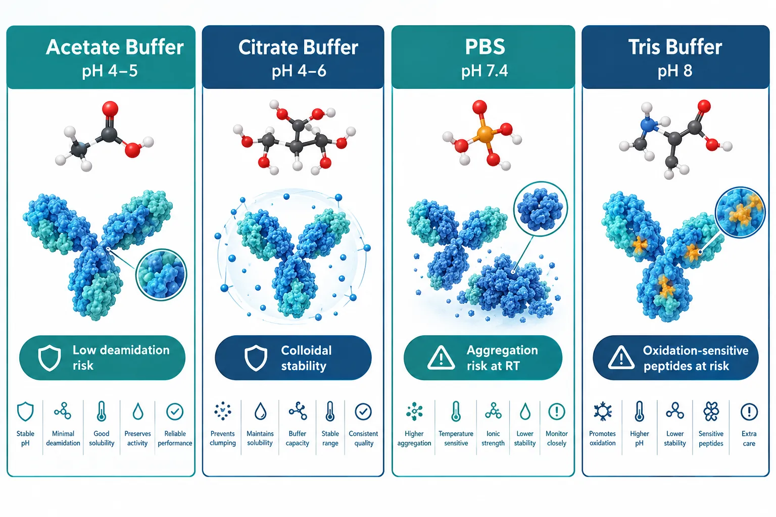
Acetate and citrate buffers at pH 4.0-6.0 have emerged as first-line vehicles for lab reconstitution. Both offer strong buffering capacity in the acidic range where most peptides show the lowest rates of deamidation and backbone hydrolysis. Citrate has the added advantage of mild metal-chelating activity, which is valuable for cysteine-containing and oxidation-sensitive sequences.

PBS (phosphate-buffered saline) at pH 7.4 remains widely used because it matches physiological conditions, but its limitations are now better characterized. At room temperature, PBS accelerates aggregation in hydrophobic peptides and offers no protection against oxidation. For short-term cell-based assays it is acceptable; for storage beyond 24 hours, it is a liability.

Tris and HEPES buffers (pH 7.5-8.0) are common in biochemistry but problematic for peptide stability. The alkaline environment accelerates deamidation of asparagine and glutamine residues and promotes beta-elimination in serine- and threonine-rich sequences.

"Buffer selection is not a formulation footnote, it is the first experimental variable that determines whether a peptide survives long enough to be tested."

Practical buffer selection by peptide class:

Peptide Class Recommended Buffer Target pH Key Risk Mitigated
GLP-class (e.g., GLP-3 analogs) Acetate or citrate 4.0-5.0 Hydrolysis, aggregation
Growth-hormone peptides (e.g., Sermorelin, CJC-1295) Acetate 4.5-5.5 Deamidation
Regenerative/antioxidant (e.g., SS-31, GHK-Cu) Citrate, pH-adjusted water 4.0-5.0 Oxidation, metal coordination
Nasal formulations (e.g., Semax) Citrate-phosphate 5.0-6.5 Mucosal compatibility, stability

Researchers working with SS-31 peptide should pay particular attention to citrate buffers, as the tetrapeptide's aromatic-cationic structure is sensitive to oxidative degradation at neutral or alkaline pH.


pH-Dependent Degradation Pathways: Mapping the Risk Zones

Every peptide has a pH-stability profile, and the degradation chemistry changes depending on where that profile sits.

Below pH 3.0: Acid-catalyzed hydrolysis of Asp-Pro bonds accelerates sharply. Peptides with proline-rich sequences or internal aspartate residues are particularly vulnerable.

pH 3.5-4.5 (optimal zone): For most peptides, this range minimizes both acid-catalyzed hydrolysis and base-catalyzed deamidation simultaneously. Solution purity data consistently show the slowest degradation rates here.

pH 5.0-6.5: Acceptable for many peptides, especially those destined for mucosal delivery. Deamidation begins to increase modestly above pH 5.5 for asparagine-containing sequences.

pH 7.0-8.0: Deamidation of Asn and Gln residues accelerates significantly. Beta-elimination in phosphorylated or glycosylated peptides also increases. Aggregation rates for hydrophobic sequences rise sharply.

Above pH 8.0: Racemization and disulfide scrambling become dominant degradation pathways. Cysteine-containing peptides are at high risk.

Researchers using GLP-3 peptide analogs should note that the long-chain fatty acid modifications common in this class increase hydrophobic aggregation risk at neutral pH, reinforcing the case for acidic reconstitution buffers.


Osmolality, Freeze, Thaw Cycles, and Practical Reconstitution Controls
Osmolality, Freeze, Thaw Cycles, and Practical Reconstitution Controls

Osmolality: The Overlooked Variable

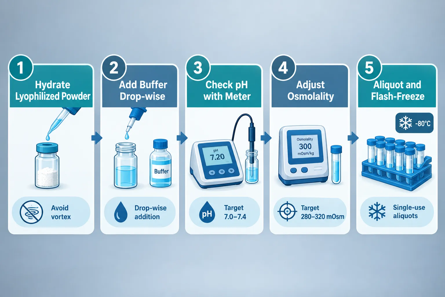
Osmolality is frequently treated as relevant only for clinical formulations, but it matters in research too. For intranasal peptides such as Semax peptide formulations, osmolality outside the 270-320 mOsm/kg range can alter mucosal absorption and confound pharmacokinetic data. For parenteral research models, hyperosmolar solutions cause cell stress artifacts that distort results.

Achieving isotonicity in an acidic buffer requires careful addition of sodium chloride or mannitol. Mannitol is preferred when freeze-drying stability is also a concern, as it acts as a lyoprotectant.

Freeze, Thaw Cycles: Quantifying the Damage

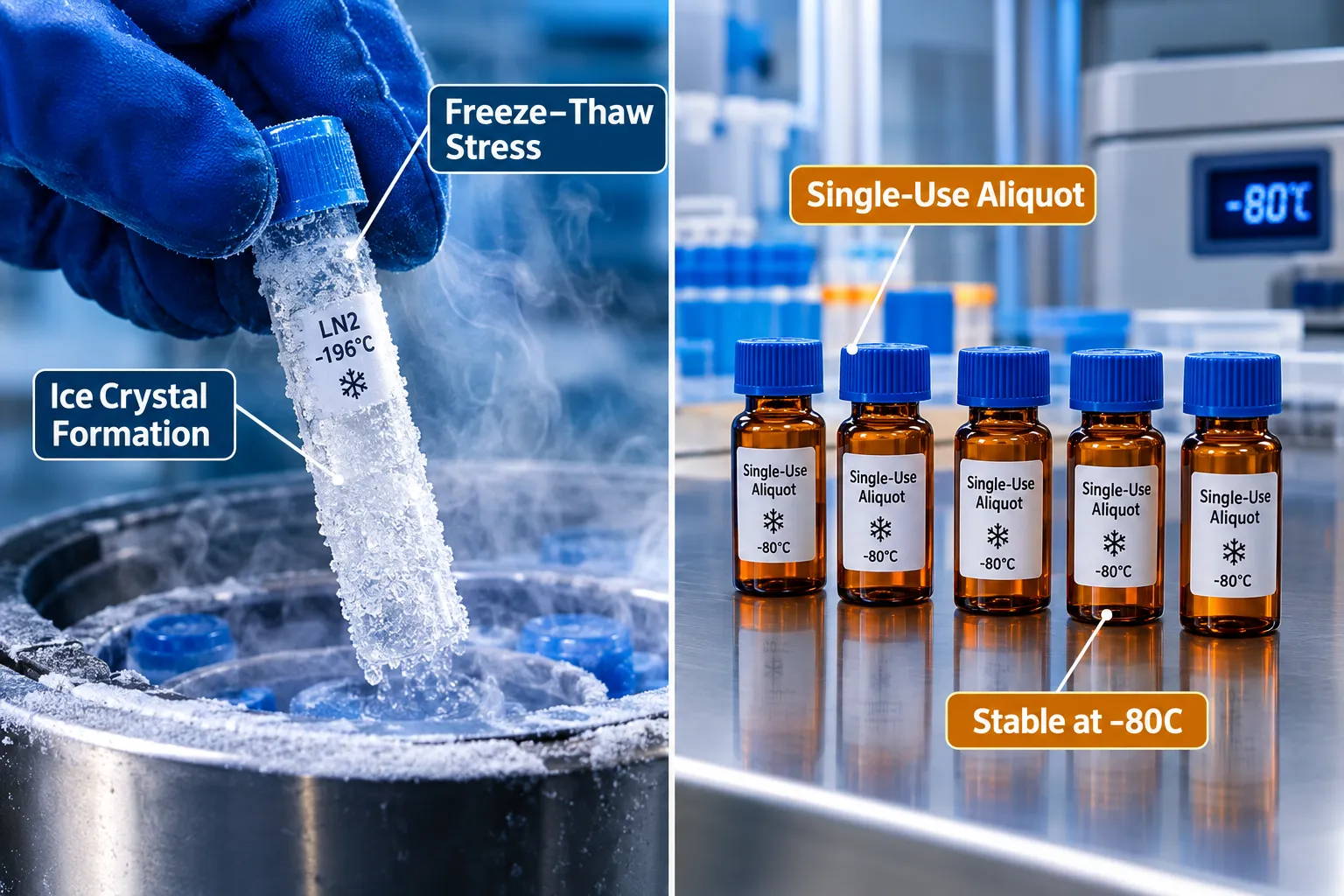
Each freeze, thaw cycle introduces two distinct stresses:

  1. Ice crystal formation, mechanical disruption of peptide aggregates and concentration effects at the ice-liquid interface
  2. Cryoconcentration, solutes concentrate in unfrozen microdomains, creating transient local pH extremes and ionic strength spikes

Studies tracking purity over repeated cycles show measurable losses beginning at cycle two for most peptides, with cumulative degradation accelerating nonlinearly by cycle four or five. The practical implication is unambiguous: single-use aliquots are not optional.

Freeze, thaw mitigation checklist:

  • Aliquot into volumes that match a single experiment
  • Use cryoprotectants (5-10% mannitol or trehalose) when lyophilization is not available
  • Flash-freeze in liquid nitrogen rather than slow-freezing at -20°C
  • Store at -80°C; avoid -20°C for samples held longer than two weeks
  • Never refreeze a thawed aliquot

For complex blends such as Tesamorelin/CJC-1295/Ipamorelin reconstitution, each component has its own freeze, thaw sensitivity, making single-use aliquoting even more critical.

Order of Addition and Mixing

Lab-scale reconstitution protocols now emphasize that order of addition matters. Adding buffer to lyophilized peptide (rather than the reverse) prevents local pH extremes that can occur when a small volume of concentrated peptide contacts a large buffer volume. Gentle swirling, not vortexing, prevents shear-induced aggregation. Sonication is reserved for peptides confirmed to be aggregation-prone, and only in short, controlled pulses.


Stability Matrix Across Peptide Classes
Stability Matrix Across Peptide Classes

The table below consolidates stability variables across four research-relevant peptide classes. It is designed as a practical bench reference.

Variable GLP-Class Analogs Growth-Hormone Peptides Regenerative Peptides (SS-31, GHK-Cu) Nasal Peptides (Semax)
Optimal pH 4.0-5.0 4.5-5.5 4.0-5.0 5.0-6.5
Preferred buffer Acetate Acetate Citrate Citrate-phosphate
Osmolality target 280-310 mOsm/kg 280-310 mOsm/kg 280-310 mOsm/kg 270-320 mOsm/kg
Max freeze, thaw cycles 1-2 2-3 1-2 1
Primary degradation risk Aggregation, hydrolysis Deamidation Oxidation Mucosal pH mismatch
Cryoprotectant recommended Mannitol Mannitol or trehalose Trehalose Mannitol
Stable at -80°C (reconstituted) Up to 3 months Up to 6 months Up to 3 months Up to 4 weeks

Researchers sourcing SS-31 peptide for sale or GHK-Cu peptides should verify that third-party purity certificates reflect post-reconstitution stability data, not just pre-reconstitution synthesis purity.

For GLP-3 peptide research, the fatty acid-modified backbone demands particular attention to aggregation at the reconstitution step. Warming to room temperature before adding buffer, rather than adding cold buffer to a cold vial, reduces nucleation of aggregates.


Conclusion

Peptide reconstitution and stability, how buffer choice, pH, osmolality, and freeze, thaw cycles affect research samples, is a discipline that rewards systematic attention. The actionable steps are clear:

  1. Select buffer based on sequence chemistry, defaulting to acetate at pH 4.0-5.0 for most classes and citrate for oxidation-sensitive peptides.
  2. Verify osmolality before any intranasal or parenteral experiment; adjust with mannitol or NaCl.
  3. Aliquot immediately after reconstitution into single-use volumes and flash-freeze.
  4. Never exceed two freeze, thaw cycles for any peptide; treat cycle one as a warning threshold.
  5. Request purity data at multiple time points and pH values from suppliers, not just a single synthesis certificate.
  6. Match the stability matrix to the peptide class before the experiment begins, not after anomalous results appear.

Implementing these controls does not require expensive equipment. It requires deliberate protocol design and an understanding that every variable in the reconstitution environment is an experimental variable.

https://www.puretestedpeptides.com/wp-content/uploads/2026/09/peptide-reconstitution-and-stability-how-buffer-choice-ph-osmolality-and-freeze.webp 1024 1536 https://www.puretestedpeptides.com/wp-content/uploads/2026/01/buy-peptides-online.jpg 2026-09-23 13:07:532026-09-23 13:07:53Peptide Reconstitution and Stability: How Buffer Choice, pH, Osmolality, and Freeze–Thaw Cycles Affect Research Samples
How to Read Metabolic Research Endpoints: A Practical Guide to A1C, LDL, Triglycerides, Waist Circumference, and hsCRP

How to Read Metabolic Research Endpoints: A Practical Guide to A1C, LDL, Triglycerides, Waist Circumference, and hsCRP

September 23, 2026/0 Comments/in Uncategorized/by

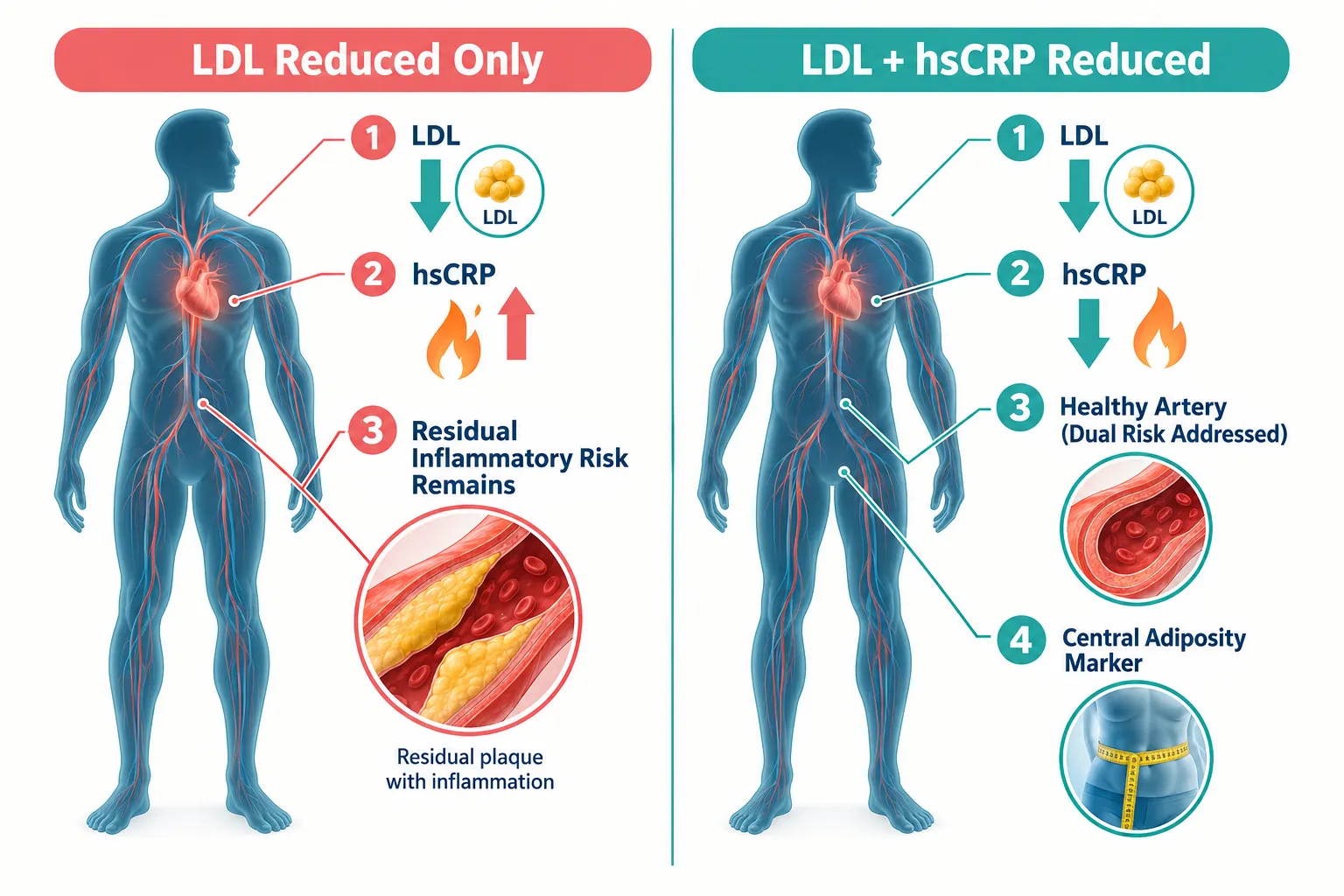
Fewer than 20% of statin-treated patients with controlled LDL-C are actually free of residual cardiovascular risk, a gap that becomes visible only when researchers look beyond a single biomarker. This practical guide to reading metabolic research endpoints covers exactly that: how to interpret A1C, LDL, triglycerides, waist circumference, and hsCRP together, especially when evaluating studies on GLP-class peptides, MOTS-c analogs, and NNMT-related compounds where endpoint selection and baseline adjustment determine whether a result is meaningful or misleading.

Key Takeaways

  • No single metabolic endpoint tells the full story; A1C, LDL, triglycerides, waist circumference, and hsCRP each capture a distinct mechanism.
  • A1C targets in 2026 are stratified by hypoglycemia risk, age, and frailty, a numeric value must be read in that clinical context.
  • LDL-C interpretation depends entirely on the patient's cardiovascular risk tier; the same number can signal success or undertreatment.
  • hsCRP is now recognized as an independent predictor of cardiovascular events and should not be treated as a secondary or optional endpoint.
  • Waist circumference anchors biochemical endpoints to visceral adiposity and insulin resistance, making it a critical companion measure.

Understanding the Five Core Endpoints

A solid reading of metabolic research endpoints starts with knowing what each marker actually measures, and what it misses.

A1C (Glycated Hemoglobin)

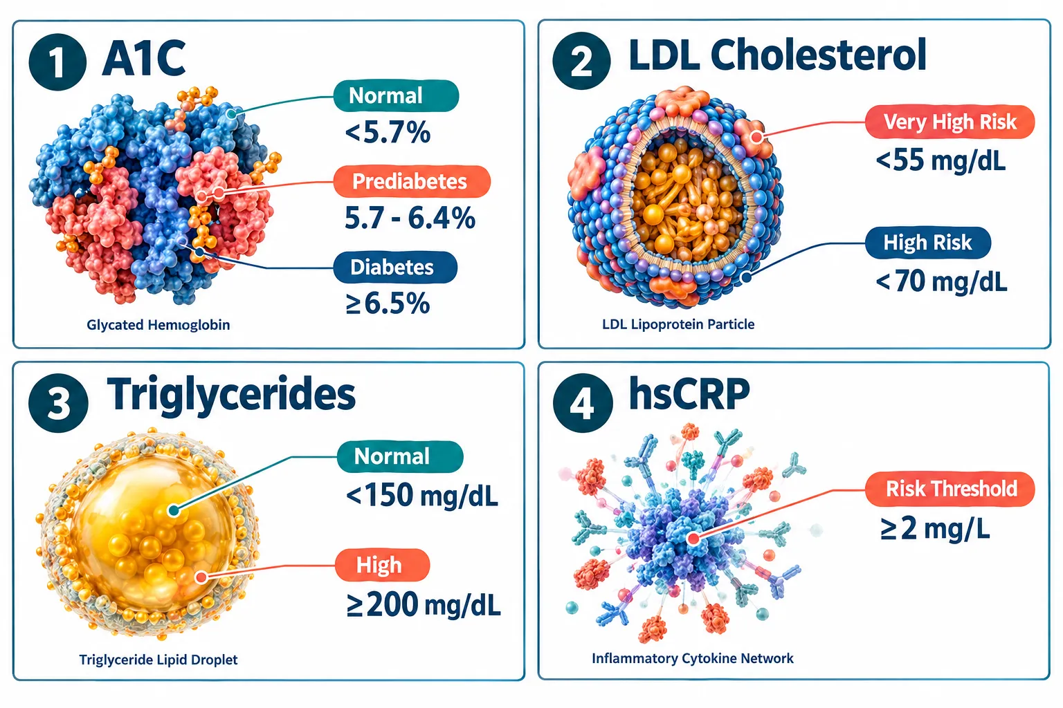
A1C reflects average blood glucose over roughly three months. Current 2026 guidance from major diabetes organizations sets stratified targets rather than a single universal number. For most adults with type 2 diabetes at low hypoglycemia risk, an A1C of 6.5% or below is considered optimal. For frail elderly individuals, those with dementia, or those with recurrent severe hypoglycemia, a range of 7.1-8.5% is considered reasonable. In pediatric studies, a target below 7% applies to most children, with 6.5% achievable in low-risk youth with type 2 diabetes.

Critically, A1C alone is no longer sufficient in modern trials. Continuous glucose monitoring metrics, particularly time in range above 70%, must accompany A1C to confirm that a lower value was achieved safely rather than through excess hypoglycemia. When reviewing GLP-class compound studies, always ask: was the A1C reduction accompanied by improved time-in-range data?

LDL Cholesterol

LDL-C thresholds are tiered by cardiovascular risk category. For low-risk individuals, a value below 116 mg/dL is acceptable. Moderate-risk patients target below 100 mg/dL. High-risk patients aim for below 70 mg/dL, and very-high-risk patients, such as those post-acute coronary syndrome, target below 55 mg/dL. In individuals with recurrent ASCVD events, targets can fall below 40 mg/dL, with evidence showing continued benefit at even lower levels without identified safety concerns.

This tiered structure means a trial reporting an LDL-C of 68 mg/dL could represent excellent control in a moderate-risk primary prevention cohort but inadequate control in a secondary prevention population. Endpoint interpretation must always reference the appropriate risk category, not an absolute number in isolation.

Triglycerides

Triglycerides are increasingly recognized as contributors to residual cardiovascular risk, particularly in patients whose LDL-C is already at target. A normal fasting triglyceride level sits below 150 mg/dL, with values above 200 mg/dL considered high and above 500 mg/dL posing a risk for pancreatitis. Current guidelines remain LDL-centric, but accumulating evidence implicates elevated triglycerides and remnant lipoproteins as independent risk factors. Upcoming guideline revisions are expected to formalize more explicit triglyceride-based goals. In studies of compounds like retatrutide endpoints or GLP-3 peptides, triglyceride data should be treated as a leading indicator of future threshold shifts, not a secondary footnote.

Waist Circumference

Waist circumference is a practical proxy for visceral adiposity and insulin resistance. Standard thresholds flag elevated risk at above 88 cm in women and above 102 cm in men, though ethnicity-adjusted values are increasingly used. In cardiometabolic trials, waist circumference anchors biochemical endpoints to actual body composition. An improvement in A1C and LDL-C without a reduction in waist circumference may indicate partial but incomplete metabolic risk modification. Research on visceral fat outcomes consistently shows that central adiposity independently predicts insulin resistance, dyslipidemia, and cardiovascular events beyond what lipid panels capture alone.

hsCRP (High-Sensitivity C-Reactive Protein)

hsCRP is the most robust available biomarker of residual inflammatory cardiovascular risk. A value at or above 2 mg/L is now classified as a risk-enhancing factor in ASCVD prevention guidelines, and universal screening in both primary and secondary prevention is recommended as of 2026. Elevated hsCRP predicts major adverse cardiovascular events over decades of follow-up, and it remains strongly predictive of recurrent events even in statin-treated patients with normal LDL-C. In trials, a substantial fall in LDL-C without a corresponding reduction in hsCRP leaves significant vascular risk unaddressed.


Understanding the Five Core Endpoints

Why One Biomarker Cannot Stand In for a Complete Mechanism

The most common error in reading metabolic research is treating a single endpoint as a surrogate for the full cardiometabolic picture. This matters especially when evaluating compounds that operate through multiple pathways.

Consider a study on a GLP-class compound that reports a 1.2% reduction in A1C. That result is clinically meaningful, but it tells nothing about whether LDL-C moved, whether hsCRP fell, or whether waist circumference changed. A compound that lowers glucose while leaving visceral fat and systemic inflammation untouched has addressed only one of several interconnected mechanisms. For context on how polypeptide compounds differ from small-molecule drugs in their cardiometabolic effects, the analysis of tesofensine, GLP-3, retatrutide, and GLP-2-T illustrates why endpoint panels must match the mechanism being tested.

The same logic applies to MOTS-c research. MOTS-c is a mitochondria-derived peptide that influences insulin sensitivity and energy metabolism. A trial measuring only A1C misses the compound's potential effects on lipid oxidation, triglyceride clearance, and inflammatory signaling. Similarly, NNMT (nicotinamide N-methyltransferase) inhibition research, relevant to compounds like 5-amino-1MQ, targets metabolic reprogramming at the cellular level, where waist circumference and hsCRP may be more sensitive endpoints than A1C in short-duration studies.

Baseline adjustment is equally critical. A trial that does not stratify participants by baseline hsCRP, cardiovascular risk tier, or waist circumference cannot isolate the compound's effect from baseline population differences. A participant with an hsCRP of 0.5 mg/L at baseline has a fundamentally different inflammatory risk profile than one entering the study at 4 mg/L. Reporting a mean hsCRP change without this stratification obscures whether the compound meaningfully reduced inflammatory risk or simply reflected a low-inflammation cohort at the outset.

Key interpretive principle: In cardiometabolic research, endpoint panels should map directly to the compound’s proposed mechanism. Selecting endpoints that the compound is unlikely to affect, or omitting endpoints that it plausibly would affect, introduces systematic bias into the interpretation of results.

Why One Biomarker Cannot Stand In for a Complete Mechanism

A Practical Framework for Reading Metabolic Research Endpoints

The following framework applies directly when reviewing trial data on cardiometabolic compounds, including those studied in the context of tesa benefits for visceral fat reduction or SS-31 elamipretide for mitochondrial and cardiometabolic function.

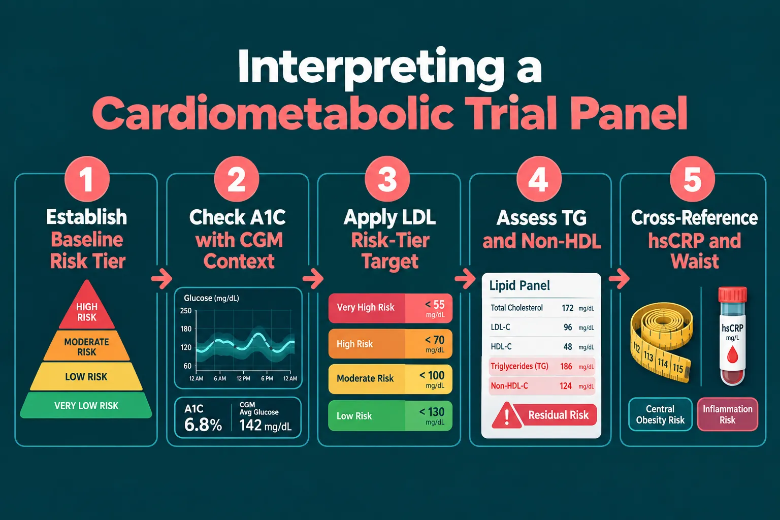
Step Action Key Question
1 Establish baseline risk tier What was the population's cardiovascular risk category at enrollment?
2 Contextualize A1C Was the change accompanied by CGM time-in-range data? Was hypoglycemia tracked?
3 Apply LDL risk-tier target Does the reported LDL-C meet the threshold appropriate for this cohort's risk level?
4 Assess triglycerides and non-HDL Were remnant lipoproteins measured alongside LDL-C?
5 Cross-reference hsCRP and waist Did inflammatory burden and central adiposity change in the same direction as biochemical endpoints?

A Practical Framework for Reading Metabolic Research Endpoints

Reading the endpoints together reveals the mechanism. A compound that lowers A1C, reduces waist circumference, and decreases hsCRP is likely acting on insulin sensitivity, visceral fat, and systemic inflammation simultaneously, a genuinely comprehensive cardiometabolic effect. A compound that lowers only LDL-C may be addressing lipid transport without touching the inflammatory or adiposity components of risk.

Non-HDL cholesterol deserves mention here. Because it captures LDL plus remnant lipoproteins, non-HDL is a more complete lipid endpoint than LDL-C alone. In UK cardiovascular prevention audits, non-HDL targets of 2.6 mmol/L or below are used alongside LDL targets to judge treatment adequacy. Trials that report only LDL-C without non-HDL leave the remnant lipoprotein contribution to risk unaccounted for.


Conclusion

Reading metabolic research endpoints accurately requires treating A1C, LDL, triglycerides, waist circumference, and hsCRP as a complementary panel rather than interchangeable alternatives. Each marker captures a distinct biological mechanism, and no single value substitutes for the full picture.

Actionable next steps for researchers and informed readers:

  • When reviewing a trial, check whether the endpoint panel matches the compound's proposed mechanism of action.
  • Confirm that A1C results are accompanied by CGM-derived time-in-range data and hypoglycemia rates.
  • Identify the study population's cardiovascular risk tier before judging whether an LDL-C value represents success or residual undertreatment.
  • Treat hsCRP as a primary endpoint in any study involving inflammatory pathways, not a supplementary add-on.
  • Flag any trial that reports improvements in biochemical markers without measuring waist circumference, central adiposity may be the missing variable explaining why risk did not fully resolve.

The field is moving toward multi-endpoint cardiometabolic panels as the standard for evaluating both pharmaceutical and peptide-based interventions. Understanding how to read those panels is the foundational skill for interpreting the next generation of metabolic research.

https://www.puretestedpeptides.com/wp-content/uploads/2026/09/how-to-read-metabolic-research-endpoints-a-practical-guide-to-a1c-ldl-triglyceri.webp 1024 1536 https://www.puretestedpeptides.com/wp-content/uploads/2026/01/buy-peptides-online.jpg 2026-09-23 13:07:522026-09-23 13:07:52How to Read Metabolic Research Endpoints: A Practical Guide to A1C, LDL, Triglycerides, Waist Circumference, and hsCRP
Tesofensine Mechanism in Appetite Research: Where Noradrenergic Modulation Fits Alongside GLP Peptides

Tesofensine Mechanism in Appetite Research: Where Noradrenergic Modulation Fits Alongside GLP Peptides

August 16, 2026/0 Comments/in Uncategorized/by

Obesity affects more than one billion people globally, yet fewer than five pharmacological mechanisms have been validated as durable appetite suppressants in controlled human trials. The tesofensine mechanism in appetite research stands out as one of the most instructive examples of how noradrenergic modulation fits alongside GLP peptides, not as a competitor, but as a mechanistically distinct layer that operates through different neural circuits to achieve overlapping metabolic goals.

Key Takeaways

  • Tesofensine inhibits reuptake of norepinephrine, dopamine, and serotonin simultaneously, with noradrenergic action playing a central role in appetite suppression.
  • Its primary weight-loss effect in research models is driven by reduced caloric intake rather than increased energy expenditure.
  • A 2024 mechanistic finding identified silencing of lateral hypothalamic GABAergic feeding neurons as a key downstream effect.
  • GLP-1 receptor pathways and central noradrenergic circuits act on distinct but converging appetite nodes, making combination research strategies scientifically plausible.
  • Cardiovascular effects remain a key variable that separates tesofensine's anti-obesity mechanism from its hemodynamic profile.

How Tesofensine Inhibits Three Monoamine Transporters

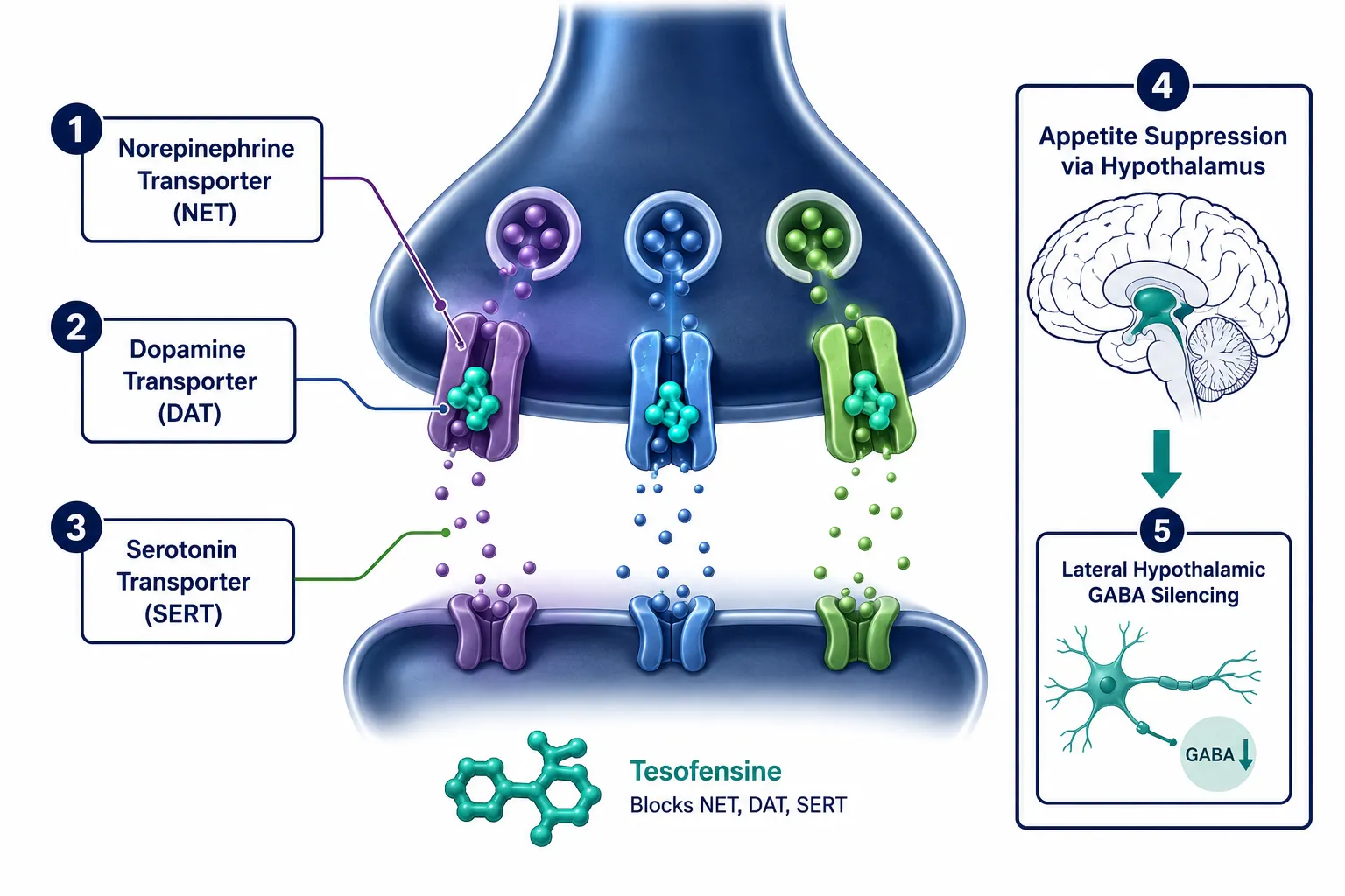
Tesofensine is a triple monoamine reuptake inhibitor. It blocks the norepinephrine transporter (NET), the dopamine transporter (DAT), and the serotonin transporter (SERT) simultaneously. Among these three targets, the noradrenergic component carries the greatest weight in appetite suppression.

How Tesofensine Inhibits Three Monoamine Transporters

When norepinephrine reuptake is blocked, synaptic norepinephrine levels rise. This activates alpha-adrenoceptors in the hypothalamus, particularly in the paraventricular nucleus, triggering a hypophagic response, meaning the drive to eat is reduced. This alpha-adrenoceptor-mediated hypophagia is well-characterized in preclinical models and aligns with human appetite sensation data showing increased satiety and fullness scores without meaningful changes in total energy expenditure.

The dopamine component adds a second layer. Elevated dopamine in mesolimbic circuits reduces food reward salience, the craving dimension of appetite, rather than purely homeostatic hunger. The serotonin component reinforces satiety through 5-HT2C receptor engagement in the hypothalamus, a pathway also targeted by earlier anti-obesity agents.

What makes tesofensine distinct is not any single transporter block, but the simultaneous elevation of all three monoamines, which produces a broader appetite-suppression profile than selective agents alone.

A notable 2024 mechanistic advance identified that tesofensine silences lateral hypothalamic GABAergic feeding neurons. These neurons normally disinhibit feeding behavior. When tesofensine suppresses their activity, the net result is a sustained reduction in meal initiation, a finding that positions the compound within modern circuit-level appetite neuroscience rather than older receptor-pharmacology frameworks.

Noradrenergic Modulation and GLP-1 Receptor Pathways: Where the Circuits Converge

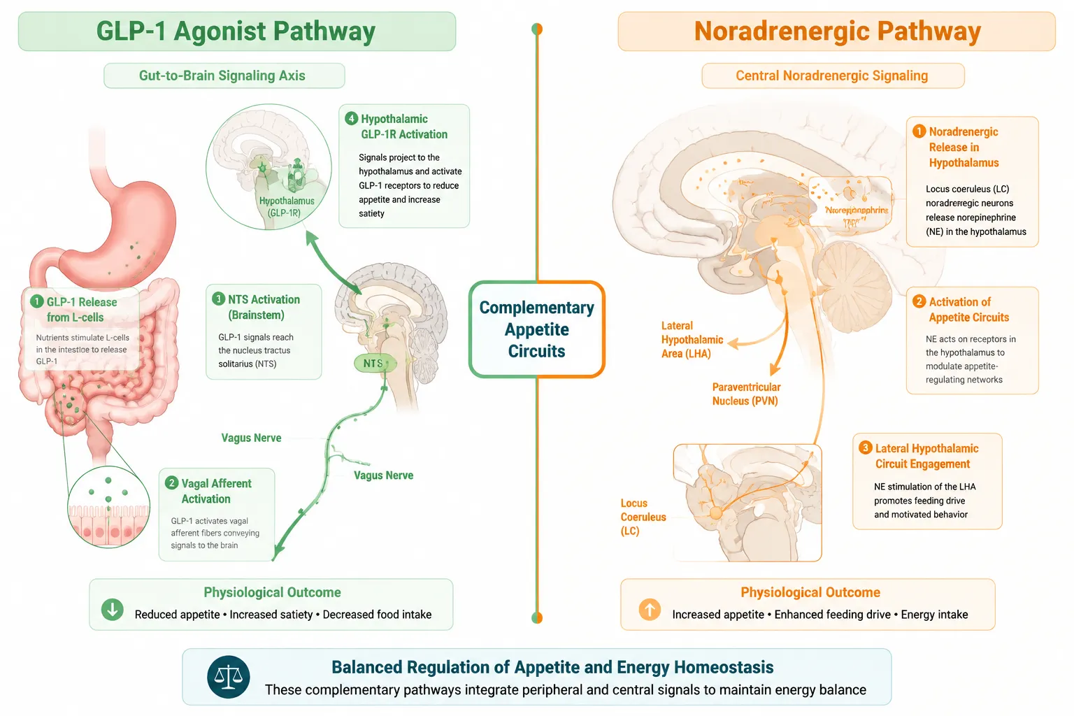
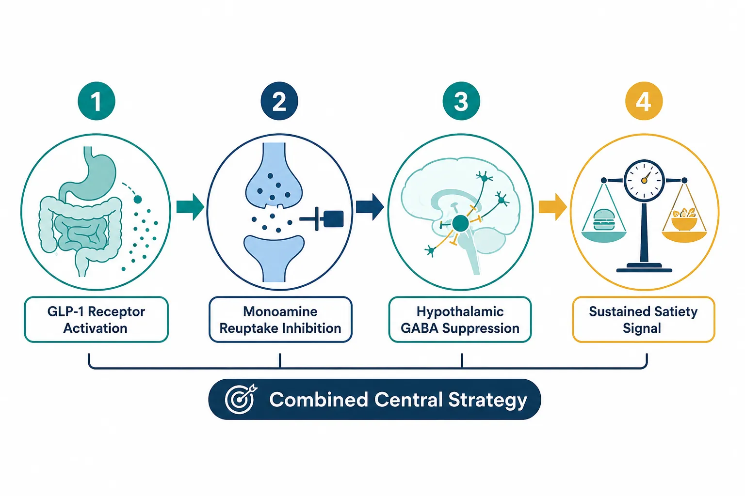
Understanding the tesofensine mechanism in appetite research requires mapping how noradrenergic modulation fits alongside GLP peptides at the circuit level. GLP-1 receptor agonists, a class that includes compounds actively studied in obesity and MASLD research, work primarily through peripheral and central GLP-1 receptors. Their appetite-suppressing signal travels from gut enteroendocrine cells via the vagus nerve to the nucleus tractus solitarius (NTS), then projects to the hypothalamus and limbic system.

Noradrenergic Modulation and GLP-1 Receptor Pathways: Where the Circuits Converge

Noradrenergic modulation, by contrast, originates centrally. Tesofensine elevates norepinephrine directly within hypothalamic synapses, bypassing the gut-brain axis that GLP-1 agonists depend on. This distinction matters for experimental design.

Researchers exploring GLP-1 peptides in obesity models are increasingly interested in whether adding a central monoamine component amplifies outcomes. The hypothalamic GABA circuits affected by tesofensine overlap anatomically with regions that express GLP-1 receptors, suggesting the two mechanisms could act synergistically rather than redundantly.

For those researching metabolic compounds, the top research peptides for metabolic health resource provides useful context on how multiple peptide classes are being evaluated alongside small-molecule agents in 2026 research designs.

A key distinction also emerges around energy expenditure. GLP-1 agonists produce modest increases in energy expenditure alongside appetite suppression. Tesofensine's weight loss in clinical data is attributed almost entirely to reduced caloric intake, not thermogenesis. This means the two approaches address appetite through different effector mechanisms even when they converge on the same hypothalamic output.

Research Insight: When noradrenergic modulation and GLP-1 receptor activation are studied in parallel models, their appetite-suppressing effects appear additive rather than redundant, a finding that supports multi-mechanism experimental designs.

Positioning Tesofensine Within Multi-Mechanism Obesity Research

The tesofensine mechanism in appetite research becomes most strategically relevant when placed alongside GLP peptides in multi-target experimental models. Research on triple-agonist compounds like retatrutide, detailed in this Retatrutide and MASLD analysis, has demonstrated that engaging multiple receptor systems simultaneously produces greater metabolic benefits than single-target approaches. Tesofensine offers a central monoamine dimension that peptide-based GLP agents do not cover.

Positioning Tesofensine Within Multi-Mechanism Obesity Research

Cardiovascular effects remain a critical variable. Norepinephrine elevation raises heart rate and blood pressure, which creates a hemodynamic profile that must be separated from the anti-obesity mechanism in research designs. This is not unique to tesofensine, adrenergic agents broadly carry this challenge, but it does mean that dosing strategies and co-administration with GLP-1 agents require careful titration in preclinical and early clinical models.

For researchers sourcing validated compounds for such studies, understanding where to buy peptides from quality-controlled suppliers is a practical starting point. Purity and documentation standards are especially important when combining small-molecule agents with peptide compounds in the same experimental protocol.

Speculative future directions, and these should be clearly framed as predictions rather than established science, point toward combined central monoamine and GLP-1 strategies as a next frontier. If lateral hypothalamic GABA silencing by tesofensine and GLP-1 receptor-mediated NTS activation both converge on paraventricular nucleus output, a rationally designed combination could produce durable appetite suppression with lower individual doses of each agent, potentially reducing cardiovascular and gastrointestinal side-effect burden. This hypothesis remains to be tested in controlled trials.

Researchers interested in the broader landscape of hormone research compounds will find that the noradrenergic-GLP-1 intersection is one of several active areas where mechanistic diversity is being deliberately engineered into next-generation obesity protocols.

For additional context on how GLP-2 and related peptide variants are being studied alongside appetite-modulating agents, the GLP-2 peptide research tag provides relevant compound documentation.

Conclusion

The tesofensine mechanism in appetite research offers a precise, centrally acting noradrenergic tool that fills a mechanistic gap that GLP peptides do not address. By blocking NET, DAT, and SERT simultaneously, tesofensine elevates hypothalamic norepinephrine, silences lateral hypothalamic GABAergic feeding neurons, and reduces caloric intake through satiety enhancement rather than energy expenditure changes.

Actionable next steps for researchers in 2026:

  • Map experimental designs to include both central monoamine endpoints and peripheral GLP-1 receptor endpoints when studying appetite suppression in obesity or MASLD models.
  • Account for cardiovascular variables separately from anti-obesity outcomes when interpreting noradrenergic data.
  • Prioritize compounds sourced with verified purity documentation when combining peptide and small-molecule agents in the same protocol.
  • Monitor emerging trial data on combination central monoamine and GLP-1 strategies as the most likely near-term advance in multi-mechanism obesity pharmacology.

The noradrenergic and incretin pathways are not rivals. They are complementary axes in a complex appetite circuit, and understanding where each one acts is the foundation for designing more effective metabolic research in the years ahead.

https://www.puretestedpeptides.com/wp-content/uploads/2026/08/tesofensine-mechanism-in-appetite-research-where-noradrenergic-modulation-fits-a.webp 1024 1536 https://www.puretestedpeptides.com/wp-content/uploads/2026/01/buy-peptides-online.jpg 2026-08-16 13:05:212026-08-16 13:05:21Tesofensine Mechanism in Appetite Research: Where Noradrenergic Modulation Fits Alongside GLP Peptides
×

Helpful Links

  • My account
  • Cart
  • Checkout
  • Refund and Returns Policy
  • Privacy Policy
  • SMS Privacy Policy
  • Login
  • My Account
  • Logout

USA Made Lab Tested Peptides

All products are sold for research, laboratory, or analytical purposes only, and are not for human consumption


Pure Tested Peptides is a chemical supplier. Pure Tested Peptides is not a compounding / chemical compounding facility as defined under 503A of the Federal Food, Drug, and Cosmetic act. Pure Tested Peptides is not an outsourcing facility as defined under 503B of the Federal Food, Drug, and Cosmetic act.

The statements made within this website have not been evaluated by the US Food and Drug Administration. The products we offer are not intended to diagnose, treat, cure or prevent any disease.

Human/Animal Consumption Prohibited. Laboratory/In-Vitro Experimental Use Only

Scroll to top Scroll to top Scroll to top