Iron Pills and Peptide Research: Separating Anemia Correction From Metabolic and Mitochondrial Effects
Fatigue disappears after iron supplementation, but corrected hemoglobin does not always explain why. Research published in 2025 and 2026 confirms that iron deficiency disrupts mitochondrial ATP synthesis, shifts metabolism toward anaerobic glycolysis, and elevates lactate, pyruvate, and succinate long before a blood count flags clinical anemia. For researchers studying metabolic peptides such as MOTS-c and 5-Amino-1MQ, that distinction matters enormously. This article examines the full scope of Iron Pills and Peptide Research: Separating Anemia Correction From Metabolic and Mitochondrial Effects, covering what the latest studies show, why baseline iron variables must be recorded in every peptide model, and how oxidative stress bridges both fields.
Key Takeaways
- Iron deficiency impairs mitochondrial electron transport and ATP output well before anemia develops, creating confounding variables in metabolic research.
- Elevated succinate, lactate, and reduced ATP serve as metabolic biomarkers that go beyond hemoglobin when evaluating iron interventions.
- Baseline ferritin, hemoglobin, and serum iron must be documented in MOTS-c, 5-Amino-1MQ, and endurance peptide studies to allow valid interpretation.
- Peptides that target mitochondrial function, such as SS-31, may interact with iron-driven oxidative stress in ways that require careful experimental design.
- High-dose iron supplementation carries oxidative risk; optimal dosing strategies balance anemia correction with protection against free-radical damage.
Beyond Hemoglobin: The Metabolic and Mitochondrial Burden of Iron Deficiency
Iron is not simply a building block for red blood cells. It is a core component of Complexes I through IV of the mitochondrial electron transport chain. When iron is insufficient, oxidative phosphorylation slows, ATP output drops, and cells compensate by burning glucose anaerobically, a metabolic shift reflected in elevated lactate and pyruvate. Succinate, a TCA-cycle intermediate, accumulates as a signal of impaired mitochondrial flux, and reactive oxygen species (ROS) production increases.
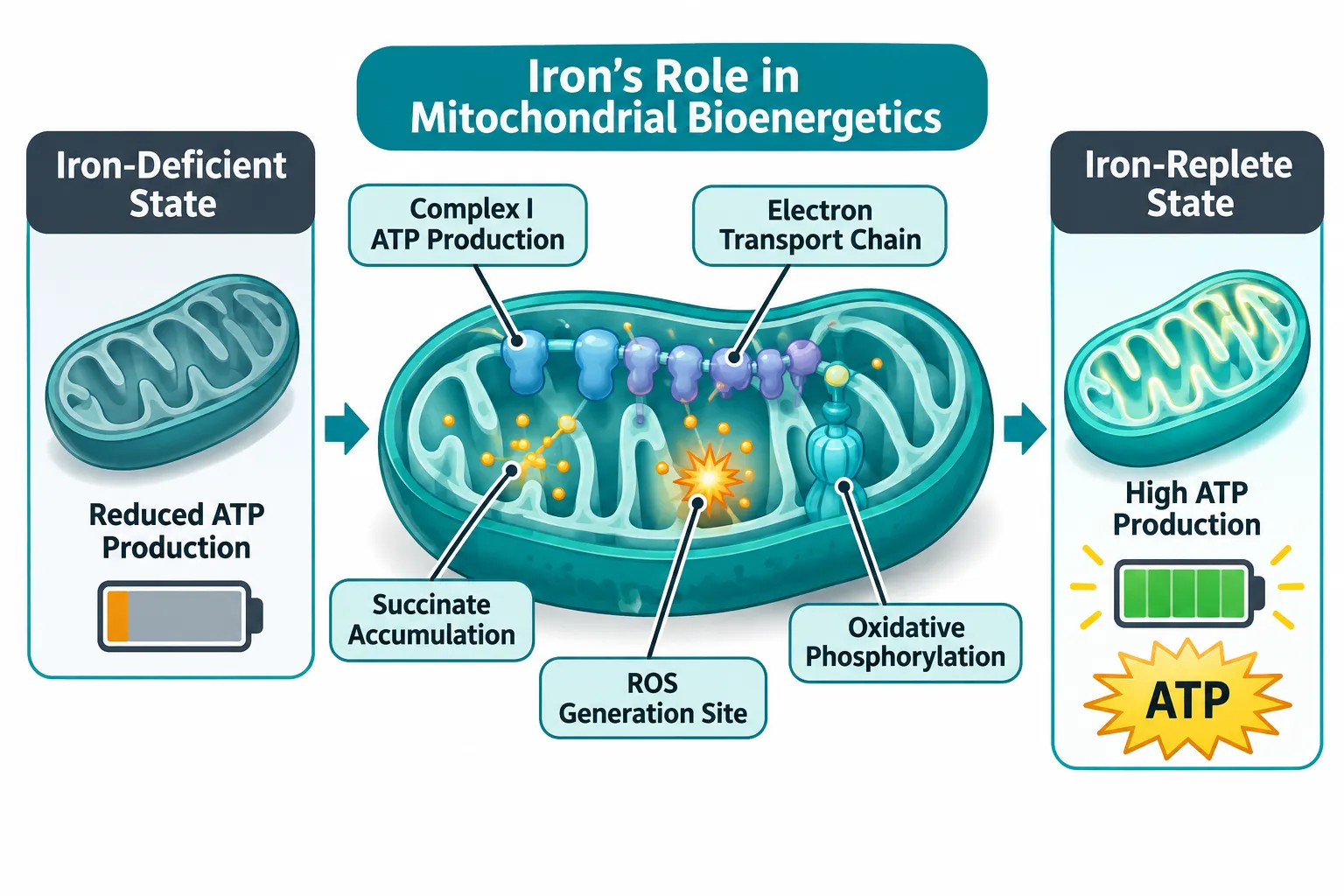
Symptomatic fatigue and reduced exercise capacity can appear in iron-deficient individuals with completely normal hemoglobin. A 2025 review confirmed that iron repletion improves muscle function and aerobic performance as independent outcomes from anemia correction, and randomized data support oral iron supplementation improving unexplained fatigue in non-anemic women. The ongoing MyoIRON clinical trial captures this directly: participants with suboptimal but non-anemic iron stores are randomized to 150 mg elemental iron every other day for 12 weeks, measuring skeletal-muscle iron storage, mitochondrial protein expression, and single-leg exercise performance alongside conventional hematologic markers. The trial is an explicit attempt at separating anemia correction from metabolic and mitochondrial effects in a living system.
A 2024 rat study reinforced the point mechanistically. Animals receiving an iron-based supplement with vitamin B6 for 30 days showed substantially increased Complex I, and Complex II, driven ATP production in gastrocnemius and cerebellar mitochondria, without a corresponding rise in basal or succinate-stimulated ROS. Mitochondrial energy output improved without worsening oxidative stress, a finding that cannot be reduced to simple erythropoiesis.
Iron Pills and Peptide Research: Why MOTS-c and 5-Amino-1MQ Studies Need Iron Variables
MOTS-c is a mitochondria-derived peptide encoded in the 12S rRNA region of mitochondrial DNA. Its primary research interest lies in metabolic regulation, specifically AMPK activation, glucose uptake, and mitochondrial biogenesis. 5-Amino-1MQ is a small-molecule inhibitor of NNMT (nicotinamide N-methyltransferase) linked to energy expenditure, fat metabolism, and NAD+ pathway regulation. Both targets sit directly within the same metabolic machinery that iron deficiency disrupts.
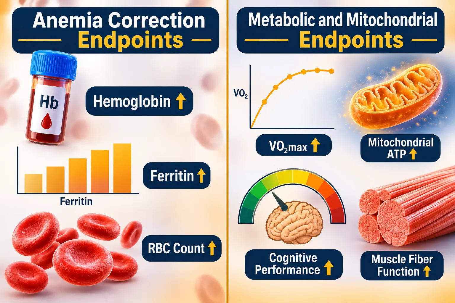
When an animal or human subject enters a MOTS-c or 5-Amino-1MQ study in an iron-deficient state, several confounders activate simultaneously:
| Variable | Iron-Deficient Effect | Peptide Research Impact |
|---|---|---|
| Mitochondrial ATP output | Reduced Complex I/II activity | Masks or mimics peptide effect on energy |
| ROS production | Elevated | Inflates oxidative stress readings |
| AMPK signaling | Activated by low energy charge | Confounds MOTS-c AMPK data |
| VO2max / aerobic capacity | Decreased | Distorts endurance endpoint comparisons |
| Muscle glycolytic shift | Increased lactate/pyruvate | Alters substrate utilization metrics |
This is not a hypothetical concern. A large 2025 intravenous iron trial in heart failure patients showed improvements in functional capacity and quality of life that were partly attributed to improved cardiac and skeletal muscle energetics, effects distinct from anemia correction. If iron status was not measured at baseline in those subjects, the metabolic improvements would have been attributed entirely to the therapeutic intervention under study.
Core principle: In any peptide model targeting metabolic or mitochondrial outcomes, an undocumented low-ferritin subject is an uncontrolled variable.
For researchers exploring SS-31 mitochondrial research themes alongside iron protocols, this overlap is especially relevant. SS-31 targets cardiolipin on the inner mitochondrial membrane, stabilizing cristae architecture and reducing ROS leak, the same ROS that iron-driven Fenton chemistry can amplify. Studies of SS31 mitochondrial dynamics therefore benefit from documented iron and oxidative-stress baselines.
Oxidative Stress, Hepcidin, and the Peptide Research Frontier
Iron Pills and Peptide Research: Separating Anemia Correction From Metabolic and Mitochondrial Effects becomes most complex at the intersection of oxidative biology. Free, unliganded iron catalyzes hydroxyl radical formation via the Fenton reaction. High-dose oral or intravenous iron therefore carries oxidative risk at the mitochondrial membrane, the very site that peptides like SS-31 are designed to protect.
A 2026 review highlighted this oxidative risk explicitly, noting that dosing strategies must balance anemia correction with prevention of cellular oxidative injury. That balance is dose- and timing-dependent, and it interacts with antioxidant peptide activity in ways that have not yet been systematically characterized.
A second complication arises in metabolic disease. In subjects with obesity, chronic inflammation upregulates hepcidin, the master iron-regulatory hormone, restricting iron availability to tissues. Iron deficiency in this population impairs attention, memory, executive function, and aerobic performance before hemoglobin falls. For peptide studies aimed at visceral fat or metabolic syndrome, areas covered extensively in visceral fat research, a subject's inflammatory iron restriction represents a silent confounder that inflates apparent peptide efficacy when iron normalizes incidentally.
Unabsorbed luminal iron from oral supplementation also alters gut microbial communities, potentially modulating inflammatory signaling and systemic metabolic tone through pathways that overlap with GLP-class peptide research. Researchers following developments in GLP-3 peptide studies should note that iron-microbiome interactions may affect metabolic endpoints used in those models.
Recommended baseline variables for any metabolic or mitochondrial peptide study:
- Serum ferritin (iron stores)
- Hemoglobin and hematocrit
- Serum iron and transferrin saturation
- C-reactive protein (to flag inflammatory iron restriction)
- Lactate and pyruvate ratios (as markers of glycolytic shift)
For neurodevelopmental models, iron is a cofactor for myelination and neurotransmitter synthesis, both energy-intensive processes. Cognitive peptide studies that omit iron status risk attributing brain-energy improvements to the peptide when simple iron repletion drove the result. Reviewing SS-31 kidney health research illustrates how organ-specific energy demands interact with mitochondrial protectants in ways that iron status modulates at the cellular level.
Conclusion
The evidence from 2025 and 2026 is unambiguous: iron deficiency alters mitochondrial function, metabolic substrate use, and oxidative stress in ways that are independent of hemoglobin status. For peptide researchers working with MOTS-c, 5-Amino-1MQ, SS-31, or endurance-performance models, iron variables are not background noise, they are primary confounders.
Actionable next steps for research teams:
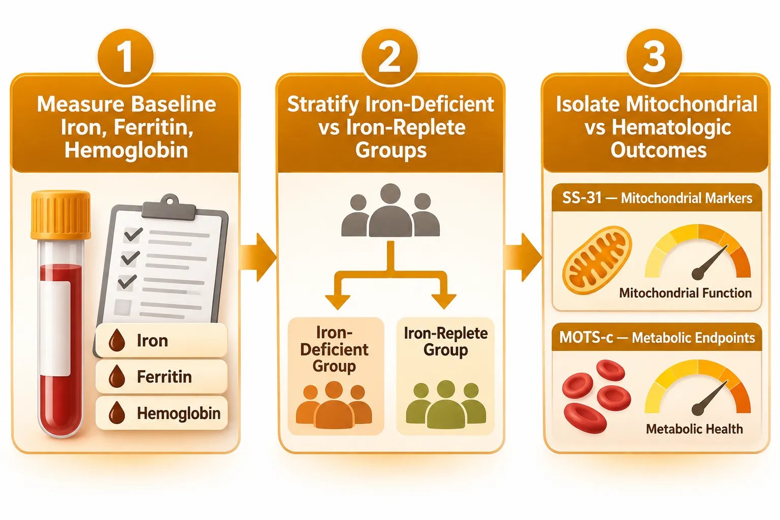
- Screen all subjects for serum ferritin, hemoglobin, transferrin saturation, and CRP before enrollment in any metabolic or mitochondrial peptide study.
- Stratify or exclude iron-deficient individuals unless iron status is itself a study variable.
- When evaluating mitochondrial peptides, record baseline lactate-to-pyruvate ratios and VO2max alongside standard iron panels.
- For studies using SS-31 or similar antioxidant peptides, document iron dosing and timing to separate peptide-driven ROS reduction from iron-dose-driven oxidative load.
- Consult published resources on SS-31 peptide benefits and LL-37 versus SS-31 comparative research for reference frameworks when designing combination protocols.
Separating anemia correction from metabolic and mitochondrial effects is not academic fine-tuning, it is the difference between a valid result and a confounded one.