Advanced Glycation End Products in Peptide Studies: Choosing Fluorescence, RAGE, and Oxidative-Stress Readouts
Roughly 50 distinct molecular species qualify as advanced glycation end products (AGEs), yet most peptide researchers rely on fewer than five assay strategies to detect them, a mismatch that quietly undermines experimental conclusions. Designing a rigorous glycation study requires matching the right detection method to the right AGE species, understanding how RAGE signaling connects to downstream inflammation, and selecting oxidative-stress readouts that complement rather than duplicate fluorescence data. This article on Advanced Glycation End Products in Peptide Studies: Choosing Fluorescence, RAGE, and Oxidative-Stress Readouts walks through each decision point so researchers can build a coherent, reproducible assay panel from the start.
Key Takeaways
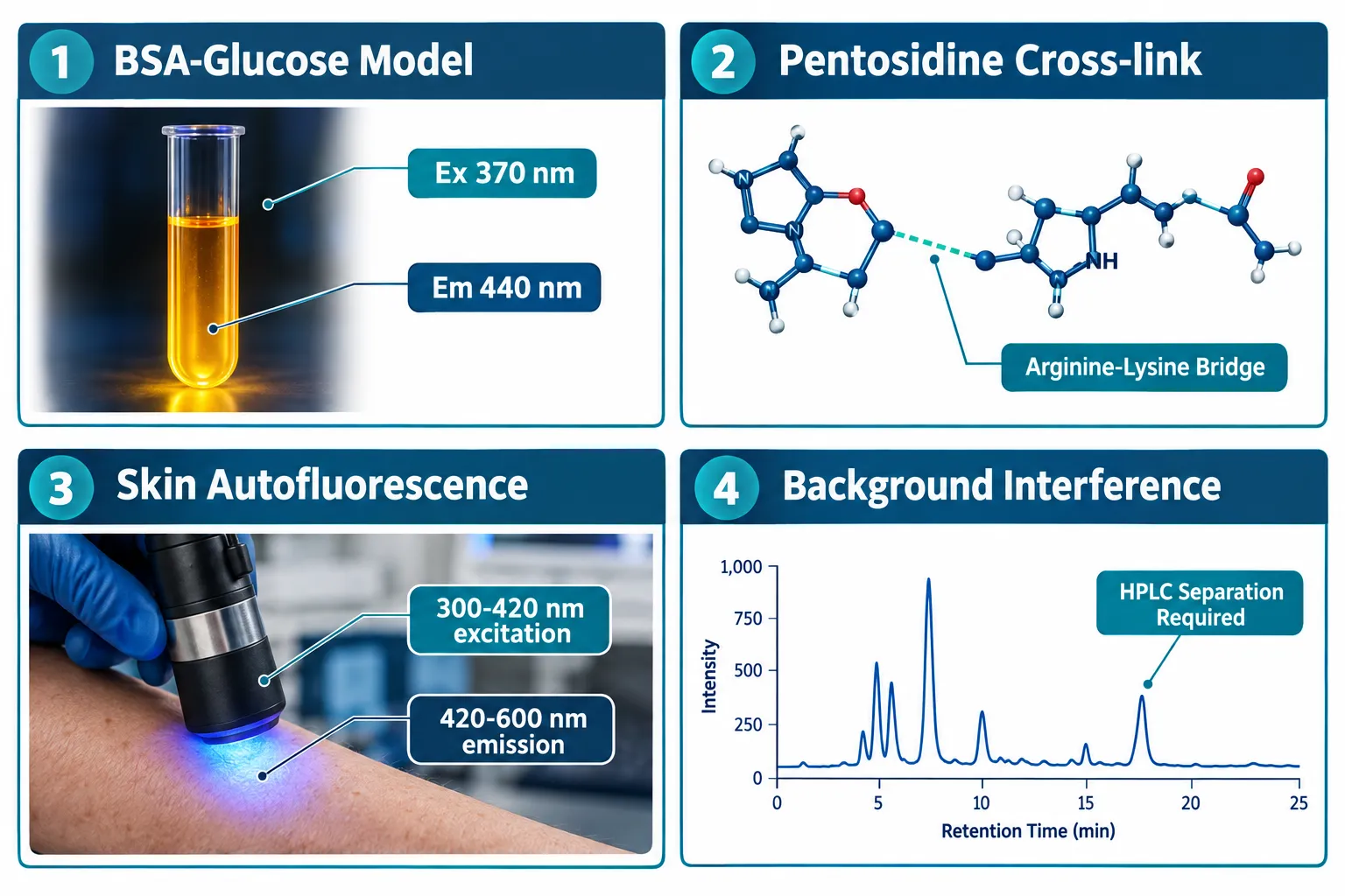
- Fluorescent AGEs (fAGEs) such as pentosidine and methylglyoxal-lysine dimer (MOLD) are detected at specific excitation/emission windows, but background fluorescence often demands HPLC or immunoassay separation.
- The RAGE signaling panel should include at minimum CML, CEL, MG-H1, sRAGE, esRAGE, HMGB1, and S100A12 for a complete picture of receptor-mediated glycation stress.
- Oxidative-stress markers (ROS, 8-OHdG, MDA) serve as orthogonal readouts that confirm AGE-driven cellular damage without redundancy.
- Peptide candidates such as SS-31 and GHK-Cu are evaluated in glycation models for their potential to modulate these pathways, though causality claims require controlled, multi-readout designs.
- Choosing the correct glycation model (BSA-glucose, HSA-methylglyoxal, or collagen-ribose) determines which AGE species dominate and therefore which assays are appropriate.
Setting Up the Glycation Model: The Foundation of Every Assay
Before selecting any readout, researchers must define the glycation model. The choice of protein substrate and reducing sugar determines which AGE species accumulate, and that dictates every downstream measurement.
Three models dominate the literature:
| Model | Primary AGEs Formed | Best Readout |
|---|---|---|
| BSA + glucose (37 °C, 4-8 weeks) | Pentosidine, CML, MOLD | Fluorescence (Ex 370/Em 440 nm) |
| HSA + methylglyoxal (short incubation) | MG-H1, CEL | LC-MS/MS or ELISA |
| Collagen + ribose | Cross-linked fAGEs, pentosidine | Fluorescence + tensile assay |
In a well-characterized in vitro study using the BSA-glucose model, novel peptides were screened for anti-glycation activity by measuring fluorescence at excitation 370 nm and emission 440 nm with a spectrofluorometer. This approach is cost-effective and scalable, but it captures only the fluorescent fraction of total AGEs.
Key point: Non-fluorescent AGEs such as pyrraline and CML will not appear in a standard fluorescence scan. If the research question involves receptor binding or cardiovascular risk markers, a fluorescence-only design is insufficient.
Fluorescence Readouts: Matching Excitation/Emission to the Target AGE
The fluorescence approach to Advanced Glycation End Products in Peptide Studies: Choosing Fluorescence, RAGE, and Oxidative-Stress Readouts is attractive because it requires no antibodies and delivers rapid throughput. However, precision depends on knowing the spectral fingerprint of each target species.
Major fluorescent AGEs and their windows:
- Pentosidine, the first characterized natural fluorescent cross-link; excitation ~335 nm, emission ~385 nm
- MOLD (methylglyoxal-lysine dimer), excitation ~370 nm, emission ~440 nm
- Arg-P, useful for arginine-derived fAGE detection; recent mass spectrometry work has identified new cross-linked structures at arginine residues that extend this category
Cutaneous AGEs exhibit autofluorescence with excitation in the 300-420 nm range and emission between 420-600 nm, enabling non-invasive skin measurements. This principle underlies commercial skin autofluorescence readers used in diabetes monitoring.
"Background fluorescence from other sample components frequently obscures results, necessitating chromatographic or immunoassay-based separation for robust quantification."
A practical solution is the flow injection assay (FIA) integrated with HPLC, which has been validated for quantifying small fluorescent AGE-modified peptides in urine samples. For researchers evaluating skin-relevant peptides, the article on research-grade Glow Blend peptide purity and copper complexes provides useful context on how copper-complexed peptides interact with skin models where autofluorescence is a concern.
RAGE Signaling Panel: Moving Beyond Fluorescence
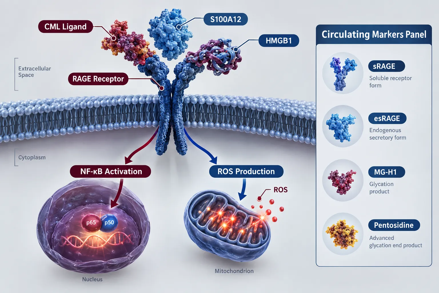
Fluorescence detects AGE accumulation, but RAGE activation is what drives pathological outcomes, NF-kB-mediated inflammation, fibrosis, and cardiomyopathy. A complete panel for Advanced Glycation End Products in Peptide Studies: Choosing Fluorescence, RAGE, and Oxidative-Stress Readouts should include both AGE ligands and RAGE-related circulating measures.
Recommended RAGE panel components:
- AGE ligands: CML, CEL, MG-H1, pentosidine
- Receptor variants: total soluble RAGE (sRAGE), endogenous secretory RAGE (esRAGE)
- Danger-associated ligands: HMGB1, S100A12
The sRAGE/esRAGE ratio is particularly informative: soluble forms act as decoy receptors, sequestering AGE ligands before they engage membrane-bound RAGE. A high sRAGE level generally correlates with reduced signaling intensity.
For peptide studies with a metabolic focus, GLP-1 peptides and related incretin-axis compounds are increasingly studied in contexts where AGE-RAGE signaling intersects with glycemic control. Similarly, mitochondria-targeted peptides like SS-31 (Szeto-Schiller peptide) are evaluated in models where RAGE-driven oxidative stress is the primary endpoint, given their proposed role in mitochondrial membrane stabilization.
Measurement methods by marker:
- CML, CEL, MG-H1: LC-MS/MS (gold standard) or competitive ELISA
- sRAGE, esRAGE: sandwich ELISA
- HMGB1, S100A12: ELISA or multiplex immunoassay
Oxidative-Stress Readouts: The Orthogonal Confirmation Layer
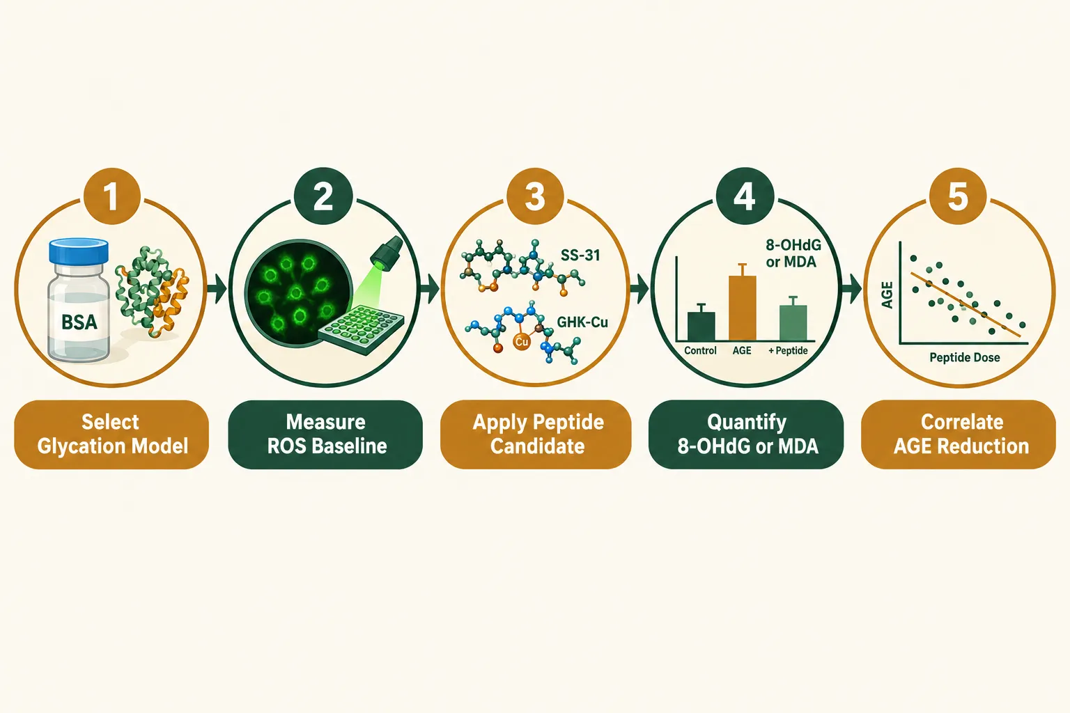
AGE formation and RAGE activation both generate reactive oxygen species (ROS), creating a self-amplifying loop. Oxidative-stress markers serve as orthogonal readouts that confirm biological impact without duplicating fluorescence or ELISA data.
Three-tier oxidative-stress readout strategy:
- ROS quantification, DCFH-DA fluorescence assay in cell-based models; captures real-time oxidative burst
- Lipid peroxidation, malondialdehyde (MDA) via TBARS assay; reflects membrane-level AGE-driven damage
- DNA oxidation, 8-hydroxy-2'-deoxyguanosine (8-OHdG) by ELISA; most specific marker of glycoxidative DNA stress
Anti-glycation agents such as metformin and pyridoxamine reduce fluorescent HSA-AGE emission intensity in HSA-dihydroxyacetone models, and this reduction correlates with decreased ROS output, demonstrating why pairing fluorescence with an ROS assay strengthens mechanistic claims.
Peptides under investigation for aging-related pathways, including those in the aging support category and mitochondria-focused compounds such as the Synergy of LL-37 and SS-31, are evaluated against these markers to assess whether observed effects align with reduced glycoxidative stress. Causal claims, however, require dose-response relationships and appropriate controls before they can be stated with confidence.
Practical tip: Always run a no-peptide glycation control and a non-glycated protein control in parallel. Without these, it is impossible to distinguish peptide-driven AGE reduction from assay variability.
Conclusion
Designing a rigorous AGE study for peptide research comes down to three sequential decisions: choose the right glycation model for the target AGE species, select fluorescence readouts matched to those species' spectral windows, and add RAGE panel markers plus orthogonal oxidative-stress assays to confirm biological relevance. Researchers should resist the temptation to rely on a single fluorescence scan as a complete answer, it captures only the fluorescent fraction and misses non-fluorescent species like CML and pyrraline that drive RAGE signaling.
Actionable next steps for 2026 research teams:
- Establish the BSA-glucose or HSA-methylglyoxal model before introducing any peptide candidate
- Run fluorescence scans at Ex 370/Em 440 nm alongside LC-MS/MS for CML and MG-H1
- Include sRAGE and esRAGE ELISAs to assess receptor-level activity
- Add at minimum one oxidative-stress readout (MDA or 8-OHdG) per experimental arm
- Document all purity and concentration data for peptide candidates to ensure reproducibility across labs