Call or Text 727-513-9780
  • Shopping Cart Shopping Cart
    0Shopping Cart
Pure Tested Peptides | America's most trusted Peptides for sale online
  • Peptides for sale
    • Oral Peptides for sale
      • Peptide Capsules for sale
      • BPC 157 Capsules 1000mcg
      • SLU-PP-332 Capsules | 1000 mcg
      • 5-Amino-1MQ 50mg Capsules
      • Tesofensine 500mcg
    • All Peptides for sale
    • Peptide Sprays
      • BPC 157 Nasal Spray Kit
      • BPC-157 TB500 Nasal Spray Kit
      • Semax Nasal Spray 10mg
      • Selank – Nasal Spray Kit – 10mg
      • Epithalon 50MG Nasal Spray Kit
      • Ipamorelin 10mg Nasal Spray
      • Klow Nasal Spray (BPC-157 + TB-500 + GHK-Cu + KPV) | 80mg
      • Hulk Nasal Spray Tesa / Ipa Blend 6/3 MG
      • Klow Nasal Spray
      • NAD + 500 mg Nasal Spray
      • PT-141 Nasal Spray Kit
    • GHRH Peptides
      • Ipa Peptides
      • CJC-1295 Peptides
        • CJC-1295 with DAC 5 mg
        • CJC-1295 without DAC 5 mg
        • CJC-1295 Ipa 10mg
      • Tesa Peptides
        • Tesa Peptide
        • Tesa 20 mg
    • GHK-Cu Peptides
      • All GHK-Cu Peptides
      • GHK-Cu 100mg
      • KLOW Peptide Blend – Buy KLOW blend online
    • BPC Peptides
      • All BPC Peptides
      • BPC-157
      • BPC-157 TB-500
      • BPC 157 capsules 1000mcg
    • SLU-PP-332 Peptides
      • All SLU-PP-332 Peptides
      • SLU-PP-332 5mg
    • GLP3 Peptides
    • PT-141 Peptides
      • PT-141 Peptides for sale
      • PT-141 10mg
      • PT-141 Nasal Spray
    • CAG Peptides
      • Lipo-C Peptide Blend
      • CAG 5mg
      • CAG 10mg
    • MOTS-C Peptides
      • MOTS-C Peptides for sale
      • MOTS-c peptide
      • MOTS-c 10mg *6 pack*
    • 5 Amino 1MQ Peptides
      • 5 Amino 1MQ Peptides for sale
      • 5-Amino-1MQ 50mg Capsules
      • 5-Amino-1MQ 5mg
    • Epithalon Peptides
      • Epithalon Peptides for sale
      • Epithalon 10mg
      • Epithalon 50mg
  • Shop
    • GLPs
      • 5-Amino-1MQ 50mg Capsules
      • 5-Amino-1MQ 5mg
      • L-Carnitine 500mg/ml
      • Tesofensine 500mcg
      • SLU-PP-332 5mg
      • MOTS-c 10mg *6 pack*
    • Epithalon & BPC Peptides
      • Epithalon 10mg
      • Epithalon 50mg
      • BPC-157
      • BPC 157 capsules 1000mcg
      • BPC-157 TB-500
      • BPC-157 TB500 Nasal Spray Kit
      • BPC 157 Nasal Spray Kit
    • BPC TB-500 & NAD+ Peptides
      • NAD+ 500 mg
      • KLOW Peptide Blend – Buy KLOW blend online
      • GLOW Peptide Blend
      • TB 500 5mg
      • BPC 157 capsules 1000mcg – Supplement
      • BPC 157 Nasal Spray Kit
      • BPC-157
      • BPC-157 TB500 Nasal Spray Kit
      • BPC-157 TB-500
      • BPC 157 capsules 1000mcg
    • LL-37 Peptide
      • LL-37 10 mg
    • MOTS-C & Selank
      • MOTS-c peptide
      • Selank 10mg
    • GHK Peptides
      • GHK-Cu 100mg
      • GLOW Peptide Blend
      • KLOW Peptide Blend – Buy KLOW blend online
  • COAs
  • Wholesale
    • Wholesale Peptides for sale
  • PTP FAQ
  • Affiliates
    • Selank Peptide: Advanced Pharmacological Mechanisms Underlying Its Anxiolytic and Nootropic Effects in Research
      • 5-Amino-1MQ Peptide: Detailed Mechanisms of NNMT Inhibition and Its Impact on Cellular Metabolism Research
        • MOTS-C Peptide: Unraveling Its Role in Mitochondrial Dynamics and Energy Metabolism Research
          • Epithalon Peptide and Telomerase Regulation: Investigating Its Impact on Cellular Senescence and Lifespan Research Models
            • GLP2-T vs GLP2 Tirz Peptide: Understanding the Naming, Mechanistic Nuances, and Research Implications for Gut Health
      • Peptides and Polypeptides in Modern Pharmacology: What Research on Metoprolol, Prednisone, and Amlodipine Reveals
        • Polypeptide Peptides vs NSAIDs: What Naproxen and Diclofenac Teach Tissue-Repair Researchers About BPC‑157 and TB‑500
          • Peptides and Polypeptides in Endocrine Pharmacology: How GLP-1, GLP-2, and GLP-3 Retatrutide Differ From Classic Drugs Like Prednisone and Amlodipine
          • 5‑Amino‑1MQ Peptide and NNMT Inhibition: How It Compares With Statins Like Atorvastatin in Adipose and Lipid Metabolism Research
      • Enclomiphene, Estrogen Receptor Signaling, and Luteinizing Phase Biology: What Hormone Researchers Should Measure
        • Mesenchymal Stem Cells, BPC‑157, and GHK‑Cu: How Tissue Repair Peptides Compare With Classic NSAIDs Like Naproxen in Injury Models
          • Peptide Calculator Use Cases Beyond Growth Hormone: Working Through GLP‑3 Retatrutide, MOTS‑c, and BPC‑157 Research Dosing
        • Polypeptide Peptides in Endocrine and Metabolic Pharmacology: Lessons From Amlodipine, Prednisone, and Metoprolol
        • Peptides Calculator for Advanced Blends: Worked Examples for Tesamorelin, CJC‑1295, and Ipamorelin Stacks
          • Peptides and Polypeptides in Human Physiology: How Molecular Size Shapes Research Applications
            • Selank Peptide: Advanced Pharmacological Mechanisms Underlying Its Anxiolytic and Nootropic Effects in Research
          • 5-Amino-1MQ Peptide: Detailed Mechanisms of NNMT Inhibition and Its Impact on Cellular Metabolism Research
            • MOTS-C Peptide: Unraveling Its Role in Mitochondrial Dynamics and Energy Metabolism Research
              • Epithalon Peptide and Telomerase Regulation: Investigating Its Impact on Cellular Senescence and Lifespan Research Models
                • GLP2-T vs GLP2 Tirz Peptide: Understanding the Naming, Mechanistic Nuances, and Research Implications for Gut Health
                  • Peptides and Polypeptides in Endocrine Pharmacology: How GLP-1, GLP-2, and GLP-3 Retatrutide Differ From Classic Drugs Like Prednisone and Amlodipine
                    • Polypeptide Peptides vs NSAIDs: What Naproxen and Diclofenac Teach Tissue-Repair Researchers About BPC‑157 and TB‑500
                      • Peptides and Polypeptides in Modern Pharmacology: What Research on Metoprolol, Prednisone, and Amlodipine Reveals
                        • 5‑Amino‑1MQ Peptide and NNMT Inhibition: How It Compares With Statins Like Atorvastatin in Adipose and Lipid Metabolism Research
                        • Enclomiphene, Estrogen Receptor Signaling, and Luteinizing Phase Biology: What Hormone Researchers Should Measure
                        • Mesenchymal Stem Cells, BPC‑157, and GHK‑Cu: How Tissue Repair Peptides Compare With Classic NSAIDs Like Naproxen in Injury Models
                        • Peptide Calculator Use Cases Beyond Growth Hormone: Working Through GLP‑3 Retatrutide, MOTS‑c, and BPC‑157 Research Dosing
                        • Polypeptide Peptides in Endocrine and Metabolic Pharmacology: Lessons From Amlodipine, Prednisone, and Metoprolol
                        • Peptides Calculator for Advanced Blends: Worked Examples for Tesamorelin, CJC‑1295, and Ipamorelin Stacks
                        • Peptides and Polypeptides in Human Physiology: How Molecular Size Shapes Research Applications
                        • Selank Peptide: Advanced Pharmacological Mechanisms Underlying Its Anxiolytic and Nootropic Effects in Research
                        • MOTS-C Peptide: Unraveling Its Role in Mitochondrial Dynamics and Energy Metabolism Research
                        • Peptides and Polypeptides in Modern Pharmacology: What Research on Metoprolol, Prednisone, and Amlodipine Reveals
                        • Peptides and Polypeptides in Endocrine Pharmacology: How GLP-1, GLP-2, and GLP-3 Retatrutide Differ From Classic Drugs Like Prednisone and Amlodipine
                        • 5-Amino-1MQ Peptide: Detailed Mechanisms of NNMT Inhibition and Its Impact on Cellular Metabolism Research
                        • Mesenchymal Stem Cells, BPC‑157, and GHK‑Cu: How Tissue Repair Peptides Compare With Classic NSAIDs Like Naproxen in Injury Models
                        • Peptide Calculator Use Cases Beyond Growth Hormone: Working Through GLP‑3 Retatrutide, MOTS‑c, and BPC‑157 Research Dosing
                        • GLP2-T vs GLP2 Tirz Peptide: Understanding the Naming, Mechanistic Nuances, and Research Implications for Gut Health
                        • Polypeptide Peptides in Endocrine and Metabolic Pharmacology: Lessons From Amlodipine, Prednisone, and Metoprolol
                        • Polypeptide Peptides vs NSAIDs: What Naproxen and Diclofenac Teach Tissue-Repair Researchers About BPC‑157 and TB‑500
                        • Peptides and Polypeptides in Human Physiology: How Molecular Size Shapes Research Applications
                        • Peptides Calculator for Advanced Blends: Worked Examples for Tesamorelin, CJC‑1295, and Ipamorelin Stacks
                        • 5‑Amino‑1MQ Peptide and NNMT Inhibition: How It Compares With Statins Like Atorvastatin in Adipose and Lipid Metabolism Research
                        • Enclomiphene, Estrogen Receptor Signaling, and Luteinizing Phase Biology: What Hormone Researchers Should Measure
                        • Epithalon Peptide and Telomerase Regulation: Investigating Its Impact on Cellular Senescence and Lifespan Research Models
                        • Best research protocol Klow blend
                        • best time to take BPC-157
                        • best time to take DSIP (Delta Sleep Inducing Peptide)
                        • best time to take CJC-1295
                        • best time to take AOD-9604
                        • best time to take Follistatin 344
                        • best time to take Ipamorelin
                        • best time to take MK-677 (Ibutamoren)
                        • best time to take Ligandrol (LGD-4033) — research compound
                        • best time to take Ostarine (MK-2866) — research compound
                        • best time to take GHK-CU
                        • best time to take TB-500
                        • best time to take MOTS-c
                        • best time to take Semax
                        • best time to take RAD-140 (Testolone) — research compound
                        • best time to take Thymosin Alpha-1
                        • best time to take PEG-MGF
                        • Biolife Plasma, Octapharma Plasma, and Research Peptides: How Plasma Donation Labs Differ From Peptide Suppliers
                        • best time to take YK-11 — research compound
                        • best time to take PT-141 (Bremelanotide)
                        • Best research protocol Klow blend
                        • 5-Amino-1MQ and MOTS-C Synergy: Metabolic Signaling, Mitochondria, and Research Design
                        • BPC-157 and TB-500: Investigating Their Combined Effects on Angiogenesis and Cellular Migration in Tissue Repair Models
                        • BPC-157 Peptide: Gut Barrier Function, Inflammation, and Tissue-Recovery Research
                        • 5‑Amino‑1MQ and MOTS‑c Synergy in Metabolic Research: Designing NNMT and Mitochondrial Biogenesis Stacks
                        • CJC-1295 with DAC vs. Without DAC: Half-Life, Release Kinetics, and Research Implications
                        • CJC‑1295 with DAC vs. Without DAC: Expanding on Half‑Life Differences Using Tesamorelin and Ipamorelin Blend Case Studies
                        • Collagen Biology and Copper‑Binding Peptides: How GHK‑Cu, Glow Blend, and Klow Blend Interact with Skin and Connective Tissue
                        • Collagen Biology and Regenerative Peptides: How GHK‑Cu, Glow Blend, and Klow Blend Affect Extracellular Matrix Research
                    • DNA, Telomeres, and Longevity Peptides: Positioning Epithalon and MOTS‑c in Genetic Aging Research
                      • Enclomiphene Citrate: serm Mechanism, Testosterone Research, and Stack Compatibility
                        • Enclomiphene vs Enclomiphene Citrate: Formulation, Bioavailability, and Research Distinctions
                        • Epithalon Peptide Research: Telomerase Activation, Aging, and Pineal Gland Function
                        • Estrogen Receptor Signaling and Enclomiphene: How Selective Modulators Compare with Classic Polypeptide Hormones
                        • GHK-Cu Peptide: Advanced Mechanisms in Extracellular Matrix Remodeling and Wound Healing Research
                        • GHK-Cu Peptide: Collagen Synthesis, Wound Repair, and Skin-Barrier Research Models
                        • GLP-1 vs GLP-3 vs GLP-2: Peptide Classification and Research Applications
                        • GLP-2 Peptide Research Guide: Gut Barrier Function, Nutrient Absorption, and Intestinal Recovery Models
                        • GLP-3 Retatrutide vs. GLP-1 Drugs: What Triple-Agonist Biology Changes in Research Models
                        • Ipamorelin and Tesamorelin Combination: Synergistic GH Secretagogue Research and Dosing Protocols
                        • GLP2 Tirz Peptide: What It Is, Why the Name Exists, and How Researchers Should Interpret It
                        • Klow Blend Peptide Nasal Spray: What the Formulation Is Trying to Do in Cognitive Research
                        • Mitochondria, NNMT Inhibition, and Peptide Modulators: Where MOTS‑c and 5‑Amino‑1MQ Fit in Cellular Energy Research
                        • MOTS-c Peptide: Mitochondrial Function, Energy Metabolism, and What Researchers Measure
                        • MOTS-c vs. 5-Amino-1MQ: Which Metabolic Research Questions Each Compound Actually Answers
                        • Nasal Spray Peptides: Bioavailability, Administration, and Semax/Selank Research Applications
                        • PT-141 Peptide Research: Mechanism of Action and Melanocortin Receptor Signaling
                        • Retatrutide for Research: Mechanism, Structure, and GLP-1/GLP-3 Dual Action
                        • Retatrutide for Obesity and Type 2 Diabetes: What the Latest Trial Data Suggest
                        • Tesofensine Peptide Research: Mechanism, Appetite Suppression, and Neuropeptide Y Pathways
  • Contact
    • Contact Customer Service
    • Text Customer Support
  • About US
  • Shop all peptides
  • Affiliate Program
    • Affiliate Signup
  • Login / Register Login / Register Page Link Login / Register Page Link
  • Click to open the search input field Click to open the search input field Search
  • Menu Menu

Tag Archive for: ss-31 peptide

Best Vitamin D3 and Mitochondrial Peptide Stacks: Optimizing Nuclear Receptor and MOTS-c Signaling Pathways

Best Vitamin D3 and Mitochondrial Peptide Stacks: Optimizing Nuclear Receptor and MOTS-c Signaling Pathways

September 18, 2026/0 Comments/in Uncategorized/by

Roughly one billion people worldwide have insufficient vitamin D levels, yet the molecular machinery that calcitriol activates inside the cell nucleus shares a striking functional overlap with a peptide encoded not in nuclear DNA but in mitochondrial DNA. That convergence is the foundation for exploring the best Vitamin D3 and mitochondrial peptide stacks: optimizing nuclear receptor and MOTS-c signaling pathways — a frontier that is generating serious interest in metabolic research circles in 2026.

Key Takeaways

  • Vitamin D3 (as calcitriol) acts through the vitamin D receptor (VDR), a nuclear receptor that directly regulates gene transcription for metabolic and immune functions.
  • MOTS-c is a mitochondria-derived peptide that activates AMPK and can translocate to the cell nucleus, giving it a genomic influence that parallels VDR signaling.
  • No published human clinical trial has yet tested a combined Vitamin D3 and MOTS-c stack; the evidence base remains mechanistic and preclinical.
  • The first true MOTS-c efficacy trial (MOTS-MET, Phase 2a) is underway but has not yet reported results.
  • Stack design in 2026 must be grounded in the available mechanistic evidence, with speculative synergies clearly labeled as such.

How Vitamin D3 Activates Nuclear Receptors

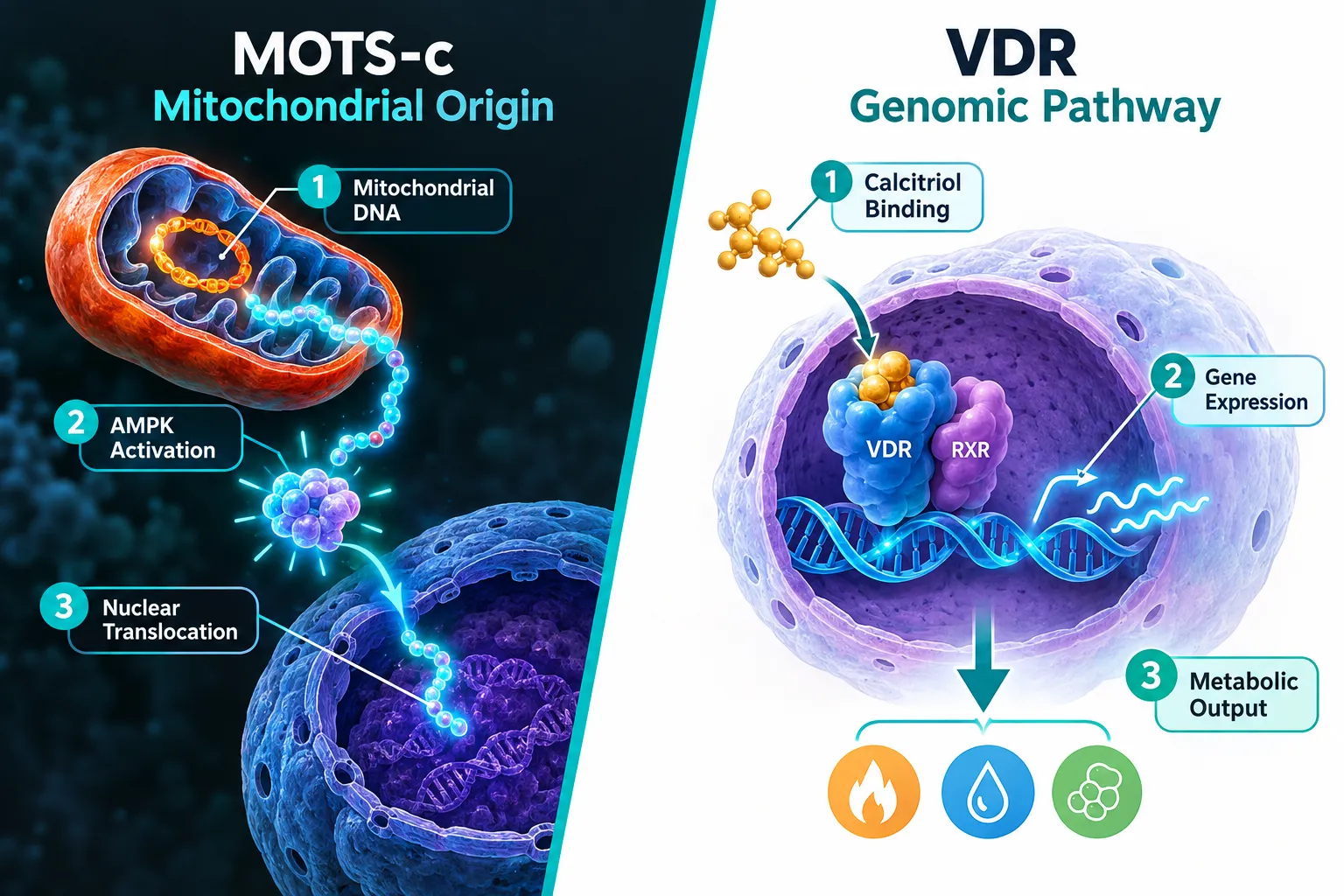
Vitamin D3 itself is biologically inert until the liver converts it to 25-hydroxyvitamin D and the kidneys complete the process by producing calcitriol (1,25-dihydroxyvitamin D3). Calcitriol is the active hormone, and its primary mechanism is genomic: it binds the vitamin D receptor (VDR), which then pairs with the retinoid X receptor (RXR) to form a heterodimer. That complex binds vitamin D response elements on DNA and switches target genes on or off.

The downstream effects are broad. VDR target genes regulate calcium homeostasis, innate immune responses, insulin secretion, and mitochondrial biogenesis. This last point is critical: calcitriol can upregulate PGC-1 alpha expression, a master regulator of mitochondrial function. That creates a direct genomic bridge between Vitamin D3 status and the health of the very organelle that produces MOTS-c.

How Vitamin D3 Activates Nuclear Receptors

Key VDR-mediated metabolic effects:

  • Improved insulin sensitivity via GLUT4 regulation
  • Reduced inflammatory cytokine expression
  • Enhanced mitochondrial biogenesis through PGC-1 alpha
  • Modulation of AMPK activity (indirectly)

MOTS-c: A Mitochondrial Peptide With Nuclear Reach

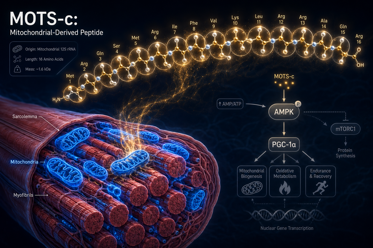
MOTS-c (Mitochondrial Open Reading Frame of the 12S rRNA type-c) is a 16-amino-acid peptide encoded within mitochondrial DNA. Its discovery challenged the assumption that mitochondria only produce energy — they also produce signaling molecules that travel to the nucleus and alter gene expression.

The primary mechanism involves AMPK activation. Under metabolic stress, MOTS-c is released from mitochondria, activates AMPK in the cytoplasm, and then translocates into the nucleus. Inside the nucleus, it binds to stress-response elements and regulates genes involved in glucose metabolism, oxidative stress defense, and longevity pathways. This nuclear translocation step makes MOTS-c functionally analogous to a nuclear receptor ligand — a remarkable parallel to how calcitriol operates through VDR.

For researchers sourcing this compound, MOTS-c 10mg is available for preclinical study purposes, and those exploring MOTS-c from Peptide Science can compare vendor specifications before purchasing.
MOTS-c: A Mitochondrial Peptide With Nuclear Reach

"MOTS-c is not simply a metabolic hormone — it is a retrograde signal from the mitochondria to the genome, recalibrating nuclear gene expression in response to bioenergetic stress."

Documented MOTS-c preclinical effects include:

Outcome Evidence Level
Improved insulin sensitivity Rodent models, strong
Reduced obesity markers CB4211 analog human trial
AMPK-dependent glucose uptake Cell and animal studies
Nuclear stress-response gene regulation Mechanistic studies
Lifespan extension in mice Preclinical only

Designing the Best Vitamin D3 and Mitochondrial Peptide Stacks: Optimizing Nuclear Receptor and MOTS-c Signaling Pathways

The rationale for combining Vitamin D3 with MOTS-c rests on three mechanistic pillars: shared AMPK involvement, convergent effects on mitochondrial biogenesis, and complementary nuclear gene regulation. However, it is important to state clearly — no published human trial has tested this combination. The MOTS-MET trial (NCT07505745), a Phase 2a study representing the first true MOTS-c efficacy trial in humans, is underway but has not yet reported data. Of nine registered human MOTS-c trial records as of 2026, only one dosing study has been completed.

What does exist is a compelling mechanistic case. Calcitriol upregulates PGC-1 alpha, which drives mitochondrial biogenesis and increases the cellular pool from which MOTS-c is produced. MOTS-c then activates AMPK, which in turn can phosphorylate and enhance VDR sensitivity. This creates a potential positive feedback loop between the two pathways.

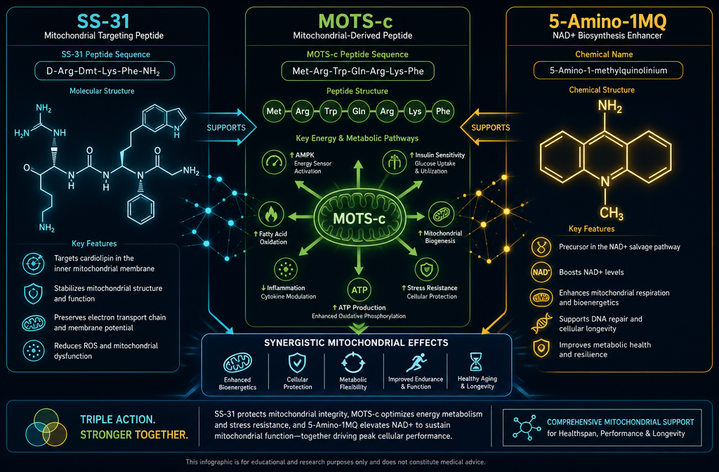
A secondary mitochondrial peptide worth considering in stack design is SS-31 (elamipretide), which targets cardiolipin on the inner mitochondrial membrane to reduce oxidative stress. Detailed research on SS-31 mitochondrial dynamics and a review of SS-31 peptide benefits can help researchers understand how this compound complements MOTS-c in a broader mitochondrial support stack. For procurement, SS-31 peptide is available for research use, and those comparing costs can review SS-31 peptide price options.

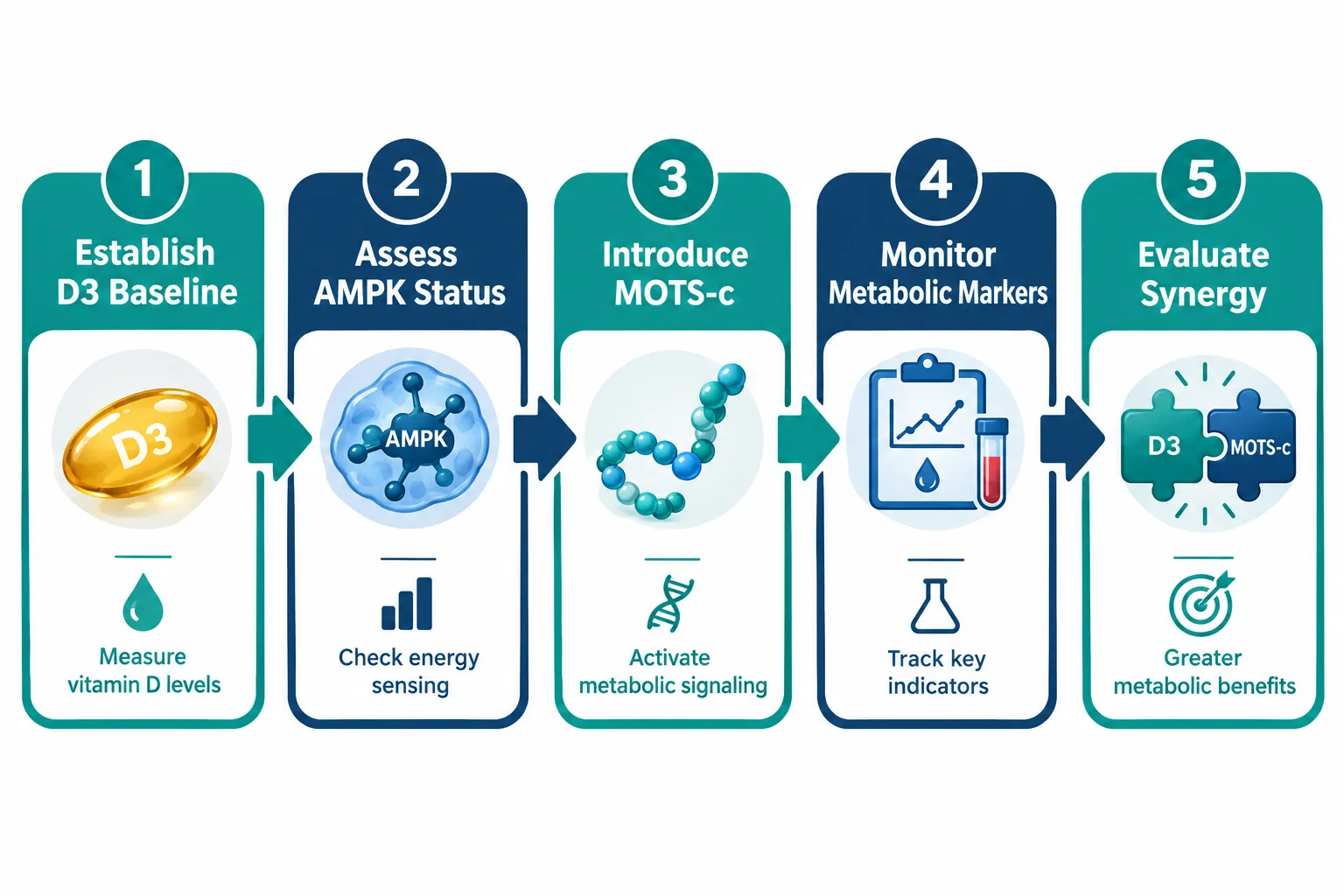
Speculative stack framework (preclinical rationale only):

  1. Optimize Vitamin D3 status first — target serum 25-OH-D levels in the 40-60 ng/mL range to ensure adequate VDR activation and PGC-1 alpha expression.
  2. Introduce MOTS-c — to leverage AMPK-mediated nuclear signaling and glucose metabolism support.
  3. Consider SS-31 — to reduce mitochondrial oxidative stress, protecting the organelle that produces MOTS-c.
  4. Monitor metabolic markers — fasting glucose, insulin sensitivity indices, and inflammatory markers.
Designing the Best Vitamin D3 and Mitochondrial Peptide Stacks: Optimizing Nuclear Receptor and MOTS-c Signaling Pathways

Evidence Gaps, Legal Context, and Research Outlook

The legal and clinical landscape for MOTS-c in 2026 remains constrained. Native MOTS-c has not received regulatory approval in any jurisdiction. The CB4211 analog — a modified version tested in a small human trial for fatty liver disease and obesity — showed early promise but remains in early-phase development. Researchers and clinicians operating outside formal trial settings face gray-market exposure when sourcing native MOTS-c, and this risk must be factored into any research protocol design.

Vitamin D3, by contrast, is fully approved, widely available, and has decades of safety data. Its nuclear receptor mechanism is among the best-characterized in human biology. This asymmetry in evidence quality is the defining practical challenge when designing the best Vitamin D3 and mitochondrial peptide stacks: optimizing nuclear receptor and MOTS-c signaling pathways for any serious research application.

Those sourcing compounds for legitimate research purposes should prioritize purity verification. Lab-tested peptides with documented certificate-of-analysis data reduce the risk of contaminant interference in mechanistic studies.

Conclusion

The convergence of calcitriol's genomic VDR signaling and MOTS-c's mitochondria-to-nucleus communication represents one of the most intellectually compelling areas in metabolic biology in 2026. The mechanistic case for a synergistic stack is coherent — shared AMPK pathways, complementary effects on mitochondrial biogenesis, and dual nuclear gene regulation make the combination theoretically attractive.

Actionable next steps for researchers:

  • Establish and document baseline Vitamin D3 status before introducing any mitochondrial peptide.
  • Follow the MOTS-MET trial (NCT07505745) for the first human efficacy data on MOTS-c.
  • Consider SS-31 as a mitochondrial oxidative stress companion in any stack protocol.
  • Source only from vendors providing independent purity verification.
  • Treat any claimed synergy between Vitamin D3 and MOTS-c as a hypothesis requiring formal trial validation, not an established clinical outcome.

The gap between mechanistic plausibility and clinical proof remains wide. Closing that gap is the work ahead.

https://www.puretestedpeptides.com/wp-content/uploads/2026/09/best-vitamin-d3-and-mitochondrial-peptide-stacks-optimizing-nuclear-receptor-and.webp 1024 1536 https://www.puretestedpeptides.com/wp-content/uploads/2026/01/buy-peptides-online.jpg 2026-09-18 13:05:442026-09-18 13:05:44Best Vitamin D3 and Mitochondrial Peptide Stacks: Optimizing Nuclear Receptor and MOTS-c Signaling Pathways
Hydration and Osmolality in Intensive Peptide Studies: The Role of Electrolyte Solutions and Liquid IV Protocols

Hydration and Osmolality in Intensive Peptide Studies: The Role of Electrolyte Solutions and Liquid IV Protocols

September 16, 2026/0 Comments/in Uncategorized/by

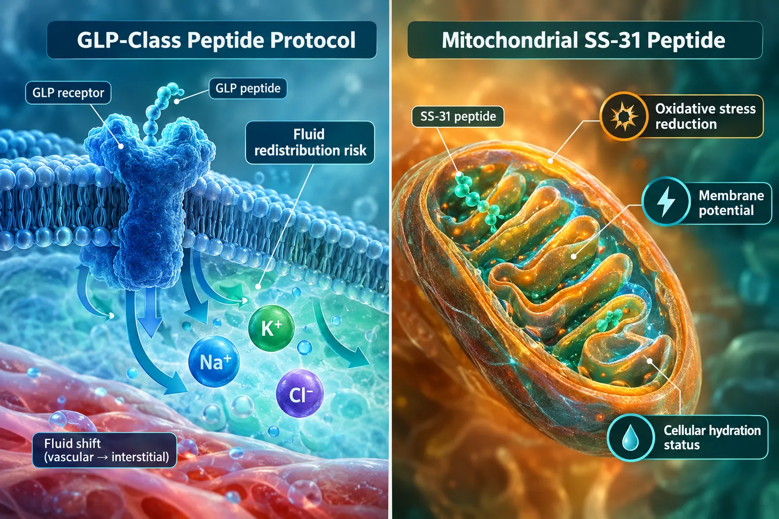
Fluid balance is rarely the headline variable in peptide research, yet it quietly determines whether a study produces clean, reproducible data or confounded results. When researchers investigate GLP-class metabolic peptides or mitochondria-targeting compounds, shifts in cellular hydration status and plasma osmolality can alter receptor binding, hormone signaling, and tissue distribution in ways that standard protocols often fail to account for. Understanding hydration and osmolality in intensive peptide studies — and the role of electrolyte solutions and liquid IV protocols in managing those variables — is therefore a foundational concern, not an afterthought.

Key Takeaways

  • Plasma osmolality targets near 285 mOsm/kg represent the physiological benchmark that electrolyte solutions in peptide research should support, not disrupt.
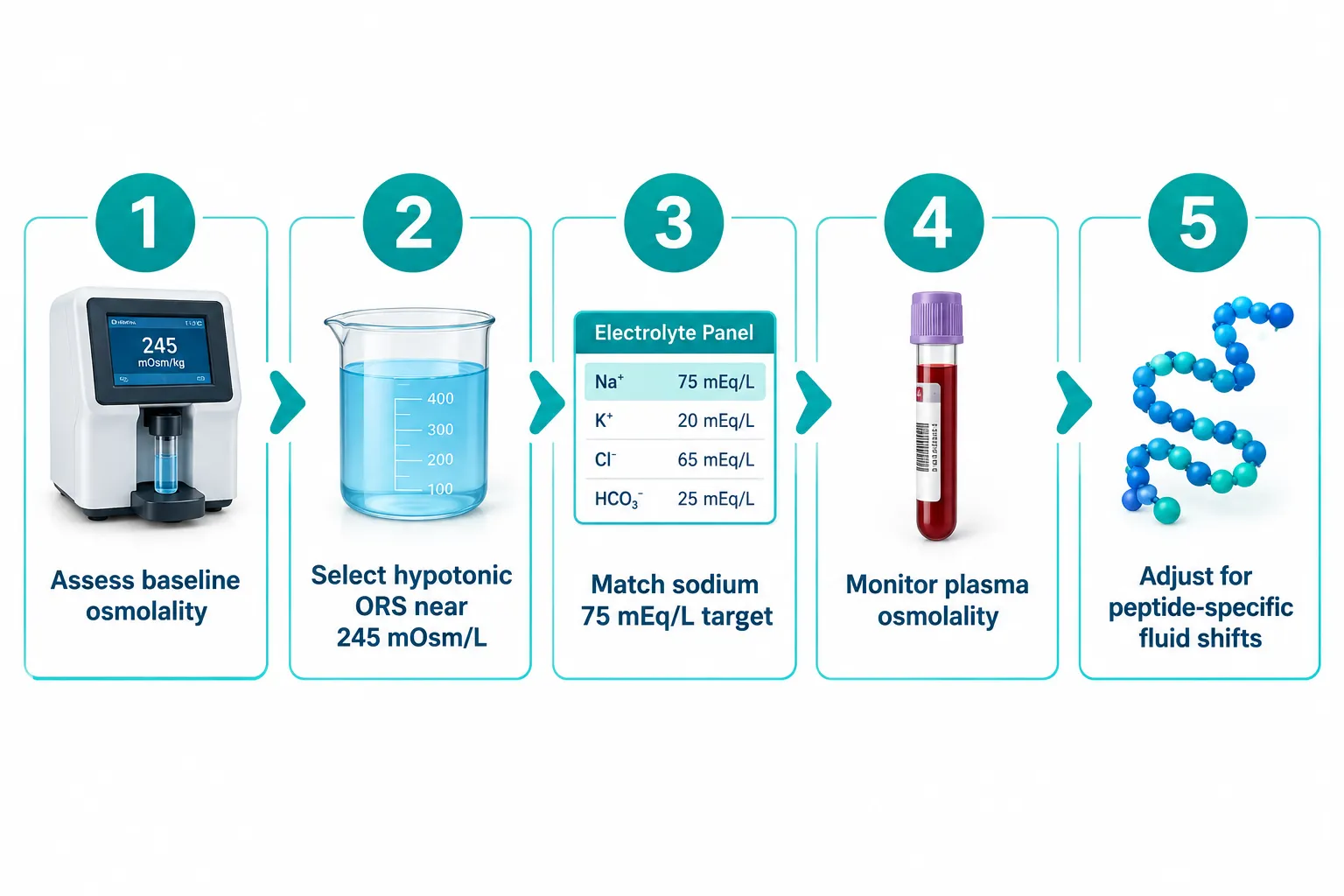
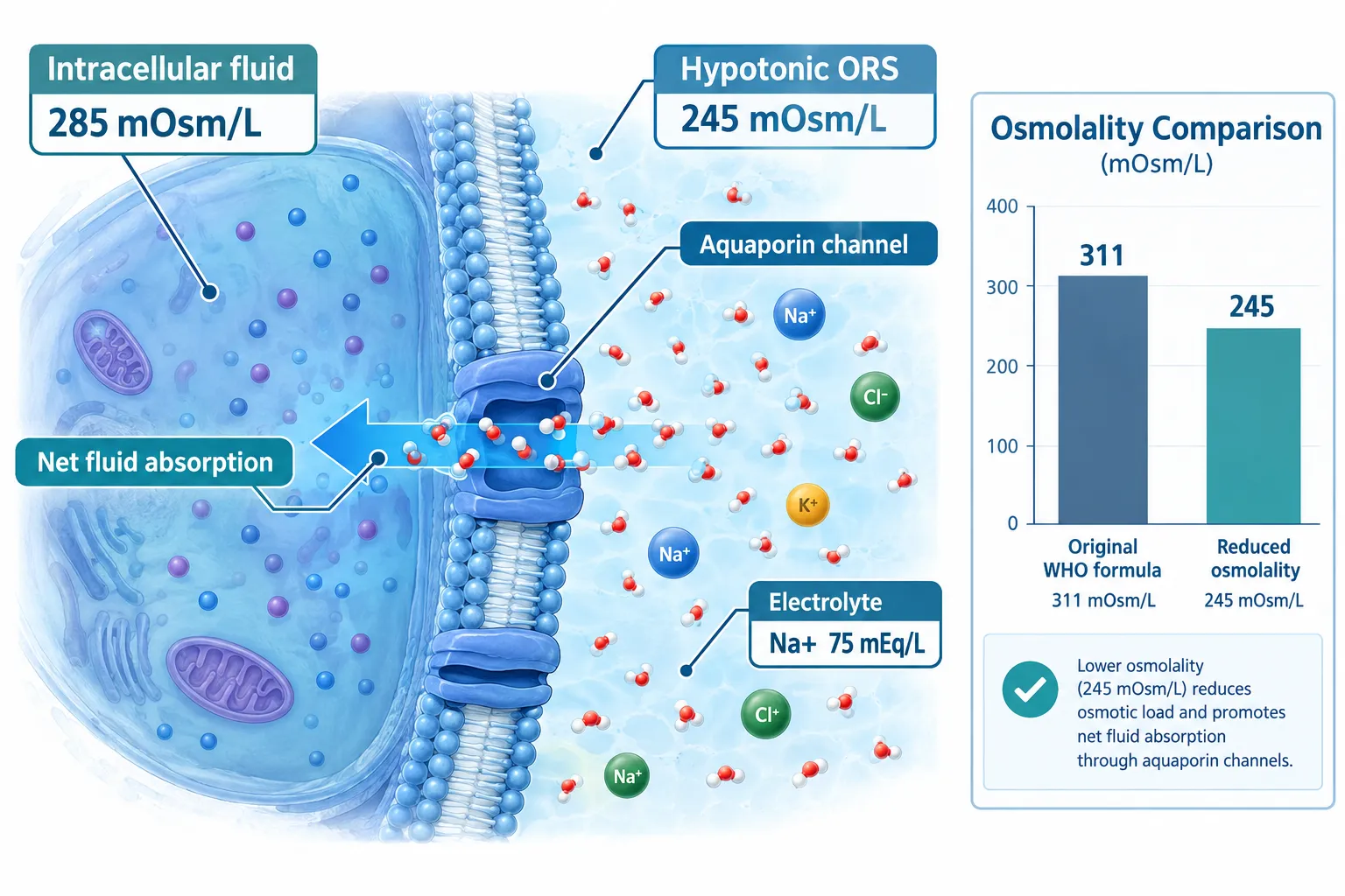
  • Current international guidelines converge on hypotonic oral rehydration solutions near 245 mOsm/L as the most effective standard for rapid, efficient rehydration.
  • GLP-class and mitochondrial peptides each carry distinct fluid-shift risks that demand osmolality-aware hydration protocols.
  • Liquid IV-style products show theoretical promise but currently lack peer-reviewed clinical evidence demonstrating superiority over properly formulated electrolyte solutions.
  • Selecting the right electrolyte solution means checking sodium content, carbohydrate load, and total osmolarity before integrating it into any intensive protocol.

Why Osmolality Matters in Intensive Peptide Research

Why Osmolality Matters in Intensive Peptide Research

Osmolality measures the concentration of dissolved particles in a fluid, expressed in milliosmoles per kilogram of water (mOsm/kg). In a living system, plasma osmolality is tightly regulated around 285 to 295 mOsm/kg. Even modest deviations — as little as 10 mOsm/kg above or below that range — trigger compensatory hormonal responses involving vasopressin, aldosterone, and the renin-angiotensin system.

For researchers working with peptides such as SS-31, which targets mitochondrial cardiolipin to reduce oxidative stress, or with GLP-receptor agonists like those explored in GLP-3R peptide formulations, these hormonal cascades are not background noise. They directly interact with the pathways under investigation. A subject or model system that enters a protocol in a mildly hypertonic or hypotonic state introduces a confounding variable that no downstream statistical correction can fully remove.

Three osmolality-related risks in peptide studies:

  • Hypertonic conditions slow gastric emptying, reduce net fluid absorption, and can falsely elevate plasma peptide concentrations by reducing distribution volume.
  • Hypotonic conditions dilute electrolytes, alter membrane potential, and may blunt receptor-mediated responses that depend on sodium-potassium gradients.
  • Fluctuating osmolality across study visits creates inter-session variability that inflates standard deviations and reduces statistical power.

Standardizing hydration inputs is therefore as important as standardizing peptide dose and timing.

Electrolyte Solutions and Osmolality Standards: What the Evidence Supports

The global benchmark for oral rehydration solution (ORS) osmolality has shifted significantly over the past two decades. The original WHO formula carried an osmolarity of approximately 311 mOsm/L with sodium at 90 mEq/L. Clinical evidence accumulated showing that this formulation, while effective at replacing electrolytes, was not optimal for net fluid absorption. The revised WHO/UNICEF standard specifies a reduced-osmolality ORS with sodium at 75 mEq/L and total osmolarity at 245 mOsm/L — a hypotonic formulation that demonstrably improves net fluid absorption and reduces gastrointestinal side effects compared with its predecessor.

Health Canada's ORS monograph reinforces this direction, specifying that total osmolarity should not exceed 280 mOsm/L and that hypotonic solutions improve clinical outcomes. Peer-reviewed pharmacotechnical analysis supports an optimal absorption window between 200 and 260 mOsm/kg, with 245 mOsm/L representing the current evidence-based sweet spot.

Key benchmark: Solutions in the 200-260 mOsm/kg range yield the greatest net fluid absorption. Hypertonic solutions above this range slow gastric emptying — a critical consideration when pairing electrolyte solutions with intensive peptide regimens.

For intensive peptide studies, this has direct implications. Commercially available ORS products span a carbohydrate content of 13.5 to 40 g/L, sodium of 45 to 75 mEq/L, and osmolarity ranging from roughly 200 to 305 mOsm/L. Selecting a product toward the upper end of that range — or using heavily sweetened sports drinks with osmolarity above 300 mOsm/L — risks slowing gastric emptying and creating transient hypertonicity that interferes with study conditions.

Practical selection criteria for electrolyte solutions in peptide protocols:

Parameter Target Range Rationale
Total osmolarity 225-260 mOsm/L Maximizes net fluid absorption
Sodium 60-75 mEq/L Matches WHO reduced-ORS standard
Glucose/carbohydrate 13.5-20 g/L Supports sodium co-transport without hypertonicity
Potassium 15-25 mEq/L Supports intracellular balance

GLP-Class and Mitochondrial Peptides: Specific Fluid-Shift Considerations

GLP-Class and Mitochondrial Peptides: Specific Fluid-Shift Considerations

Not all peptides interact with fluid balance in the same way. Understanding hydration and osmolality in intensive peptide studies requires mapping the specific fluid-shift risks of each peptide class.

GLP-receptor peptides — including agents studied alongside compounds like GLP-3R 30mg formulations and broader cardiometabolic peptide models — influence gastric emptying rate, gut motility, and fluid secretion in the gastrointestinal tract. These effects mean that subjects in GLP-focused protocols may absorb oral fluids at altered rates, making the osmolality of any co-administered electrolyte solution especially consequential. A hypertonic solution that would merely slow absorption in a resting subject could produce meaningful fluid redistribution in a GLP-stimulated gut.

Mitochondria-targeting peptides such as SS-31 operate at the level of the inner mitochondrial membrane, modulating oxidative phosphorylation and reactive oxygen species. Research on SS-31 peptide benefits and SS-31 research considerations highlights that mitochondrial function is sensitive to cellular hydration status. Dehydration reduces mitochondrial membrane potential and amplifies oxidative stress — the very pathology SS-31 is designed to study. Running an SS-31 protocol without a controlled hydration baseline risks confounding the primary endpoint.

Growth hormone-releasing peptides like tesa influence body composition and fluid compartmentalization through IGF-1-mediated pathways. Sodium and water retention are recognized downstream effects of growth hormone axis activation, meaning that plasma osmolality monitoring should be built into any extended tesa protocol.

Liquid IV Protocols: Promise, Evidence Gaps, and Practical Guidance

Liquid IV Protocols: Promise, Evidence Gaps, and Practical Guidance

Liquid IV-style products — high-sodium, glucose-containing sachets marketed on cellular transport technology — have attracted attention as rapid rehydration tools for intensive protocols. The theoretical basis is sound: sodium-glucose co-transport (SGLT1) in the small intestine can accelerate fluid uptake when the sodium-to-glucose ratio is optimized, and a well-formulated product near 245 mOsm/L could theoretically outperform plain water in restoring plasma osmolality after exercise-induced dehydration.

The clinical evidence, however, remains thin. A registered randomized crossover trial (NCT06063655) is tracking body weight, urine osmolality, plasma osmolality, and blood electrolytes following exercise-induced dehydration with Liquid I.V. rehydration, but as of 2026 no peer-reviewed results have been published. An earlier poster study from Washington State University Vancouver compared plasma osmolarity after plain water versus Liquid I.V. in mildly dehydrated participants and predicted no significant difference between groups — though this remains an undergraduate-level poster rather than a peer-reviewed clinical trial.
For peptide researchers, the practical takeaway is straightforward: any liquid IV protocol should be evaluated on its actual osmolarity value, sodium content, and carbohydrate load against the 245 mOsm/L benchmark before adoption. A product that clusters near that target with sodium around 75 mEq/L is defensible. A heavily sweetened product above 300 mOsm/L is not, regardless of marketing claims.

Conclusion

Hydration and osmolality in intensive peptide studies — and the role of electrolyte solutions and liquid IV protocols — deserve the same methodological rigor applied to dosing, timing, and endpoint selection. The evidence base is clear: hypotonic electrolyte solutions near 245 mOsm/L, with sodium around 75 mEq/L and modest glucose content, provide the most efficient and gastrointestinally tolerable rehydration platform currently available.

Actionable next steps for researchers:

  1. Measure baseline plasma osmolality in all subjects before peptide administration and flag any value outside 280-295 mOsm/kg.
  2. Select an electrolyte solution with documented osmolarity at or below 260 mOsm/L — check the product specification sheet, not just the label claims.
  3. For GLP-class protocols, account for altered gastric emptying when timing oral fluid administration relative to peptide dosing.
  4. For mitochondrial peptide studies, treat cellular hydration status as a primary covariate, not a background variable.
  5. Treat liquid IV-style products as potentially useful tools but require osmolarity data before incorporating them into any standardized protocol.

Fluid balance is not a peripheral concern in peptide research. It is a core experimental variable — and managing it precisely is what separates reproducible science from noise.

https://www.puretestedpeptides.com/wp-content/uploads/2026/09/hydration-and-osmolality-in-intensive-peptide-studies-the-role-of-electrolyte-so.webp 1024 1536 https://www.puretestedpeptides.com/wp-content/uploads/2026/01/buy-peptides-online.jpg 2026-09-16 13:06:542026-09-16 13:06:54Hydration and Osmolality in Intensive Peptide Studies: The Role of Electrolyte Solutions and Liquid IV Protocols
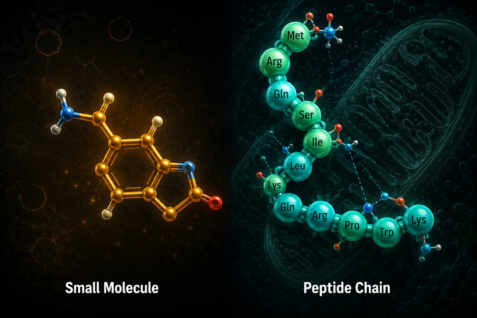
Research-Use Only Peptides: How "Peptides" Differ From Classic Small-Molecule Drugs Like Prednisone and Atorvastatin in Lab Design

Research-Use Only Peptides: How “Peptides” Differ From Classic Small-Molecule Drugs Like Prednisone and Atorvastatin in Lab Design

August 27, 2026/0 Comments/in Uncategorized/by

Only about 40 oral peptide drugs have ever reached clinical approval worldwide, a striking contrast to the thousands of approved small-molecule drugs that fill every pharmacy shelf. That gap is not a failure of biology; it is a direct result of how profoundly research-use only peptides differ from classic small-molecule drugs like prednisone and atorvastatin in lab design, stability, and experimental logic.

Understanding those differences is essential for any researcher sourcing, handling, or building assays around compounds such as BPC-157, MOTS-c, GLP-3, or SS-31.

Key Takeaways

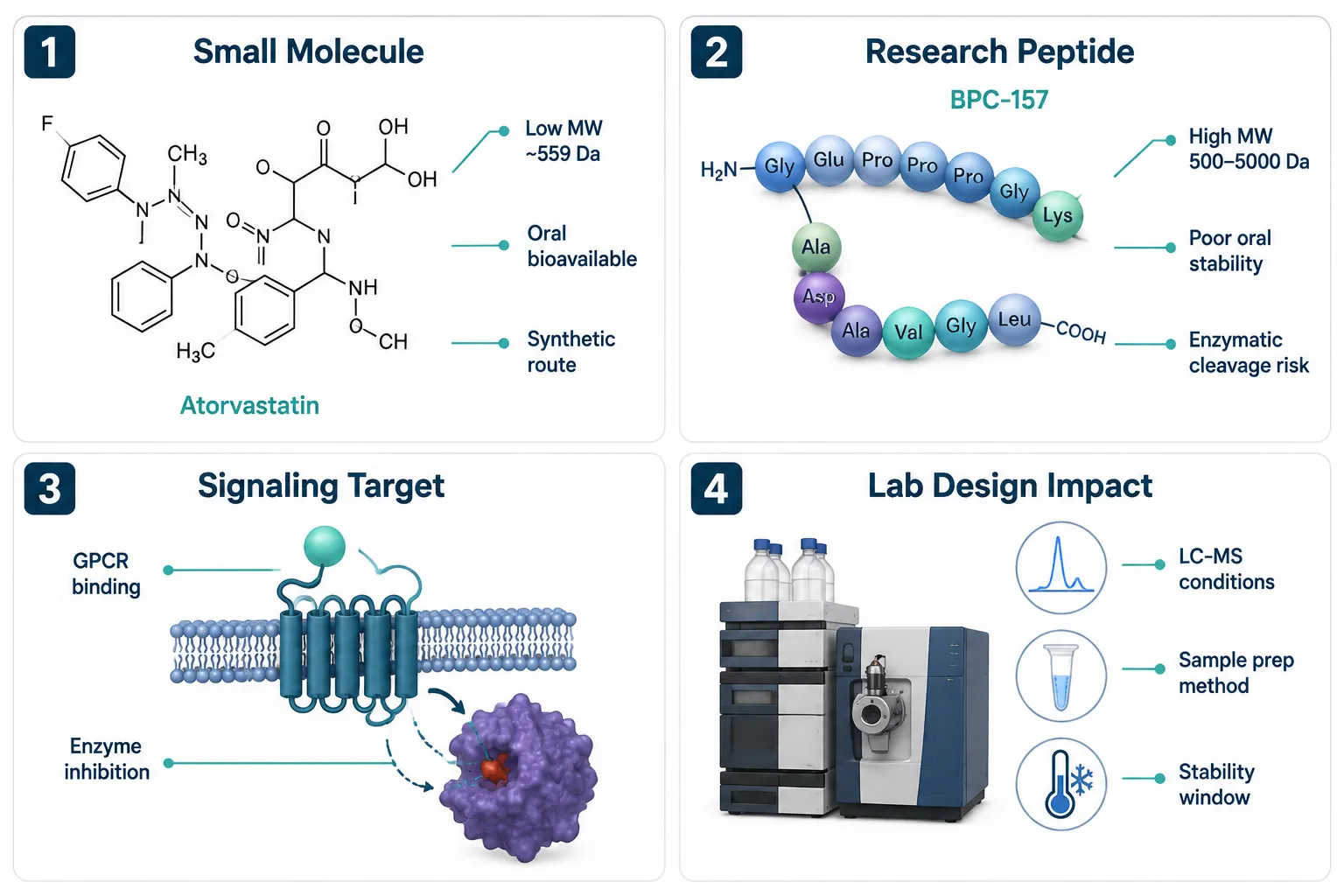
  • Research-use only (RUO) peptides are chains of amino acids with molecular weights typically between 500 and 5,000 Da, far larger and more structurally complex than small molecules like atorvastatin (559 Da) or prednisone (358 Da).
  • Small molecules are generally orally bioavailable and metabolically stable; peptides are highly susceptible to enzymatic cleavage and require specialized formulation and storage.
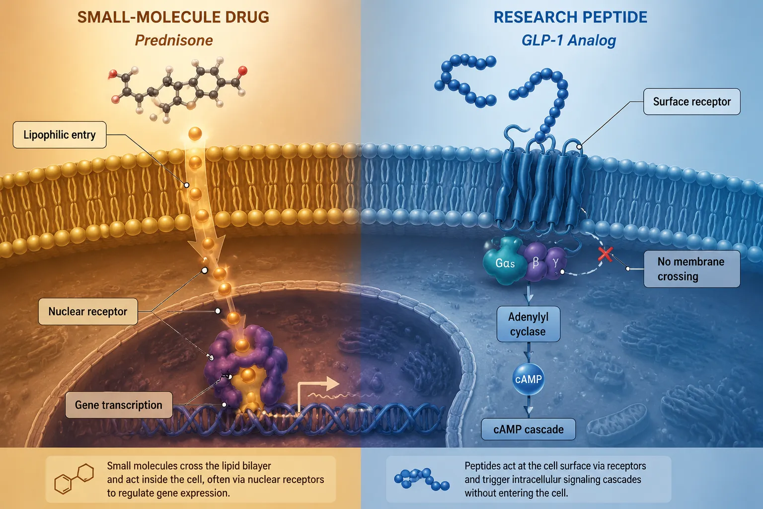
  • Peptides act primarily at cell-surface receptors or extracellular targets, while many classic small molecules penetrate cells or nuclei directly.
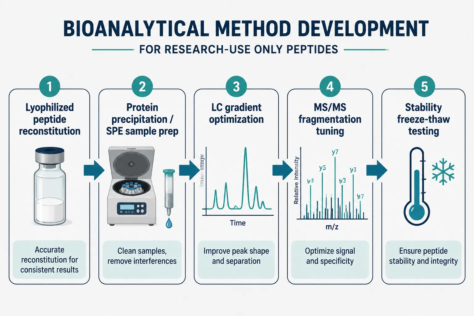
  • Bioanalytical methods for RUO peptides demand different LC-MS conditions, sample preparation strategies, and stability testing protocols compared to small-molecule assays.
  • The regulatory boundary between RUO labeling and therapeutic use is tightening in 2026, making proper sourcing and documentation critical for compliant research.

Structural Foundations: Size, Sequence, and Complexity

The most immediate difference between research-use only peptides and classic small-molecule drugs like prednisone and atorvastatin in lab design is sheer molecular size.

Structural Foundations: Size, Sequence, and Complexity

Prednisone is a steroid with a molecular weight of roughly 358 Da and a rigid, four-ring carbon scaffold. Atorvastatin (Lipitor) weighs about 559 Da and inhibits HMG-CoA reductase through a well-defined binding pocket. Both molecules are small enough to be synthesized in a few chemical steps and characterized quickly by standard NMR or HPLC methods.

Research peptides occupy a different structural tier entirely:

Compound Type Approx. MW Chain Length
Prednisone Small molecule 358 Da N/A
Atorvastatin Small molecule 559 Da N/A
BPC-157 Research peptide ~1,419 Da 15 amino acids
SS-31 Research peptide ~639 Da 4 amino acids
MOTS-c Research peptide ~2,174 Da 16 amino acids
GLP-1 analog Research peptide ~3,300 Da 30 amino acids

Even the shortest research peptides carry multiple chiral centers, hydrogen-bond donors, and rotatable bonds that make them far more sensitive to environmental conditions than a steroid or statin.

A key principle in peptide lab design: molecular complexity drives every downstream decision, from storage temperature to the LC gradient used in bioanalysis.

Because peptide bonds are hydrolyzed by proteases found in plasma, gut lumen, and even standard laboratory buffers, stability is never assumed. Researchers working with SS-31 peptides or similar mitochondria-targeting compounds must account for degradation windows that simply do not apply to a statin dissolved in DMSO.

How Peptides Signal Differently Than Small-Molecule Drugs

How Peptides Signal Differently Than Small-Molecule Drugs

Classic small molecules often work by entering cells or even nuclei. Prednisone, after conversion to prednisolone, diffuses across the plasma membrane and binds cytoplasmic glucocorticoid receptors. The complex then translocates to the nucleus and modulates gene transcription directly. Atorvastatin reaches its target enzyme inside hepatocytes through active transport.

Most research peptides cannot follow that path. Their size and hydrophilicity prevent passive membrane diffusion. Instead, they act at:

  • Cell-surface G-protein-coupled receptors (GPCRs), as seen with GLP-1 peptide analogs that activate incretin receptors
  • Extracellular matrix proteins, as with BPC-157, which appears to interact with growth factor receptors and angiogenic pathways
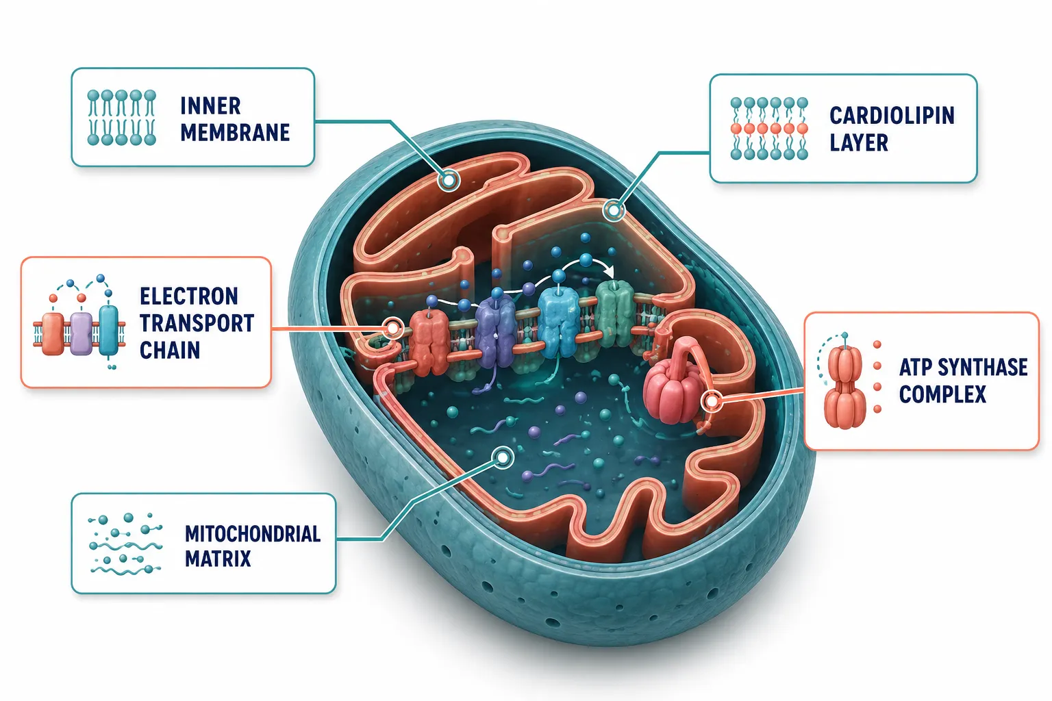
  • Mitochondrial membrane interfaces, as with SS-31, which associates with cardiolipin on the inner mitochondrial membrane without entering the matrix

This distinction reshapes every aspect of assay design. A researcher cannot simply measure nuclear translocation or enzyme inhibition with the same endpoint used for a steroid. Functional readouts, cAMP accumulation, receptor internalization, mitochondrial membrane potential, must replace or supplement traditional biochemical endpoints.

For peptides with less-characterized mechanisms, such as MOTS-c or 5-Amino-1MQ (a small-molecule/peptide-adjacent NNMT inhibitor), researchers must build multi-endpoint assays that capture pathway-level responses rather than a single molecular event.

Detailed considerations for specific compounds are covered in resources like SS-31 10mg research peptide considerations and the PT-141 peptide research context QA and controls guide.

Bioanalytical and Formulation Challenges Unique to RUO Peptides

Bioanalytical and Formulation Challenges Unique to RUO Peptides

When a researcher builds a method around atorvastatin, they benefit from decades of published HPLC-UV and LC-MS/MS data, stable reference standards, and predictable protein binding. Peptides offer none of those shortcuts.

Key bioanalytical differences include:

  1. Sample preparation, Protein precipitation alone is often insufficient. Solid-phase extraction (SPE) or mixed-mode sorbents are needed to recover hydrophilic peptides from plasma matrices without co-eluting interferences.

  2. LC conditions, Peptides require shallow, extended gradient programs on C18 or C8 columns with ion-pairing reagents (e.g., trifluoroacetic acid or heptafluorobutyric acid) to achieve adequate retention and peak shape.

  3. MS/MS fragmentation, Peptide precursor ions are multiply charged. Method developers must select the correct charge state and optimize collision energy for each unique sequence, a step irrelevant for single-charged small molecules.

  4. Stability testing, Freeze-thaw cycles, bench-top stability, and long-term frozen stability must all be validated separately. Peptides can degrade within hours at room temperature, while prednisone tablets remain stable for years on a shelf.

  5. Reconstitution and storage, Most RUO peptides are supplied lyophilized. Reconstitution solvent, concentration, and aliquot size must be defined before any experiment begins. Resources such as the AOD-9604 sale research method notes, storage and traceability page illustrate how seriously vendors and researchers must treat these variables.

Researchers sourcing compounds should consult verified suppliers. Guidance on where to buy peptides for research purposes highlights purity documentation and certificate-of-analysis standards that distinguish compliant RUO supply from unverified sources.

The 2026 Regulatory Context

The FDA has continued tightening its position on RUO labeling throughout 2026. Compounds sold as research-use only must not be marketed with therapeutic intent, and enforcement actions have targeted suppliers who blur that line. Researchers must ensure that procurement, labeling, and internal documentation all reflect the non-clinical, laboratory-only nature of the work. Pure Tested Peptides represents the kind of supplier model that prioritizes third-party purity testing and transparent RUO documentation to meet this evolving standard.

Conclusion

The differences between research-use only peptides and classic small-molecule drugs like prednisone and atorvastatin in lab design are not superficial. They span molecular architecture, receptor pharmacology, bioanalytical methodology, and regulatory classification.

Actionable next steps for researchers:

  • Treat every peptide as structurally unique, do not transfer small-molecule assay conditions without validation.
  • Build stability testing into the experimental plan from day one, not as an afterthought.
  • Select suppliers who provide third-party purity data and clear RUO documentation; explore wholesale peptides for sale options only from vendors with traceable quality systems.
  • Review compound-specific method notes before designing LC-MS/MS workflows.
  • Stay current with FDA guidance updates in 2026, particularly around peptide compounding and bulk substance classification.

Understanding these distinctions is what separates rigorous, reproducible peptide research from experiments that fail at the method level before the biology is ever tested.

https://www.puretestedpeptides.com/wp-content/uploads/2026/08/research-use-only-peptides-how-peptides-differ-from-classic-small-molecule-drugs.webp 1024 1536 https://www.puretestedpeptides.com/wp-content/uploads/2026/01/buy-peptides-online.jpg 2026-08-27 13:04:552026-08-27 13:04:55Research-Use Only Peptides: How “Peptides” Differ From Classic Small-Molecule Drugs Like Prednisone and Atorvastatin in Lab Design
Cellular Energy and Research Peptides: Why ATP Readouts Matter in Mitochondrial Studies

Cellular Energy and Research Peptides: Why ATP Readouts Matter in Mitochondrial Studies

August 20, 2026/0 Comments/in Uncategorized/by

Mitochondria generate roughly 90 percent of the ATP a mammalian cell requires to survive, yet for decades researchers lacked a fast, reliable way to watch that production change in real time. The convergence of cellular energy and research peptides, and specifically the question of why ATP readouts matter in mitochondrial studies, has become one of the most active methodological discussions in preclinical biology in 2026. Understanding the biology behind ATP measurement is essential before interpreting any peptide-related mitochondrial data.

Key Takeaways

  • ATP concentration is the most direct proxy for mitochondrial metabolic activity available to researchers today.
  • The luciferin-luciferase bioluminescence reaction is the gold-standard method for quantifying cellular ATP in high-throughput formats.
  • Compartment-specific luciferase probes now allow researchers to distinguish mitochondrial ATP from cytosolic ATP in living cells.
  • Mitochondrial-targeted peptides such as SS-31 and MOTS-c are evaluated partly through ATP-linked bioenergetic endpoints in both preclinical and clinical settings.
  • Timing, signal stability, and assay dynamic range are critical variables that determine whether an ATP readout is genuinely quantitative.

Why ATP Is the Right Proxy for Mitochondrial Activity

Why ATP Is the Right Proxy for Mitochondrial Activity

Adenosine triphosphate is not simply a fuel molecule, it is a real-time indicator of how well the entire oxidative phosphorylation chain is functioning. When mitochondria are stressed, damaged, or pharmacologically targeted, ATP output drops before most other measurable parameters shift. That sensitivity is exactly why intracellular ATP measurement is now established as a primary proxy for mitochondrial activity in research settings.

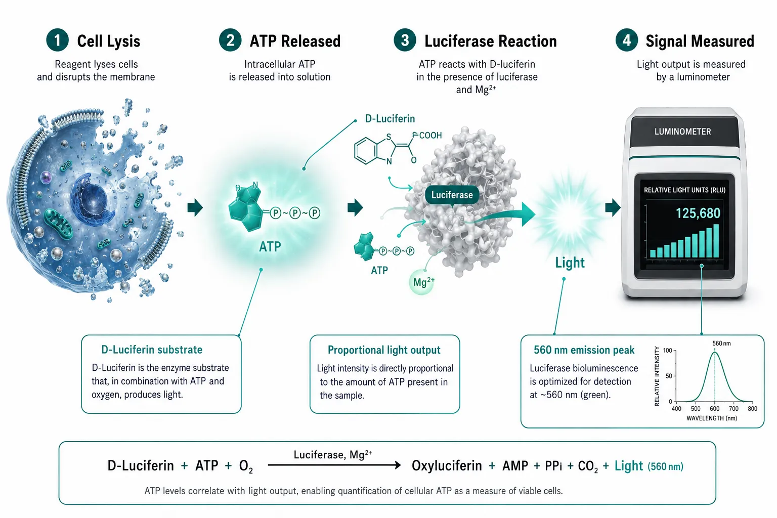
The dominant detection method is the luciferin-luciferase bioluminescence assay. Firefly luciferase catalyzes a reaction between D-luciferin and ATP, producing light. Because luminescence intensity is directly proportional to ATP concentration when ATP is the limiting reagent, the assay delivers a quantitative signal without requiring radioactive tracers or complex instrumentation. Most current protocols use a single working reagent that simultaneously lyses cells and generates luminescence, with measurements taken within one minute to prevent signal drift.

A standard workflow looks like this:

  1. Add equal volumes of sample and working solution to a 96-well plate.
  2. Incubate at 25 degrees Celsius for a fixed period (commonly 10 minutes per validated protocols).
  3. Read luminescence immediately to capture peak signal before kinetic decay.

Timing matters. Even a two-minute delay after adding the reaction mixture can introduce measurable error. Researchers building mitochondrial assay panels must treat the ATP readout as a time-sensitive endpoint, not a stable colorimetric measurement.

One additional consideration is dynamic range. When cell density is high or mitochondrial activity is elevated, the luminescent signal can saturate. Adjusting substrate volume or diluting lysate before adding the luciferase reagent is standard practice to keep measurements within the linear range of the assay.

Compartment-Specific Measurement and the Role of Cellular Energy and Research Peptides in Mitochondrial Studies

Compartment-Specific Measurement and the Role of Cellular Energy and Research Peptides in Mitochondrial Studies

A whole-cell ATP readout captures the sum of all nucleotide pools, cytosolic, mitochondrial, and pericellular. For many screening applications that aggregate signal is sufficient. But when the research question is specifically about how a peptide alters mitochondrial energy production, a whole-cell number can obscure the answer.

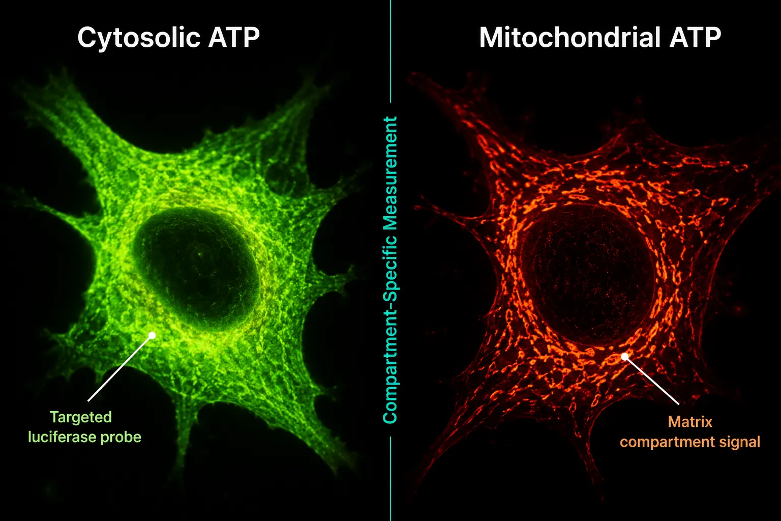
Targeted luciferase chimeras solve this problem. By fusing a luciferase gene to a mitochondrial matrix-targeting sequence, researchers can direct the reporter protein to a specific subcellular compartment. Luminescence from that probe reflects only the ATP pool in that location. The same strategy works for the cytosol and pericellular space, enabling simultaneous multi-compartment profiling across multi-day experiments.

This level of resolution matters for evaluating SS-31 mitochondrial dynamics because the peptide's proposed mechanism involves direct interaction with cardiolipin in the inner mitochondrial membrane. A whole-cell ATP assay might show a modest aggregate increase, while a matrix-targeted probe could reveal a substantially larger improvement confined to the mitochondrial compartment, a distinction with real mechanistic significance.

Beyond single-nucleotide assays, dual-detection platforms now allow simultaneous quantitation of both GTP and ATP from the same sample well. This matters because GTP is a direct product of the TCA cycle succinyl-CoA synthetase reaction, making it an independent indicator of mitochondrial metabolic flux. Combining GTP and ATP readouts in a single luminescent assay provides a more complete picture of cellular energy metabolism than either measurement alone.

For researchers mapping metabolic pathway dependency, ATP assays also help distinguish how much a cell relies on glycolysis versus oxidative phosphorylation. By selectively inhibiting one pathway and measuring the ATP response, investigators can characterize a cell line's bioenergetic phenotype, information that is directly relevant when screening peptide candidates for metabolic effects. Readers exploring that intersection may find the top 5 research peptides for metabolic health guide a useful companion resource.

Translating ATP Readouts to Peptide Research: Clinical and Preclinical Implications

Translating ATP Readouts to Peptide Research: Clinical and Preclinical Implications

The question of why ATP readouts matter in mitochondrial studies becomes most concrete when examining how mitochondrial-targeted peptides are actually evaluated in research programs. Two peptides illustrate the point clearly.

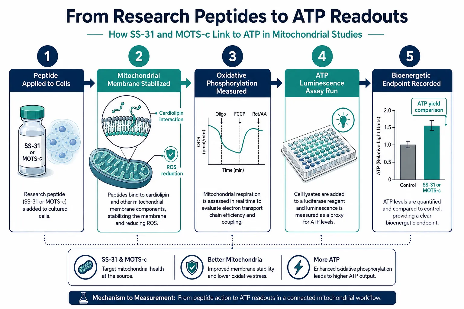
Elamipretide (SS-31) is a small, cell-permeable tetrapeptide that associates with cardiolipin in the inner mitochondrial membrane. Preclinical data consistently show that it improves mitochondrial respiration, reduces reactive oxygen species, and enhances ATP production. In clinical heart failure trials, even when primary endpoints such as infarct size reduction were not met, improvements in mitochondrial function and reductions in cardiac injury biomarkers were observed. This pattern suggests that ATP-linked bioenergetic measures may be more sensitive indicators of therapeutic effect than some anatomical endpoints. Two ongoing Phase 3 trials, ReNEW and ReGAIN, are expected to report data in 2026, and bioenergetic endpoints will be central to interpreting those results. Researchers can review the SS-31 mechanism and research overview for additional background on its mitochondrial targets.

MOTS-c is a mitochondria-encoded peptide that has entered human trials, with Phase 1 completion projected for mid-2026 and Phase 2 initiation anticipated later in the year. Mitochondrial respiratory capacity is a planned endpoint, and ATP and respiration measures are expected to quantify metabolic responses. The SS-31 mitochondrial research themes resource provides relevant context on how mitochondrial endpoints are structured across similar peptide programs.

The broader implication is that ATP assays are transitioning from purely preclinical screening tools to clinically meaningful biomarkers. As late-2026 trial data accumulate, consistent improvements in ATP-linked markers alongside clinical outcomes would further validate ATP readouts as surrogate endpoints for mitochondrial peptide therapies.

For research teams sourcing compounds for these studies, working with lab-tested peptides ensures that purity data are available to separate compound-related effects from assay artifacts, a non-trivial concern when ATP luminescence is the primary readout.

Peptide Primary Mitochondrial Target ATP-Related Endpoint Trial Stage (2026)
Elamipretide (SS-31) Cardiolipin / inner membrane ATP production, ROS reduction Phase 3 (ReNEW, ReGAIN)
MOTS-c Mitochondrial genome / AMPK Respiratory capacity, insulin sensitivity Phase 1 completion / Phase 2 initiation

Conclusion

ATP concentration is not a peripheral metric in mitochondrial research, it is the most direct, quantifiable signal of whether the organelle is doing its job. The luciferin-luciferase platform has made high-throughput ATP measurement practical, but reliable data require strict attention to timing, dynamic range, and compartment specificity. As the fields of cellular energy and research peptides converge more tightly, ATP readouts are becoming the common language between bench assays and clinical endpoints.

Actionable next steps for research teams:

  • Validate assay timing protocols before comparing treatment groups; even small delays introduce quantitative error.
  • Consider compartment-targeted luciferase probes when the research question is specifically about mitochondrial (not total cellular) ATP.
  • Pair ATP assays with GTP or oxygen consumption measurements to capture a fuller bioenergetic profile.
  • When evaluating mitochondrial peptides such as SS-31, design studies to capture ATP-linked secondary endpoints alongside primary anatomical or functional measures.
  • Source compounds from verified suppliers and review the SS-31 kidney health research literature to understand how ATP endpoints have been applied across different tissue models.

The 2026 clinical readouts from ongoing mitochondrial peptide trials will test whether ATP-based bioenergetic markers can carry the weight of surrogate endpoints. The methodology to support that claim is already in place.

https://www.puretestedpeptides.com/wp-content/uploads/2026/08/cellular-energy-and-research-peptides-why-atp-readouts-matter-in-mitochondrial-s.webp 1024 1536 https://www.puretestedpeptides.com/wp-content/uploads/2026/01/buy-peptides-online.jpg 2026-08-20 13:05:152026-08-20 13:05:15Cellular Energy and Research Peptides: Why ATP Readouts Matter in Mitochondrial Studies
Mitochondria and Research Peptides: What Labs Measure When Studying Cellular Energy Signaling

Mitochondria and Research Peptides: What Labs Measure When Studying Cellular Energy Signaling

August 18, 2026/0 Comments/in Uncategorized/by

Fewer than 1% of the roughly 1,500 proteins inside a human mitochondrion have been fully characterized for their role in energy output, yet that small fraction already underpins some of the most active areas in peptide research today. Understanding Mitochondria and Research Peptides: What Labs Measure When Studying Cellular Energy Signaling is no longer a niche concern for biochemists alone. In 2026, it sits at the center of metabolic disease research, rare disease trials, and next-generation therapeutic design.

Key Takeaways

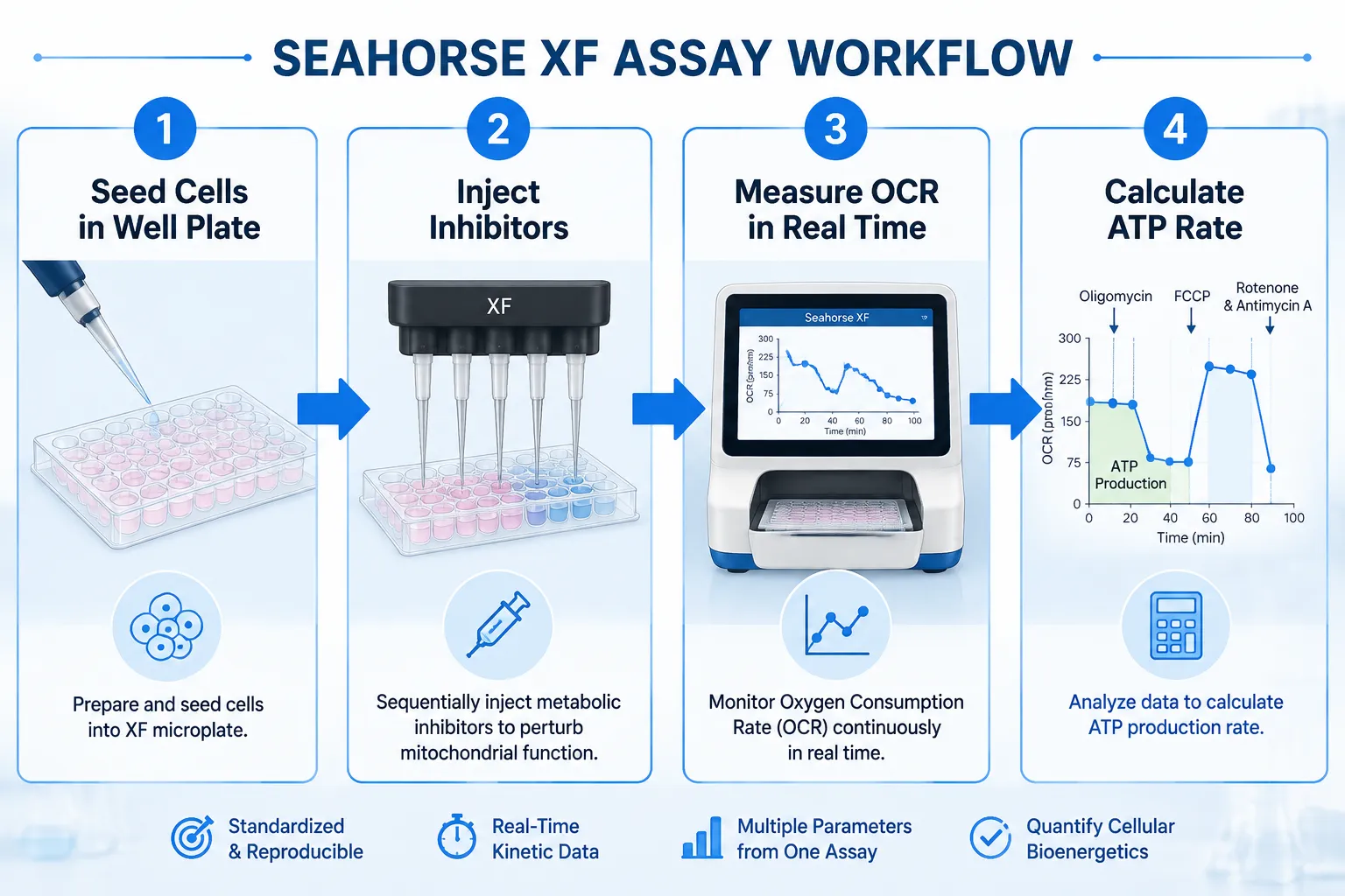
  • Mitochondrial function is measured through oxygen consumption rate (OCR) and ATP production assays, most commonly using Seahorse XF technology.
  • Research peptides such as SS-31 and MOTS-c target distinct nodes in the mitochondrial energy network and produce measurable, quantifiable effects.
  • Labs convert raw OCR data into ATP production rates using established bioenergetic equations, enabling direct comparison across studies.
  • Mitochondrial targeting sequences (MTS) are engineered design features that labs validate through membrane potential and proteomics assays.
  • Peptide purity and certificate of analysis standards directly affect the reliability of cellular energy signaling data.

Why Mitochondrial Energy Signaling Demands Precise Measurement

Why Mitochondrial Energy Signaling Demands Precise Measurement

Mitochondria are not static power generators. They are dynamic organelles that constantly shift their output in response to nutrient availability, stress signals, and intercellular communication. When a research peptide enters this environment, it can alter membrane potential, modulate electron transport chain activity, or change the rate at which ATP synthase produces adenosine triphosphate.

Labs studying cellular energy signaling need quantitative endpoints, not qualitative impressions. The most widely adopted platform for this work is the Seahorse XF Analyzer, which measures oxygen consumption rate (OCR) and extracellular acidification rate (ECAR) in live cells in real time. These two metrics together reveal how a cell balances oxidative phosphorylation against glycolysis, a balance that research peptides can shift in measurable ways.

"The OCR value alone tells you how hard the mitochondria are working. The ATP production rate tells you how efficiently that work translates into usable energy."

Beyond OCR, labs also track:

  • Mitochondrial membrane potential (using dyes such as JC-1 or TMRE)
  • Reactive oxygen species (ROS) output via fluorescent probes
  • NAD+/NADH ratios as indicators of redox balance
  • Mitochondrial fusion and fission dynamics through live-cell imaging

A 2025 study examining LRRK2 gene mutations used this exact framework to quantify ATP deficits in patient-derived neurons, demonstrating how OCR-to-ATP conversion math can anchor a disease mechanism to a specific molecular target.

Core Assays in Mitochondria and Research Peptides Studies

Core Assays in Mitochondria and Research Peptides Studies

The Seahorse XF Mito Stress Test

The standard Seahorse mito stress test injects three compounds in sequence, oligomycin, FCCP, and rotenone/antimycin A, each blocking a different step in the respiratory chain. The resulting OCR curve reveals:

Parameter What It Reflects
Basal respiration Baseline mitochondrial workload
ATP-linked respiration OCR directly coupled to ATP synthesis
Maximal respiration Full electron transport chain capacity
Spare respiratory capacity Metabolic reserve under stress
Proton leak Membrane integrity and uncoupling

Labs convert ATP-linked OCR to an ATP production rate by applying a stoichiometric factor derived from the P/O ratio, roughly 2.73 ATP per oxygen atom consumed during oxidative phosphorylation. This conversion is critical when comparing peptide-treated cells against controls.

Proteomics and Interaction Networks

SS-31 (elamipretide) has become a model compound for understanding how a peptide reshapes the mitochondrial protein interaction landscape. Proteomics studies have mapped SS-31's binding to cardiolipin on the inner mitochondrial membrane, revealing downstream stabilization of cristae architecture and suppression of cytochrome c release. Labs use co-immunoprecipitation and proximity labeling (BioID) to build these interaction networks.

For researchers sourcing this compound, the SS-31 10mg research peptide considerations page outlines purity and handling requirements that directly affect assay reproducibility. Additional context on SS-31 mitochondrial dynamics research is also available for deeper background.

Comparing SS-31 and MOTS-c: Two Distinct Signaling Profiles

Comparing SS-31 and MOTS-c: Two Distinct Signaling Profiles

In 2026 analyses, researchers have drawn a sharper line between SS-31 and MOTS-c, two peptides that both influence mitochondrial energy output but through fundamentally different mechanisms.

SS-31 acts at the inner mitochondrial membrane. It binds cardiolipin, reduces ROS production, and stabilizes the electron transport chain supercomplexes. Its primary measurable effect is an increase in ATP-linked OCR and a reduction in proton leak, outcomes directly visible in Seahorse assay data. Clinical trials in Barth syndrome and primary mitochondrial myopathy (MMPOWER-3) used functional endpoints such as the six-minute walk test and fatigue scores alongside these bioenergetic markers, though MMPOWER-3 showed limited efficacy improvements over placebo.

MOTS-c, by contrast, is a mitochondria-derived peptide that translocates to the nucleus under metabolic stress. It activates AMPK signaling, upregulates antioxidant gene expression, and shifts cellular metabolism toward glucose utilization. Labs measure its effects through AMPK phosphorylation assays, gene expression panels, and glucose uptake assays rather than pure OCR data.

This distinction matters for experimental design. Researchers exploring metabolic health applications can review the top 5 research peptides for metabolic health to understand how these compounds compare in applied research contexts.

Engineering Mitochondrial Targeting Sequences

Beyond naturally occurring peptides, labs now engineer mitochondrial targeting sequences (MTS), short amphipathic helical peptides that guide attached cargo into the mitochondrial matrix. Key metrics labs validate include:

  • Import efficiency (measured by protease protection assays)
  • Membrane potential dependence (collapsed by CCCP treatment)
  • Submitochondrial localization (outer membrane vs. matrix)

Plant-derived peptides such as roseltide rT1 have served as structural models for MTS design, demonstrating that even non-mammalian sequences can modulate ATP production in cell-free and cell-based systems. Reference standards for these comparisons are discussed in detail in the Bachem and reference standards guide for building robust peptide benchmarks.

Data Quality and Peptide Sourcing in Energy Signaling Research

The reliability of any cellular energy signaling dataset depends on the quality of the peptide used. Impurities in a research-grade compound can independently alter OCR, membrane potential, or ROS output, confounding results in ways that are difficult to detect post hoc.

Labs should require:

  • Certificate of Analysis (CoA) with HPLC purity above 98%
  • Mass spectrometry confirmation of molecular weight
  • Endotoxin testing results for cell-based assays
  • Lot-specific documentation for reproducibility across experiments

Researchers can find guidance on evaluating supplier documentation through peptide CoA standards and documentation and peptide supplier comparison resources. For those studying kidney-specific mitochondrial applications, SS-31 kidney health research provides organ-specific context for interpreting bioenergetic data.

Conclusion

The intersection of Mitochondria and Research Peptides: What Labs Measure When Studying Cellular Energy Signaling is defined by rigorous quantitative methods, Seahorse OCR profiling, ATP rate calculations, proteomics interaction mapping, and MTS validation assays. SS-31 and MOTS-c exemplify how two peptides targeting the same organelle can produce entirely different measurable signatures, requiring distinct experimental frameworks.

Actionable next steps for research teams:

  1. Standardize OCR-to-ATP conversion using published P/O ratios before comparing datasets across labs.
  2. Pair Seahorse assay data with at least one orthogonal endpoint (membrane potential or ROS) to validate findings.
  3. Confirm peptide purity via CoA and mass spectrometry before any cell-based energy assay.
  4. Select peptide compounds based on the specific node of the energy signaling pathway under investigation, membrane-targeted versus nuclear-translocating mechanisms require different readouts.
  5. Document lot numbers and storage conditions for every experiment to support reproducibility.

As mitochondrial peptide research matures in 2026, the labs that invest in measurement precision will produce the datasets that hold up to scrutiny, and drive the field forward.

https://www.puretestedpeptides.com/wp-content/uploads/2026/08/mitochondria-and-research-peptides-what-labs-measure-when-studying-cellular-ener.webp 1024 1536 https://www.puretestedpeptides.com/wp-content/uploads/2026/01/buy-peptides-online.jpg 2026-08-18 13:08:342026-08-18 13:08:34Mitochondria and Research Peptides: What Labs Measure When Studying Cellular Energy Signaling
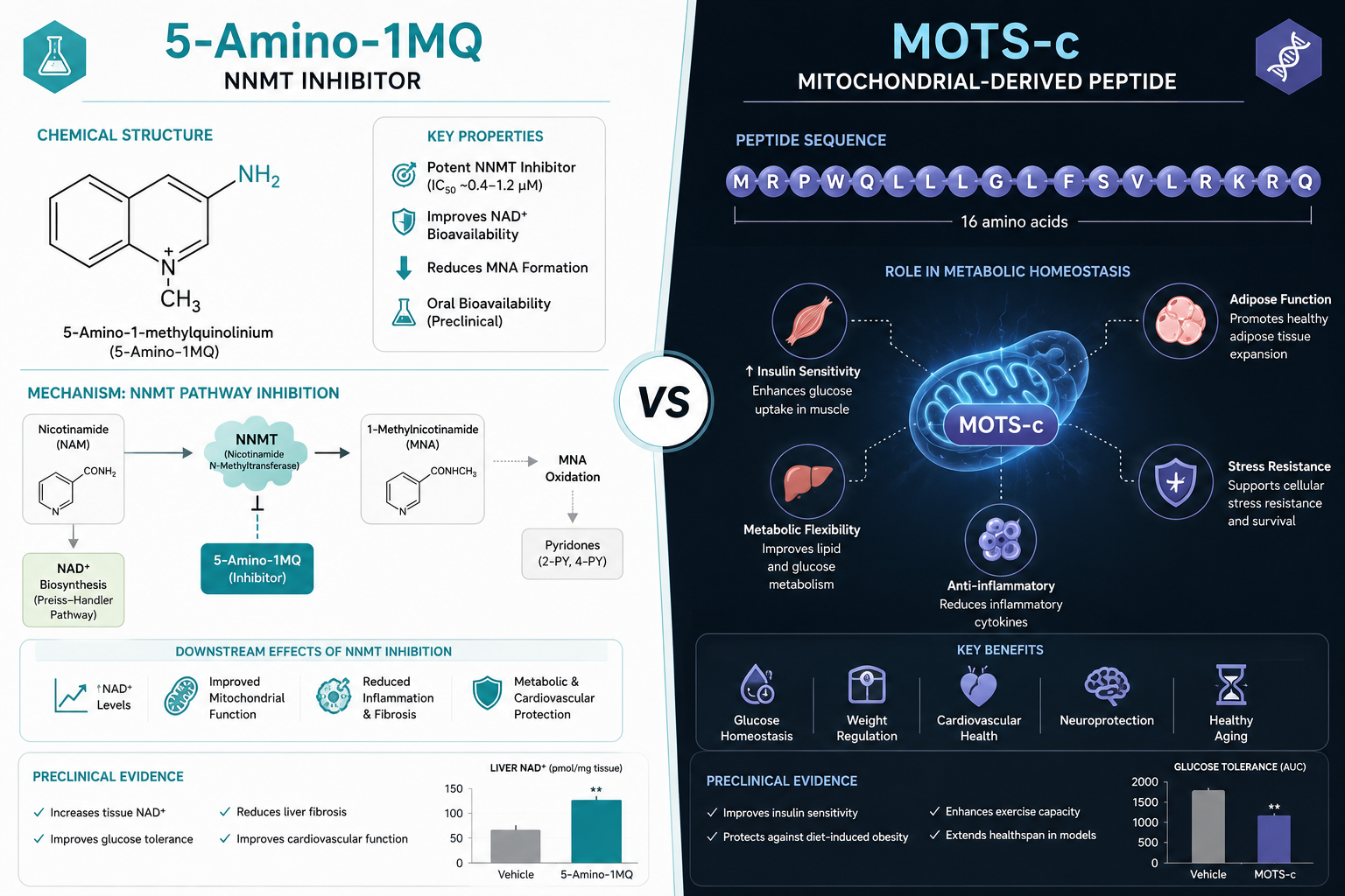
5-Amino-1MQ Peptide: Mechanism, Metabolic Research, and How It Differs From Mitochondrial Peptides

5-Amino-1MQ Peptide: Mechanism, Metabolic Research, and How It Differs From Mitochondrial Peptides

August 10, 2026/0 Comments/in Uncategorized/by

}

Only about 15% of ingested NAD+ precursors reach intracellular compartments where they can actually drive energy metabolism, a bottleneck that has pushed researchers toward upstream enzyme inhibitors as a more direct intervention point. That upstream target is NNMT, and the compound drawing the most research attention in 2026 is 5-Amino-1MQ. This article breaks down the 5-Amino-1MQ peptide: mechanism, metabolic research, and how it differs from mitochondrial peptides, answering the mechanism questions that efficacy summaries typically skip.

Key Takeaways

  • 5-Amino-1MQ is technically a small-molecule NNMT inhibitor, not a peptide, though it is frequently grouped with metabolic peptide stacks in research literature.
  • Its primary mechanism involves blocking NNMT-driven NAD+ consumption, which raises intracellular NAD+ availability and activates SIRT1 signaling.
  • Preclinical models show significant effects on adipocyte differentiation, lipid accumulation, and energy expenditure.
  • Mitochondrial peptides such as MOTS-c and SS-31 work through distinct receptor-level and membrane-targeting pathways that do not overlap with NNMT inhibition.
  • Understanding these mechanistic differences matters for designing multi-compound research protocols.

Professional () hero image with SHORT (≤42 chars): '5-Amino-1MQ: Mechanism & Metabolic Research', white on a deep navy

What Is 5-Amino-1MQ and Why the "Peptide" Label Persists

Before diving into mechanism, a classification note is worth making. 5-Amino-1MQ, full name 5-amino-1-methylquinolinium, is a small-molecule inhibitor, not a peptide. It has no amino acid chain, no peptide bond, and no receptor-binding motif typical of endogenous peptides. The "peptide" label persists because researchers and suppliers frequently group it with metabolic peptide stacks, and because its functional territory overlaps with compounds like MOTS-c.

This distinction matters for protocol design. For a broader look at how different compound classes interact at the cellular level, the overview of peptides mechanism from GLP-3 retatrutide to CJC-1295 and MOTS-c provides useful framing.

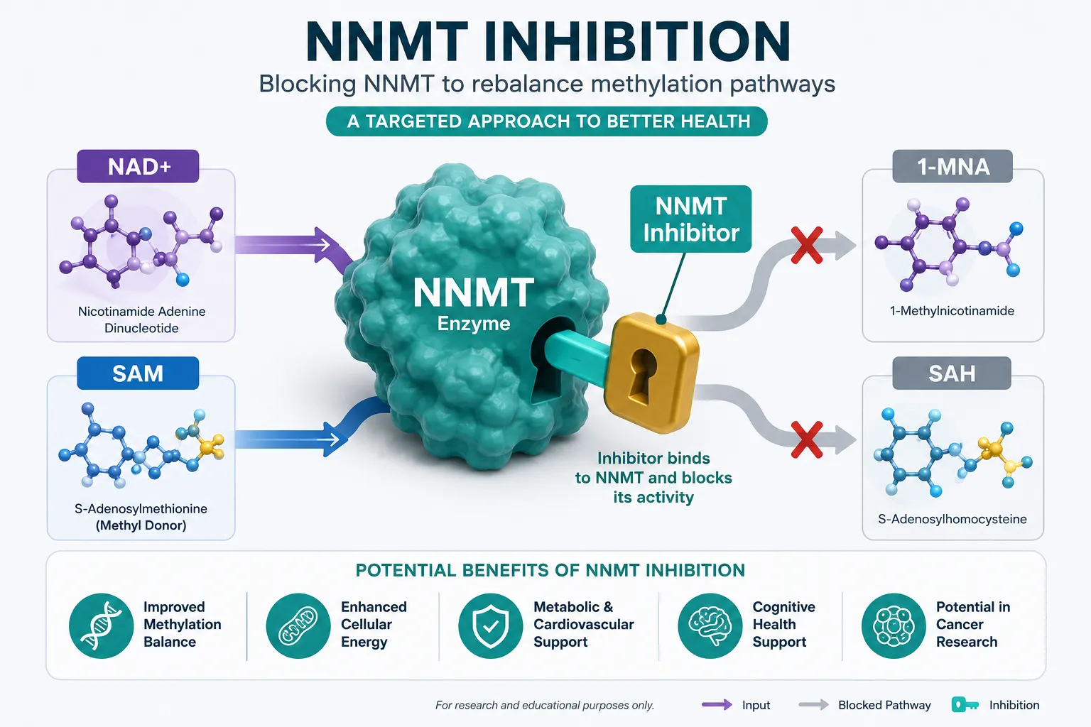
5-Amino-1MQ's molecular target is nicotinamide N-methyltransferase (NNMT), an enzyme highly expressed in adipose tissue that consumes S-adenosylmethionine (SAM) and NAD+ precursors during methylation reactions. When NNMT is overactive, it depletes both SAM and the NAD+ pool, suppressing SIRT1 activity and impairing mitochondrial function.

The Core Mechanism: NNMT Inhibition and NAD+ Restoration

The Core Mechanism: NNMT Inhibition and NAD+ Restoration

The mechanistic chain is straightforward once broken into steps:

  1. NNMT inhibition, 5-Amino-1MQ binds competitively to the NNMT active site, reducing the enzyme's ability to methylate nicotinamide.
  2. NAD+ precursor conservation, With less nicotinamide consumed by NNMT, more substrate feeds into the NAD+ biosynthesis pathway via NAMPT.
  3. SIRT1 activation, Elevated intracellular NAD+ activates SIRT1, a deacetylase that regulates metabolic gene expression, mitochondrial biogenesis, and fat oxidation.
  4. SAM preservation, Reduced NNMT activity also conserves SAM, supporting methylation reactions involved in epigenetic regulation and one-carbon metabolism.

"The compound does not donate NAD+ directly, it removes the enzymatic drain that prevents NAD+ from accumulating in the first place."

This indirect restoration model is mechanistically different from NAD+ precursor supplementation (NMN, NR), which adds substrate without addressing the enzymatic drain. For a deeper look at how NAD+ interacts with mitochondrial peptide research, the article on adenosine triphosphate and mitochondrial peptides including MOTS-c and 5-Amino-1MQ covers ATP production endpoints in detail.

Key Molecular Effects Observed in Preclinical Models

Effect Observed Outcome
NNMT inhibition Reduced nicotinamide methylation in adipocytes
Intracellular NAD+ Elevated in treated cell lines
SIRT1 activity Upregulated downstream of NAD+ increase
Adipocyte lipid accumulation Reduced in differentiation assays
Energy expenditure markers Increased in diet-induced obesity models

Metabolic Research Findings: Adipose Tissue and Energy Balance

Metabolic Research Findings: Adipose Tissue and Energy Balance

Preclinical research on 5-Amino-1MQ has concentrated on white adipose tissue (WAT), where NNMT expression is highest. In rodent models of diet-induced obesity, NNMT inhibition with 5-Amino-1MQ has been associated with:

  • Reduced fat mass without significant lean mass changes
  • Increased expression of thermogenic markers in adipose depots
  • Improved insulin sensitivity in metabolically compromised models
  • Upregulation of mitochondrial biogenesis genes

These findings position 5-Amino-1MQ within a broader class of metabolic research tools that target energy balance from the cellular level upward. Researchers comparing it against appetite-modulating compounds should note that its mechanism is entirely peripheral, there is no central nervous system component in current models. For contrast, the article on tesofensine and metabolic research comparing noradrenergic appetite modulators with GLP-3 peptides illustrates how centrally acting compounds differ in study design.

The peptides and polypeptides overview connecting DNA, mitochondria, and research compounds like MOTS-c and 5-Amino-1MQ also contextualizes where NNMT inhibitors fit within the broader mitochondrial research landscape.

How 5-Amino-1MQ Differs From Mitochondrial Peptides

How 5-Amino-1MQ Differs From Mitochondrial Peptides

This is where the 5-Amino-1MQ peptide: mechanism, metabolic research, and how it differs from mitochondrial peptides question becomes most practically relevant for researchers designing stacks or comparative studies.

Mitochondrial peptides, including MOTS-c, Humanin, and SS-31, are short amino acid sequences encoded in mitochondrial DNA or designed to target mitochondrial membranes. Their mechanisms include:

  • MOTS-c: Translocates to the nucleus under metabolic stress, activating AMPK and regulating folate and methionine metabolism
  • SS-31 (Elamipretide): Targets cardiolipin on the inner mitochondrial membrane, reducing oxidative stress and improving electron transport chain efficiency
  • Humanin: Binds cell-surface receptors and acts as a cytoprotective signaling molecule

5-Amino-1MQ, by contrast:

  • Has no amino acid structure
  • Does not interact with mitochondrial membranes directly
  • Does not bind peptide receptors
  • Works entirely through enzyme inhibition in the cytoplasm

This means the two compound classes are mechanistically complementary rather than redundant. A protocol pairing 5-Amino-1MQ with MOTS-c, for example, could theoretically address both the NAD+ depletion problem (via NNMT inhibition) and the downstream mitochondrial signaling deficit (via MOTS-c's AMPK activation). Researchers interested in SS-31's distinct membrane-targeting mechanism can explore SS-31 peptide research resources for comparison data.

For researchers sourcing compounds for metabolic studies, lab-tested peptides with verified purity documentation are essential for reproducible results.

Conclusion

5-Amino-1MQ occupies a unique position in the 2026 metabolic research landscape: it is not a peptide, but it operates in the same functional territory as mitochondrial peptides by restoring the NAD+ environment that those peptides depend on. Its mechanism, competitive NNMT inhibition leading to NAD+ conservation, SIRT1 activation, and improved adipose tissue metabolism, is well-defined at the preclinical level and mechanistically distinct from compounds like MOTS-c or SS-31.

Actionable next steps for researchers:

  • Review NNMT expression data in your specific tissue model before including 5-Amino-1MQ in a protocol
  • Consider pairing with a mitochondrial peptide to address both upstream NAD+ availability and downstream membrane-level function
  • Verify compound purity through third-party COA documentation before initiating any in vitro or in vivo work
  • Design controls that isolate NNMT inhibition from NAD+ precursor supplementation to avoid confounded endpoints

Understanding the mechanistic boundaries of each compound class, not just their reported outcomes, is what separates rigorous research design from assumption-driven stacking.

https://www.puretestedpeptides.com/wp-content/uploads/2026/08/5-amino-1mq-peptide-mechanism-metabolic-research-and-how-it-differs-from-mitocho.webp 1024 1536 https://www.puretestedpeptides.com/wp-content/uploads/2026/01/buy-peptides-online.jpg 2026-08-10 13:03:502026-08-10 13:03:505-Amino-1MQ Peptide: Mechanism, Metabolic Research, and How It Differs From Mitochondrial Peptides
Peptides and Polypeptides Explained: Connecting DNA, Mitochondria, and Modern Research-Use Compounds Like MOTS-c and 5-Amino-1MQ

Peptides and Polypeptides Explained: Connecting DNA, Mitochondria, and Modern Research-Use Compounds Like MOTS-c and 5-Amino-1MQ

August 4, 2026/0 Comments/in Uncategorized/by

Every protein in the human body, from the enzymes digesting food to the antibodies fighting infection, begins as a short chain of amino acids called a peptide. That single biological fact connects classical genetics, cellular energy production, and an entirely new generation of research compounds now drawing serious scientific attention in 2026.

This guide on Peptides and Polypeptides Explained: Connecting DNA, Mitochondria, and Modern Research-Use Compounds Like MOTS-c and 5-Amino-1MQ bridges foundational biology with cutting-edge investigational molecules, giving researchers and curious readers a clear, connected picture.

Key Takeaways

  • Peptides are short amino acid chains; polypeptides are longer chains that fold into functional proteins.
  • DNA encodes the instructions that ribosomes use to assemble every peptide and polypeptide in the body.
  • Mitochondria produce their own small peptides, including MOTS-c, that regulate metabolism and stress responses.
  • 5-Amino-1MQ is a small-molecule research compound studied for its role in metabolic enzyme inhibition, often discussed alongside mitochondria-targeting peptides.
  • Both MOTS-c and 5-Amino-1MQ remain strictly research-use compounds; neither is approved for human therapeutic use.

Key Takeaways

From DNA to Peptides: The Biological Blueprint

What Are Peptides and Polypeptides?

A peptide is a molecule made of two or more amino acids linked by peptide bonds. The naming follows a simple size rule:

Term Amino Acid Count Example
Dipeptide 2 Carnosine
Oligopeptide 3-20 GLP-1 (7 residues)
Polypeptide 20-50+ Growth hormone fragments
Protein 50+ (folded) Insulin, collagen

The line between "polypeptide" and "protein" is functional rather than strict, proteins are polypeptides that have folded into a defined three-dimensional shape.

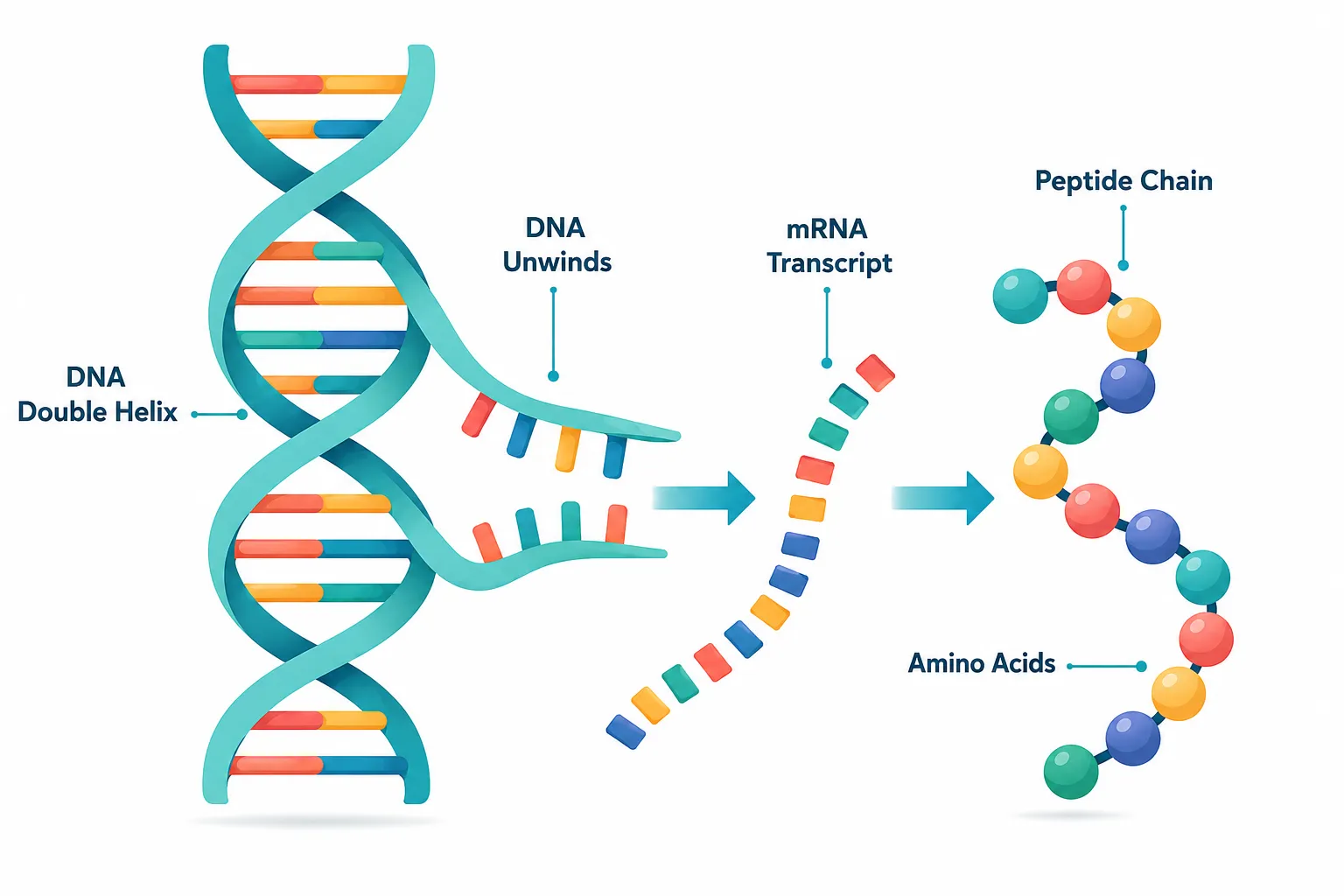
How DNA Encodes Peptide Sequences

DNA stores genetic information as sequences of nucleotide bases (A, T, G, C). When a gene is expressed:

  1. Transcription converts the DNA sequence into messenger RNA (mRNA).
  2. Translation uses ribosomes to read mRNA codons and assemble the corresponding amino acids.
  3. The resulting chain is a polypeptide, which may be cleaved, modified, or folded into its final form.

This process is the origin of every peptide the body produces naturally, including the mitochondria-derived peptides now attracting intense research interest.

"The ribosome is essentially a molecular factory reading a blueprint written in DNA and outputting a peptide product."

Researchers studying BDNF peptides and neuroprotective compounds rely on this same transcription-translation logic to understand how target sequences are designed and synthesized.

How DNA Encodes Peptide Sequences

Mitochondria as Peptide Factories: MOTS-c and the Energy Connection

Why Mitochondria Matter Beyond ATP

Most biology courses teach mitochondria as the cell's power plants, organelles that convert nutrients into adenosine triphosphate (ATP) through oxidative phosphorylation. What is less commonly taught is that mitochondria carry their own DNA (mtDNA), separate from nuclear DNA, and that this mtDNA encodes a small family of bioactive peptides called mitochondria-derived peptides (MDPs).

MOTS-c (Mitochondrial Open Reading Frame of the 12S rRNA-c) is the most studied MDP. It is a 16-amino-acid peptide encoded within the 12S ribosomal RNA gene of mtDNA. Preclinical research has examined MOTS-c in the context of:

  • Metabolic regulation and insulin sensitivity
  • Exercise-induced signaling pathways
  • Cellular stress responses and longevity-associated pathways

Another well-studied MDP, Humanin, has been investigated for neuroprotective properties, illustrating how the mitochondrial genome produces peptides with diverse systemic roles.

For researchers interested in mitochondria-targeted molecules, the SS-31 mitochondrial research overview provides a useful parallel, SS-31 is a synthetic tetrapeptide designed to concentrate in the inner mitochondrial membrane and is among the most cited mitochondria-targeting research peptides available today.

5-Amino-1MQ: A Small Molecule in the Metabolic Research Space

5-Amino-1MQ (5-amino-1-methylquinolinium) is not a peptide, it is a small organic molecule. It is included in this discussion because it targets NNMT (nicotinamide N-methyltransferase), an enzyme involved in NAD+ metabolism and fat cell differentiation. By inhibiting NNMT, 5-Amino-1MQ is hypothesized in preclinical models to:

  • Raise intracellular NAD+ precursor availability
  • Reduce lipid accumulation in adipocytes
  • Interact with metabolic pathways that overlap with those regulated by MOTS-c

This mechanistic overlap, both compounds influencing mitochondrial energy metabolism through different entry points, explains why they are frequently discussed together in metabolic research literature.

Researchers exploring this space also review SS-31 peptide research considerations for comparative context on how mitochondria-targeting compounds are evaluated.

5-Amino-1MQ: A Small Molecule in the Metabolic Research Space

Modern Research-Use Compounds: Context, Sourcing, and Responsible Use

The Research Compound Landscape in 2026

The category of research-use peptides and polypeptides has expanded considerably. Compounds once confined to academic laboratory settings are now more accessible to qualified researchers, creating both opportunity and responsibility. Key categories include:

  • Growth hormone secretagogues, such as those explored in GHRP-2 versus Sermorelin comparisons
  • Metabolic peptides, including GLP-1 analogs studied in generational research sourcing contexts
  • Mitochondria-targeted peptides, SS-31 and related compounds available through dedicated SS-31 research peptide resources
  • Repair and recovery peptides, such as the TB-500 and BPC-157 combination studied in tissue-repair research

Sourcing and Purity Standards

For any research application, purity and third-party verification are non-negotiable. Researchers should prioritize suppliers that provide:

  • Certificate of Analysis (CoA) from independent laboratories
  • High-performance liquid chromatography (HPLC) purity data
  • Mass spectrometry verification of molecular identity

Those evaluating suppliers can consult peptide supplier comparison resources to understand how to interpret third-party testing documentation.

Important disclaimer: MOTS-c, 5-Amino-1MQ, SS-31, and all compounds discussed in this article are research-use only. They are not approved by the FDA or equivalent regulatory bodies for human therapeutic use. All research must comply with applicable institutional and legal guidelines.

Conclusion

Understanding Peptides and Polypeptides Explained: Connecting DNA, Mitochondria, and Modern Research-Use Compounds Like MOTS-c and 5-Amino-1MQ requires holding two ideas at once: the elegant simplicity of how DNA encodes amino acid sequences, and the remarkable complexity of what those sequences do once assembled. Mitochondria are no longer just power plants, they are peptide-producing organelles whose outputs like MOTS-c may influence metabolism, aging, and stress resilience. Small molecules like 5-Amino-1MQ extend that conversation into enzyme inhibition and NAD+ biology.

Actionable next steps for researchers:

  • Review primary literature on MOTS-c (Lee et al., Cell Metabolism) and NNMT inhibition before designing protocols.
  • Verify supplier purity credentials before sourcing any research compound, consult where to buy peptides guidance for evaluation criteria.
  • Cross-reference mitochondria-targeting peptides such as SS-31 through SS-31 mitochondrial dynamics research to build comparative context.
  • Stay current with regulatory updates in 2026, as the research peptide landscape continues to evolve rapidly.

The biology connecting DNA, mitochondria, and modern research compounds is not abstract, it is the foundation every serious investigator needs before working with these molecules.

https://www.puretestedpeptides.com/wp-content/uploads/2026/08/peptides-and-polypeptides-explained-connecting-dna-mitochondria-and-modern-resea.webp 1024 1536 https://www.puretestedpeptides.com/wp-content/uploads/2026/01/buy-peptides-online.jpg 2026-08-04 13:05:022026-08-04 13:05:02Peptides and Polypeptides Explained: Connecting DNA, Mitochondria, and Modern Research-Use Compounds Like MOTS-c and 5-Amino-1MQ

Tag Archive for: ss-31 peptide

Best Research Peptides for Mitochondrial Health: A Comparison of MOTS-c, 5-Amino-1MQ, and Emerging Compounds

Best Research Peptides for Mitochondrial Health: A Comparison of MOTS-c, 5-Amino-1MQ, and Emerging Compounds

June 20, 2026/0 Comments/by Pure Tested

Mitochondrial dysfunction now appears in the mechanistic pathway of over 50 human diseases, from type 2 diabetes to neurodegeneration — yet the pharmacological toolkit for directly targeting these organelles remained thin until the last decade. The field of best research peptides for mitochondrial health: a comparison of MOTS-c, 5-Amino-1MQ, and emerging compounds has moved quickly, giving researchers a growing menu of targeted molecules to evaluate. This article breaks down the leading candidates, their mechanisms, and what distinguishes each for preclinical study design in 2026.

Key Takeaways

  • MOTS-c is a 16-amino-acid mitochondrial-derived peptide that activates AMPK, reduces oxidative stress, and declines naturally with age.
  • 5-Amino-1MQ targets NNMT enzyme inhibition, influencing NAD+ metabolism and energy expenditure at the cellular level.
  • SS-31 (elamipretide) protects the inner mitochondrial membrane and is one of the most studied structural mitochondrial peptides.
  • Researchers should evaluate purity, mechanism specificity, and study context when selecting among these compounds.
  • Emerging molecules such as SLU-PP-332 and humanin analogs are expanding the mitochondrial peptide research landscape.

Key Takeaways

MOTS-c: The Mitochondrial-Derived Peptide Redefining Metabolic Research

MOTS-c is encoded within the mitochondrial genome itself — a distinction that separates it from most synthetic research peptides. This 16-amino-acid peptide translocates to the nucleus under metabolic stress and exercise, where it activates antioxidant response elements and regulates stress-adaptation genes.

Key mechanisms of MOTS-c:

  • Inhibits the folate cycle and de novo purine biosynthesis
  • Activates AMPK, the master cellular energy sensor
  • Upregulates PGC-1alpha, promoting mitochondrial biogenesis
  • Reduces reactive oxygen species (ROS) emission and protein oxidative damage

Research shows that MOTS-c levels increase in skeletal muscle, systemic circulation, and the hypothalamus following exercise. Critically, circulating MOTS-c declines with age, which correlates with reduced insulin sensitivity, increased adiposity, and impaired muscle homeostasis. Exogenous MOTS-c administration in animal models has reversed age-dependent and diet-induced insulin resistance.

"MOTS-c acts as a molecular signal linking mitochondrial stress to whole-body metabolic adaptation — a property no synthetic small molecule fully replicates."

For researchers building study frameworks around this peptide, the MOTS-c mitochondrial research themes resource provides a useful orientation to current experimental directions. Those interested in mechanistic depth can also explore MOTS-c and mitochondrial dynamics for pathway-level detail.


MOTS-c: The Mitochondrial-Derived Peptide Redefining Metabolic Research

Comparing the Best Research Peptides for Mitochondrial Health: A Comparison of MOTS-c, 5-Amino-1MQ, and Emerging Compounds

5-Amino-1MQ: NNMT Inhibition and NAD+ Metabolism

5-Amino-1MQ is a small-molecule NNMT (nicotinamide N-methyltransferase) inhibitor rather than a peptide in the classical sense, but it is routinely grouped with research peptides given its metabolic targeting profile. NNMT consumes SAM (S-adenosylmethionine) and reduces NAD+ precursor availability. By blocking NNMT, 5-Amino-1MQ effectively raises intracellular NAD+ levels, which supports mitochondrial electron transport chain efficiency.

Comparison table: MOTS-c vs. 5-Amino-1MQ

Feature MOTS-c 5-Amino-1MQ
Origin Mitochondrial genome Synthetic small molecule
Primary target AMPK / PGC-1alpha NNMT enzyme
NAD+ effect Indirect (via AMPK) Direct (via NNMT inhibition)
Oxidative stress reduction Demonstrated Under active study
Age-related decline Yes Not applicable

SS-31 (Elamipretide): Structural Mitochondrial Protection

SS-31 targets cardiolipin on the inner mitochondrial membrane, stabilizing cristae architecture and improving ATP synthesis efficiency. Unlike MOTS-c, SS-31 does not rely on nuclear translocation — it acts directly at the membrane. Researchers studying kidney, cardiac, or skeletal muscle models frequently pair SS-31 with MOTS-c to address both structural and signaling dimensions of mitochondrial health. The SS-31 and MOTS-c research tag reflects this growing interest in combinatorial study designs.

For kidney-specific mitochondrial research, the SS-31 kidney health research page offers relevant preclinical context.


SS-31 (Elamipretide): Structural Mitochondrial Protection

Emerging Compounds and Sourcing Considerations

Humanin, SLU-PP-332, and Beyond

The mitochondrial-derived peptide (MDP) family extends beyond MOTS-c. Humanin and SHLP2 (small humanin-like peptides) are encoded in the same mitochondrial 16S rRNA region and show cytoprotective effects in neuronal and cardiomyocyte models. SLU-PP-332 is an ERR-alpha/gamma agonist that mimics exercise-induced mitochondrial gene expression — a distinct but complementary mechanism. Researchers interested in this compound can review the SLU-PP-332 metabolic research overview for study design notes.

Longevity-oriented research programs increasingly stack these compounds. The longevity peptide research framework outlines how multiple mitochondrial targets can be addressed within a single experimental protocol.

Sourcing and Purity Standards

Compound quality is non-negotiable in mitochondrial research. ROS-sensitive assays and AMPK phosphorylation readouts are highly vulnerable to contaminant interference. Researchers should prioritize suppliers with documented certificate of analysis (COA) data and reference standard benchmarking. The Bachem and reference standards guide addresses how to evaluate peptide purity against validated benchmarks.

For researchers building broader metabolic study panels, the MOTS-c and elamipretide comparison page provides a useful side-by-side of two of the field's most studied mitochondrial compounds.


Conclusion

Selecting among the best research peptides for mitochondrial health requires matching mechanism to research question. MOTS-c is the strongest candidate for studies targeting AMPK activation, age-related metabolic decline, and exercise physiology. 5-Amino-1MQ suits protocols focused on NAD+ metabolism and NNMT-driven energy regulation. SS-31 remains the reference compound for inner mitochondrial membrane integrity. Emerging molecules like SLU-PP-332 and humanin analogs are broadening the toolkit further.

Actionable next steps for researchers:

  1. Define the specific mitochondrial pathway under investigation before compound selection.
  2. Obtain COA-verified peptides from suppliers using validated reference standards.
  3. Consider combinatorial designs (e.g., MOTS-c plus SS-31) for multi-target mitochondrial studies.
  4. Monitor the MDP literature actively — this field is advancing rapidly in 2026.
https://www.puretestedpeptides.com/wp-content/uploads/2026/06/Best-Research-Peptides-for-Mitochondrial-Health-A-Comparison-of-MOTS-c-5-Amino-1MQ-and-Emerging-Compounds.png 1024 1536 Pure Tested https://www.puretestedpeptides.com/wp-content/uploads/2026/01/buy-peptides-online.jpg Pure Tested2026-06-20 13:04:512026-07-20 15:02:39Best Research Peptides for Mitochondrial Health: A Comparison of MOTS-c, 5-Amino-1MQ, and Emerging Compounds
Peptides and Polypeptides in Cell Biology: How Experimental Peptides Interact With DNA, Mitochondria, and Hormone Receptors

Peptides and Polypeptides in Cell Biology: How Experimental Peptides Interact With DNA, Mitochondria, and Hormone Receptors

June 4, 2026/0 Comments/by Pure Tested

{"cover":"Professional landscape format (1536×1024) hero image with bold text overlay: 'Peptides & Polypeptides in Cell Biology' as main headline and subtitle 'How Experimental Peptides Interact With DNA, Mitochondria, and Hormone Receptors' in extra large 68pt bold white sans-serif font with deep navy semi-transparent overlay box, centered upper-third composition. Background shows a stunning macro scientific illustration of a glowing double-helix DNA strand intertwined with a luminous mitochondrion and molecular peptide chain structures, rendered in electric blue, teal, and white on deep navy background. Magazine cover aesthetic, editorial quality, high contrast, cinematic lighting, 2026 research theme.","content":["Detailed landscape format (1536×1024) scientific illustration showing intracellular peptide-DNA interaction: a close-up cross-section of a cell nucleus with glowing peptide chains (labeled EL28, PepH, Pep5) binding to a double-helix DNA strand. Molecular bonds shown as luminous gold nodes. Background features a translucent cell membrane with proteasome structures. Color palette: deep purple, gold, white. Clean lab-diagram aesthetic with annotated arrows, editorial quality, high contrast lighting from within the nucleus.","Detailed landscape format (1536×1024) visualization of mitochondrial targeting by peptides: a dramatic bird's-eye cross-section of a mitochondrion with its cristae structure clearly visible in electric teal and orange. Amphipathic peptide chains (MOTS-c labeled) are shown penetrating the outer mitochondrial membrane and localizing to the inner membrane. Surrounding cytoplasm shows cell-penetrating peptide vectors in motion. Deep charcoal background, bioluminescent glow effect, annotated molecular pathway arrows, editorial scientific illustration quality.","Detailed landscape format (1536×1024) diagram of hormone receptor signal transduction: a stylized cell surface showing multiple G protein-coupled receptors (GPCRs) with peptide hormone ligands docking onto receptor binding sites. Intracellular cascade shown with cAMP second messenger molecules, protein kinase activation chain, and nuclear receptor binding to DNA strand in the background. Color scheme: crimson red, white, slate blue. Clean infographic-style layout with labeled pathway steps, modern sans-serif annotations, editorial quality, high contrast."]

Professional landscape hero image () with : "Peptides and Polypeptides in Cell Biology: How Experimental Peptides Interact

Roughly 30% of all FDA-approved drugs work by targeting G protein-coupled receptors — proteins that respond directly to peptide signals. That single statistic reveals how deeply peptides and polypeptides in cell biology are woven into the machinery of life, and why research into experimental peptides has accelerated so sharply in 2026.

This article walks through the core mechanisms: how short amino acid chains reach the cell nucleus, penetrate mitochondrial membranes, and dock onto hormone receptors to trigger downstream signaling cascades.


Key Takeaways

  • Intracellular peptides such as EL28, PepH, and Pep5 interact directly with DNA-associated proteins and are studied as drug prototypes.
  • Peptide hormones are hydrophilic and cannot cross the lipid bilayer, so they bind cell surface receptors and activate second messengers like cyclic AMP.
  • Experimental peptides including MOTS-c can localize to mitochondria and influence energy regulation pathways.
  • GPCRs are the primary receptor family for peptide hormones and represent a major pharmacological target class.
  • Research-grade peptides such as CJC-1295 and GLP-1 analogs operate through receptor-mediated signaling with measurable downstream effects on gene expression.

Peptides and Polypeptides in Cell Biology: The Structural Foundation

Peptides and Polypeptides in Cell Biology: The Structural Foundation

A peptide is a chain of two or more amino acids linked by peptide bonds. A polypeptide is simply a longer chain — typically more than 50 residues. When folded into functional shapes, polypeptides become proteins. The distinction matters in research because short peptides often behave differently from full proteins: they can slip through membranes, evade immune detection, and reach targets that larger molecules cannot.

Intracellular Peptides and DNA Interaction

Inside the cell, certain peptides operate in the nucleus itself. Intracellular peptides derived from proteasomal degradation — including EL28 (from proteasome regulatory subunit 4), PepH (from Histone H2B), and Pep5 (from cyclin D2) — have been identified as functional modulators of protein-protein interactions linked to gene regulation. These are not merely degradation byproducts; they act as prototype drug candidates because they already exist in the cellular environment and interact with DNA-associated machinery.

This opens a compelling research angle: if naturally occurring intracellular peptides can modulate transcription-linked proteins, then synthetic analogs designed to mimic or block those interactions could influence gene expression with high precision.


Mitochondrial Targeting: How Experimental Peptides Reach the Powerhouse

Mitochondrial Targeting: How Experimental Peptides Reach the Powerhouse

Mitochondria are not passive energy factories. They participate in intracrine signaling — internal signaling loops that influence cell survival, metabolism, and apoptosis. Peptides including angiotensin II and transforming growth factor-beta have been detected inside mitochondria, suggesting that peptide signaling extends well beyond the cell surface.

More recently, amphipathic proline-rich cell-penetrating peptides have been engineered to cross the plasma membrane and localize specifically to mitochondria. These vectors carry therapeutic payloads or act directly on mitochondrial membranes to stabilize cristae architecture and reduce oxidative stress.

MOTS-c, a mitochondria-derived peptide encoded in mitochondrial DNA, is one of the most studied examples. Research into MOTS-c mitochondrial research themes shows that it translocates to the nucleus under metabolic stress and regulates gene expression — a striking example of cross-compartment peptide signaling. The compound MOTS-c and SLU-PP-332 pairing has also attracted attention for its potential effects on mitochondrial biogenesis pathways.

The SS-31 peptide (elamipretide) represents another mitochondria-targeted research compound. Its mechanism centers on cardiolipin stabilization within the inner mitochondrial membrane. Detailed research considerations are covered in this SS-31 10mg research peptide overview, and its broader mitochondrial dynamics are explored in SS-31 mitochondrial dynamics research.


Hormone Receptors and Signal Transduction: Where Peptides Meet Cell Biology

Hormone Receptors and Signal Transduction: Where Peptides Meet Cell Biology

Because peptide hormones are hydrophilic, they cannot diffuse through the fatty lipid bilayer of the cell membrane. Instead, they bind to receptors on the cell surface, which then relay the signal inward.

Three Major Receptor Classes for Peptide Hormones

Receptor Type Mechanism Example Peptide
G protein-coupled receptors (GPCRs) Activate G proteins, trigger cAMP GLP-1, GIP
Enzyme-linked receptors Direct kinase activation Insulin, IGF-1
Ion channel receptors Gate ion flow Neuropeptides

GPCRs dominate peptide hormone pharmacology. When a peptide ligand binds, the receptor activates a G protein, which in turn stimulates adenylyl cyclase to produce cyclic AMP (cAMP). This second messenger activates protein kinases that phosphorylate downstream targets — ultimately altering metabolism, proliferation, or secretion.

Research into GLP-1 dual receptor agonism and GIP receptor importance illustrates how next-generation peptide drugs exploit this pathway. Similarly, CJC-1295 research demonstrates GPCR-mediated growth hormone secretion through GHRH receptor activation.

Steroid hormones follow a different route — they diffuse through the membrane and bind nuclear receptors that act directly as transcription factors, binding DNA to switch genes on or off. Experimental peptides that mimic steroid hormone behavior are therefore studied for their potential to regulate gene expression without the systemic side effects of steroids.


Conclusion

Understanding peptides and polypeptides in cell biology — how experimental peptides interact with DNA, mitochondria, and hormone receptors — is no longer purely academic. In 2026, this knowledge directly informs the design of research-grade compounds targeting metabolic disease, mitochondrial dysfunction, and endocrine signaling.

Actionable next steps for researchers:

  • Review mitochondria-targeted compounds such as SS-31 and MOTS-c for models of intracellular peptide delivery.
  • Study GPCR-mediated pathways when evaluating GLP-1, GIP, and secretagogue peptides like CJC-1295 and ipamorelin.
  • Examine intracellular peptide prototypes (EL28, PepH) as templates for nucleus-targeted drug design.
  • Explore the full peptides research catalog to identify compounds relevant to specific signaling pathways.

The cell is not a black box. Peptides are the keys — and mapping how they fit each lock is the central challenge of modern molecular biology.


https://www.puretestedpeptides.com/wp-content/uploads/2026/06/Peptides-and-Polypeptides-in-Cell-Biology-How-Experimental-Peptides-Interact-With-DNA-Mitochondria-and-Hormone-Receptors.png 672 1024 Pure Tested https://www.puretestedpeptides.com/wp-content/uploads/2026/01/buy-peptides-online.jpg Pure Tested2026-06-04 13:04:432026-07-20 15:04:08Peptides and Polypeptides in Cell Biology: How Experimental Peptides Interact With DNA, Mitochondria, and Hormone Receptors
5-Amino-1MQ Peptide Research: NNMT Inhibition, Fat Metabolism, and Why It Is Often Paired With Mitochondrial Stacks

5-Amino-1MQ Peptide Research: NNMT Inhibition, Fat Metabolism, and Why It Is Often Paired With Mitochondrial Stacks

June 4, 2026/0 Comments/by Pure Tested

Nicotinamide N-methyltransferase, or NNMT, is overexpressed in the adipose tissue of individuals with obesity at rates roughly two to four times higher than in lean controls — a biochemical pattern that has made it one of the more compelling metabolic targets in current research. At the center of that research sits 5-Amino-1MQ, a small-molecule NNMT inhibitor that has attracted growing interest for its role in fat metabolism and energy regulation. This article breaks down 5-Amino-1MQ peptide research: NNMT inhibition, fat metabolism, and why it is often paired with mitochondrial stacks — covering the core biology, the metabolic rationale, and how researchers are thinking about combination protocols.

Key Takeaways

  • 5-Amino-1MQ is a selective NNMT inhibitor, not a true peptide, though it is commonly grouped with peptide-based metabolic compounds in research contexts.
  • NNMT regulates the methyl economy of cells; inhibiting it raises SAM levels and shifts adipose tissue toward greater energy expenditure.
  • Preclinical data suggest NNMT inhibition can reduce fat mass, improve insulin sensitivity, and support a shift from white to beige adipose phenotype.
  • Mitochondrial peptides such as SS-31 and MOTS-c are frequently studied alongside 5-Amino-1MQ because they address complementary steps in the same metabolic pathway.
  • Research into this compound remains at the preclinical stage; no approved clinical applications exist as of 2026.

Key Takeaways

Understanding NNMT and What 5-Amino-1MQ Actually Does

Despite being called a peptide in many research discussions, 5-Amino-1MQ is technically a small-molecule compound — a methylquinolinium derivative. The distinction matters because its mechanism is enzymatic inhibition rather than receptor binding in the conventional peptide sense. However, it is routinely grouped with peptide-based metabolic stacks because it targets overlapping biological pathways.

NNMT's core function is to transfer methyl groups from S-adenosylmethionine (SAM) to nicotinamide, producing S-adenosylhomocysteine (SAH) and 1-methylnicotinamide. This process consumes methyl groups that would otherwise support epigenetic regulation, NAD+ recycling, and mitochondrial signaling. When NNMT activity is high — as it tends to be in obese adipose tissue — the methyl pool is depleted, and cellular energy metabolism slows.

By selectively blocking NNMT, 5-Amino-1MQ preserves SAM availability. The downstream effects observed in preclinical models include:

  • Increased NAD+ and NADH cycling
  • Upregulation of thermogenic gene expression in adipose tissue
  • Reduced lipid accumulation in fat cells
  • Improved insulin sensitivity markers

"NNMT sits at a metabolic crossroads — its inhibition does not simply block one pathway but redistributes methyl currency across multiple energy-sensing systems."

This broad upstream influence is precisely why 5-Amino-1MQ peptide research has attracted attention beyond simple fat-loss applications.


Understanding NNMT and What 5-Amino-1MQ Actually Does

NNMT Inhibition, Fat Metabolism, and the Adipose Tissue Connection

The adipose tissue findings from 5-Amino-1MQ research are among its most discussed features. In mouse models, NNMT inhibition has been associated with a shift in white adipose tissue toward a beige or brown-like phenotype — a process sometimes called "beiging." Beige adipocytes express higher levels of uncoupling protein 1 (UCP1), which dissipates energy as heat rather than storing it as fat.

Key metabolic outcomes observed in preclinical studies:

Outcome Direction
Body fat mass Decreased
Lean mass Preserved or increased
Insulin sensitivity Improved
SAM/SAH ratio Increased
UCP1 expression Upregulated

This metabolic profile makes 5-Amino-1MQ relevant to researchers studying AOD-9604 metabolic research and other compounds targeting adipose function. It also connects naturally to GLP-1 and incretin research themes, since both pathways converge on insulin sensitivity and energy partitioning.

Researchers studying MOTS-c and metabolic flexibility have noted similar adipose remodeling effects, which has prompted interest in whether combining these compounds produces additive or synergistic outcomes.


NNMT Inhibition, Fat Metabolism, and the Adipose Tissue Connection

Why 5-Amino-1MQ Is Often Paired With Mitochondrial Stacks

The pairing of 5-Amino-1MQ with mitochondrial peptides is not arbitrary. It reflects a layered approach to metabolic research where each compound addresses a distinct step in the same energy-production hierarchy.

The rationale works like this:

  1. 5-Amino-1MQ preserves the methyl pool and raises NAD+ availability — setting the biochemical conditions for efficient mitochondrial function.
  2. SS-31 (Elamipretide) targets cardiolipin on the inner mitochondrial membrane, stabilizing electron transport chain efficiency. Research on SS-31 mitochondrial research themes highlights its role in reducing oxidative stress at the mitochondrial level.
  3. MOTS-c is a mitochondria-derived peptide that activates AMPK and supports glucose uptake in skeletal muscle — complementing the insulin-sensitizing effects of NNMT inhibition.

The combination of MOTS-c and SS-31 (Elamipretide) has already been explored in preclinical contexts, and 5-Amino-1MQ is increasingly discussed as a third layer in such stacks.

Researchers also note that NAD+ availability — which NNMT inhibition supports — is directly relevant to NAD+ scientific evidence and the broader sirtuin/AMPK signaling network that mitochondrial peptides also engage.

For those reviewing broader metabolic peptide combinations, IPA muscle and fat research themes offer additional context on how growth hormone secretagogues interact with fat oxidation pathways that 5-Amino-1MQ may also influence.


Conclusion

5-Amino-1MQ occupies a unique position in metabolic research: it acts upstream of both fat storage and mitochondrial efficiency by preserving the methyl economy that both systems depend on. The preclinical evidence for NNMT inhibition — reduced fat mass, beige adipose conversion, improved insulin sensitivity, and elevated NAD+ cycling — provides a mechanistic basis for why researchers pair it with mitochondrial peptides like SS-31 and MOTS-c.

Actionable next steps for researchers:

  • Review the preclinical NNMT inhibition literature before designing any combination protocol.
  • Examine SS-31 and MOTS-c data independently to understand where their mechanisms overlap with and differ from 5-Amino-1MQ.
  • Source compounds only from verified, third-party-tested suppliers to ensure research-grade purity.
  • Treat all findings as preclinical; no human clinical approvals exist for 5-Amino-1MQ as of 2026.

The mechanistic logic behind 5-Amino-1MQ peptide research — NNMT inhibition, fat metabolism, and mitochondrial stack pairing — is coherent and well-grounded in cell biology. As research matures, this compound is likely to remain a central figure in metabolic and longevity-focused peptide discussions.


https://www.puretestedpeptides.com/wp-content/uploads/2026/06/5-Amino-1MQ-Peptide-Research-NNMT-Inhibition-Fat-Metabolism-and-Why-It-Is-Often-Paired-With-Mitochondrial-Stacks.png 672 1024 Pure Tested https://www.puretestedpeptides.com/wp-content/uploads/2026/01/buy-peptides-online.jpg Pure Tested2026-06-04 13:04:412026-07-20 15:04:095-Amino-1MQ Peptide Research: NNMT Inhibition, Fat Metabolism, and Why It Is Often Paired With Mitochondrial Stacks
×

Helpful Links

  • My account
  • Cart
  • Checkout
  • Refund and Returns Policy
  • Privacy Policy
  • SMS Privacy Policy
  • Login
  • My Account
  • Logout

USA Made Lab Tested Peptides

All products are sold for research, laboratory, or analytical purposes only, and are not for human consumption


Pure Tested Peptides is a chemical supplier. Pure Tested Peptides is not a compounding / chemical compounding facility as defined under 503A of the Federal Food, Drug, and Cosmetic act. Pure Tested Peptides is not an outsourcing facility as defined under 503B of the Federal Food, Drug, and Cosmetic act.

The statements made within this website have not been evaluated by the US Food and Drug Administration. The products we offer are not intended to diagnose, treat, cure or prevent any disease.

Human/Animal Consumption Prohibited. Laboratory/In-Vitro Experimental Use Only

Scroll to top Scroll to top Scroll to top