

ACE Inhibitors Explained: How Lisinopril and Angiotensin-Converting Enzyme Signaling Can Confound Peptide Vascular Studies
Nearly one in three adults worldwide takes a medication that directly reshapes the body's peptide signaling landscape — yet most vascular peptide studies fail to account for it. Angiotensin-converting enzyme (ACE) inhibitors, led by the widely prescribed lisinopril, do far more than lower blood pressure. They fundamentally alter the concentrations of angiotensin II, bradykinin, and a cascade of downstream peptides that researchers use as biomarkers and endpoints. Understanding ACE inhibitors explained through the lens of vascular peptide research is not just useful — it is essential for producing reproducible, interpretable data.
Key Takeaways
- ACE inhibitors like lisinopril block the conversion of angiotensin I to angiotensin II, but also elevate bradykinin, Ac-SDKP, and angiotensin-(1-7), altering the entire vascular peptide environment.
- Background ACE inhibitor use in study subjects creates a shifted peptide "baseline" that can make experimental results misleading if not controlled for.
- Dietary and research-use peptides can independently inhibit ACE, adding another layer of confounding in vascular studies.
- ACE inhibition measurably reduces collagen-related peptide biomarkers, affecting tissue-remodeling endpoints.
- Rigorous study design requires screening, stratification, and biomarker adjustment for ACE inhibitor exposure.
How ACE Biology Drives Vascular Peptide Signaling
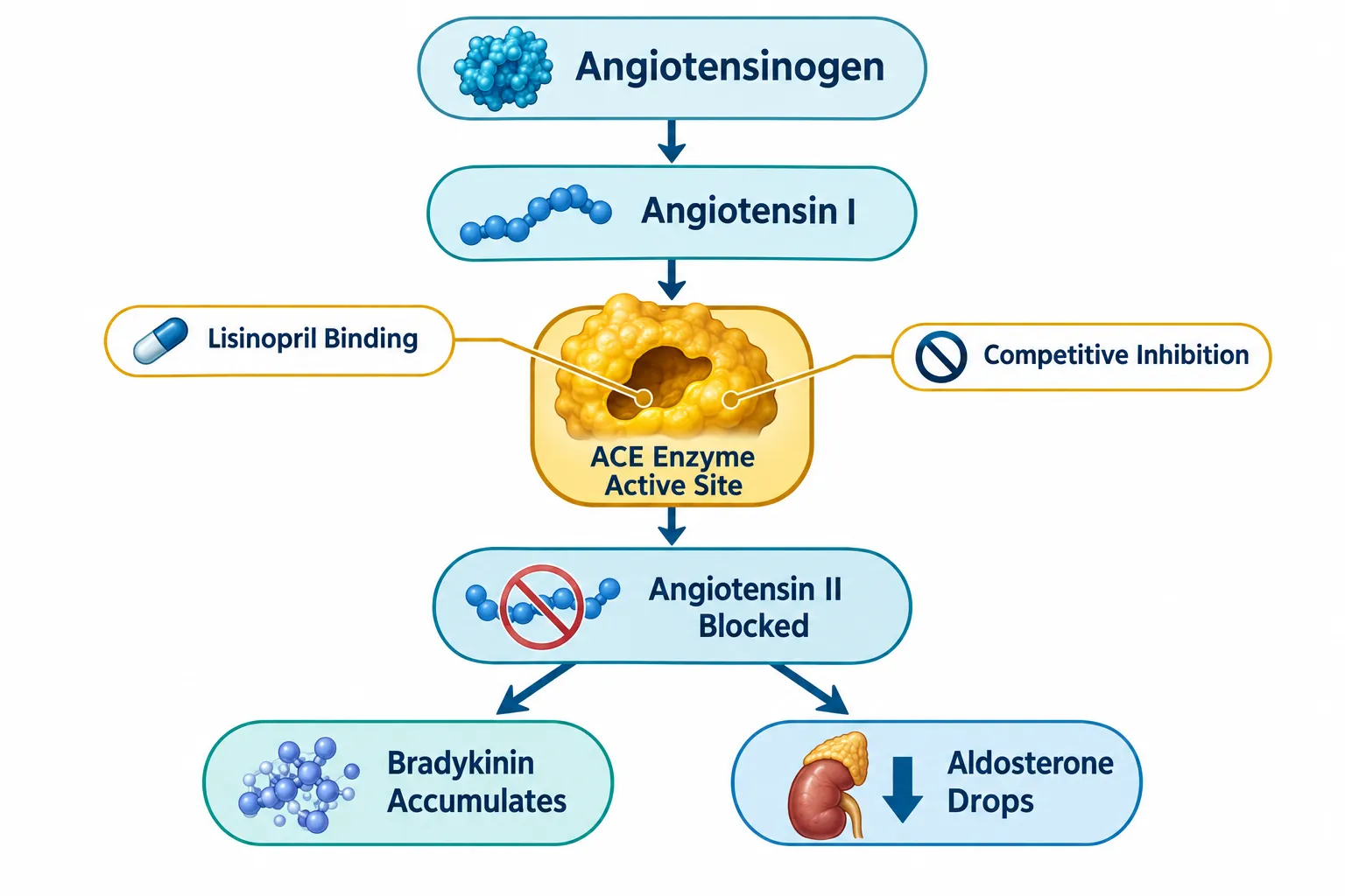
The renin-angiotensin-aldosterone system (RAAS) is one of the most studied hormonal axes in cardiovascular medicine. At its center sits angiotensin-converting enzyme — a peptidyl dipeptidase anchored to vascular endothelium. ACE cleaves two amino acids from angiotensin I to generate angiotensin II, a potent vasoconstrictor that raises blood pressure, stimulates aldosterone release, and promotes vascular smooth-muscle contraction.
Lisinopril binds competitively to the ACE active site, blocking this conversion. The result is a drop in circulating angiotensin II and a parallel reduction in aldosterone and vasopressor activity. Blood pressure falls. But the enzymatic blockade does not stop there.
ACE also degrades bradykinin, a vasodepressor peptide that promotes vasodilation and natriuresis. When ACE is inhibited, bradykinin accumulates. Current regulatory labeling for lisinopril acknowledges that elevated bradykinin may contribute to the drug's therapeutic effects, though its precise role remains under investigation. Beyond bradykinin, ACE inhibition raises levels of:
| Peptide Species | Direction of Change | Vascular Effect |
|---|---|---|
| Angiotensin II | Decreases | Less vasoconstriction |
| Bradykinin | Increases | Vasodilation |
| Ac-SDKP | Increases | Anti-fibrotic, vasodilatory |
| Angiotensin-(1-7) | Increases | Vasodilation, anti-proliferative |
This multi-peptide shift is not a side effect — it is a core feature of ACE inhibitor pharmacology. For researchers studying vascular peptide biology, it represents a significant source of confounding.
"Any background ACE inhibition — whether from a drug or a dietary source — can substantially skew vascular peptide profiles in experimental studies."
ACE Inhibitors Explained: Why Lisinopril Confounds Peptide Vascular Studies
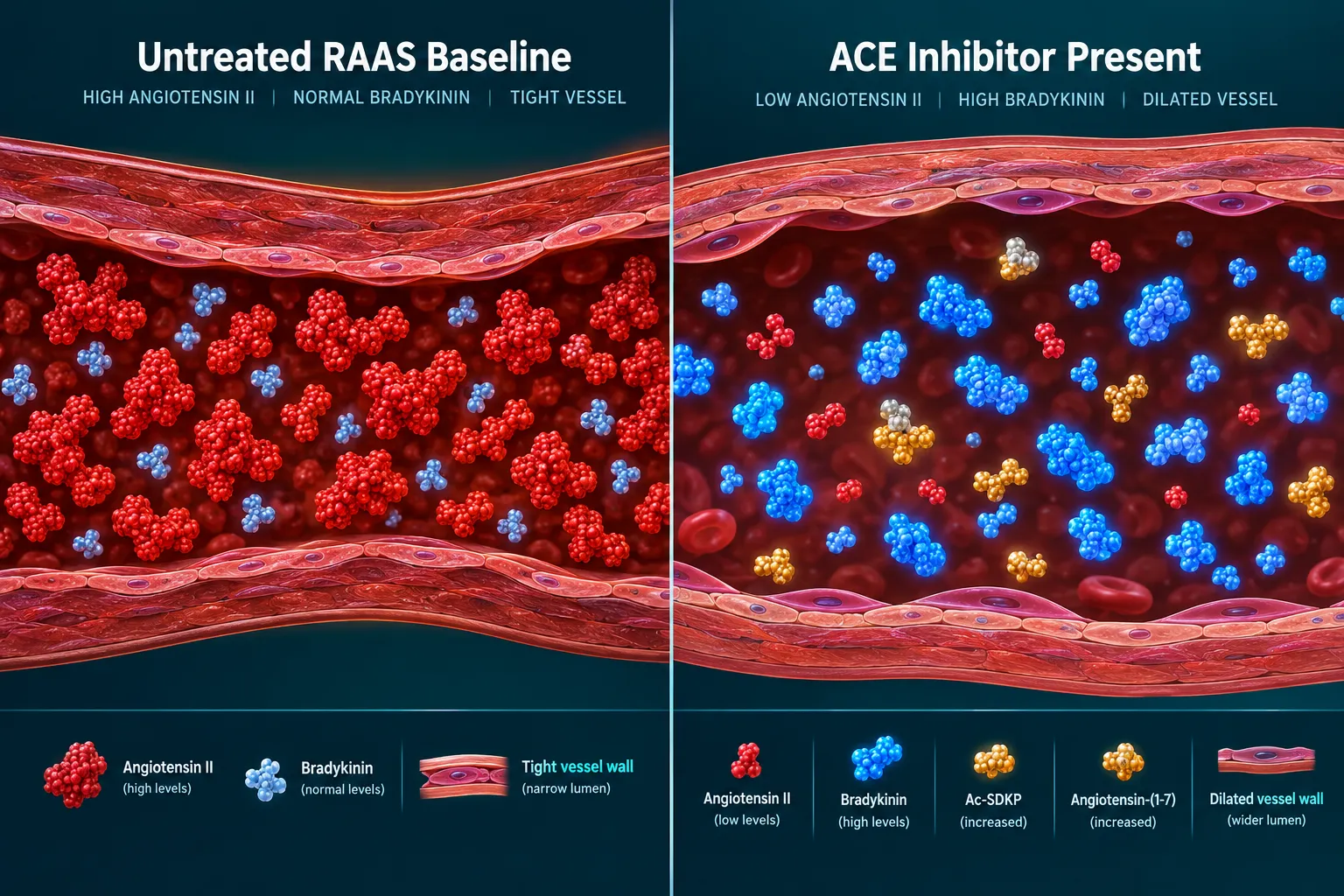
The confounding problem becomes clearest when examining what happens to the peptide environment inside study subjects already taking lisinopril. Their "baseline" is not a neutral RAAS state. It is a pharmacologically shifted landscape with suppressed angiotensin II, elevated kinins, and altered angiotensin peptide degradation kinetics.
Research published in 2026 confirms that ACE-inhibiting agents significantly alter the degradation kinetics and half-life of upstream angiotensin peptides, including angiotensin-(1-12). This means that not just angiotensin II, but the entire spectrum of RAAS peptides, is affected. Studies that enroll subjects on lisinopril without stratification will pool two biologically distinct peptide environments.
Three specific confounding mechanisms stand out:
- Shifted angiotensin II levels — The primary endpoint in many vascular studies, angiotensin II, is directly suppressed. Effect sizes will be compressed or reversed.
- Elevated bradykinin and Ac-SDKP — These vasodilatory peptides oppose the actions of test compounds, masking or amplifying observed vascular responses.
- Altered collagen-related peptide biomarkers — ACE inhibitor therapy produces mean reductions of roughly 20% in serum procollagen III peptide (PIIIP) and significant decreases in other collagen markers. Studies using these peptides to assess tissue remodeling will find systematically lower values in ACE-inhibitor-exposed subjects.
This last point matters for cardiometabolic research. Peptide-based biomarkers of myocardial fibrosis and vascular remodeling are widely used in heart failure and hypertension trials. If ACE inhibitor status is not documented and controlled, the signal is corrupted.
For researchers exploring peptide compounds with vascular or mitochondrial protective properties — such as those described in resources on SS31 peptide benefits or the synergy of LL-37 and SS31 — understanding the pharmacological background of study subjects is critical to interpreting results accurately.
Dietary and Research Peptides as Hidden ACE Inhibitors
A less-recognized confounding source is the peptides themselves. Food-derived and synthetic peptides can independently inhibit ACE at pharmacologically relevant concentrations.
Broccoli-derived peptides — including HLVNK, LTEVR, and LLDGR — have been shown to reduce intracellular angiotensin II and inhibit ERK and p38 MAPK phosphorylation in vascular smooth-muscle cells. Marine muscle protein hydrolysate yields peptides such as APFLAG with measurable ACE IC50 values near 69.5 µM, acting through SRC and STAT3 signaling. These are the same pathways affected by lisinopril.
This creates a layered confounding problem in peptide vascular studies:
- Pharmaceutical ACE inhibitors shift the subject's baseline peptide environment.
- Dietary ACE-inhibitory peptides from food sources can mimic or augment drug effects.
- Research-use peptides under investigation may themselves carry ACE-inhibitory activity that interacts with background drug therapy.
Researchers reviewing foundational peptide mechanisms will find the Peptides 101 for research-use only buyers guide a useful reference for understanding how peptide structure drives receptor selectivity. Similarly, building robust peptide benchmarks with Bachem reference standards is directly relevant to establishing clean experimental controls.
For studies involving metabolic peptides, understanding how GLP-related compounds interact with vascular tone — as explored in resources on GLP peptides for sale — adds another dimension to study design. Likewise, researchers examining cardiometabolic endpoints should consult data on tesa peptide benefits for context on growth-hormone-releasing peptides and vascular outcomes.
Practical Controls for ACE Inhibitor Confounding in Vascular Research
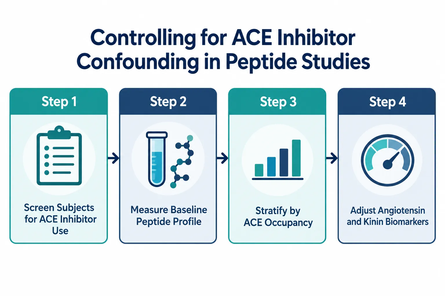
Addressing ACE inhibitor confounding requires deliberate study design. The following protocol elements are now considered best practice in rigorous vascular peptide research as of 2026:
Screening and Enrollment
- Document all ACE inhibitor and ARB use at enrollment.
- Exclude or separately stratify subjects on lisinopril or equivalent agents.
- Screen for dietary patterns high in ACE-inhibitory peptide sources.
Biomarker Adjustment
- Measure baseline bradykinin, Ac-SDKP, and angiotensin-(1-7) alongside angiotensin II.
- Use angiotensin-(1-12) degradation kinetics as a proxy for ACE occupancy.
- Adjust collagen-related peptide endpoints (PIIIP, PIP, PICP) for ACE inhibitor status.
Imaging and Occupancy Measurement
- Nuclear imaging tools using lisinopril radioconjugates can now quantify tissue ACE occupancy directly, allowing researchers to correct for pre-existing ACE inhibitor binding in concurrent peptide pharmacodynamic measurements.
These controls are not optional refinements — they are the difference between interpretable data and noise. Lisinopril remains antihypertensive even in low-renin states, meaning its vascular effects span a broad clinical spectrum that cannot be predicted from renin status alone.
Conclusion
ACE inhibitors explained in full reveal a pharmacological footprint that extends far beyond blood pressure control. Lisinopril and its class alter angiotensin II, bradykinin, Ac-SDKP, angiotensin-(1-7), and collagen-related peptide biomarkers simultaneously. For vascular and cardiometabolic peptide researchers, this creates a multi-layered confounding problem that corrupts endpoints, compresses effect sizes, and undermines reproducibility.
Actionable next steps for researchers in 2026:
- Implement mandatory ACE inhibitor screening in all vascular peptide study enrollment protocols.
- Add kinin and angiotensin-(1-7) panels to standard biomarker batteries.
- Treat dietary ACE-inhibitory peptide exposure as a covariate, not background noise.
- Consult updated reference standards and benchmark documentation before finalizing study designs.
- Anticipate that regulatory and journal reviewers will increasingly require ACE inhibitor status disclosure in vascular peptide trial reports.
The field is moving toward whole-RAAS and kinin-system peptidome profiling as standard practice. Researchers who build these controls into their protocols now will produce data that stands up to scrutiny — and advances the science of peptide vascular biology rather than obscuring it.


Leave a Reply
Want to join the discussion?Feel free to contribute!