

BPC-157 Peptide Research: Separating Angiogenesis, Barrier Repair, and Inflammatory Signaling
Fewer than 15 amino acids long, BPC-157 generates more mechanistic debate per residue than almost any other peptide in preclinical science. As 2026 research programs expand across vascular, epithelial, and immune models, the central challenge in BPC-157 peptide research: separating angiogenesis, barrier repair, and inflammatory signaling is no longer whether effects exist, it is how to measure each pathway cleanly without confounding the others.
Key Takeaways
- BPC-157 activates at least three distinct biological readouts: vascular remodeling, epithelial barrier restoration, and cytokine-level immune modulation.
- New 2026 mechanistic work links the FBXO22-BACH1 axis to BPC-157-driven angiogenesis, shifting the debate from "stimulating" to "organizing" blood vessel growth.
- Intestinal tight-junction repair is the most replicated barrier-repair finding, but corneal and mucosal models are extending the picture.
- NF-kB suppression and downstream cytokine reduction represent the clearest inflammatory signaling readout, though pathway cross-talk complicates interpretation.
- Rigorous experimental design, orthogonal assays, appropriate controls, and staged time points, is essential for drawing pathway-specific conclusions.
The Angiogenesis Question: Stimulating or Organizing?
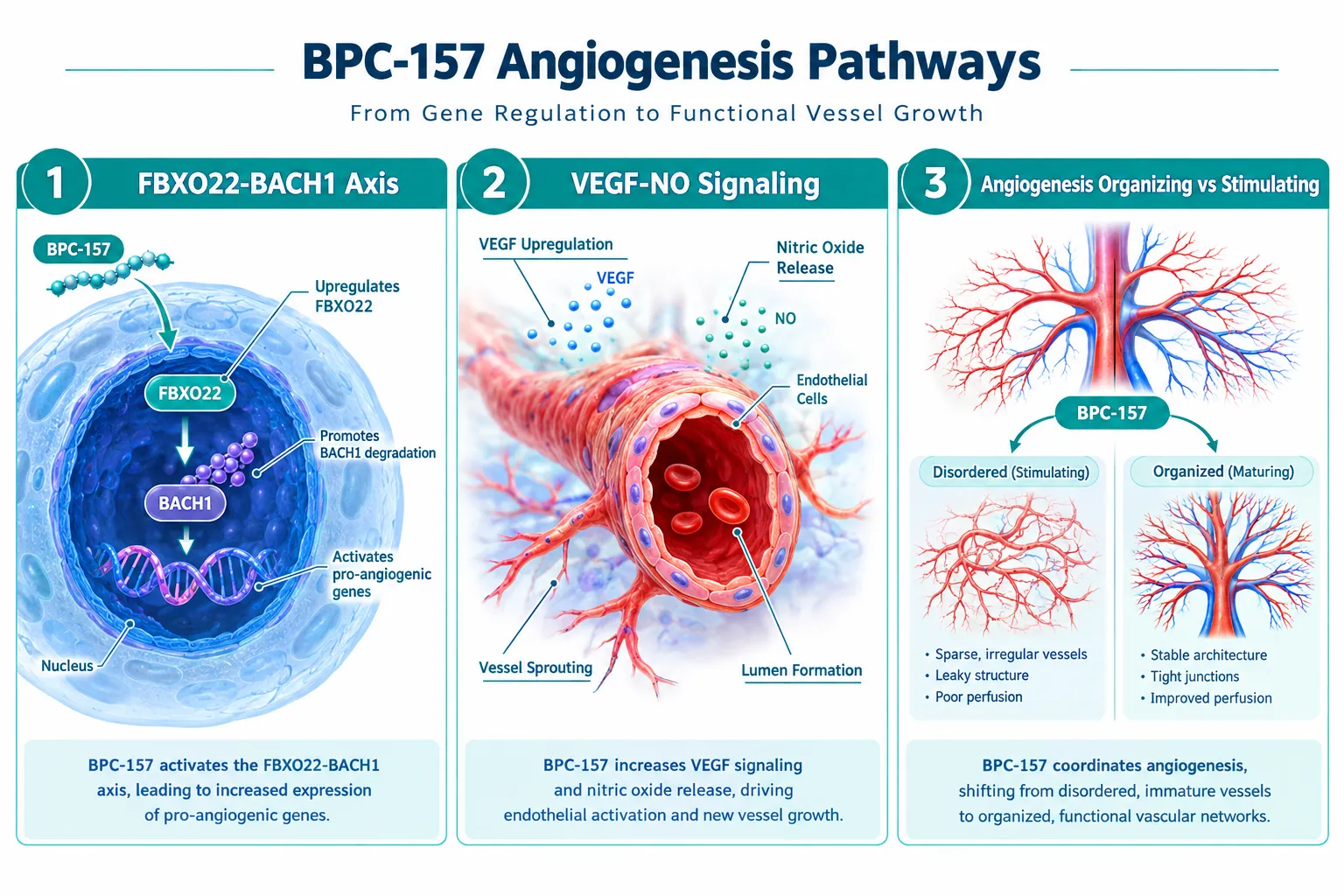
The oldest debate in BPC-157 peptide research centers on what the compound actually does to blood vessels. Early models consistently showed accelerated capillary formation in wound beds, which led to the label "pro-angiogenic." More precise 2026 mechanistic work complicates that framing significantly.
The FBXO22-BACH1 signaling axis has emerged as a key upstream node. FBXO22, an E3 ubiquitin ligase, targets BACH1 for proteasomal degradation. BACH1 normally represses a suite of pro-vascular genes. When BPC-157 appears to facilitate FBXO22 activity, BACH1 levels drop, and genes governing vessel sprouting and stabilization become more accessible. Critically, this is a regulatory event, not a raw amplification of VEGF output.
The VEGF-nitric oxide (NO) axis remains central to the vascular story. BPC-157 consistently upregulates endothelial nitric oxide synthase (eNOS) in preclinical models, driving local NO production that relaxes vessel walls and supports lumen formation. However, the effect appears dose-sensitive and context-dependent, disordered vasculature in ischemic tissue tends to become more organized, not simply more abundant.
Key distinction: "Angiogenesis-organizing" describes a process that improves vessel architecture and perfusion efficiency. "Angiogenesis-stimulating" implies net new vessel mass regardless of context. The evidence in 2026 leans toward the former.
Recommended assay approach for vascular readouts:
| Readout | Preferred Assay | Critical Control |
|---|---|---|
| Capillary density | CD31 immunostaining | Vehicle-matched sham |
| Vessel maturation | Alpha-SMA co-staining | Time-matched baseline |
| NO production | DAF-FM diacetate fluorescence | eNOS inhibitor (L-NAME) |
| BACH1 degradation | Western blot / co-IP | FBXO22 knockdown line |
Researchers exploring related vascular peptide mechanisms may find useful context in IPA Sermorelin Stack Research, which addresses growth-factor-driven vascular signaling in adjacent model systems.
Barrier Repair: Gut, Cornea, and Mucosal Models
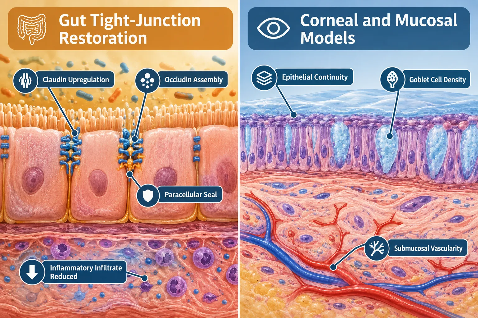
Intestinal barrier restoration is the most replicated finding in BPC-157 peptide research: separating angiogenesis, barrier repair, and inflammatory signaling requires treating this pathway as mechanistically independent from the vascular work, even though both involve tissue remodeling.
In gut models, BPC-157 consistently accelerates tight-junction reassembly following chemically induced permeability damage. The proteins most clearly affected are claudin family members and occludin, which form the paracellular seal between enterocytes. Upregulation of these proteins reduces translocation of luminal antigens into submucosal tissue, which in turn lowers the inflammatory stimulus that would otherwise perpetuate barrier disruption.
This creates an important experimental pitfall: if a study measures inflammation reduction after BPC-157 administration in a leaky-gut model, the result may reflect barrier repair rather than direct immune modulation. Separating these effects requires:
- Measuring tight-junction protein expression at early time points (6-12 hours) before cytokine changes peak
- Using a barrier-intact inflammatory model as a parallel arm
- Confirming permeability with FITC-dextran flux assays independent of histology
Beyond the gut, barrier-repair activity has been demonstrated in corneal epithelial models and oral mucosal tissue. In corneal studies, BPC-157 accelerates re-epithelialization after abrasion, with effects on goblet cell density and submucosal vascularity that parallel gut findings. This cross-tissue consistency suggests a conserved epithelial repair mechanism rather than organ-specific pharmacology.
For researchers sourcing peptides with documented purity for barrier-model work, the discussion at Where to Buy Research-Grade Glow Blend Peptide covers purity standards and skin-model compatibility considerations relevant to epithelial assay design.
Thymosin Alpha 4 Peptide research offers a useful comparative framework, as thymosin beta-4 also promotes actin cytoskeleton remodeling in epithelial repair, providing an orthogonal positive control for barrier-reassembly studies.
Inflammatory Signaling: NF-kB, Cytokines, and Pathway Cross-Talk
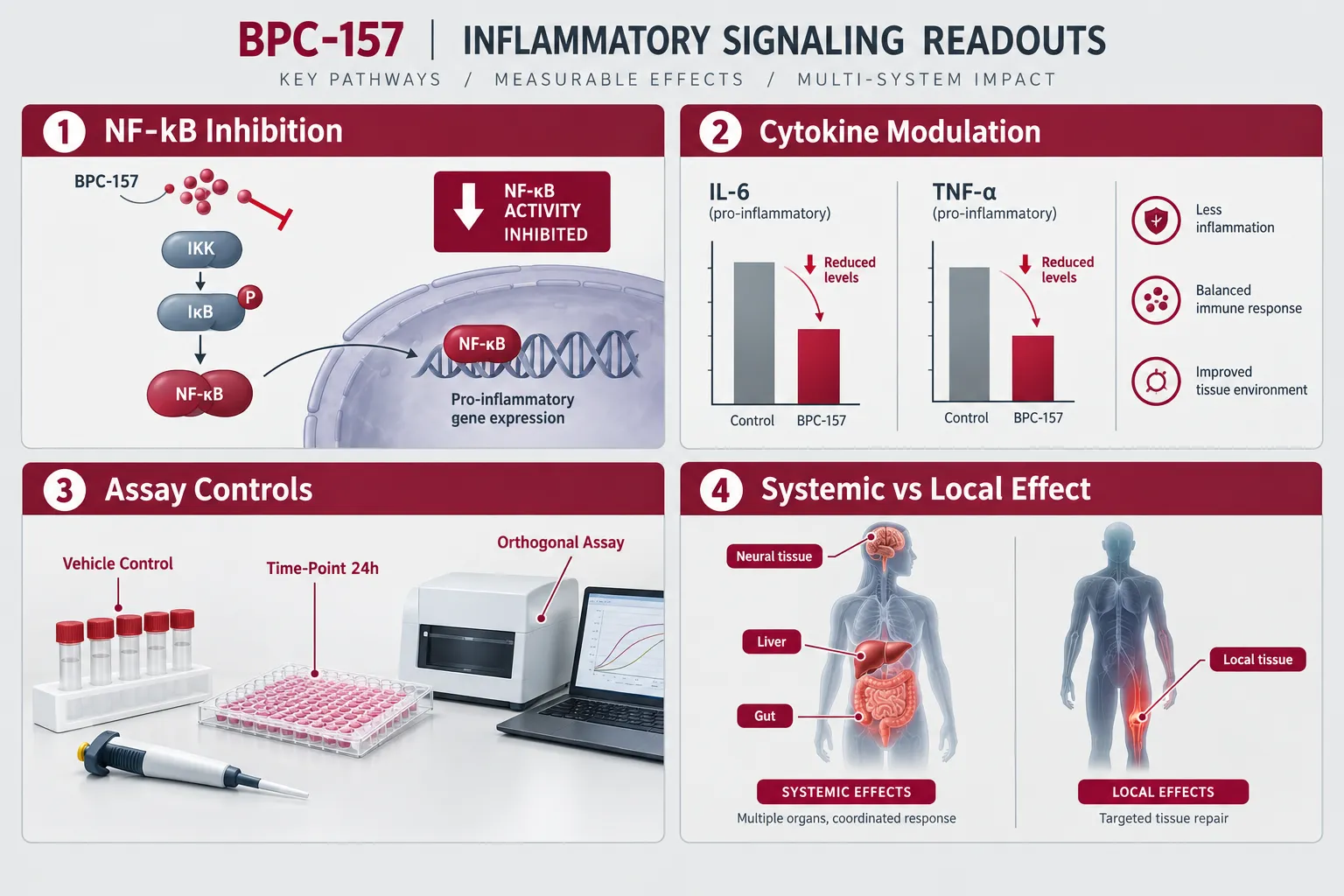
The inflammatory signaling arm of BPC-157 peptide research: separating angiogenesis, barrier repair, and inflammatory signaling demands the most careful experimental architecture, because NF-kB sits at the intersection of all three pathways.
BPC-157 consistently suppresses NF-kB nuclear translocation in preclinical inflammatory models. Downstream, this reduces transcription of TNF-alpha, IL-6, and IL-1-beta, the primary cytokines driving both acute tissue damage and chronic low-grade inflammation. In orthopedic and sports-medicine models, this anti-inflammatory profile has generated significant interest for tendon and ligament repair applications, though clinical data in humans remains minimal as of 2026.
The cross-talk problem is real. Consider this cascade:
- BPC-157 repairs the gut barrier
- Reduced antigen translocation lowers systemic LPS burden
- Lower LPS burden reduces TLR4-NF-kB activation
- Measured cytokine reduction appears to be a direct anti-inflammatory effect
Without a barrier-intact arm in the study design, the cytokine reduction is easily misattributed. Orthogonal assay strategies that isolate each pathway are not optional, they are methodologically required for publishable mechanistic claims.
Recommended time-point staging for inflammatory readouts:
- 0-6 hours: Measure NF-kB translocation and immediate cytokine mRNA
- 12-24 hours: Assess protein-level cytokine secretion (ELISA)
- 48-72 hours: Evaluate tissue histology and immune cell infiltration
- Day 7+: Confirm resolution versus suppression using resolution-phase markers (e.g., lipoxin A4)
Cross-system applications, including neurodegeneration models where NF-kB-driven neuroinflammation is a primary target, and liver fibrosis models where stellate cell activation is NF-kB-dependent, are expanding the research footprint. SS-31 Mitochondrial Research Themes provides relevant context for researchers examining mitochondrial-inflammatory crosstalk in these overlapping disease models.
Safety and regulatory framing remains an open issue. BPC-157 is not approved for human use in any jurisdiction as of 2026, and regulatory agencies have flagged uncertainty around systemic vascular effects at higher doses. The "regeneration vs. risk" framing that dominates industry discussion should not substitute for controlled dose-response data in validated models.
Conclusion
Advancing BPC-157 peptide research means resisting the temptation to report a single unified "healing effect." The vascular, epithelial, and immune readouts are real, but they are distinct, they interact, and they require purpose-built experimental designs to separate cleanly.
Actionable next steps for research teams in 2026:
- Design studies with three parallel arms: vascular-only, barrier-intact inflammatory, and full-model, to isolate pathway contributions
- Use FBXO22-BACH1 Western blots and eNOS fluorescence assays as orthogonal vascular controls alongside CD31 staining
- Stage time points explicitly: tight-junction protein expression before cytokine peaks, and resolution markers after the acute phase
- Include barrier-flux assays (FITC-dextran) in any gut-inflammation model to prevent misattribution of cytokine changes
- Cross-reference findings against structurally related repair peptides, including Thymosin Alpha 4 Peptide, to build mechanistic confidence through orthogonal validation
The peptide's small size belies its signaling complexity. Rigorous pathway separation is not a methodological luxury, it is the foundation on which any translational claim must eventually stand.


Leave a Reply
Want to join the discussion?Feel free to contribute!