Call or Text 727-513-9780
  • Shopping Cart Shopping Cart
    0Shopping Cart
Pure Tested Peptides | America's most trusted Peptides for sale online
  • Peptides for sale
    • Oral Peptides for sale
      • Peptide Capsules for sale
      • BPC 157 Capsules 1000mcg
      • SLU-PP-332 Capsules | 1000 mcg
      • 5-Amino-1MQ 50mg Capsules
      • Tesofensine 500mcg
    • All Peptides for sale
    • Peptide Sprays
      • BPC 157 Nasal Spray Kit
      • BPC-157 TB500 Nasal Spray Kit
      • Semax Nasal Spray 10mg
      • Selank – Nasal Spray Kit – 10mg
      • Epithalon 50MG Nasal Spray Kit
      • Ipamorelin 10mg Nasal Spray
      • Klow Nasal Spray (BPC-157 + TB-500 + GHK-Cu + KPV) | 80mg
      • Hulk Nasal Spray Tesa / Ipa Blend 6/3 MG
      • Klow Nasal Spray
      • NAD + 500 mg Nasal Spray
      • PT-141 Nasal Spray Kit
    • GHRH Peptides
      • Ipa Peptides
      • CJC-1295 Peptides
        • CJC-1295 with DAC 5 mg
        • CJC-1295 without DAC 5 mg
        • CJC-1295 Ipa 10mg
      • Tesa Peptides
        • Tesa Peptide
        • Tesa 20 mg
    • GHK-Cu Peptides
      • All GHK-Cu Peptides
      • GHK-Cu 100mg
      • KLOW Peptide Blend – Buy KLOW blend online
    • BPC Peptides
      • All BPC Peptides
      • BPC-157
      • BPC-157 TB-500
      • BPC 157 capsules 1000mcg
    • SLU-PP-332 Peptides
      • All SLU-PP-332 Peptides
      • SLU-PP-332 5mg
    • GLP3 Peptides
    • PT-141 Peptides
      • PT-141 Peptides for sale
      • PT-141 10mg
      • PT-141 Nasal Spray
    • CAG Peptides
      • Lipo-C Peptide Blend
      • CAG 5mg
      • CAG 10mg
    • MOTS-C Peptides
      • MOTS-C Peptides for sale
      • MOTS-c peptide
      • MOTS-c 10mg *6 pack*
    • 5 Amino 1MQ Peptides
      • 5 Amino 1MQ Peptides for sale
      • 5-Amino-1MQ 50mg Capsules
      • 5-Amino-1MQ 5mg
    • Epithalon Peptides
      • Epithalon Peptides for sale
      • Epithalon 10mg
      • Epithalon 50mg
  • Shop
    • GLPs
      • 5-Amino-1MQ 50mg Capsules
      • 5-Amino-1MQ 5mg
      • L-Carnitine 500mg/ml
      • Tesofensine 500mcg
      • SLU-PP-332 5mg
      • MOTS-c 10mg *6 pack*
    • Epithalon & BPC Peptides
      • Epithalon 10mg
      • Epithalon 50mg
      • BPC-157
      • BPC 157 capsules 1000mcg
      • BPC-157 TB-500
      • BPC-157 TB500 Nasal Spray Kit
      • BPC 157 Nasal Spray Kit
    • BPC TB-500 & NAD+ Peptides
      • NAD+ 500 mg
      • KLOW Peptide Blend – Buy KLOW blend online
      • GLOW Peptide Blend
      • TB 500 5mg
      • BPC 157 capsules 1000mcg – Supplement
      • BPC 157 Nasal Spray Kit
      • BPC-157
      • BPC-157 TB500 Nasal Spray Kit
      • BPC-157 TB-500
      • BPC 157 capsules 1000mcg
    • LL-37 Peptide
      • LL-37 10 mg
    • MOTS-C & Selank
      • MOTS-c peptide
      • Selank 10mg
    • GHK Peptides
      • GHK-Cu 100mg
      • GLOW Peptide Blend
      • KLOW Peptide Blend – Buy KLOW blend online
  • COAs
  • Wholesale
    • Wholesale Peptides for sale
  • PTP FAQ
  • Affiliates
    • Selank Peptide: Advanced Pharmacological Mechanisms Underlying Its Anxiolytic and Nootropic Effects in Research
      • 5-Amino-1MQ Peptide: Detailed Mechanisms of NNMT Inhibition and Its Impact on Cellular Metabolism Research
        • MOTS-C Peptide: Unraveling Its Role in Mitochondrial Dynamics and Energy Metabolism Research
          • Epithalon Peptide and Telomerase Regulation: Investigating Its Impact on Cellular Senescence and Lifespan Research Models
            • GLP2-T vs GLP2 Tirz Peptide: Understanding the Naming, Mechanistic Nuances, and Research Implications for Gut Health
      • Peptides and Polypeptides in Modern Pharmacology: What Research on Metoprolol, Prednisone, and Amlodipine Reveals
        • Polypeptide Peptides vs NSAIDs: What Naproxen and Diclofenac Teach Tissue-Repair Researchers About BPC‑157 and TB‑500
          • Peptides and Polypeptides in Endocrine Pharmacology: How GLP-1, GLP-2, and GLP-3 Retatrutide Differ From Classic Drugs Like Prednisone and Amlodipine
          • 5‑Amino‑1MQ Peptide and NNMT Inhibition: How It Compares With Statins Like Atorvastatin in Adipose and Lipid Metabolism Research
      • Enclomiphene, Estrogen Receptor Signaling, and Luteinizing Phase Biology: What Hormone Researchers Should Measure
        • Mesenchymal Stem Cells, BPC‑157, and GHK‑Cu: How Tissue Repair Peptides Compare With Classic NSAIDs Like Naproxen in Injury Models
          • Peptide Calculator Use Cases Beyond Growth Hormone: Working Through GLP‑3 Retatrutide, MOTS‑c, and BPC‑157 Research Dosing
        • Polypeptide Peptides in Endocrine and Metabolic Pharmacology: Lessons From Amlodipine, Prednisone, and Metoprolol
        • Peptides Calculator for Advanced Blends: Worked Examples for Tesamorelin, CJC‑1295, and Ipamorelin Stacks
          • Peptides and Polypeptides in Human Physiology: How Molecular Size Shapes Research Applications
            • Selank Peptide: Advanced Pharmacological Mechanisms Underlying Its Anxiolytic and Nootropic Effects in Research
          • 5-Amino-1MQ Peptide: Detailed Mechanisms of NNMT Inhibition and Its Impact on Cellular Metabolism Research
            • MOTS-C Peptide: Unraveling Its Role in Mitochondrial Dynamics and Energy Metabolism Research
              • Epithalon Peptide and Telomerase Regulation: Investigating Its Impact on Cellular Senescence and Lifespan Research Models
                • GLP2-T vs GLP2 Tirz Peptide: Understanding the Naming, Mechanistic Nuances, and Research Implications for Gut Health
                  • Peptides and Polypeptides in Endocrine Pharmacology: How GLP-1, GLP-2, and GLP-3 Retatrutide Differ From Classic Drugs Like Prednisone and Amlodipine
                    • Polypeptide Peptides vs NSAIDs: What Naproxen and Diclofenac Teach Tissue-Repair Researchers About BPC‑157 and TB‑500
                      • Peptides and Polypeptides in Modern Pharmacology: What Research on Metoprolol, Prednisone, and Amlodipine Reveals
                        • 5‑Amino‑1MQ Peptide and NNMT Inhibition: How It Compares With Statins Like Atorvastatin in Adipose and Lipid Metabolism Research
                        • Enclomiphene, Estrogen Receptor Signaling, and Luteinizing Phase Biology: What Hormone Researchers Should Measure
                        • Mesenchymal Stem Cells, BPC‑157, and GHK‑Cu: How Tissue Repair Peptides Compare With Classic NSAIDs Like Naproxen in Injury Models
                        • Peptide Calculator Use Cases Beyond Growth Hormone: Working Through GLP‑3 Retatrutide, MOTS‑c, and BPC‑157 Research Dosing
                        • Polypeptide Peptides in Endocrine and Metabolic Pharmacology: Lessons From Amlodipine, Prednisone, and Metoprolol
                        • Peptides Calculator for Advanced Blends: Worked Examples for Tesamorelin, CJC‑1295, and Ipamorelin Stacks
                        • Peptides and Polypeptides in Human Physiology: How Molecular Size Shapes Research Applications
                        • Selank Peptide: Advanced Pharmacological Mechanisms Underlying Its Anxiolytic and Nootropic Effects in Research
                        • MOTS-C Peptide: Unraveling Its Role in Mitochondrial Dynamics and Energy Metabolism Research
                        • Peptides and Polypeptides in Modern Pharmacology: What Research on Metoprolol, Prednisone, and Amlodipine Reveals
                        • Peptides and Polypeptides in Endocrine Pharmacology: How GLP-1, GLP-2, and GLP-3 Retatrutide Differ From Classic Drugs Like Prednisone and Amlodipine
                        • 5-Amino-1MQ Peptide: Detailed Mechanisms of NNMT Inhibition and Its Impact on Cellular Metabolism Research
                        • Mesenchymal Stem Cells, BPC‑157, and GHK‑Cu: How Tissue Repair Peptides Compare With Classic NSAIDs Like Naproxen in Injury Models
                        • Peptide Calculator Use Cases Beyond Growth Hormone: Working Through GLP‑3 Retatrutide, MOTS‑c, and BPC‑157 Research Dosing
                        • GLP2-T vs GLP2 Tirz Peptide: Understanding the Naming, Mechanistic Nuances, and Research Implications for Gut Health
                        • Polypeptide Peptides in Endocrine and Metabolic Pharmacology: Lessons From Amlodipine, Prednisone, and Metoprolol
                        • Polypeptide Peptides vs NSAIDs: What Naproxen and Diclofenac Teach Tissue-Repair Researchers About BPC‑157 and TB‑500
                        • Peptides and Polypeptides in Human Physiology: How Molecular Size Shapes Research Applications
                        • Peptides Calculator for Advanced Blends: Worked Examples for Tesamorelin, CJC‑1295, and Ipamorelin Stacks
                        • 5‑Amino‑1MQ Peptide and NNMT Inhibition: How It Compares With Statins Like Atorvastatin in Adipose and Lipid Metabolism Research
                        • Enclomiphene, Estrogen Receptor Signaling, and Luteinizing Phase Biology: What Hormone Researchers Should Measure
                        • Epithalon Peptide and Telomerase Regulation: Investigating Its Impact on Cellular Senescence and Lifespan Research Models
                        • Best research protocol Klow blend
                        • best time to take BPC-157
                        • best time to take DSIP (Delta Sleep Inducing Peptide)
                        • best time to take CJC-1295
                        • best time to take AOD-9604
                        • best time to take Follistatin 344
                        • best time to take Ipamorelin
                        • best time to take MK-677 (Ibutamoren)
                        • best time to take Ligandrol (LGD-4033) — research compound
                        • best time to take Ostarine (MK-2866) — research compound
                        • best time to take GHK-CU
                        • best time to take TB-500
                        • best time to take MOTS-c
                        • best time to take Semax
                        • best time to take RAD-140 (Testolone) — research compound
                        • best time to take Thymosin Alpha-1
                        • best time to take PEG-MGF
                        • Biolife Plasma, Octapharma Plasma, and Research Peptides: How Plasma Donation Labs Differ From Peptide Suppliers
                        • best time to take YK-11 — research compound
                        • best time to take PT-141 (Bremelanotide)
                        • Best research protocol Klow blend
                        • 5-Amino-1MQ and MOTS-C Synergy: Metabolic Signaling, Mitochondria, and Research Design
                        • BPC-157 and TB-500: Investigating Their Combined Effects on Angiogenesis and Cellular Migration in Tissue Repair Models
                        • BPC-157 Peptide: Gut Barrier Function, Inflammation, and Tissue-Recovery Research
                        • 5‑Amino‑1MQ and MOTS‑c Synergy in Metabolic Research: Designing NNMT and Mitochondrial Biogenesis Stacks
                        • CJC-1295 with DAC vs. Without DAC: Half-Life, Release Kinetics, and Research Implications
                        • CJC‑1295 with DAC vs. Without DAC: Expanding on Half‑Life Differences Using Tesamorelin and Ipamorelin Blend Case Studies
                        • Collagen Biology and Copper‑Binding Peptides: How GHK‑Cu, Glow Blend, and Klow Blend Interact with Skin and Connective Tissue
                        • Collagen Biology and Regenerative Peptides: How GHK‑Cu, Glow Blend, and Klow Blend Affect Extracellular Matrix Research
                    • DNA, Telomeres, and Longevity Peptides: Positioning Epithalon and MOTS‑c in Genetic Aging Research
                      • Enclomiphene Citrate: serm Mechanism, Testosterone Research, and Stack Compatibility
                        • Enclomiphene vs Enclomiphene Citrate: Formulation, Bioavailability, and Research Distinctions
                        • Epithalon Peptide Research: Telomerase Activation, Aging, and Pineal Gland Function
                        • Estrogen Receptor Signaling and Enclomiphene: How Selective Modulators Compare with Classic Polypeptide Hormones
                        • GHK-Cu Peptide: Advanced Mechanisms in Extracellular Matrix Remodeling and Wound Healing Research
                        • GHK-Cu Peptide: Collagen Synthesis, Wound Repair, and Skin-Barrier Research Models
                        • GLP-1 vs GLP-3 vs GLP-2: Peptide Classification and Research Applications
                        • GLP-2 Peptide Research Guide: Gut Barrier Function, Nutrient Absorption, and Intestinal Recovery Models
                        • GLP-3 Retatrutide vs. GLP-1 Drugs: What Triple-Agonist Biology Changes in Research Models
                        • Ipamorelin and Tesamorelin Combination: Synergistic GH Secretagogue Research and Dosing Protocols
                        • GLP2 Tirz Peptide: What It Is, Why the Name Exists, and How Researchers Should Interpret It
                        • Klow Blend Peptide Nasal Spray: What the Formulation Is Trying to Do in Cognitive Research
                        • Mitochondria, NNMT Inhibition, and Peptide Modulators: Where MOTS‑c and 5‑Amino‑1MQ Fit in Cellular Energy Research
                        • MOTS-c Peptide: Mitochondrial Function, Energy Metabolism, and What Researchers Measure
                        • MOTS-c vs. 5-Amino-1MQ: Which Metabolic Research Questions Each Compound Actually Answers
                        • Nasal Spray Peptides: Bioavailability, Administration, and Semax/Selank Research Applications
                        • PT-141 Peptide Research: Mechanism of Action and Melanocortin Receptor Signaling
                        • Retatrutide for Research: Mechanism, Structure, and GLP-1/GLP-3 Dual Action
                        • Retatrutide for Obesity and Type 2 Diabetes: What the Latest Trial Data Suggest
                        • Tesofensine Peptide Research: Mechanism, Appetite Suppression, and Neuropeptide Y Pathways
  • Contact
    • Contact Customer Service
    • Text Customer Support
  • About US
  • Shop all peptides
  • Affiliate Program
    • Affiliate Signup
  • Login / Register Login / Register Page Link Login / Register Page Link
  • Click to open the search input field Click to open the search input field Search
  • Menu Menu

Tag Archive for: peptide aging research

Epithalon in Cellular-Aging Models: Separating Telomerase Activity, Telomere Length, DNA Damage, and Senescence

Epithalon in Cellular-Aging Models: Separating Telomerase Activity, Telomere Length, DNA Damage, and Senescence

October 14, 2026/0 Comments/in Uncategorized/by

Fewer than 1% of peptides studied in aging research generate data across four distinct cellular endpoints simultaneously, yet Epithalon, a synthetic tetrapeptide derived from the pineal gland protein epithalamin, has accumulated a body of evidence touching telomerase activation, telomere elongation, DNA damage reduction, and senescence delay. The challenge for researchers in 2026 is not whether Epithalon affects these pathways, but how to cleanly separate them. Conflating a short-term telomerase signal with genuine telomere lengthening, or mistaking reduced oxidative damage for a true senescence delay, leads to inflated claims and flawed experimental design. This article presents a structured, longitudinal assay framework for studying Epithalon in cellular-aging models: separating telomerase activity, telomere length, DNA damage, and senescence with methodological rigor.

Key Takeaways

  • Telomerase activation and telomere lengthening are distinct, time-separated events that require different assays and measurement windows.
  • Epithalon produces approximately 30-35% telomere extension in cell models, but this effect takes weeks to manifest after initial telomerase signaling.
  • DNA damage reduction appears to operate through a mechanism partially independent of telomere dynamics.
  • Senescence delay, measured by SA-beta-galactosidase activity and p21 expression, represents a downstream phenotypic outcome rather than a primary mechanism.
  • As of 2026, Epithalon holds no regulatory approval; all findings remain in the preclinical and early exploratory human-data stage.

The Four Endpoints Are Not the Same Thing

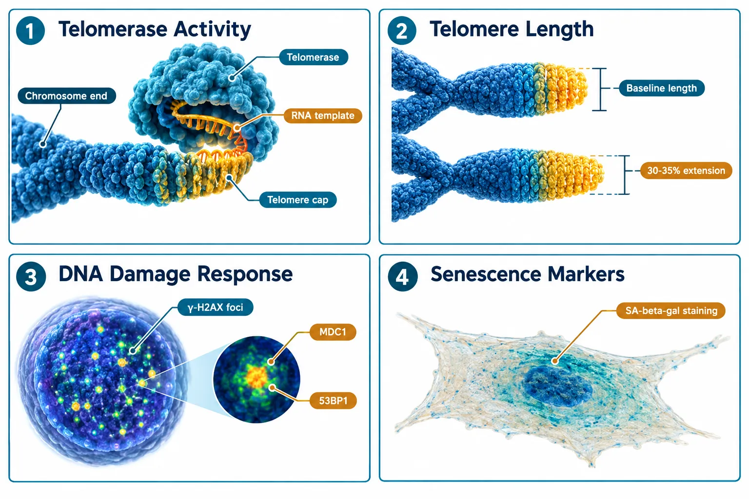
A common error in Epithalon literature is treating telomerase activity as a proxy for telomere length. These are related but temporally and mechanistically distinct. Telomerase is an enzyme; its activity can spike within 24 to 72 hours of peptide exposure. Telomere length, by contrast, reflects cumulative replication history and changes over days to weeks, not hours.
The Four Endpoints Are Not the Same Thing

Similarly, DNA damage response, quantified through gamma-H2AX foci counts or comet assay tail moment, reflects genomic stress that may or may not be telomere-driven. Senescence, the terminal arrest of proliferating cells, is a downstream phenotype shaped by all three upstream signals combined. Researchers studying SS-31 mechanism and research alongside Epithalon have noted similar endpoint-separation challenges in mitochondrial peptide work, underscoring that this is a broader methodological issue in peptide aging research.

The four endpoints and their primary assays:

Endpoint Primary Assay Measurement Window
Telomerase activity TRAP assay, RT-qPCR for hTERT 24-72 hours
Telomere length Q-FISH, Southern blot TRF 2-6 weeks
DNA damage Comet assay, gamma-H2AX foci 48 hours to 2 weeks
Senescence SA-beta-gal, p21/p16 expression 3-8 weeks

Building a Longitudinal Assay Plan for Epithalon in Cellular-Aging Models

The most defensible experimental design treats each endpoint as a separate dependent variable with its own measurement schedule. Collapsing them into a single time point produces noise, not signal.
Building a Longitudinal Assay Plan for Epithalon in Cellular-Aging Models

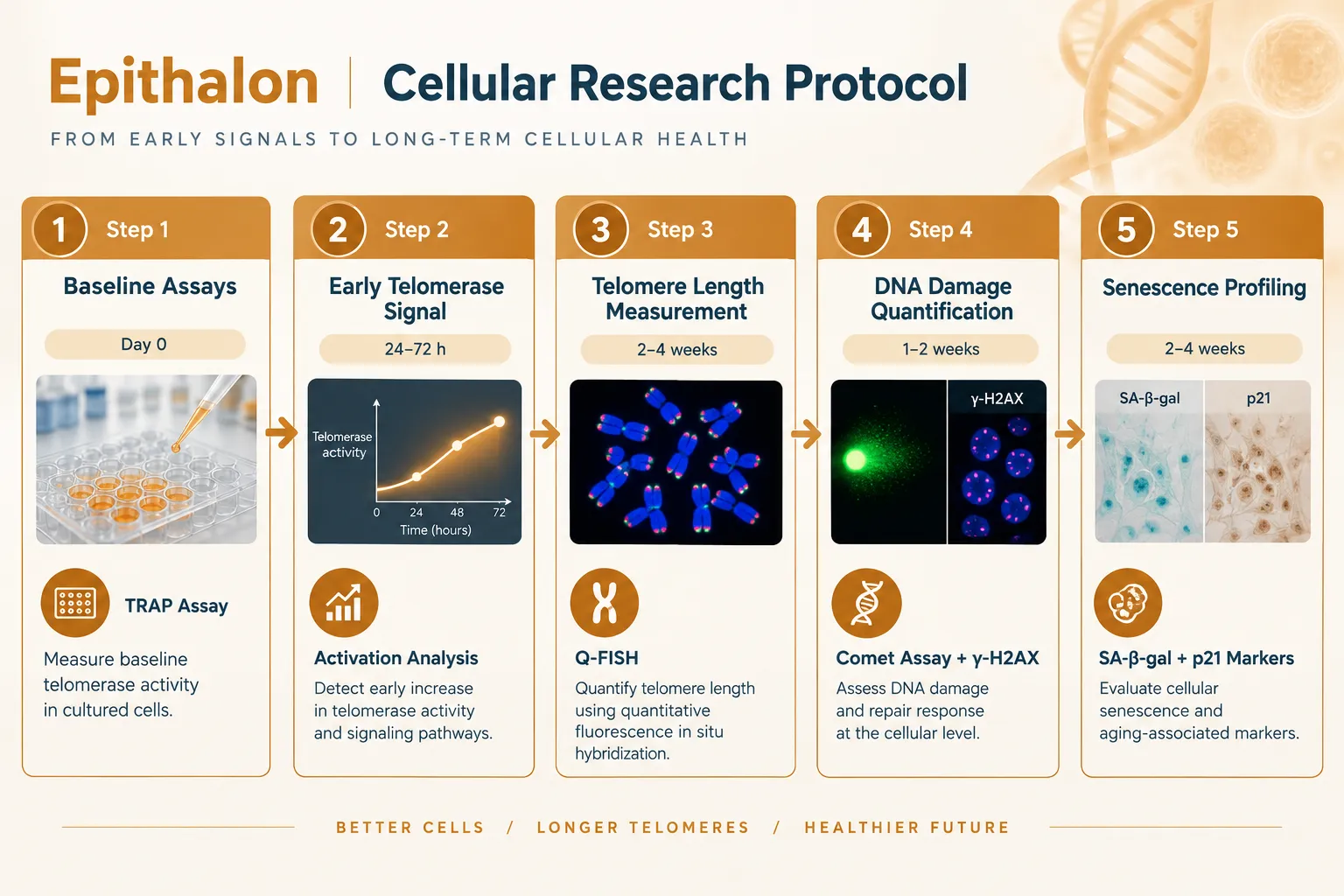
A five-stage longitudinal framework:

Stage 1, Baseline (Day 0). Establish pre-treatment values for all four endpoints in the same cell population. Human diploid fibroblasts (IMR-90) and WI-38 lines are well-validated for this purpose. Record passage number, population doubling level, and baseline SA-beta-galactosidase positivity.

Stage 2, Early telomerase signal (24-72 hours). Apply TRAP assay and hTERT mRNA quantification. Magnitude of activation in published cell models has ranged from modest to substantial depending on cell type and peptide concentration. This window captures the enzymatic signal but says nothing about whether telomeres have actually lengthened.

Stage 3, Telomere length measurement (Weeks 2-4). Use quantitative fluorescence in situ hybridization (Q-FISH) or terminal restriction fragment (TRF) analysis. Independent replication work from multiple groups has reported approximately 30-35% telomere extension relative to untreated controls at this stage. Critically, this measurement must be taken at a population doubling interval, not a fixed calendar interval, to control for proliferation rate differences.

Stage 4, DNA damage quantification (Days 3-14). Run parallel comet assays and gamma-H2AX immunofluorescence. Evidence from multiple cell-model studies suggests that Epithalon reduces markers of oxidative DNA damage even in cells where telomere lengthening is modest, pointing to a partially independent mechanism, possibly involving antioxidant enzyme upregulation rather than telomere-mediated genomic stability.

Stage 5, Senescence profiling (Weeks 4-8). Quantify SA-beta-galactosidase-positive cells, p21 and p16 protein expression, and secretory phenotype markers (IL-6, IL-8 as SASP indicators). Extended replicative lifespan, the number of population doublings before crisis, is the gold-standard functional readout. Studies have reported meaningful delays in replicative senescence onset in Epithalon-treated cultures.

Key insight: Running all five stages in parallel, rather than sequentially, allows researchers to build a causal map rather than a simple correlation between peptide exposure and aging phenotype.

For researchers designing controlled experimental models, the quality-control workflow considerations discussed in TB-500 controlled experimental models and QC workflow offer transferable lessons on blinding, passage controls, and contamination prevention.


Evidence Across Model Systems: What the Data Actually Show

Separating Epithalon in cellular-aging models from broader claims requires looking at each model type individually.
Evidence Across Model Systems: What the Data Actually Show

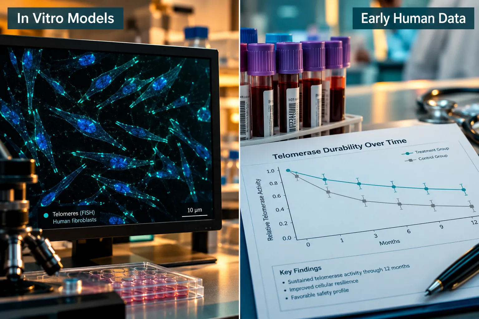
In vitro cell models provide the most mechanistically detailed data. Telomerase activation is consistently observed. Telomere extension at the 30-35% range has been replicated by independent groups including work published from Brunel University and affiliated researchers in 2025-2026. DNA damage markers decrease. Senescence is delayed. The effect size varies by cell type, with fetal lung fibroblasts showing stronger responses than primary skin fibroblasts.

Animal aging models extend findings to whole-organism endpoints. Rodent studies have reported increased mean and maximum lifespan, reduced lipid peroxidation markers, and improved immune function in aged animals. These outcomes are consistent with the cellular data but introduce confounders, diet, housing stress, and genetic background, that make direct mechanistic attribution difficult.

Reproductive models represent a newer frontier. A 2025 study using bovine cumulus-oocyte complexes found that Epithalon treatment improved oocyte competence and reduced oxidative stress markers, suggesting that telomere-protective effects extend to non-somatic cell types with high replicative demand.

Early human data remain limited. Peripheral blood mononuclear cell studies have detected telomerase activity increases following Epithalon administration, with some evidence of durability extending weeks beyond the treatment period. These findings are preliminary and have not been replicated in adequately powered trials.

Researchers interested in how peptide sourcing quality affects experimental reproducibility can find relevant context in resources on where to buy SS-31 and Epithalon online and the importance of purity verification for research-grade compounds. Related mitochondrial aging research on SS-31 mitochondrial dynamics provides useful comparative context for understanding oxidative-stress endpoints.


Safety, Cancer Risk, and the Hype-Evidence Gap

No discussion of Epithalon in cellular-aging models: separating telomerase activity, telomere length, DNA damage, and senescence is complete without addressing the cancer risk question. Telomerase activation is a double-edged signal. While it protects normal somatic cells from replicative senescence, it is also a hallmark of most cancer cells. Preclinical data on Epithalon have not shown tumor-promoting activity in standard carcinogenicity assays, and some studies report anti-tumor effects in aged animals. However, long-term safety data in humans are absent.

The regulatory position as of 2026 is clear: Epithalon has no approved therapeutic indication in any major jurisdiction. It is available as a research compound, and its use in human subjects outside formal clinical trial protocols raises significant ethical and safety concerns.

The expert consensus in late 2026 is cautiously interested but appropriately skeptical. The mechanistic data are real and replicable. The translational gap, from cell culture to clinically meaningful human outcomes, remains wide. Researchers and commentators who collapse this gap prematurely do a disservice to a genuinely interesting area of science.


Conclusion

The value of studying Epithalon in cellular-aging models lies precisely in the discipline of keeping its four major endpoints, telomerase activity, telomere length, DNA damage, and senescence, analytically separate. A longitudinal assay plan that stages measurements across the appropriate biological windows, uses validated assays for each endpoint, and controls for proliferation rate and passage number will produce data that actually advances understanding. The 30-35% telomere extension finding is worth replicating rigorously. The DNA damage reduction signal deserves mechanistic dissection independent of telomere biology. The senescence delay data need larger, better-controlled models before translational claims are warranted.

Actionable next steps for researchers:

  • Design experiments with a minimum five-stage longitudinal structure, not a single endpoint snapshot.
  • Always measure telomerase activity and telomere length in separate assay windows, never use one as a proxy for the other.
  • Include DNA damage endpoints (comet assay, gamma-H2AX) as independent variables, not secondary observations.
  • Source research-grade Epithalon with documented purity certificates and replicate findings across at least two cell lines before drawing mechanistic conclusions.
  • Track the evolving regulatory and safety literature; the field is moving quickly in 2026 and responsible research requires staying current.
https://www.puretestedpeptides.com/wp-content/uploads/2026/10/epithalon-in-cellular-aging-models-separating-telomerase-activity-telomere-lengt.webp 1024 1536 https://www.puretestedpeptides.com/wp-content/uploads/2026/01/buy-peptides-online.jpg 2026-10-14 13:04:142026-10-14 13:04:14Epithalon in Cellular-Aging Models: Separating Telomerase Activity, Telomere Length, DNA Damage, and Senescence
Epithalon Peptide: Telomerase Activation and Cellular Senescence Research Applications

Epithalon Peptide: Telomerase Activation and Cellular Senescence Research Applications

August 30, 2026/0 Comments/in Uncategorized/by

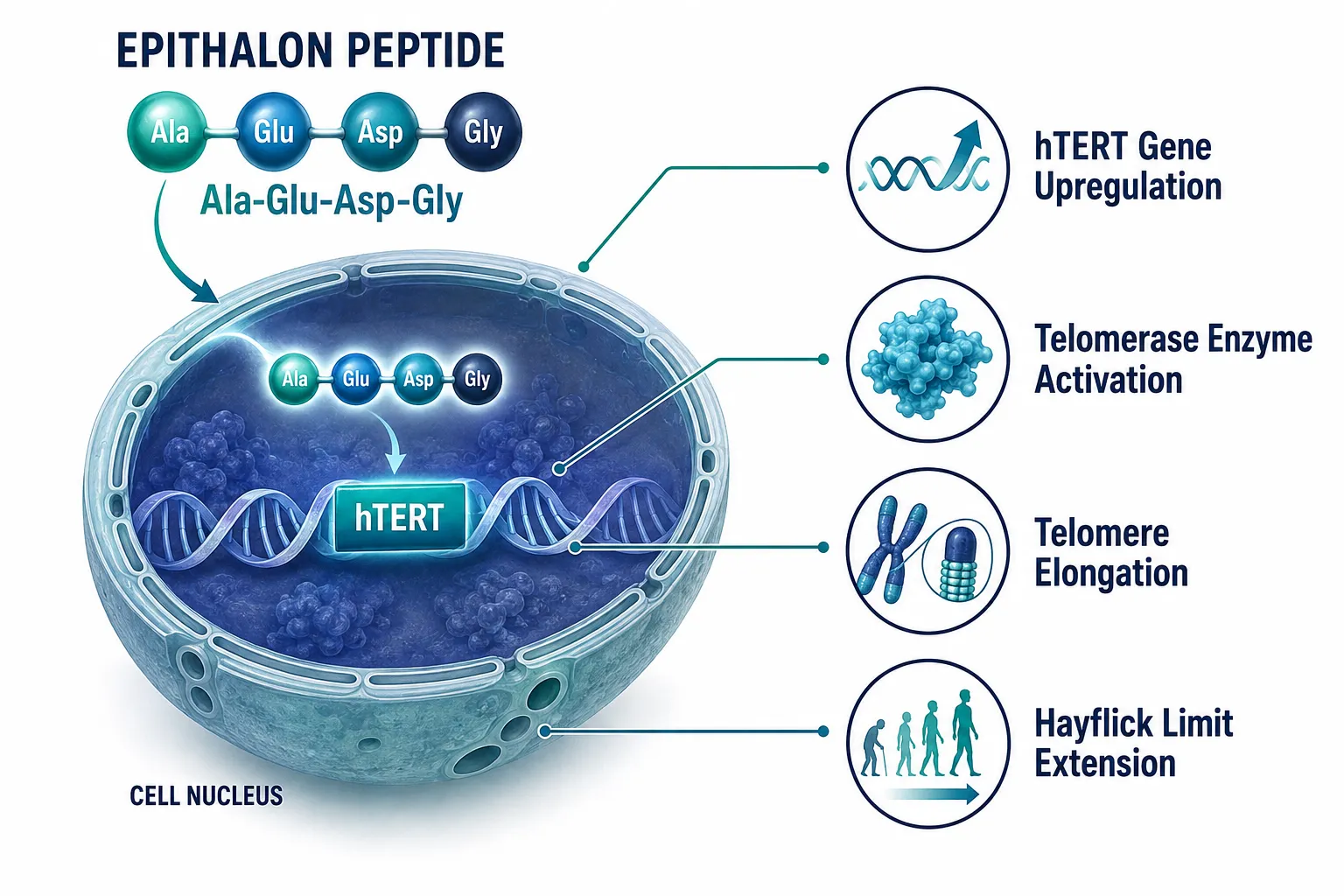
A tetrapeptide made of just four amino acids, Alanine, Glutamic acid, Aspartic acid, and Glycine, has generated decades of scientific debate over whether it holds a key to slowing cellular aging at its most fundamental level. Epithalon peptide: telomerase activation and cellular senescence research applications sit at the center of that debate, drawing attention from gerontologists, reproductive biologists, and translational researchers alike. As of 2026, the compound remains strictly a research tool, yet the mechanistic data emerging from cell-line studies continues to sharpen understanding of how telomere dynamics govern the aging process.

Key Takeaways

  • Epithalon (Ala-Glu-Asp-Gly) activates telomerase by upregulating the catalytic subunit hTERT, producing measurable telomere elongation in human somatic cell cultures.
  • In vitro studies report telomere length increases from roughly 2.4 kb to as much as 8 kb at doses of 0.1-1.0 micrograms per milliliter over three weeks.
  • Rodent models have reported lifespan extensions of approximately 12-24%, though no controlled human clinical trials have replicated these findings.
  • Epithalon carries no FDA, EMA, or MHRA approval; it was removed from the FDA Category 2 bulk drug list in April 2026 and is scheduled for regulatory review in July 2026.
  • Human evidence is currently graded as low-quality (Grade D), meaning researchers must treat all findings as preliminary and hypothesis-generating only.

Mechanism of Action: How Epithalon Activates Telomerase

Mechanism of Action: How Epithalon Activates Telomerase

Understanding telomerase activation is essential before interpreting Epithalon's research profile. Telomerase is a ribonucleoprotein enzyme that adds repetitive nucleotide sequences to the ends of chromosomes, counteracting the progressive shortening that occurs with each cell division. In most adult somatic cells, telomerase activity is suppressed, which is a primary driver of replicative senescence.

Epithalon's primary mechanism involves inducing expression of hTERT, the catalytic subunit of telomerase, in human somatic cell cultures. A 2026 mechanistic review confirmed that this induction increases telomerase enzymatic activity to a degree sufficient to extend cellular lifespan beyond the Hayflick limit in vitro. A 2024 study at the Institute of Bioregulation and Gerontology in St. Petersburg quantified this effect: telomerase activity increased by approximately 30-40% in human fibroblast cultures within 72 hours, with the most pronounced changes occurring during the G1 phase of the cell cycle.

Key mechanistic steps observed in research models:

  • Epithalon binds to regulatory regions influencing hTERT gene transcription
  • Increased hTERT mRNA is detected within hours of exposure
  • Telomerase enzymatic activity rises in a dose-dependent pattern
  • Telomere elongation follows over days to weeks of sustained exposure

These findings position Epithalon as a valuable signaling peptides research tool for dissecting the upstream regulation of telomerase in normal aging cells.

Epithalon Peptide: Telomerase Activation and Cellular Senescence Research Applications in Cell-Line Studies

Epithalon Peptide: Telomerase Activation and Cellular Senescence Research Applications in Cell-Line Studies

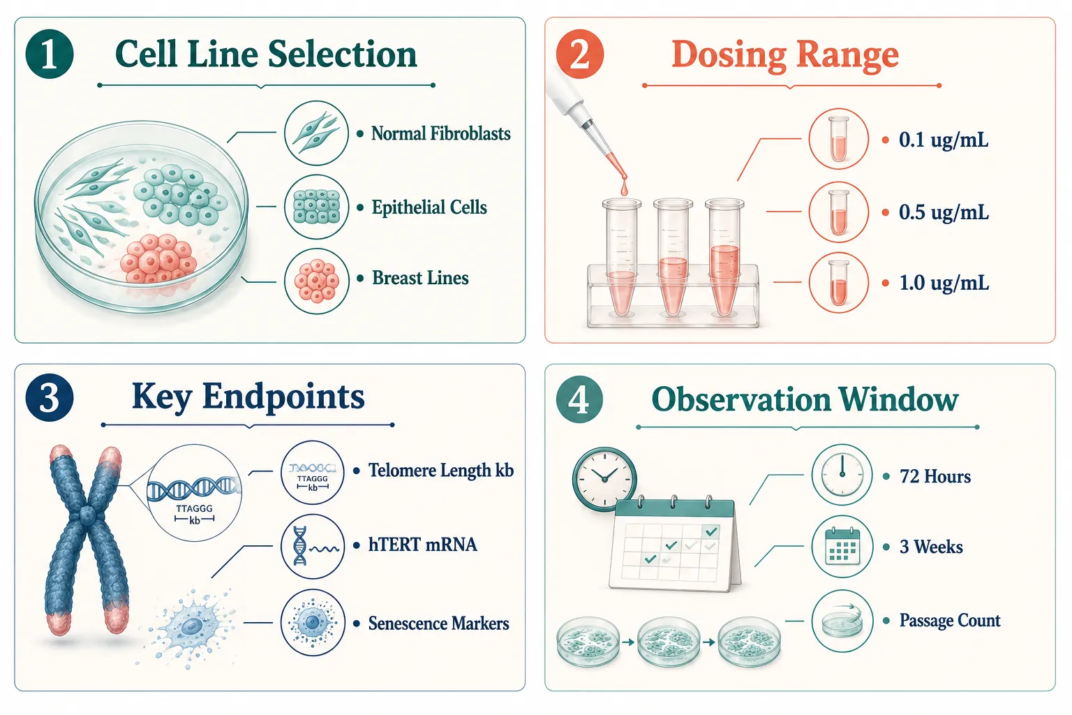
The most rigorous recent work comes from a 2025 Brunel University replication study. Researchers treated four human cell lines, two breast cancer lines and two normal mammary epithelial lines, with Epithalon at concentrations ranging from 0.1 to 1.0 micrograms per milliliter for three weeks. The results showed dose-dependent telomere elongation, with baseline telomere lengths near 2.4 kilobases extending to approximately 8 kilobases in some lines. Critically, the authors framed Epithalon as a tool compound for telomere biology research, not a clinically validated therapy.

A separate 2025 human cell-line study confirmed that Epithalon increased telomere length in normal epithelial and fibroblast cells by upregulating both hTERT mRNA and telomerase activity, corroborating earlier Russian data in a Western laboratory context.

Tracking senescence markers alongside telomere measurements is considered best practice in this research area. Useful endpoints for study design include:

Endpoint Measurement Method Relevance
Telomere length (kb) Q-FISH or Southern blot Direct senescence indicator
hTERT mRNA expression RT-qPCR Mechanistic confirmation
Beta-galactosidase activity Histochemical staining Classic senescence marker
Reactive oxygen species Fluorescent probes Oxidative stress component
Cell passage number Manual counting Replicative lifespan proxy

For researchers designing experiments, a translational research design framework that pairs molecular endpoints with functional senescence assays will yield the most interpretable data.

Epithalon Peptide: Telomerase Activation and Cellular Senescence Research Applications Beyond Standard Cell Lines

Epithalon Peptide: Telomerase Activation and Cellular Senescence Research Applications Beyond Standard Cell Lines

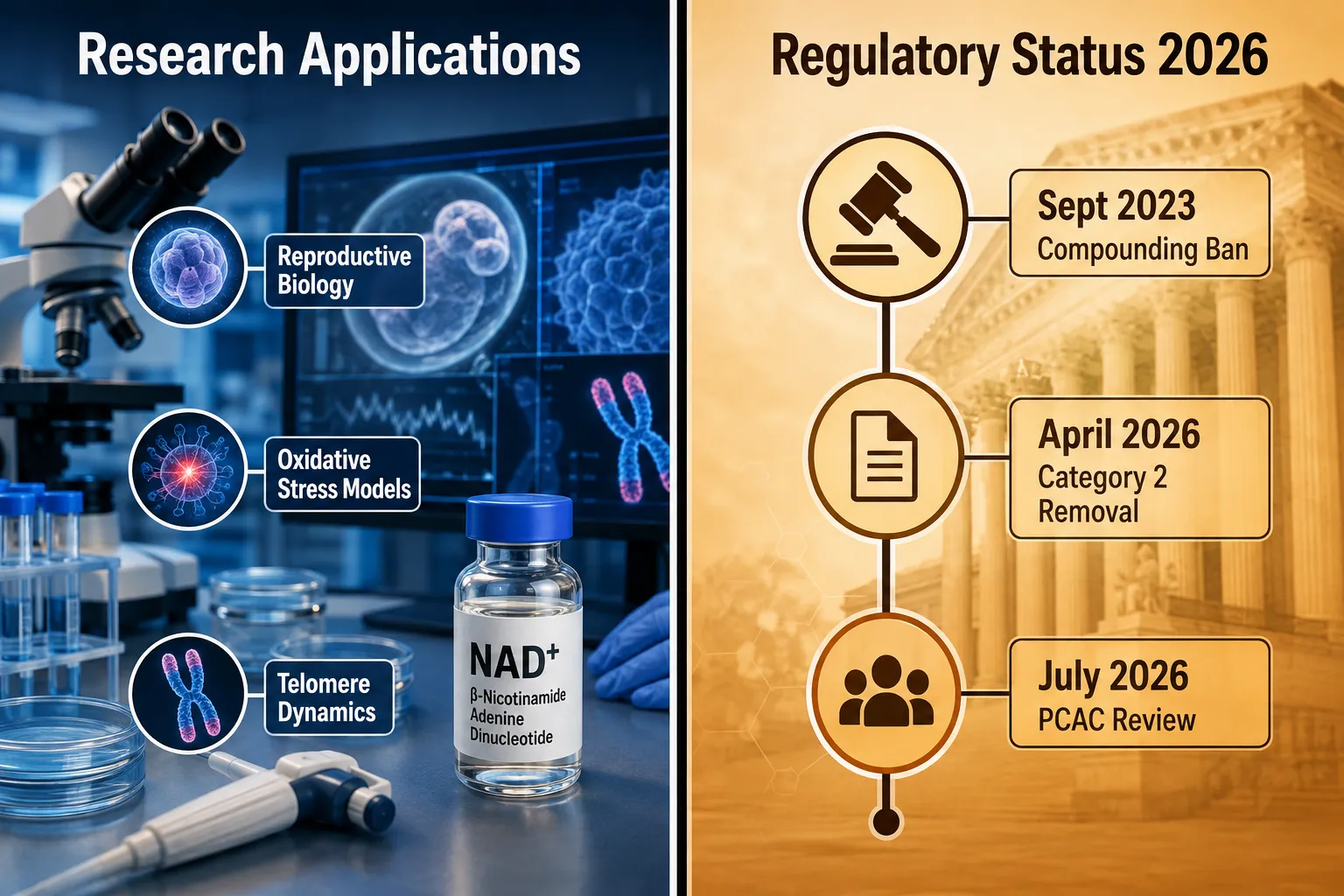
Research interest in Epithalon has expanded beyond standard fibroblast and epithelial models. A 2025 study by Ullah et al. demonstrated that Epithalon stimulates telomerase activity in bovine cumulus cells and cumulus-oocyte complexes, opening a pathway for studying reproductive aging and in vitro oocyte senescence. A complementary 2022 oocyte study found that appropriately dosed Epithalon can reduce oxidative stress-related damage associated with post-ovulatory aging, suggesting relevance to experimental models of oxidative stress-induced cellular senescence.

These findings connect to broader skin biology research and tissue recovery research contexts, where controlling cellular senescence in specialized cell populations is a growing priority.

Regulatory and safety context researchers must understand in 2026:

  • Epithalon has no FDA, EMA, or MHRA approval and no active IND, NDA, or BLA filing
  • It was banned from U.S. compounding pharmacies in September 2023 due to concerns including immunogenicity, aggregation risk, and insufficient clinical data
  • The FDA removed Epithalon from its Category 2 bulk drug substances list effective April 22, 2026, with a Pharmacy Compounding Advisory Committee review scheduled for July 24, 2026
  • Telomerase activation carries a theoretical long-term carcinogenic risk that regulators have flagged as a key concern
  • All human evidence is currently classified as Grade D, based primarily on small Soviet-era and Russian cohort data with no modern Phase 3 trials

"In vitro telomerase activation should not be equated with proven clinical anti-aging effects, the mechanistic data is compelling, but the clinical translation gap remains wide."

Researchers exploring therapeutic peptides in aging models should build study designs that explicitly account for this gap, using Epithalon as a mechanistic probe rather than a presumed intervention.

Conclusion

Epithalon peptide: telomerase activation and cellular senescence research applications represent one of the most mechanistically detailed areas of peptide aging biology available to researchers in 2026. The compound reliably upregulates hTERT, increases telomerase activity by measurable margins, and produces telomere elongation across multiple human cell-line models. Rodent lifespan data adds biological plausibility, and emerging reproductive biology findings expand the experimental toolkit further.

Actionable next steps for researchers:

  1. Design studies with paired molecular endpoints (hTERT mRNA, telomerase activity) and functional senescence assays (beta-galactosidase, passage number) to build interpretable datasets.
  2. Use dose ranges of 0.1-1.0 micrograms per milliliter as a validated starting point, with observation windows of at least 72 hours for acute mechanistic work and three weeks for telomere length outcomes.
  3. Monitor the July 2026 PCAC review outcomes, as regulatory conclusions will shape future research access and compounding pathways.
  4. Frame all findings within the Grade D human evidence classification and avoid extrapolating in vitro telomerase activation to clinical anti-aging conclusions.
  5. Pair Epithalon with established senescence marker panels to contribute data that moves the field toward higher evidence grades.

The science is genuinely interesting. The regulatory and safety landscape demands that researchers approach it with rigorous methodology and transparent reporting.

https://www.puretestedpeptides.com/wp-content/uploads/2026/08/epithalon-peptide-telomerase-activation-and-cellular-senescence-research-applica-2.webp 672 1008 https://www.puretestedpeptides.com/wp-content/uploads/2026/01/buy-peptides-online.jpg 2026-08-30 13:14:152026-08-30 13:14:15Epithalon Peptide: Telomerase Activation and Cellular Senescence Research Applications
×

Helpful Links

  • My account
  • Cart
  • Checkout
  • Refund and Returns Policy
  • Privacy Policy
  • SMS Privacy Policy
  • Login
  • My Account
  • Logout

USA Made Lab Tested Peptides

All products are sold for research, laboratory, or analytical purposes only, and are not for human consumption


Pure Tested Peptides is a chemical supplier. Pure Tested Peptides is not a compounding / chemical compounding facility as defined under 503A of the Federal Food, Drug, and Cosmetic act. Pure Tested Peptides is not an outsourcing facility as defined under 503B of the Federal Food, Drug, and Cosmetic act.

The statements made within this website have not been evaluated by the US Food and Drug Administration. The products we offer are not intended to diagnose, treat, cure or prevent any disease.

Human/Animal Consumption Prohibited. Laboratory/In-Vitro Experimental Use Only

Scroll to top Scroll to top Scroll to top