

Cardiovascular Stress Markers in Growth Hormone Research: Monitoring Lisinopril and ACE Pathways Alongside Secretagogues
Fewer than 30% of researchers tracking growth hormone secretagogue protocols routinely measure ACE-related cardiovascular endpoints, a gap that carries real physiological consequences. As interest in GHRH analogs and synthetic secretagogues accelerates in 2026, the intersection of cardiovascular stress markers in growth hormone research and blood pressure regulation via the renin-angiotensin-aldosterone system (RAAS) has become one of the most clinically relevant monitoring challenges in the field.
When secretagogues elevate IGF-1 and promote fluid retention, the cardiovascular system absorbs much of that load. Lisinopril, an ACE inhibitor, is frequently co-administered to manage that burden, but the interaction between these two pathways demands a structured, marker-driven approach.
Key Takeaways
- Growth hormone secretagogues raise IGF-1 and can promote sodium and fluid retention, increasing cardiac preload and blood pressure.
- Lisinopril and other ACE inhibitors blunt RAAS activation but introduce their own monitoring needs, including potassium elevation and renal function shifts.
- Tracking cardiovascular stress markers, hemodynamic, structural, biomarker, and vascular, is essential when combining these agents in research protocols.
- NT-proBNP, hs-troponin, seated and ambulatory blood pressure, and echocardiographic data form the core monitoring panel.
- Integrated monitoring of the ACE pathway alongside secretagogue use reduces the risk of undetected cardiac stress.
How Secretagogues Interact With the Cardiovascular System
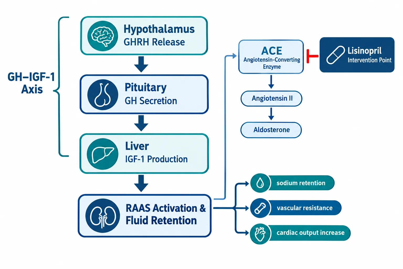
Growth hormone secretagogues, including GHRH analogs, ghrelin mimetics, and synthetic GH-releasing peptides, stimulate pituitary GH release, which in turn drives hepatic IGF-1 production. This cascade carries meaningful cardiovascular effects that researchers must account for.
IGF-1 elevation promotes myocardial growth, increases cardiac output, and enhances vascular smooth muscle responsiveness. In the short term, these effects can look beneficial. Over longer exposures, however, elevated IGF-1 contributes to left ventricular hypertrophy and arterial stiffness, both established markers of cardiovascular stress.
Ghrelin and its synthetic analogs add another layer. Research consistently shows that ghrelin-type secretagogues exert vasodilatory, anti-ischemic, and anti-apoptotic effects on cardiac tissue. They reduce peripheral vascular resistance and may lower blood pressure acutely. However, this vasodilation can interact unpredictably with ACE inhibitors, raising the risk of orthostatic hypotension.
Fluid retention is a particularly important concern. GH directly stimulates renal sodium reabsorption via the IGF-1 receptor, expanding plasma volume. This increases cardiac preload and, over time, elevates systolic blood pressure. Researchers working with IPA Sermorelin stack protocols should factor this mechanism into their baseline cardiovascular assessments.
The RAAS responds to this expanded volume by modulating aldosterone and angiotensin II levels. This is precisely where lisinopril enters the picture.
Lisinopril and ACE Pathway Dynamics in Secretagogue Protocols
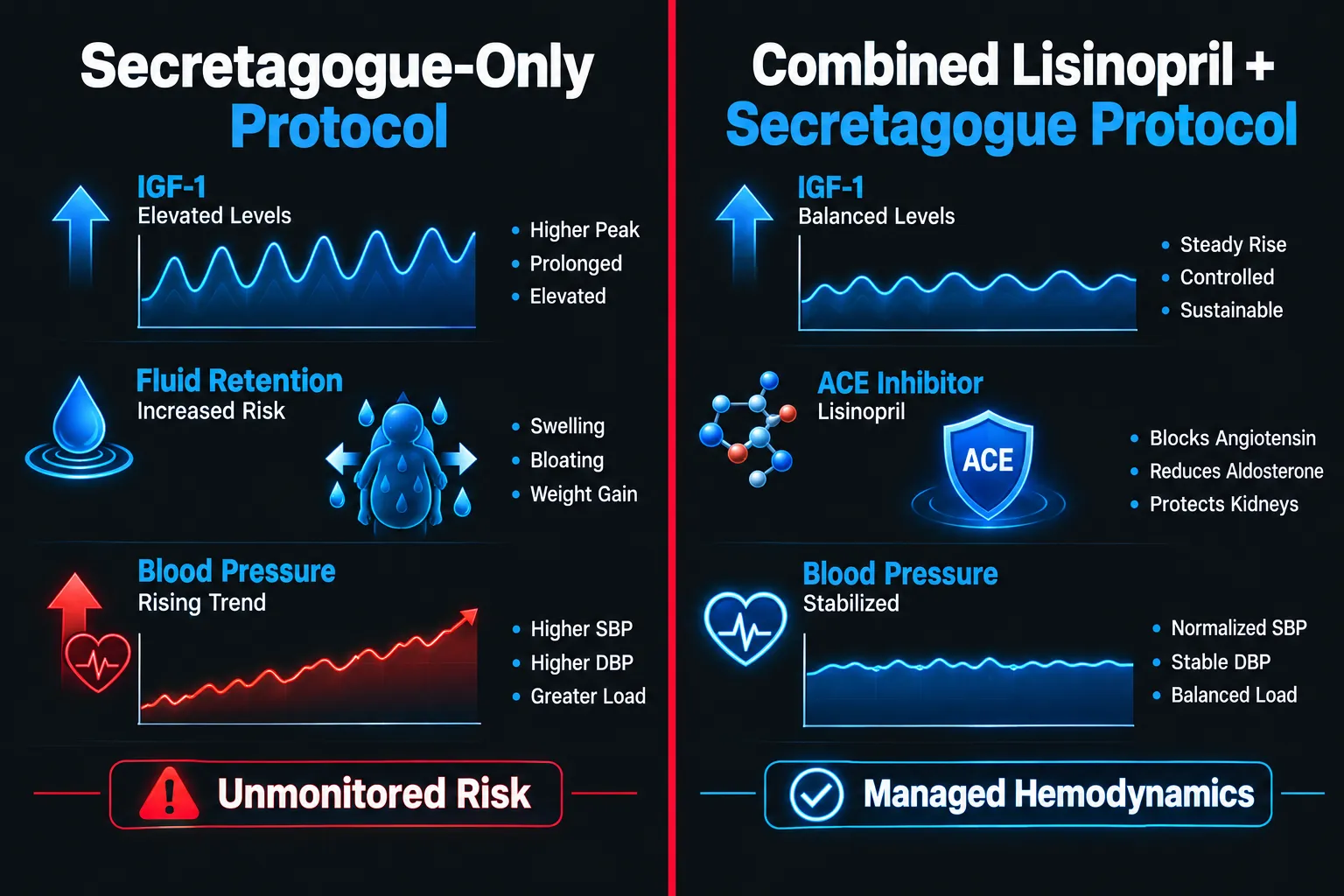
Lisinopril blocks ACE, the enzyme that converts angiotensin I to the potent vasoconstrictor angiotensin II. By reducing angiotensin II, lisinopril lowers peripheral resistance, reduces aldosterone-driven sodium retention, and decreases cardiac afterload. These are exactly the mechanisms that counteract GH-induced fluid retention.
"The ACE pathway and the GH/IGF-1 axis do not operate in isolation, co-administration of lisinopril alongside secretagogues creates a dynamic cardiovascular environment that requires active, structured monitoring."
However, ACE inhibition introduces its own variables. Potassium levels rise because aldosterone suppression reduces urinary potassium excretion. Renal perfusion pressure can drop, particularly in subjects with any pre-existing renal sensitivity. Bradykinin accumulates, which may amplify the vasodilatory effects already present from ghrelin-type secretagogues.
Key interaction points to monitor:
- Blood pressure response may be exaggerated when ghrelin-type secretagogues and lisinopril are combined
- Potassium can climb into hyperkalemic ranges without regular electrolyte testing
- Renal function (creatinine, eGFR) may shift as RAAS suppression alters glomerular filtration dynamics
- Fluid balance changes can mask or amplify biomarker readings
Researchers exploring peptide combinations that modulate metabolic and cardiovascular pathways, such as those reviewed in tesofensine noradrenergic and incretin-based pathway research, will recognize that multi-pathway interactions demand multi-marker monitoring frameworks.
Core Cardiovascular Stress Markers for Combined RAAS-GH Axis Monitoring
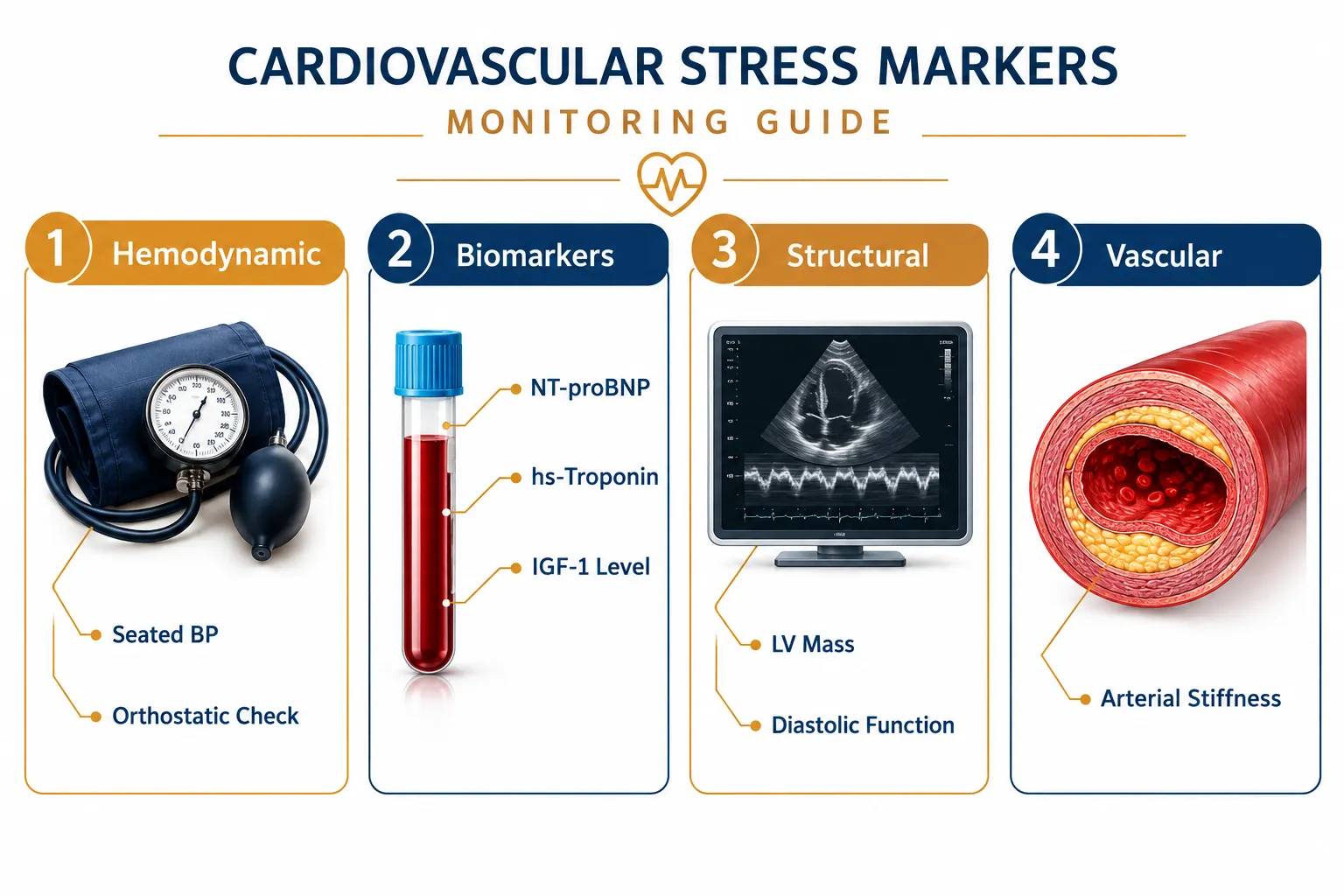
Monitoring cardiovascular stress markers in growth hormone research alongside lisinopril and ACE pathway modulation requires a structured, four-domain approach.
Hemodynamic Markers
| Marker | Measurement Method | Key Concern |
|---|---|---|
| Seated blood pressure | Manual or automated sphygmomanometry | Hypertension from GH fluid load |
| Ambulatory blood pressure | 24-hour ABPM device | Masked hypertension patterns |
| Orthostatic blood pressure | Supine to standing protocol | Hypotension risk with ACE + ghrelin |
| Resting heart rate | ECG or pulse oximetry | Tachycardia from volume shifts |
Structural Markers
Echocardiography provides the most direct window into cardiac remodeling. Left ventricular mass index, wall thickness, and both systolic and diastolic function should be assessed at baseline and at defined intervals. Diastolic dysfunction often precedes systolic changes and can appear early in GH-excess states.
Biomarker Panel
NT-proBNP is the most sensitive biomarker for detecting early cardiac wall stress and volume overload. Elevated levels signal that the heart is under pressure before symptoms appear. High-sensitivity troponin (hs-troponin) identifies subclinical myocardial injury.
Additional biomarkers include:
- IGF-1 (to calibrate secretagogue dosing and exposure)
- Serum creatinine and eGFR (renal safety on ACE inhibitor)
- Potassium and sodium (electrolyte balance under RAAS suppression)
- Fasting glucose and lipid panel (metabolic context)
Research on mitochondrial cardioprotection compounds such as those examined in SS-31 mitochondrial research highlights how cellular stress markers can complement systemic biomarker panels in cardiovascular monitoring.
Vascular Markers
Arterial stiffness indices, measured via pulse wave velocity or augmentation index, capture changes in large-vessel compliance that echocardiography and biomarkers may miss. Endothelial function assessment adds further resolution in longer-duration protocols.
Practical Monitoring Frequency and Protocol Design
Effective cardiovascular stress monitoring in growth hormone research is not a one-time assessment. A tiered frequency model works best:
Baseline (before protocol initiation): Full panel, hemodynamic, echocardiographic, biomarker, and vascular measures.
Early phase (weeks 2-4): Blood pressure, heart rate, potassium, creatinine, and eGFR. This window captures the most acute RAAS-GH interaction effects.
Mid-protocol (weeks 6-12): Repeat biomarker panel including NT-proBNP and hs-troponin. Reassess IGF-1 to confirm secretagogue response is within target range.
End of protocol: Full repeat of baseline panel plus echocardiographic comparison for structural changes.
Researchers working with GLP-based peptide compounds, including those available as GLP-3 Reta 10mg or GLP-3 Reta 20mg, should apply similarly structured cardiovascular monitoring, as GLP receptor pathways also influence cardiac output and vascular tone.
Conclusion
Cardiovascular stress markers in growth hormone research represent a non-negotiable component of responsible secretagogue protocol design, particularly when lisinopril and ACE pathway modulation are part of the picture. The interaction between GH-driven fluid retention, IGF-1-mediated cardiac remodeling, and ACE inhibitor-induced RAAS suppression creates a layered hemodynamic environment that no single marker can fully capture.
Actionable next steps for researchers:
- Establish a full cardiovascular baseline before initiating any secretagogue-lisinopril combination protocol.
- Prioritize NT-proBNP, hs-troponin, potassium, and eGFR as the minimum biomarker panel throughout the study period.
- Use ambulatory blood pressure monitoring to detect masked hypertension or exaggerated hypotensive responses.
- Schedule echocardiographic assessment at baseline and protocol end to quantify structural changes.
- Adjust secretagogue dosing based on IGF-1 levels and correlate with hemodynamic trends rather than treating them as independent variables.
Structured monitoring transforms cardiovascular risk from an unknown variable into a manageable, measurable research parameter.


Leave a Reply
Want to join the discussion?Feel free to contribute!