

Prednisone and Research-Use Peptides: How Glucocorticoid Exposure Can Distort Metabolic, Immune, and Tissue-Repair Readouts
Roughly one in five research subjects enrolled in metabolic or immune-modulating studies carries an active or recent glucocorticoid prescription, yet fewer than half of published peptide protocols formally account for this exposure. The problem of Prednisone and Research-Use Peptides: How Glucocorticoid Exposure Can Distort Metabolic, Immune, and Tissue-Repair Readouts is not theoretical. When a subject is taking prednisone, the drug reshapes glucose handling, cytokine profiles, body composition, and wound biology simultaneously, making it nearly impossible to attribute changes in those endpoints to a peptide compound without deliberate experimental controls.
This guide is written for researchers, protocol designers, and informed buyers of research-use peptide compounds who need a clear, actionable map of where glucocorticoid confounding is most severe and how to design around it.
Key Takeaways
- Prednisone induces a "pseudo-metabolic syndrome" that directly mimics or masks outcomes targeted by metabolic peptides.
- Standard inflammatory biomarkers, CRP, leukocyte count, neutrophil ratio, are dose-dependently suppressed by glucocorticoids, distorting immune readouts.
- Tissue-repair and wound-healing endpoints are particularly vulnerable because glucocorticoids both dampen acute inflammation and impair collagen remodeling.
- Anabolic and sarcopenia-targeted peptides face the strongest confounding from glucocorticoid-driven muscle catabolism.
- Glucocorticoid exposure should be treated as a core design variable, not background noise, in any peptide research protocol.
How Glucocorticoids Create a Pseudo-Metabolic Syndrome
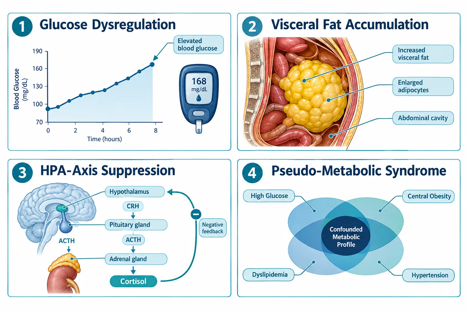
Prednisone does not simply suppress inflammation. At doses commonly used in clinical and research-adjacent settings, it triggers a cascade of metabolic changes that closely resemble metabolic syndrome: elevated fasting glucose, increased visceral adiposity, dyslipidemia, and insulin resistance. For researchers studying cardiometabolic peptide compounds, this is a direct collision.
The core metabolic confounders introduced by prednisone include:
| Endpoint | Glucocorticoid Effect | Research Risk |
|---|---|---|
| Fasting glucose | Elevated (hepatic gluconeogenesis) | Masks GLP-class peptide efficacy |
| Visceral fat | Increased via lipid redistribution | Distorts body-composition endpoints |
| Insulin sensitivity | Reduced | Confounds metabolic peptide response |
| Appetite / caloric intake | Increased | Alters weight-loss peptide readouts |
| Cortisol / HPA axis | Suppressed endogenous output | Disrupts hormonal biomarker panels |
The HPA-axis suppression point deserves particular attention. When exogenous glucocorticoids are present, endogenous cortisol production falls. This changes the hormonal baseline against which peptide pharmacodynamics are measured. For GH-analogue peptides and compounds that interact with the growth hormone axis, this hormonal shift can produce entirely misleading dose-response curves.
Researchers studying visceral fat redistribution should also note that glucocorticoid-driven adiposity preferentially accumulates centrally, the same compartment tracked in visceral fat research models. Without a glucocorticoid-naive control arm, it becomes impossible to separate peptide-driven changes from steroid-driven ones.
Immune Biomarker Distortion: What Prednisone Does to CRP, Cytokines, and Leukocytes

The immune-modulating effects of prednisone are both well-documented and deeply problematic for peptide research. The drug produces strong, dose-dependent suppression of nearly every standard inflammatory biomarker used in research protocols.
Biomarkers most severely distorted by glucocorticoid exposure:
- C-reactive protein (CRP): Hepatic CRP synthesis is directly suppressed, producing artificially low readings that can mimic anti-inflammatory peptide effects.
- Interleukin-6 (IL-6) and TNF-alpha: Both are blunted by glucocorticoid receptor activation, masking inflammatory resolution that a peptide may or may not be driving.
- Leukocyte and neutrophil counts: Prednisone causes a characteristic leukocytosis with neutrophilia and lymphopenia, a pattern that can be mistaken for an immune-activation signal from a peptide compound.
- Tissue-resident immune cells: Recent work confirms that glucocorticoids selectively modulate tissue-resident macrophages and mast cells in ways that differ from circulating immune cells. This matters enormously for regenerative peptides like BPC-157 and TB-500, where local tissue immune activity is the primary readout.
"When prednisone is present, the immune landscape a peptide encounters is fundamentally different from the one it was designed to modulate. Treating that as a minor covariate is a protocol error."
The practical consequence is that an immune-modulating peptide may appear highly effective, completely ineffective, or even counterproductive depending entirely on the glucocorticoid dose the subject carries. Without stratifying subjects by steroid exposure, or excluding active users entirely, the data cannot be interpreted with confidence.
Tissue-Repair and Anabolic Readouts: Where Glucocorticoid Confounding Is Most Severe
Wound healing and tissue repair represent perhaps the most complex confounding scenario in Prednisone and Research-Use Peptides: How Glucocorticoid Exposure Can Distort Metabolic, Immune, and Tissue-Repair Readouts. Glucocorticoids produce a paradoxical two-phase effect on repair biology.
Phase 1 (acute): Inflammation is suppressed, which can make a wound-healing peptide appear to accelerate early healing simply because the steroid has already cleared the inflammatory field.
Phase 2 (remodeling): Glucocorticoids impair fibroblast proliferation, reduce collagen synthesis, and suppress angiogenesis. A peptide that promotes collagen remodeling may show blunted or absent efficacy, not because it failed, but because the steroid has suppressed the very cellular machinery the peptide targets.
For anabolic and sarcopenia-targeted peptides, the problem is equally stark. Glucocorticoids drive skeletal muscle catabolism through multiple mechanisms: they upregulate muscle-specific ubiquitin ligases, suppress IGF-1 signaling, and reduce protein synthesis rates. Any peptide designed to promote lean mass retention or muscle repair is working against a pharmacologically imposed catabolic state. The net readout will underestimate peptide efficacy, sometimes dramatically.
Mitochondrial function is also affected. Glucocorticoids impair mitochondrial biogenesis in muscle tissue, which is directly relevant for compounds studied in SS-31 mitochondrial research contexts.
Designing Around Glucocorticoid Confounding: Practical Protocol Controls
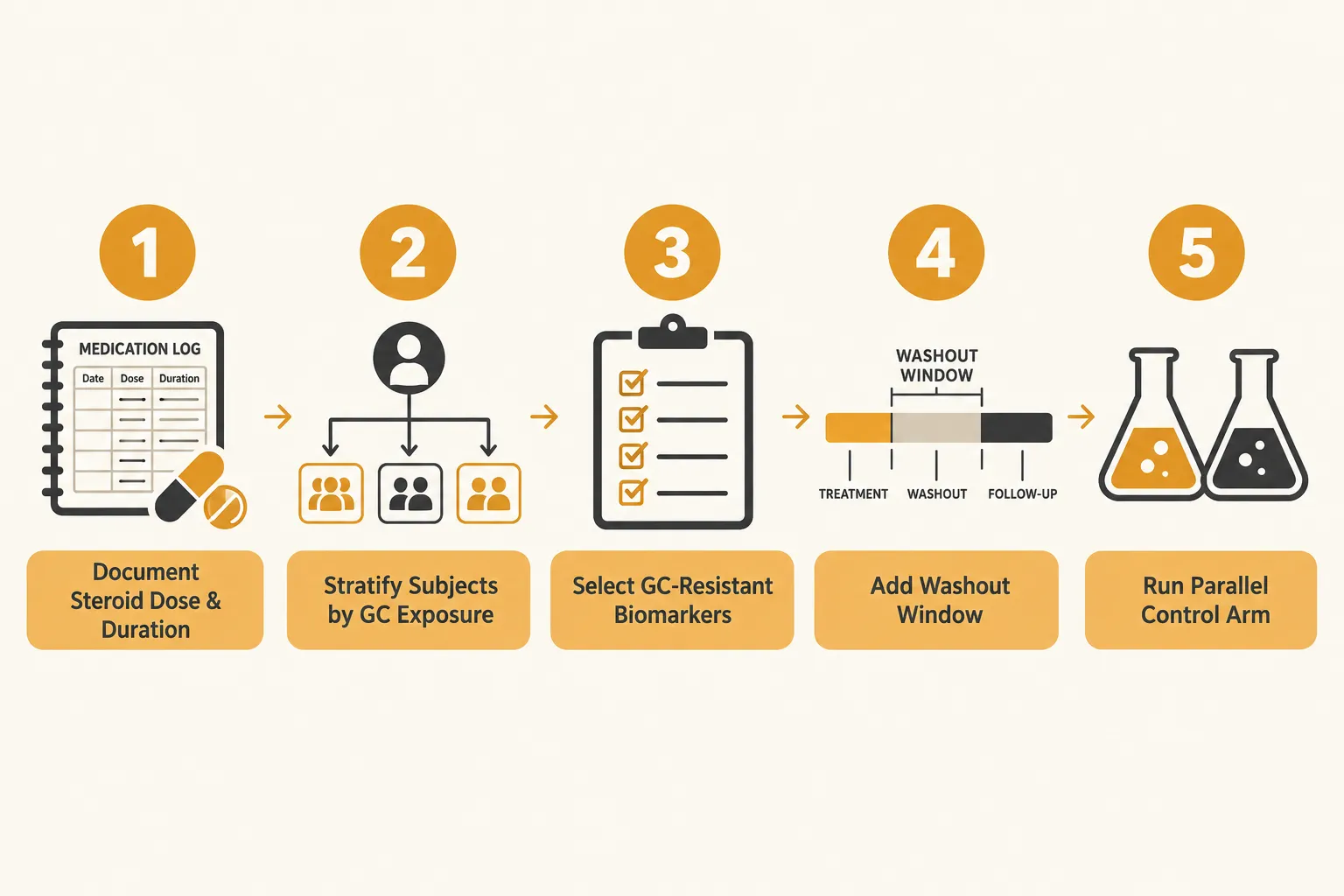
Researchers in 2026 have several validated strategies to reduce or detect glucocorticoid confounding:
- Screen and stratify at enrollment. Collect steroid use history, current dose, and duration. Stratify rather than exclude where possible to preserve sample size.
- Use glucocorticoid-resistant biomarkers where available. Some cytokines and repair markers are less sensitive to steroid suppression and can serve as secondary endpoints.
- Build in washout windows. For subjects who can safely discontinue steroids, a documented washout period before peptide exposure begins reduces confounding substantially.
- Run parallel control arms. A glucocorticoid-exposed control arm receiving vehicle only allows direct quantification of steroid-driven endpoint changes.
- Report steroid exposure as a primary covariate. Statistical models should treat glucocorticoid dose as a continuous variable, not a binary yes/no flag.
Conclusion
The relationship between Prednisone and Research-Use Peptides: How Glucocorticoid Exposure Can Distort Metabolic, Immune, and Tissue-Repair Readouts is now well enough characterized that ignoring it constitutes a preventable design flaw. Across glucose metabolism, inflammatory cytokines, collagen remodeling, body composition, and immune-cell measurements, prednisone introduces quantifiable, directional biases that can make an effective peptide look inert, or an inert one look effective.
Actionable next steps for research protocol designers:
- Add glucocorticoid screening to all enrollment checklists for metabolic, immune, or repair-focused peptide studies.
- Pre-specify steroid dose as a covariate in the statistical analysis plan before data collection begins.
- Consult updated 2026 biomarker guidance on glucocorticoid-resistant immune endpoints when designing immune-modulating peptide studies.
- Review compound-specific confounding risk, peptides targeting GH axis, collagen synthesis, or mitochondrial function carry the highest vulnerability.
Treating glucocorticoid exposure as a core design variable rather than background noise is the single highest-leverage methodological improvement available to most peptide research teams today.