

GLP-2-T and GLP2 Tirz Peptides: Measuring Intestinal Barrier Function Without Confounding Motility
Roughly 70% of the body's immune interface sits along the intestinal epithelium, a single-cell layer held together by tight junction proteins that can be disrupted in hours. That vulnerability makes accurate barrier measurement critical, yet most in vivo gut assays pick up motility signals alongside permeability signals, blurring the picture. Research into GLP-2-T and GLP2 Tirz peptides: measuring intestinal barrier function without confounding motility demands a cleaner experimental lens, one focused on permeability tracers, junction protein profiling, and organoid morphology rather than transit-time endpoints alone.
Key Takeaways
- GLP-2 enhances intestinal barrier integrity through IGF-1 and PI3K/Akt/mTORC1 signaling, directly upregulating tight junction proteins such as occludin, claudin-1, and ZO-1.
- Motility effects of GLP-2 and related peptides can confound in vivo permeability data; isolating barrier outcomes requires motility-independent readouts.
- Transepithelial electrical resistance (TER), fluorescent tracer flux, and tight junction protein profiling are the gold-standard motility-independent assays.
- Intestinal organoids and isolated epithelial monolayers allow researchers to study GLP-2-based peptide effects on barrier structure without peristaltic interference.
- Separating barrier endpoints from motility endpoints is essential for interpreting next-generation multi-agonist peptides like GLP-2-T and GLP2 Tirz compounds.
How GLP-2 Signals to the Intestinal Barrier
GLP-2 (glucagon-like peptide-2) is produced by L-cells in the distal intestine and acts as a master regulator of mucosal integrity. Its receptor is expressed on enteric neurons, subepithelial myofibroblasts, and enteroendocrine cells, not directly on absorptive enterocytes. This indirect architecture means GLP-2 recruits IGF-1 as a downstream mediator, which then activates PI3K/Akt/mTORC1 signaling within the epithelium.
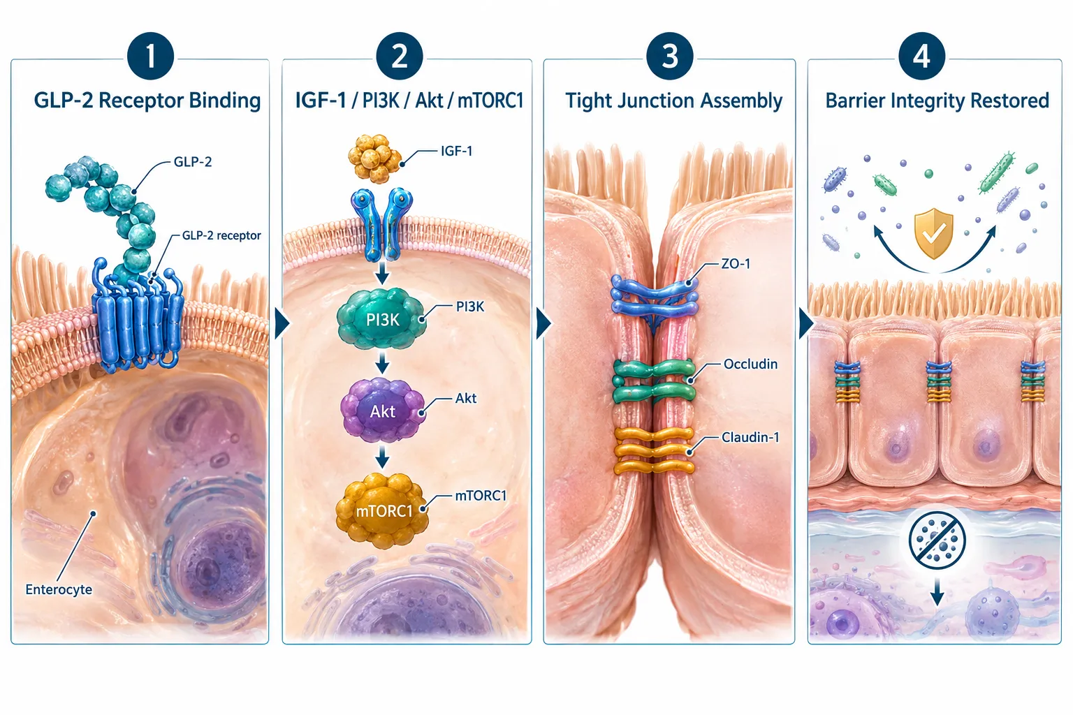
The downstream result is rapid upregulation of three key tight junction proteins:
| Protein | Role in Barrier |
|---|---|
| Occludin | Seals paracellular space against macromolecules |
| Claudin-1 | Controls ion and water flux selectivity |
| ZO-1 | Scaffolds junction complex to actin cytoskeleton |
Barrier improvement can be measurable within minutes to hours of GLP-2 exposure, far faster than structural changes in villus height or crypt depth. This speed is important: it means barrier assays must capture early molecular events, not just histological endpoints. Understanding this pathway is foundational for anyone exploring the GLP-3 peptide signaling pathway and related glucagon-family mechanisms.
Key insight: GLP-2's barrier effect is mechanistically separable from its motility effect because they operate through different receptor populations and different downstream effectors.
Why Motility Confounds Barrier Measurement, and How to Avoid It
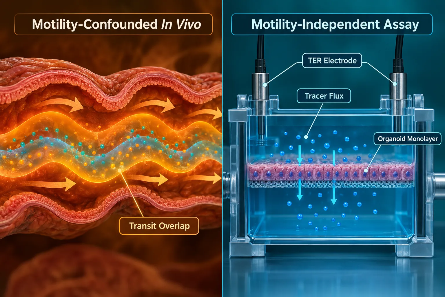
GLP-2 slows intestinal transit. That is useful clinically, it extends nutrient contact time and supports absorption, but it creates a measurement problem. When researchers measure gut permeability in a living animal after GLP-2 dosing, slower transit means luminal tracers spend more time in contact with the mucosa. That extended contact artificially reduces apparent permeability, mimicking a barrier-tightening effect even if no molecular junction changes occurred.
This confound is the central challenge when evaluating GLP-2-T and GLP2 Tirz peptides: measuring intestinal barrier function without confounding motility.
Motility-Independent Assay Toolkit
Three readout categories sidestep the motility problem entirely:
1. Transepithelial Electrical Resistance (TER)
TER measures how well an epithelial monolayer resists ionic current. Higher resistance equals tighter junctions. Performed in Ussing chambers or Transwell inserts on isolated tissue or organoid-derived monolayers, TER is completely independent of peristalsis.
2. Fluorescent Tracer Flux
FITC-dextran (typically 4 kDa) is applied to the apical surface of isolated epithelium. The rate at which it crosses to the basolateral side reflects paracellular permeability. Because the tissue is excised or the organoid is controlled, transit time is irrelevant.
3. Tight Junction Protein Profiling
Western blot, immunofluorescence, or proximity ligation assays quantify occludin, claudin-1, and ZO-1 expression and localization. A peptide that genuinely tightens the barrier will show increased junctional protein density at the membrane, a molecular endpoint with no motility component.
For context on how related peptide classes are being studied, the Peptides 101 guide for research-use-only buyers covers structural and mechanistic frameworks that apply across GLP-family compounds.
Osmotic and Intake Controls
Two additional confounds must be controlled in any barrier study:
- Osmotic load: High-osmolality solutions draw water into the lumen, mechanically stretching tight junctions and inflating permeability readings. Vehicle and tracer solutions should be iso-osmotic to luminal fluid.
- Food and water intake: GLP-2 suppresses appetite in some models. Reduced intake lowers luminal content and can alter barrier hydration. Pair-feeding controls or continuous enteral nutrition protocols normalize this variable.
Organoids, Epithelial Monolayers, and the Future of GLP-2-T Research
Intestinal organoids, three-dimensional epithelial structures grown from intestinal stem cells, have transformed barrier research. They recapitulate villus-crypt architecture, express all major tight junction proteins, and respond to GLP-2 pathway agonists. Critically, they have no enteric nervous system and no peristaltic activity, making them ideal for isolating direct epithelial effects of novel peptides.
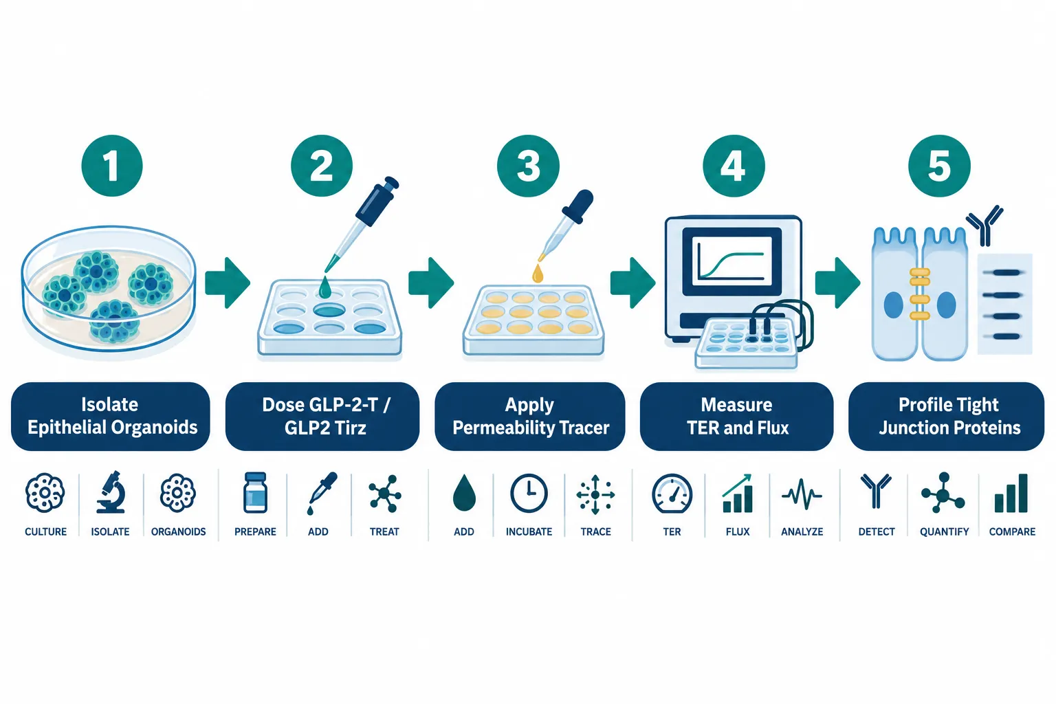
For GLP-2-T and GLP2 Tirz peptides specifically, the peer-reviewed literature does not yet contain dedicated mechanistic studies as of 2026. These appear to be emerging research-grade compounds in the glucagon-family multi-agonist space. Based on GLP-2's established biology, a rigorous experimental protocol for evaluating such peptides would include:
- Organoid monolayer preparation, seed organoid-derived cells on permeable Transwell supports until confluent
- Peptide dosing, apply GLP-2-T or GLP2 Tirz at graded concentrations to the basolateral compartment (mimicking systemic delivery)
- TER measurement, record resistance at baseline, 1 hour, 4 hours, and 24 hours
- Tracer flux assay, add apical FITC-dextran at the 4-hour time point; sample basolateral fluid at 30-minute intervals
- Junction protein profiling, fix and stain for ZO-1, occludin, and claudin-1 at study end; quantify membrane vs. cytoplasmic distribution
This design generates motility-independent barrier data while controlling for osmotic load and intake variables. Researchers exploring related GLP-3 peptides for sale in the glucagon family may find this protocol framework directly transferable, given overlapping receptor biology.
Short bowel syndrome research has already validated this approach for GLP-2 agonists: permeability and absorption metrics, not motility metrics, drove the key efficacy conclusions. Livestock studies using histology and tight-junction markers rather than transit endpoints add further proof-of-concept that barrier and motility are separable outcomes.
For researchers sourcing compounds for these studies, reviewing wholesale peptides for sale options and understanding purity standards is an important pre-study step. Purity directly affects TER and tracer flux signal quality; contaminants can independently alter epithelial resistance. Those looking for related reference compounds may also find value in reviewing IPA peptides as additional intestinal-integrity research tools.
Conclusion
Separating intestinal barrier function from motility effects is not a minor methodological detail, it is the difference between measuring what a peptide does to tight junctions and measuring what it does to transit time. For GLP-2-T and GLP2 Tirz peptides: measuring intestinal barrier function without confounding motility, the path forward is clear.
Actionable next steps for researchers:
- Prioritize TER and FITC-dextran flux assays in isolated epithelium or organoid monolayers as primary barrier endpoints
- Run separate, parallel motility assessments (bead transit, manometry) rather than combining them with permeability readouts
- Include iso-osmotic vehicle controls and pair-feeding protocols in every study design
- Profile ZO-1, occludin, and claudin-1 at the membrane level to confirm molecular barrier changes
- Consult resources such as the Peptides Doctor Near Me directory when translating preclinical findings toward supervised research applications
As multi-agonist GLP-family peptides continue to evolve, the assay toolkit described here will be essential for generating clean, interpretable data on intestinal barrier biology.


Leave a Reply
Want to join the discussion?Feel free to contribute!