

Collagen Signaling and Copper Peptides: What Researchers Measure With GHK-Cu and Related Skin Models
{"cover":"Professional landscape format (1536×1024) hero image with bold text overlay 'Collagen Signaling and Copper Peptides' in crisp white sans-serif centered on a deep teal semi-transparent overlay panel, 8% safe margins from every edge, no character touches the border. Background: macro editorial photograph of layered dermal tissue cross-section rendered as a luminous scientific illustration, collagen fiber bundles in pale gold and ivory weaving through a blue-tinted extracellular matrix, with copper-toned molecular structures floating in the foreground. Studio lighting, high contrast, magazine-cover aesthetic, 2026 editorial science journal quality.","content":["Annotated mechanism-of-action diagram, landscape format (1536×1024), showing the GHK-Cu collagen signaling pathway inside a fibroblast cell. Four labeled stages arranged left to right: Stage 1 label 'GHK-Cu Binds Receptor', Stage 2 label 'TGF-beta Activation', Stage 3 label 'Procollagen Synthesis', Stage 4 label 'MMP Regulation'. Thin callout lines point from each stage to illustrated molecular icons, a copper-peptide helix, a receptor protein, collagen triple-helix, and enzyme scissors. Clean white background, teal and navy palette, bold sans-serif labels inside 5% safe margin, no full sentences, editorial biomedical illustration quality.","Split-screen landscape format (1536×1024) comparison image: left panel labeled 'In Vitro Fibroblast Culture' shows a microscope view of human dermal fibroblasts with fluorescent green collagen fiber staining and a callout label 'Procollagen I Assay'; right panel labeled 'Ex Vivo Skin Biopsy' shows a histology cross-section of dermis with labeled callouts 'Dermal Density Imaging', 'Hydroxyproline Quantification', and 'Gene Expression Readout'. Dividing line in copper-gold tone, cool clinical lighting on left, warm amber histology stain on right, sharp editorial contrast, biomedical research aesthetic, 5% safe margins on all labels.","Numbered step-by-step process flow diagram, landscape format (1536×1024), illustrating a wound-healing research protocol using copper peptides. Five horizontal steps with illustrated icons: Step 1 'Wound Model Setup' showing a murine skin diagram, Step 2 'GHK-Cu Hydrogel Application' showing a syringe and gel matrix, Step 3 'Inflammatory Marker Sampling' showing cytokine icons IL-6 and TNF-alpha, Step 4 'Re-epithelialization Scoring' showing a healed tissue cross-section, Step 5 'Collagen Density Endpoint' showing a bar graph. Arrows connecting each step, copper and slate-blue color palette, bold 1-4 word labels per step, clean white background, editorial scientific infographic style, all labels inside 5% safe margin."]

A single tripeptide, glycine-histidine-lysine, naturally present in human plasma drops by more than 60% between the ages of 20 and 60. That decline tracks closely with measurable losses in dermal collagen density, and it is precisely why collagen signaling and copper peptides have become a serious focus in skin biology research. When GHK binds copper to form GHK-Cu, the resulting complex interacts with fibroblasts, matrix-remodeling enzymes, and gene-expression networks in ways that researchers are now quantifying with increasing precision.
Key Takeaways
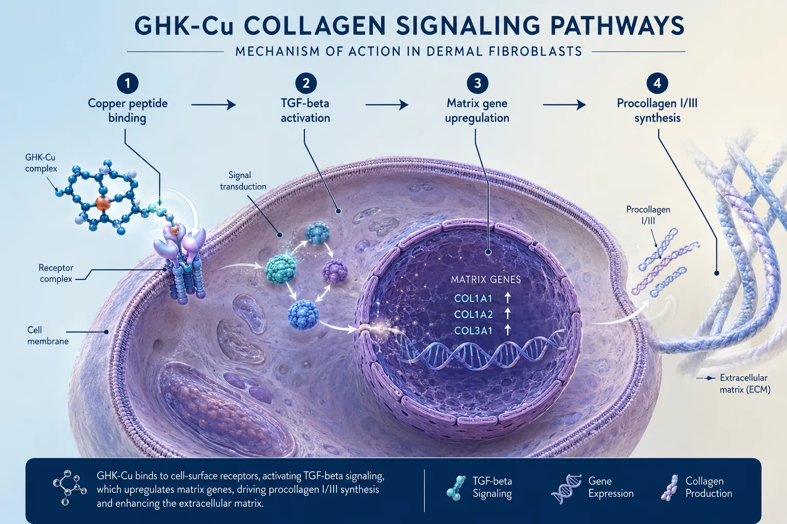
- GHK-Cu activates TGF-beta pathways in fibroblasts, driving measurable increases in procollagen I and III synthesis at nanomolar concentrations.
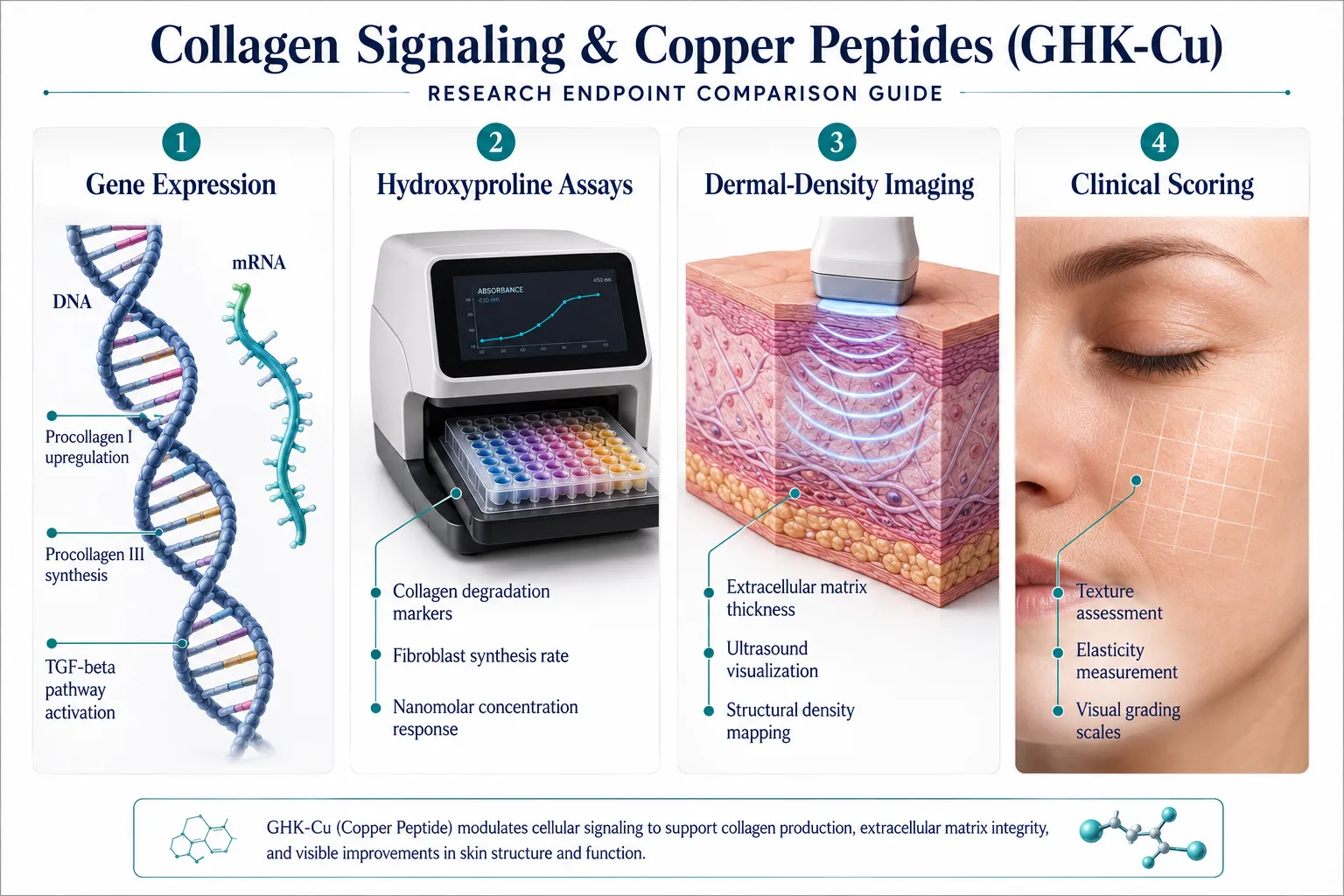
- Researchers use multiple endpoint types, gene expression, hydroxyproline assays, dermal-density imaging, and clinical scoring, to characterize collagen signaling responses.
- Wound-healing murine models and ex vivo biopsy systems are the most common preclinical platforms for studying copper peptide activity.
- Concentration matters: fibroblast culture studies show a bell-shaped dose-response curve, with optimal effects typically between 1 nM and 10 nM.
- Phase 2 clinical trial designs in 2026 are incorporating re-epithelialization speed and procollagen levels as co-primary endpoints, signaling growing regulatory interest.
The Biology Behind GHK-Cu and Collagen Signaling
GHK-Cu does not act as a simple collagen precursor. Its influence on collagen signaling and copper peptides research is primarily regulatory. The complex binds to cell-surface receptors and initiates intracellular cascades involving transforming growth factor-beta (TGF-beta), a master regulator of extracellular matrix production. When TGF-beta signaling is upregulated, fibroblasts increase transcription of COL1A1 and COL3A1, the genes encoding the alpha chains of collagen types I and III, the two most abundant structural collagens in adult dermis.
Beyond collagen synthesis, GHK-Cu modulates matrix metalloproteinases (MMPs). MMPs are enzymes that degrade collagen and other matrix proteins. Healthy tissue remodeling requires a balance between synthesis and degradation. Research in fibroblast cultures shows that GHK-Cu simultaneously increases TIMP-1 and TIMP-2 (tissue inhibitors of metalloproteinases) while suppressing MMP-1 and MMP-2 activity. The net result is a shift toward matrix accumulation rather than breakdown, a measurable outcome that makes GHK-Cu particularly relevant in aged or photodamaged skin models.
Key signaling targets identified in fibroblast culture studies:
| Target | Direction of Change | Measurement Method |
|---|---|---|
| Procollagen I | Increase | ELISA, Sircol assay |
| Procollagen III | Increase | Immunofluorescence |
| MMP-1 (collagenase) | Decrease | Zymography, qPCR |
| TIMP-1 | Increase | Western blot |
| TGF-beta1 | Increase | ELISA |
Researchers sourcing compounds for these studies often consult a GHK-Cu peptide purchase and copper peptide research sourcing guide to ensure purity specifications are met before running assays, since trace contaminants can distort dose-response curves significantly.
How Researchers Measure Collagen Signaling and Copper Peptides in Skin Models
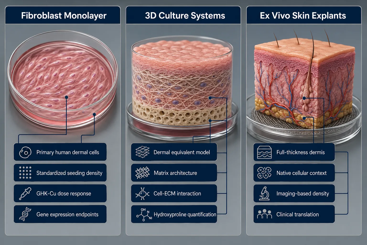
The choice of model system determines which endpoints are accessible. Three primary platforms dominate the published literature.
Fibroblast Monolayer and 3D Culture Systems
Primary human dermal fibroblasts remain the workhorse model. Researchers seed cells at standardized density, apply GHK-Cu at concentrations ranging from 0.1 nM to 1 µM, and harvest supernatants or cell lysates at 24, 48, and 72 hours. Procollagen I C-terminal propeptide (PICP) in the conditioned medium is the most common output, measured by competitive ELISA. Hydroxyproline content, a collagen-specific amino acid, is quantified after acid hydrolysis using the chloramine-T colorimetric method.
3D collagen gel contraction assays add a mechanical dimension: fibroblasts embedded in a collagen lattice contract the gel over 48-72 hours, and the degree of contraction reflects cytoskeletal activation and matrix remodeling capacity. GHK-Cu consistently increases contraction rates compared to untreated controls, a finding reproducible across multiple laboratory groups.
Ex Vivo Skin Biopsy and Dermal-Density Imaging
Human skin punch biopsies maintained in organ culture allow researchers to apply GHK-Cu to a structurally intact tissue. Histological sections stained with Masson's trichrome or picrosirius red under polarized light reveal collagen fiber organization and density. High-frequency ultrasound and optical coherence tomography (OCT) provide non-destructive dermal-density measurements, generating quantitative echogenicity scores that correlate with collagen content.
Gene-expression profiling from biopsy RNA adds an epigenetic layer. Microarray and RNA-seq datasets from photoaged biopsy models treated with GHK-Cu show upregulation of not only collagen genes but also decorin, fibronectin, and laminin, structural glycoproteins that organize the collagen scaffold. This breadth of transcriptional response distinguishes GHK-Cu from simpler collagen-stimulating agents and explains why it appears frequently alongside other regenerative peptides in comparative studies. Researchers interested in how tissue-repair peptides compare across platforms may find the BPC-157 core peptides documentation and first research guide a useful parallel reference.
Clinical Trial Endpoints: Photoaging and Wound Models
Randomized controlled trials measuring collagen signaling and copper peptides outcomes in human skin use a layered endpoint strategy. A 2025 meta-analysis of randomized trials in skin aging identified procollagen I serum levels, clinical photoaging scores (Glogau scale), and investigator-assessed wrinkle depth as the most commonly reported primary outcomes. Effect sizes across trials were modest but statistically consistent, particularly for periorbital fine lines and overall skin firmness.
A 2026 Phase 2 trial in acute wound re-epithelialization is using re-epithelialization speed (days to wound closure) and biopsy-confirmed collagen density at day 14 as co-primary endpoints, a design that reflects growing regulatory interest in objective tissue-level evidence. A 2025 infected wound murine hydrogel model demonstrated that GHK-Cu delivered in a carboxymethyl cellulose matrix reduced IL-6 and TNF-alpha levels at wound sites by approximately 40% while increasing collagen deposition scores by 35% versus vehicle control, a combined inflammatory and structural endpoint that is becoming standard in preclinical wound research.
Safety data across skin models are consistently favorable. Fibroblast viability assays at concentrations up to 100 µM show no significant cytotoxicity. Clinical trials report mild, transient erythema as the most common adverse event, with no systemic signals detected. For researchers building multi-peptide study panels, lab tested peptides with documented purity certificates are essential for maintaining assay integrity across experimental arms.
"The value of GHK-Cu in skin research is not that it does one thing well, it is that it touches matrix synthesis, degradation control, and inflammatory regulation simultaneously, making it a useful probe for studying coordinated tissue repair."
Researchers comparing copper peptide endpoints with other repair-focused compounds sometimes cross-reference findings from BPC-157 and TB-500 peptide research given the overlapping wound-healing readouts used across both compound classes.
Conclusion
Collagen signaling and copper peptides represent one of the more mechanistically rich areas of skin biology research in 2026. GHK-Cu activates TGF-beta pathways, modulates MMP/TIMP balance, and upregulates a broad suite of matrix genes, all of which are measurable using established laboratory methods ranging from ELISA and hydroxyproline assays to high-frequency ultrasound and RNA-seq.
Actionable next steps for researchers:
- Select the model system that matches your endpoint priority: fibroblast culture for molecular readouts, ex vivo biopsy for structural endpoints, murine hydrogel models for inflammatory plus collagen co-endpoints.
- Standardize GHK-Cu concentration ranges (1-10 nM for synthesis endpoints; up to 1 µM for safety profiling) before designing dose-response experiments.
- Include both synthesis markers (PICP, hydroxyproline) and degradation markers (MMP-1, TIMP-1) to capture the full matrix-remodeling picture.
- Consult a verified copper peptide research sourcing guide to confirm peptide purity and batch consistency before initiating assays.
- Consider pairing GHK-Cu endpoints with data from other repair peptides reviewed in the top 5 research peptides for metabolic health buyer's guide to contextualize findings within broader regenerative biology.
The field is moving toward multi-endpoint trial designs that demand both molecular and clinical evidence. Researchers who build rigorous, reproducible measurement frameworks now will be best positioned to contribute to that evolving standard.


Leave a Reply
Want to join the discussion?Feel free to contribute!