

MOTS-c and Slupp332 in Skeletal-Muscle Regeneration Models: Mitochondrial Flux, AMPK Readouts, and Fiber-Type Controls
Skeletal muscle accounts for roughly 40% of human body mass, yet its regenerative capacity declines sharply with age, a process tightly linked to deteriorating mitochondrial function. The emerging research on MOTS-c and Slupp332 in skeletal-muscle regeneration models: mitochondrial flux, AMPK readouts, and fiber-type controls places two distinct but potentially complementary compounds at the center of that biology. Understanding what each peptide does, how energy-sensing pathways translate their activity into measurable outputs, and what the current preclinical evidence actually shows is essential for researchers designing credible muscle-regeneration studies in 2026.
Key Takeaways
- MOTS-c is a mitochondria-derived peptide that activates AMPK, suppresses mTORC1, and upregulates PGC-1alpha to influence energy metabolism and muscle resilience.
- Slupp332 acts as a selective PPAR-delta agonist, driving oxidative gene programs associated with slow-twitch, fatigue-resistant fiber profiles.
- Mitochondrial flux, measured as oxygen consumption rate and spare respiratory capacity, is a primary functional readout in regeneration models using these compounds.
- Fiber-type correlations currently reflect observational data rather than confirmed direct causation in most published models.
- Rigorous vehicle controls, matched dosing windows, and validated AMPK phosphorylation assays are non-negotiable for reproducible results.
Mechanisms Behind MOTS-c in Skeletal-Muscle Regeneration Models
MOTS-c (mitochondrial open reading frame of the 12S ribosomal RNA type-c) is a 16-amino-acid peptide encoded within the mitochondrial genome. Its origin is unusual: unlike nuclear-encoded myokines, MOTS-c is produced inside the organelle itself, giving it a direct link to mitochondrial stress states. When mitochondrial function is strained, by exercise, nutrient scarcity, or cellular aging, MOTS-c is secreted into the cytoplasm and, critically, can translocate into the nucleus to modulate gene expression.
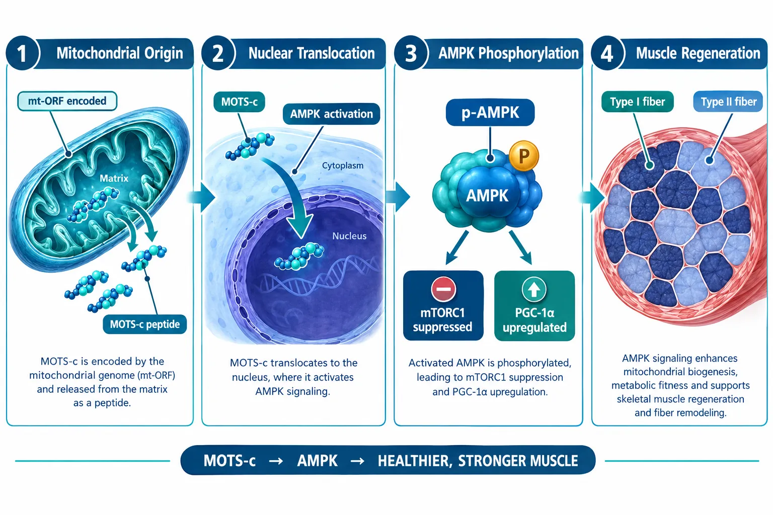
In skeletal muscle, the signaling cascade proceeds roughly as follows:
- Mitochondrial stress triggers MOTS-c release into the cytoplasm.
- MOTS-c activates AMP-activated protein kinase (AMPK), the master cellular energy sensor.
- Phosphorylated AMPK suppresses mTORC1, reducing anabolic overhead during recovery phases.
- Simultaneously, AMPK upregulates PGC-1alpha, the transcriptional coactivator governing mitochondrial biogenesis and oxidative metabolism.
Endurance-training studies in humans confirm that exercise-induced MOTS-c secretion from skeletal muscle correlates with the magnitude and duration of the AMPK, PGC-1alpha response. This positions MOTS-c not merely as a biomarker but as an active regulator of the metabolic adaptations researchers want to capture in regeneration models. Contextual factors, including age, baseline mitochondrial density, and training status, significantly modulate MOTS-c plasma levels, which has meaningful implications for experimental design.
For researchers exploring related mitochondrial peptide biology, the Mots C Elamipretide overview provides a useful comparative reference. Additional context on overlapping mitochondrial signaling compounds can be found in SS-31 mitochondrial research themes.
AMPK Readouts and Mitochondrial Flux: Interpreting Experimental Data
Quantifying AMPK activity and mitochondrial flux correctly is where many regeneration studies introduce avoidable error. The table below summarizes the primary assay categories used alongside MOTS-c and Slupp332 research protocols.
| Readout Category | Assay Method | Key Metric |
|---|---|---|
| AMPK activation | Western blot (p-AMPK / total AMPK) | Phosphorylation ratio |
| Mitochondrial flux | Seahorse XF respirometry | OCR, spare respiratory capacity |
| PGC-1alpha expression | RT-qPCR / immunofluorescence | mRNA and protein levels |
| Fiber-type composition | Immunohistochemistry (MHC isoforms) | Type I / IIa / IIx ratio |
| Glucose metabolism | Isotope tracing (U-13C-glucose) | Lactate-to-pyruvate flux |
Slupp332 approaches muscle fiber biology from a different angle. As a selective PPAR-delta (peroxisome proliferator-activated receptor delta) agonist, it activates oxidative gene programs independently of acute AMPK phosphorylation. PPAR-delta drives expression of genes associated with fatty acid oxidation, mitochondrial biogenesis, and slow-twitch muscle fiber identity, which is why fiber-type composition often appears as a secondary endpoint in Slupp332 protocols.
"The distinction between AMPK-mediated flux and PPAR-delta-driven transcription is not trivial, these are parallel but non-identical routes to oxidative remodeling, and conflating them leads to misattributed results."
When running both compounds in the same model, researchers must use phospho-AMPK western blots as the primary MOTS-c readout and separate PPAR-delta target gene panels (CPTIA, UQCRB, PDK4) for Slupp332. Oxygen consumption rate (OCR) measured via Seahorse respirometry provides a common functional endpoint that reflects the combined oxidative output.
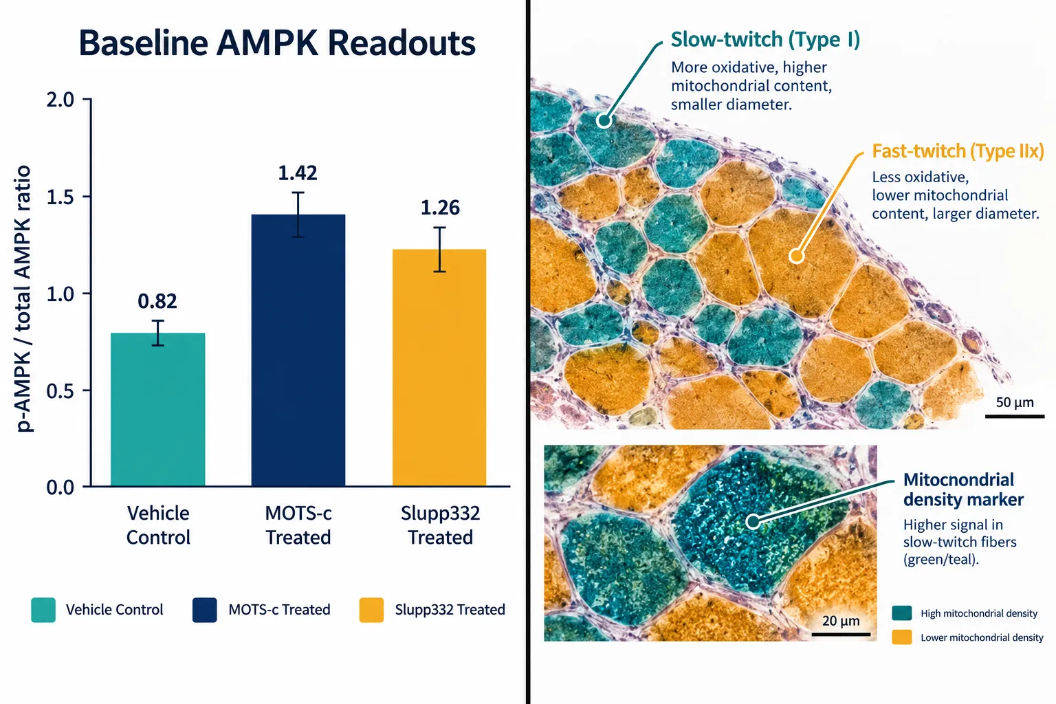
Cardiac remodeling models have served as useful analogs here, AMPK activation patterns in cardiomyocytes under stress closely mirror those seen in regenerating skeletal muscle satellite cells, providing cross-tissue validation logic for AMPK-dependent signaling. For related mitochondrial research considerations, the SS-31 10mg research peptide considerations article outlines practical experimental parameters that translate well to multi-peptide muscle protocols.
Fiber-Type Controls and Regeneration Timing in Preclinical Models
The fiber-type angle in research on MOTS-c and Slupp332 in skeletal-muscle regeneration models deserves careful framing. Current data show correlational shifts, increased Type I (slow-oxidative) fiber markers following treatment, rather than confirmed causal reprogramming. The cardiotoxin (CTX) injury model remains the standard for inducing synchronized regeneration in rodent muscle, providing clean timing windows for peptide administration.
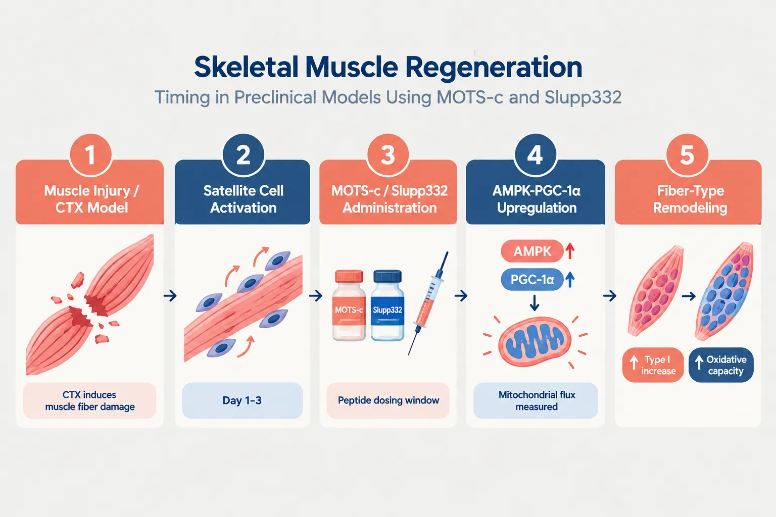
Critical design considerations for fiber-type and regeneration protocols:
- Vehicle controls must match solvent composition exactly, both MOTS-c (typically saline) and Slupp332 (often DMSO-based vehicles) require separate matched controls to isolate compound-specific effects.
- Dosing window matters enormously. MOTS-c shows greatest AMPK phosphorylation when administered during the proliferative phase (Day 2-5 post-CTX), while Slupp332's transcriptional effects on fiber-type markers become measurable in the differentiation phase (Day 5-14).
- Age-matched cohorts are essential. MOTS-c circulating levels decline with age, meaning young-adult controls will show different baseline AMPK responsiveness than aged muscle models studying sarcopenia-relevant endpoints.
- Nuclear translocation assays (fractionation western blot or proximity ligation assay) should be included when MOTS-c gene-regulatory effects are a primary hypothesis, as cytoplasmic AMPK activation and nuclear AMPK-driven transcription represent distinct mechanistic layers.
Neuropathic pain and spinal-cord repair models have independently validated AMPK-driven stress resilience pathways that overlap with skeletal-muscle regeneration logic, broadening the interpretive framework for MOTS-c AMPK readouts beyond pure muscle biology.
Research teams sourcing compounds for controlled preclinical work should prioritize purity-verified material. Reviewing lab tested peptides options and related SS-31 mechanism and research resources can help establish a baseline for sourcing standards applicable to MOTS-c studies.
Conclusion
Research on MOTS-c and Slupp332 in skeletal-muscle regeneration models: mitochondrial flux, AMPK readouts, and fiber-type controls is producing increasingly granular mechanistic data, but translating that data into reliable results demands methodological discipline. The actionable next steps for researchers are clear:
- Standardize AMPK readouts with validated p-AMPK / total AMPK western blot ratios rather than relying solely on downstream markers.
- Use Seahorse respirometry to capture oxygen consumption rate as a functional complement to gene expression data.
- Design separate vehicle controls for MOTS-c and Slupp332 given their different solvent requirements.
- Select dosing windows aligned to the proliferative versus differentiation phases of the CTX regeneration timeline.
- Interpret fiber-type data as correlational until mechanistic causation studies using fiber-type-specific knockouts are completed.
The intersection of mitochondria-derived signaling peptides and PPAR-delta pharmacology offers a compelling experimental framework for next-generation muscle-regeneration research in 2026 and beyond.


Leave a Reply
Want to join the discussion?Feel free to contribute!