5-Amino-1MQ and MOTS-c in Skeletal Muscle: How to Measure Glucose Uptake, Mitochondrial Flux, and Myotube Viability Together
Skeletal muscle consumes more than 80 percent of insulin-stimulated glucose in the human body, yet most in vitro assay designs test only one metabolic variable at a time. That single-readout approach creates a dangerous blind spot: a compound can appear to boost glucose uptake simply because it stresses mitochondria or reduces cell number, not because it genuinely improves metabolic capacity. Studying 5-Amino-1MQ and MOTS-c in skeletal muscle together demands a coordinated, three-readout strategy that keeps cytotoxicity, glucose handling, and mitochondrial flux in view simultaneously.
Key Takeaways
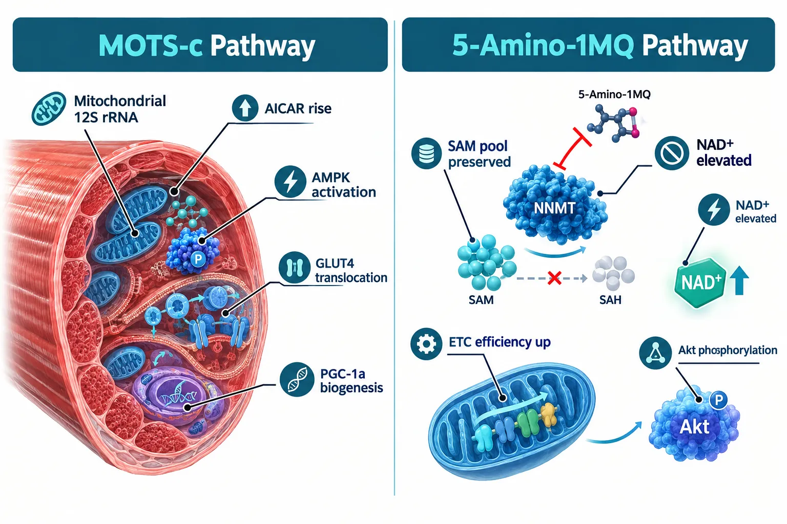
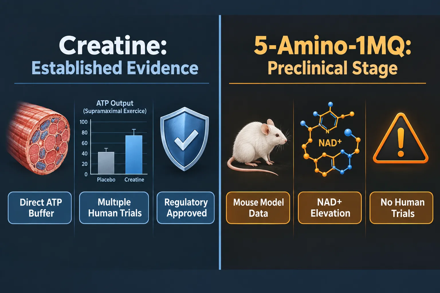
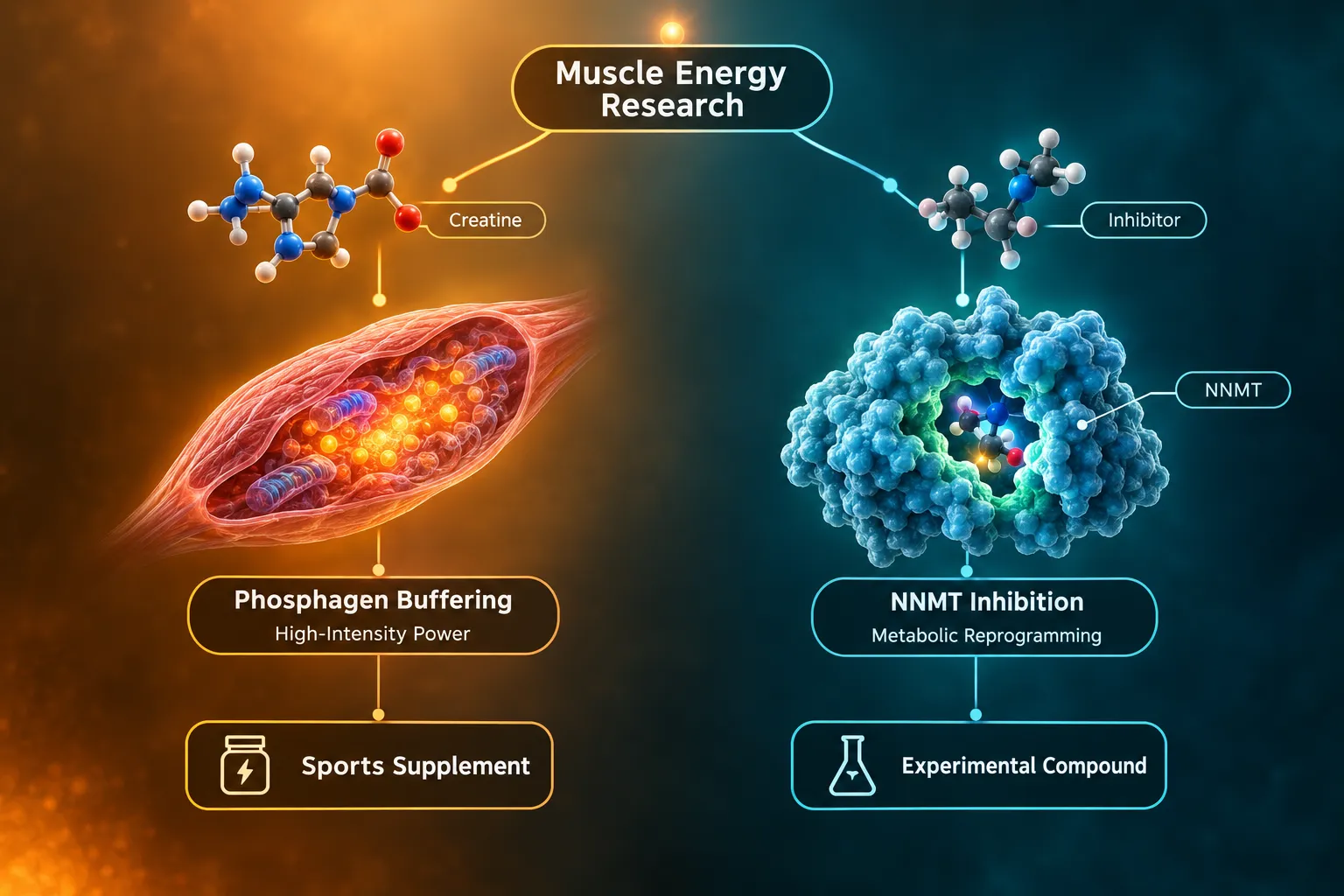
- 5-Amino-1MQ inhibits NNMT to raise NAD+ availability, while MOTS-c activates AMPK and CK2 signaling, targeting overlapping but distinct nodes in muscle metabolism.
- Neither compound has published co-administration data in skeletal muscle as of late 2026, making rigorous assay design essential before drawing mechanistic conclusions.
- Measuring glucose uptake, mitochondrial oxygen consumption rate (OCR), and myotube viability in parallel prevents false-positive metabolic readings caused by cytotoxicity or mitochondrial stress.
- Assay sequence matters: viability checks must bookend the protocol, not just follow it.
- Differentiation state and passage number of myotubes are major confounders that must be standardized before any treatment is applied.
The Mechanistic Case for Combining 5-Amino-1MQ and MOTS-c
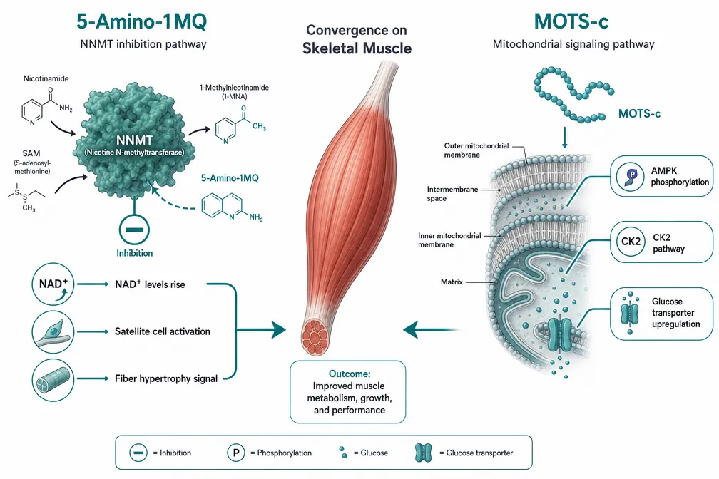
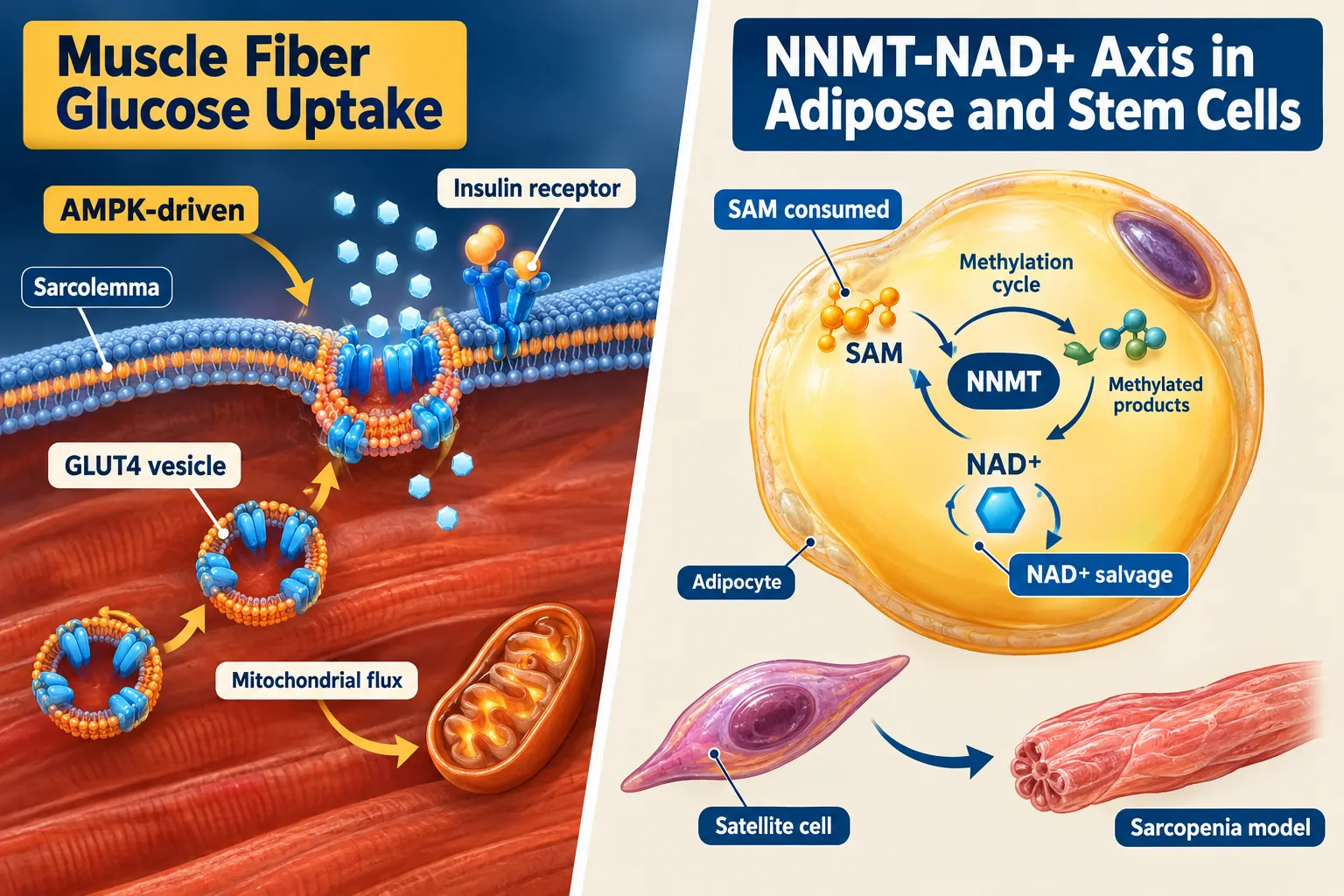
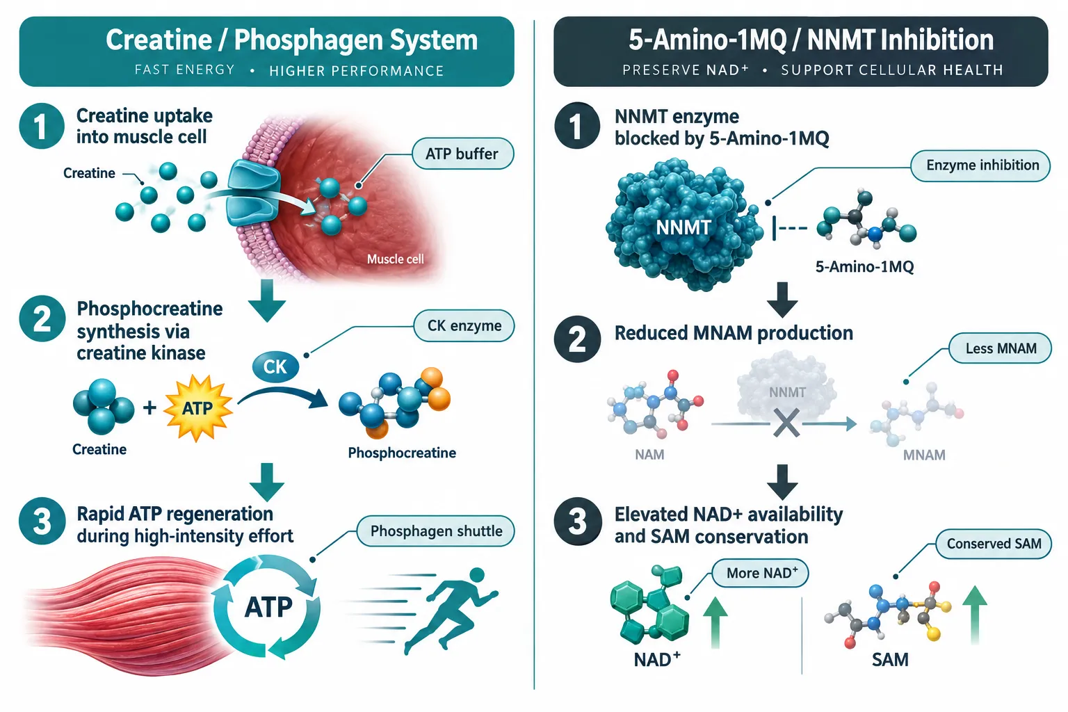
5-Amino-1MQ is a small-molecule inhibitor of nicotinamide N-methyltransferase (NNMT), an enzyme highly expressed in aged and metabolically compromised muscle tissue. When NNMT is inhibited, the methyl donor pool shifts, NAD+ precursor availability increases, and downstream sirtuin and PARP signaling becomes more active. In aged rodent models, this change correlates with satellite cell re-entry into the cell cycle, greater fiber cross-sectional area, and measurable improvements in grip strength. Early mitochondrial respiration data suggest that NNMT inhibition also raises electron transport chain capacity, though the dose-response relationship in human primary myotubes is still being characterized as of 2026.
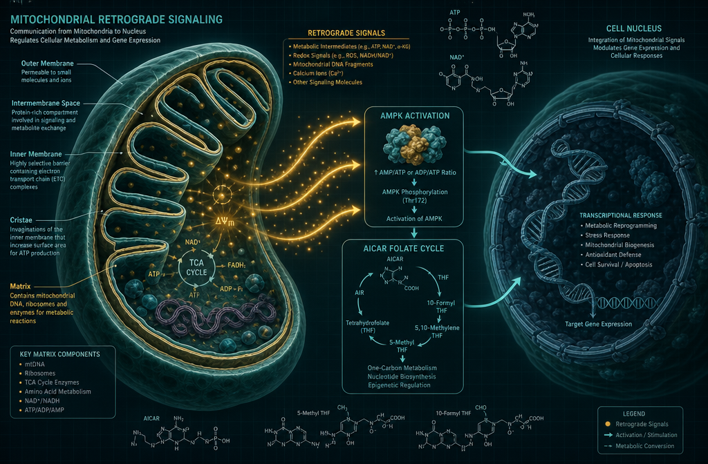
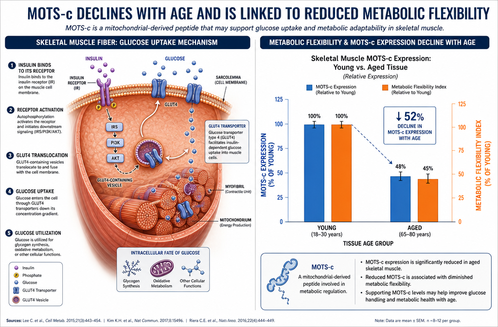
MOTS-c is a mitochondria-derived peptide encoded within the 12S rRNA region of the mitochondrial genome. Its best-characterized action in skeletal muscle is AMPK activation, which drives GLUT4 translocation and glucose uptake independent of insulin. More recent work has identified CK2 (casein kinase 2) as a direct upstream activator of MOTS-c nuclear translocation, broadening its role from a metabolic peptide to a stress-responsive transcriptional co-regulator. MOTS-c also reduces muscle atrophy triggered by immobilization, an effect tied to suppression of atrogene expression rather than to direct anabolic signaling.
Because 5-Amino-1MQ works primarily through epigenetic and NAD+ remodeling while MOTS-c acts through rapid kinase signaling, the two agents target the same metabolic outcome, improved muscle glucose handling and mitochondrial efficiency, through non-overlapping pathways. That complementarity is exactly why parallel assay measurement is non-negotiable: additive effects, antagonism, or off-target cytotoxicity each produce distinct fingerprints only visible when all three readouts run concurrently.
Researchers interested in mitochondrial-targeted peptides may also find value in reviewing SS-31 mitochondrial dynamics research, which provides a useful methodological reference for flux-based assay design in muscle cells.
Building the Assay Matrix: Sequence, Tools, and Confounders
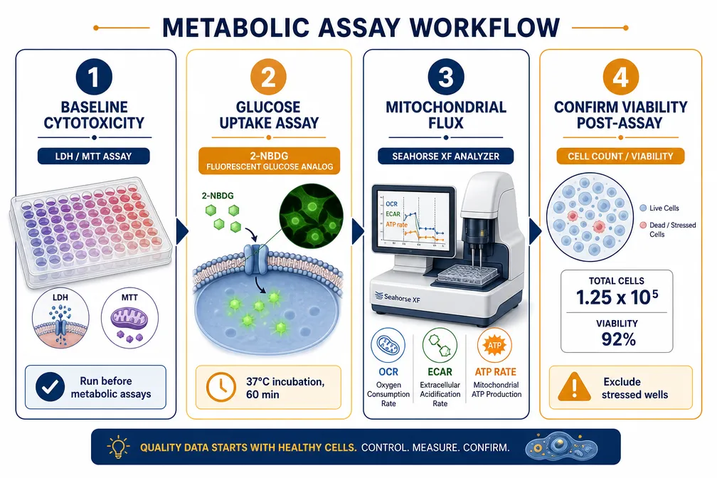
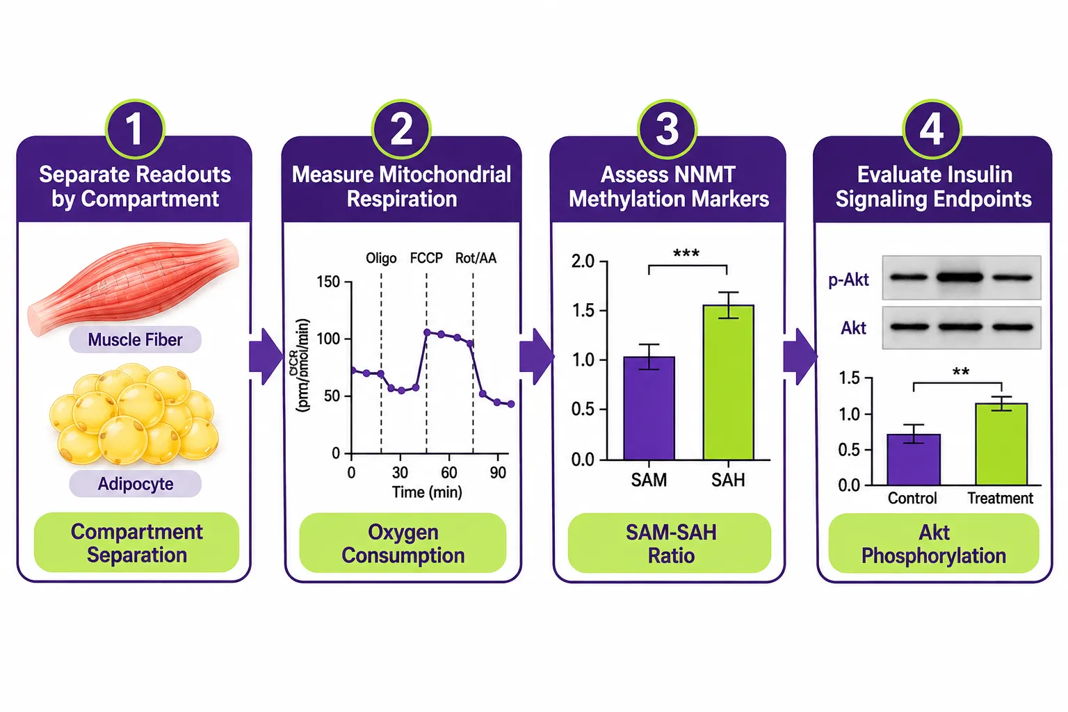
The table below outlines the recommended assay architecture for studying 5-Amino-1MQ and MOTS-c in skeletal muscle: how to measure glucose uptake, mitochondrial flux, and myotube viability together within a single experimental run.
| Assay Step | Method | Key Output | Why It Matters Here |
|---|---|---|---|
| Pre-treatment viability | MTT or CellTiter-Glo | Baseline ATP/cell count | Confirms uniform differentiation across wells |
| Glucose uptake | 2-NBDG fluorescent analog or 3H-2-DG | Relative glucose flux per cell | Distinguishes GLUT4 translocation from passive diffusion |
| Mitochondrial flux | Seahorse XF Analyzer (OCR/ECAR) | Basal respiration, ATP-linked OCR, spare capacity | Separates genuine OXPHOS gain from uncoupling artifacts |
| ROS quantification | MitoSOX or CM-H2DCFDA | Mitochondrial superoxide level | Elevated ROS can inflate apparent glucose uptake |
| Post-treatment viability | LDH release assay | Membrane integrity / cytotoxicity | Flags wells where metabolic signal reflects cell death, not improvement |
Differentiation State as a Hidden Confounder
Primary human skeletal muscle cells must be differentiated for at least five to seven days before treatment. Underdifferentiated cultures express low levels of GLUT4 and lack the contractile architecture needed for meaningful MOTS-c responses. Passage number should stay below passage six; beyond that, satellite cell identity markers decline and NNMT expression patterns shift, potentially masking 5-Amino-1MQ effects.
Dosing Windows and Timing
For 5-Amino-1MQ, the research literature in myotube models points toward a 24- to 48-hour pre-incubation window at sub-micromolar concentrations to allow NAD+ pool remodeling before metabolic readouts are collected. MOTS-c, being a peptide, acts more acutely: significant AMPK activation is detectable within 30 to 60 minutes. A staggered dosing design, MOTS-c added during the assay window after overnight 5-Amino-1MQ pre-loading, avoids timing confounds and mirrors a biologically plausible sequence. For labs sourcing mitochondrial peptides for such work, SS-31 peptide research considerations offer a practical quality-control reference for peptide-based in vitro protocols.
Interpreting the Triple-Readout Results Without Misreading the Data
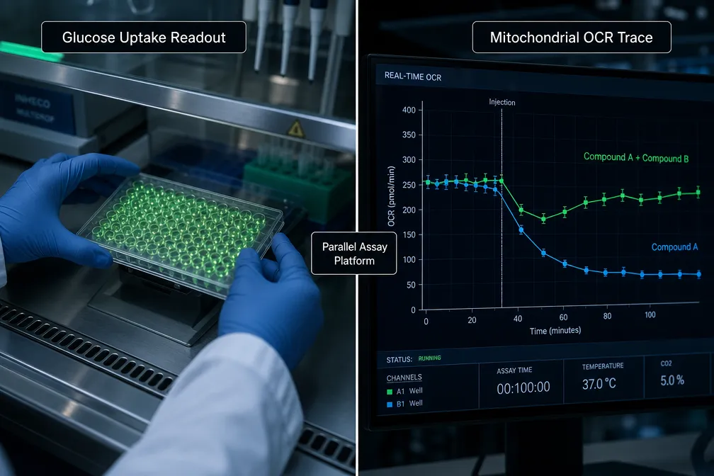
The central value of measuring 5-Amino-1MQ and MOTS-c in skeletal muscle: how to measure glucose uptake, mitochondrial flux, and myotube viability together is the ability to cross-reference outcomes and eliminate artifactual signals.
Three common misinterpretation scenarios to guard against:
-
Elevated glucose uptake with reduced cell viability, LDH release rises alongside glucose signal. This pattern indicates membrane disruption is driving passive glucose influx, not active transporter-mediated uptake. Discard the glucose data from those wells.
-
Increased OCR with elevated ROS and suppressed ATP-linked respiration, This is the classic mitochondrial uncoupling artifact. Oxygen consumption rises, but it is dissipated as heat rather than converted to ATP. A compound appearing to "boost metabolism" under this pattern is actually inducing mitochondrial stress.
-
No change in glucose uptake but improved spare respiratory capacity, This result is genuinely informative. It suggests the compound improved mitochondrial resilience without altering basal glucose flux, a distinct physiological outcome worth reporting separately.
A key interpretive principle: improvements in OCR are only metabolically meaningful when ATP production rate rises in parallel and LDH remains at baseline levels.
Researchers exploring multi-peptide experimental designs may also find the synergy of LL-37 and SS-31 research useful as a methodological template for interpreting combined-agent assay results. For labs sourcing research peptides, reviewing where to buy SS-31 and Epithalon provides guidance on quality standards relevant to any peptide used in metabolic assays. Those interested in the broader mitochondrial peptide landscape should also consult the SS-31 mechanism and research overview for cross-compound context. For labs working with lab-tested peptides, purity documentation is a prerequisite before any flux assay results can be considered interpretable.
Conclusion
The only reliable way to study 5-Amino-1MQ and MOTS-c in skeletal muscle: how to measure glucose uptake, mitochondrial flux, and myotube viability together is to treat all three readouts as a single integrated panel, not as separate experiments. The immediate actionable steps for any research team are:
- Standardize differentiation to day 5-7, passage 3-5, before any compound treatment.
- Run pre- and post-treatment viability assays in every experiment, not as an afterthought.
- Include ROS quantification alongside OCR to distinguish genuine OXPHOS improvement from uncoupling.
- Apply staggered dosing (5-Amino-1MQ overnight pre-load, MOTS-c acute treatment) to respect each agent's pharmacodynamic timeline.
- Cross-reference all five data layers before drawing conclusions about metabolic improvement.
As of 2026, no published study has used this combined assay framework with both compounds simultaneously. That gap is not a reason for caution, it is an open invitation for rigorous, well-designed research.