

Amlodipine in Peptide Research: Separating Calcium-Channel Effects From Blood-Pressure and Vascular Readouts
Fewer than one in five preclinical peptide studies that co-administer a calcium-channel blocker formally account for the drug's independent vascular effects, a methodological gap that can quietly invalidate entire datasets. When amlodipine in peptide research enters the experimental picture, the challenge is not simply adjusting for lower blood pressure. It is disentangling at least three overlapping biological layers: direct L-type calcium-channel blockade, blood-pressure-dependent vascular changes, and blood-pressure-independent vascular remodeling. Getting these layers wrong means attributing amlodipine's known anti-proliferative and anti-atherosclerotic actions to the peptide under study, or, just as problematically, masking a real peptide signal behind the drug's dominant pharmacology.
Key Takeaways
- Amlodipine blocks L-type calcium channels in vascular smooth muscle, reducing intracellular Ca²⁺ and causing arterial relaxation independently of any peptide co-administered in a study.
- The drug produces vascular benefits, including inhibited smooth-muscle cell proliferation and slowed atherosclerosis, that are separate from its blood-pressure-lowering effect.
- At clinically relevant concentrations, amlodipine does not activate CRAC channels and does not interfere with fluorescence-based Ca²⁺ imaging dyes such as Cal-520, Fluo-4, or Fura-Red.
- Researchers should pre-specify which readouts are calcium-channel-mediated, blood-pressure-dependent, or peptide-specific before data collection begins.
- Orthogonal biomarkers, endothelial nitric oxide, arterial compliance, and vascular remodeling indices, can help isolate a peptide's contribution from amlodipine's background pharmacology.
How Amlodipine Works: The Calcium-Channel Mechanism Researchers Must Account For
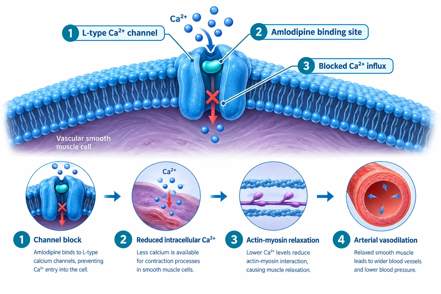
Amlodipine is a dihydropyridine calcium antagonist. Its primary action is inhibiting the transmembrane influx of calcium ions through L-type voltage-gated channels, predominantly in vascular smooth muscle cells. The result is reduced intracellular Ca²⁺, diminished actin-myosin interaction, smooth-muscle relaxation, and arterial vasodilation. Its effect on vascular smooth muscle is substantially greater than its effect on cardiac muscle, a selectivity profile that matters when interpreting any cardiovascular or vascular readout in a peptide experiment.
This mechanism is not trivial background noise. It is a dominant pharmacological signal. Any peptide co-administered with amlodipine that also influences vascular tone, intracellular calcium, or smooth-muscle behavior will generate data that reflects both agents simultaneously unless the study design explicitly separates them.
What this means practically:
- Vascular tone measurements (e.g., wire myography, pressure myography) will reflect amlodipine's baseline relaxation effect.
- Intracellular Ca²⁺ flux assays will show a suppressed resting Ca²⁺ level before the peptide is even introduced.
- Any peptide with vasodilatory properties may appear to have a blunted or absent effect simply because the vascular bed is already partially relaxed.
Researchers working with mitochondria-targeted peptides such as those explored in SS-31 10mg research peptide considerations should note that oxidative-stress and membrane-potential readouts can also be indirectly affected when intracellular calcium homeostasis is altered by a co-administered channel blocker.
Separating Calcium-Channel Effects From Blood-Pressure and Vascular Readouts: A Practical Framework
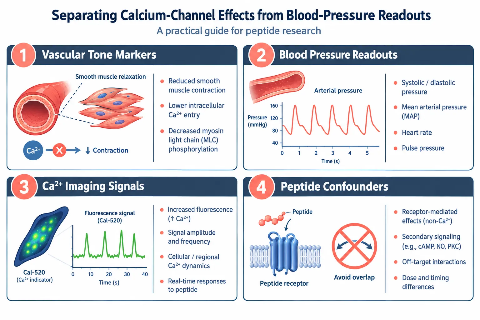
The central challenge in amlodipine in peptide research, separating calcium-channel effects from blood-pressure and vascular readouts, requires a tiered classification of every endpoint before data collection begins.
Tier 1: Blood-Pressure-Dependent Readouts
These are endpoints that change because amlodipine lowers arterial pressure. They include:
- Brachial or carotid blood pressure values
- Cardiac afterload indices
- Glomerular filtration rate changes driven by reduced perfusion pressure
Practical step: Include a vehicle-treated, amlodipine-only control group at the same dose. Any change in these readouts in that group represents the amlodipine baseline, not a peptide effect.
Tier 2: Blood-Pressure-Independent Vascular Effects
Contemporary cardiovascular literature consistently identifies a second category of amlodipine actions that persist even when blood pressure is matched to comparator agents. These include:
| Effect | Mechanism | Research Implication |
|---|---|---|
| Smooth-muscle cell anti-proliferation | Direct Ca²⁺ signaling suppression | May confound peptide anti-proliferative claims |
| Anti-atherosclerotic remodeling | Reduced oxidative stress in vessel wall | Arterial stiffness data needs amlodipine-only control |
| Improved arterial compliance | Structural vascular changes | Central pulse-wave velocity requires correction |
For peptide studies examining vascular remodeling, such as those involving VIP peptide mechanisms or vasoactive signaling pathways, these BP-independent effects represent the most likely source of confounding.
Tier 3: Calcium-Channel-Specific Molecular Readouts
This tier covers direct molecular measurements of L-type channel activity and downstream Ca²⁺ signaling. A key methodological advantage here: at therapeutic plasma concentrations of approximately 0.5 µM, amlodipine does not activate calcium-release-activated calcium (CRAC) channels. This means CRAC-mediated signaling events attributed to a peptide are not being driven by amlodipine itself.
Additionally, fluorescence-based Ca²⁺ imaging using dyes such as Cal-520, Fluo-4, and Fura-Red is not confounded by amlodipine fluorescence at these concentrations. This is a meaningful green light for researchers using live-cell calcium imaging alongside peptide stimulation assays.
"Knowing which calcium compartment amlodipine does and does not touch is the single most useful piece of information a peptide researcher can carry into experimental design."
Researchers examining peptide signaling pathways, including those studying GLP-3 peptide signaling pathway interactions, benefit from this CRAC-channel clarity because receptor-operated calcium entry can then be attributed with greater confidence to the peptide's receptor engagement rather than to the channel blocker.
Designing the Experiment: Isolating Peptide-Specific Signals
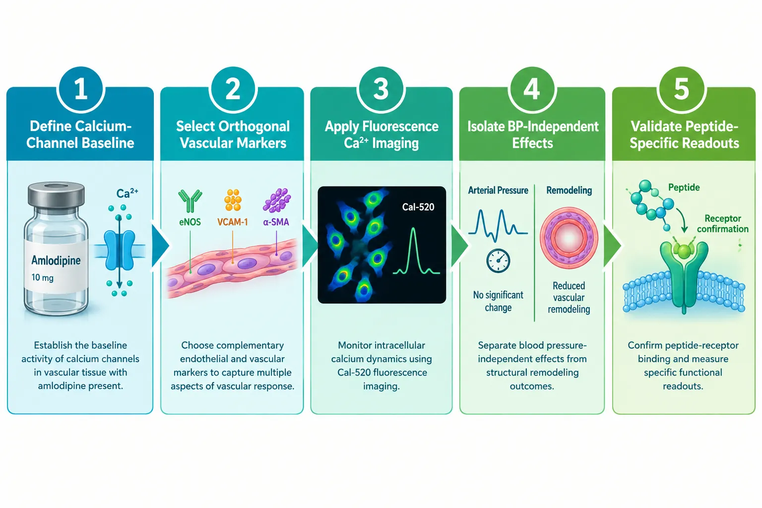
A rigorous experimental framework for amlodipine in peptide research, one that genuinely separates calcium-channel effects from blood-pressure and vascular readouts, involves five concrete design decisions.
Step 1, Define the calcium-channel baseline. Run amlodipine-only controls at the intended study concentration. Measure intracellular Ca²⁺, vascular tone, and any remodeling markers. This establishes the floor the peptide must be measured against.
Step 2, Select orthogonal vascular markers. Choose at least two endpoints that amlodipine does not directly regulate. Endothelial nitric oxide synthase (eNOS) activity, circulating endothelin-1 levels, and specific peptide receptor phosphorylation states are examples. These serve as peptide-specific signal channels.
Step 3, Apply fluorescence Ca²⁺ imaging with confidence. Given that amlodipine does not interfere with Cal-520, Fluo-4, or Fura-Red at relevant concentrations, live-cell imaging remains a valid tool. Pair it with a CRAC-channel inhibitor control to confirm that any observed Ca²⁺ flux is L-type-independent when the peptide is active.
Step 4, Separate central from peripheral blood pressure. Peripheral brachial pressure can be identical across treatment groups while central aortic pressure and pulse-wave velocity differ. Use central hemodynamic measurements to capture amlodipine's BP-independent arterial effects separately from peptide-driven changes.
Step 5, Validate with receptor-blocking controls. If the peptide of interest acts through a specific receptor, as is the case with many research peptides including those reviewed in LL-37 versus SS-31 benefits of each peptide comparisons, include a receptor-antagonist arm. Any vascular effect that disappears with receptor blockade but persists with amlodipine alone is attributable to the peptide.
Proper peptide dosing protocols also matter here: dose-response curves for the peptide should be generated in both the presence and absence of amlodipine to detect any shift in potency or efficacy caused by the background calcium-channel blockade.
For researchers sourcing compounds for controlled in vitro work, ensuring purity and verified composition is non-negotiable. Resources such as guidance on where to buy research-grade peptides evaluating purity underscore why compound quality directly affects the reliability of mechanistic conclusions.
Conclusion
Amlodipine is a well-characterized L-type calcium-channel blocker with a pharmacological footprint that extends well beyond simple blood-pressure reduction. In any peptide research context, failing to account for its anti-proliferative, anti-atherosclerotic, and Ca²⁺-suppressive actions risks misattributing drug effects to the peptide, or obscuring genuine peptide biology beneath a dominant pharmacological background.
Actionable next steps for researchers in 2026:
- Pre-classify every planned endpoint into one of the three tiers: blood-pressure-dependent, BP-independent vascular, or calcium-channel-specific molecular.
- Include an amlodipine-only control group at the study concentration in every experimental arm.
- Use fluorescence-based Ca²⁺ imaging with confidence at therapeutic concentrations, but pair it with CRAC-channel controls to confirm the signal source.
- Add orthogonal endothelial and receptor-specific biomarkers that amlodipine does not directly regulate.
- Run full dose-response curves for the peptide both with and without amlodipine present to detect potency shifts.
Rigorous separation of these layers does not complicate peptide research, it protects it. The result is data that can be defended, reproduced, and built upon.


Leave a Reply
Want to join the discussion?Feel free to contribute!