Call or Text 727-513-9780
  • Shopping Cart Shopping Cart
    0Shopping Cart
Pure Tested Peptides | America's most trusted Peptides for sale online
  • Peptides for sale
    • Oral Peptides for sale
      • Peptide Capsules for sale
      • BPC 157 Capsules 1000mcg
      • SLU-PP-332 Capsules | 1000 mcg
      • 5-Amino-1MQ 50mg Capsules
      • Tesofensine 500mcg
    • All Peptides for sale
    • Peptide Sprays
      • BPC 157 Nasal Spray Kit
      • BPC-157 TB500 Nasal Spray Kit
      • Semax Nasal Spray 10mg
      • Selank – Nasal Spray Kit – 10mg
      • Epithalon 50MG Nasal Spray Kit
      • Ipamorelin 10mg Nasal Spray
      • Klow Nasal Spray (BPC-157 + TB-500 + GHK-Cu + KPV) | 80mg
      • Hulk Nasal Spray Tesa / Ipa Blend 6/3 MG
      • Klow Nasal Spray
      • NAD + 500 mg Nasal Spray
      • PT-141 Nasal Spray Kit
    • GHRH Peptides
      • Ipa Peptides
      • CJC-1295 Peptides
        • CJC-1295 with DAC 5 mg
        • CJC-1295 without DAC 5 mg
        • CJC-1295 Ipa 10mg
      • Tesa Peptides
        • Tesa Peptide
        • Tesa 20 mg
    • GHK-Cu Peptides
      • All GHK-Cu Peptides
      • GHK-Cu 100mg
      • KLOW Peptide Blend – Buy KLOW blend online
    • BPC Peptides
      • All BPC Peptides
      • BPC-157
      • BPC-157 TB-500
      • BPC 157 capsules 1000mcg
    • SLU-PP-332 Peptides
      • All SLU-PP-332 Peptides
      • SLU-PP-332 5mg
    • GLP3 Peptides
    • PT-141 Peptides
      • PT-141 Peptides for sale
      • PT-141 10mg
      • PT-141 Nasal Spray
    • CAG Peptides
      • Lipo-C Peptide Blend
      • CAG 5mg
      • CAG 10mg
    • MOTS-C Peptides
      • MOTS-C Peptides for sale
      • MOTS-c peptide
      • MOTS-c 10mg *6 pack*
    • 5 Amino 1MQ Peptides
      • 5 Amino 1MQ Peptides for sale
      • 5-Amino-1MQ 50mg Capsules
      • 5-Amino-1MQ 5mg
    • Epithalon Peptides
      • Epithalon Peptides for sale
      • Epithalon 10mg
      • Epithalon 50mg
  • Shop
    • GLPs
      • 5-Amino-1MQ 50mg Capsules
      • 5-Amino-1MQ 5mg
      • L-Carnitine 500mg/ml
      • Tesofensine 500mcg
      • SLU-PP-332 5mg
      • MOTS-c 10mg *6 pack*
    • Epithalon & BPC Peptides
      • Epithalon 10mg
      • Epithalon 50mg
      • BPC-157
      • BPC 157 capsules 1000mcg
      • BPC-157 TB-500
      • BPC-157 TB500 Nasal Spray Kit
      • BPC 157 Nasal Spray Kit
    • BPC TB-500 & NAD+ Peptides
      • NAD+ 500 mg
      • KLOW Peptide Blend – Buy KLOW blend online
      • GLOW Peptide Blend
      • TB 500 5mg
      • BPC 157 capsules 1000mcg – Supplement
      • BPC 157 Nasal Spray Kit
      • BPC-157
      • BPC-157 TB500 Nasal Spray Kit
      • BPC-157 TB-500
      • BPC 157 capsules 1000mcg
    • LL-37 Peptide
      • LL-37 10 mg
    • MOTS-C & Selank
      • MOTS-c peptide
      • Selank 10mg
    • GHK Peptides
      • GHK-Cu 100mg
      • GLOW Peptide Blend
      • KLOW Peptide Blend – Buy KLOW blend online
  • COAs
  • Wholesale
    • Wholesale Peptides for sale
  • PTP FAQ
  • Affiliates
    • Selank Peptide: Advanced Pharmacological Mechanisms Underlying Its Anxiolytic and Nootropic Effects in Research
      • 5-Amino-1MQ Peptide: Detailed Mechanisms of NNMT Inhibition and Its Impact on Cellular Metabolism Research
        • MOTS-C Peptide: Unraveling Its Role in Mitochondrial Dynamics and Energy Metabolism Research
          • Epithalon Peptide and Telomerase Regulation: Investigating Its Impact on Cellular Senescence and Lifespan Research Models
            • GLP2-T vs GLP2 Tirz Peptide: Understanding the Naming, Mechanistic Nuances, and Research Implications for Gut Health
      • Peptides and Polypeptides in Modern Pharmacology: What Research on Metoprolol, Prednisone, and Amlodipine Reveals
        • Polypeptide Peptides vs NSAIDs: What Naproxen and Diclofenac Teach Tissue-Repair Researchers About BPC‑157 and TB‑500
          • Peptides and Polypeptides in Endocrine Pharmacology: How GLP-1, GLP-2, and GLP-3 Retatrutide Differ From Classic Drugs Like Prednisone and Amlodipine
          • 5‑Amino‑1MQ Peptide and NNMT Inhibition: How It Compares With Statins Like Atorvastatin in Adipose and Lipid Metabolism Research
      • Enclomiphene, Estrogen Receptor Signaling, and Luteinizing Phase Biology: What Hormone Researchers Should Measure
        • Mesenchymal Stem Cells, BPC‑157, and GHK‑Cu: How Tissue Repair Peptides Compare With Classic NSAIDs Like Naproxen in Injury Models
          • Peptide Calculator Use Cases Beyond Growth Hormone: Working Through GLP‑3 Retatrutide, MOTS‑c, and BPC‑157 Research Dosing
        • Polypeptide Peptides in Endocrine and Metabolic Pharmacology: Lessons From Amlodipine, Prednisone, and Metoprolol
        • Peptides Calculator for Advanced Blends: Worked Examples for Tesamorelin, CJC‑1295, and Ipamorelin Stacks
          • Peptides and Polypeptides in Human Physiology: How Molecular Size Shapes Research Applications
            • Selank Peptide: Advanced Pharmacological Mechanisms Underlying Its Anxiolytic and Nootropic Effects in Research
          • 5-Amino-1MQ Peptide: Detailed Mechanisms of NNMT Inhibition and Its Impact on Cellular Metabolism Research
            • MOTS-C Peptide: Unraveling Its Role in Mitochondrial Dynamics and Energy Metabolism Research
              • Epithalon Peptide and Telomerase Regulation: Investigating Its Impact on Cellular Senescence and Lifespan Research Models
                • GLP2-T vs GLP2 Tirz Peptide: Understanding the Naming, Mechanistic Nuances, and Research Implications for Gut Health
                  • Peptides and Polypeptides in Endocrine Pharmacology: How GLP-1, GLP-2, and GLP-3 Retatrutide Differ From Classic Drugs Like Prednisone and Amlodipine
                    • Polypeptide Peptides vs NSAIDs: What Naproxen and Diclofenac Teach Tissue-Repair Researchers About BPC‑157 and TB‑500
                      • Peptides and Polypeptides in Modern Pharmacology: What Research on Metoprolol, Prednisone, and Amlodipine Reveals
                        • 5‑Amino‑1MQ Peptide and NNMT Inhibition: How It Compares With Statins Like Atorvastatin in Adipose and Lipid Metabolism Research
                        • Enclomiphene, Estrogen Receptor Signaling, and Luteinizing Phase Biology: What Hormone Researchers Should Measure
                        • Mesenchymal Stem Cells, BPC‑157, and GHK‑Cu: How Tissue Repair Peptides Compare With Classic NSAIDs Like Naproxen in Injury Models
                        • Peptide Calculator Use Cases Beyond Growth Hormone: Working Through GLP‑3 Retatrutide, MOTS‑c, and BPC‑157 Research Dosing
                        • Polypeptide Peptides in Endocrine and Metabolic Pharmacology: Lessons From Amlodipine, Prednisone, and Metoprolol
                        • Peptides Calculator for Advanced Blends: Worked Examples for Tesamorelin, CJC‑1295, and Ipamorelin Stacks
                        • Peptides and Polypeptides in Human Physiology: How Molecular Size Shapes Research Applications
                        • Selank Peptide: Advanced Pharmacological Mechanisms Underlying Its Anxiolytic and Nootropic Effects in Research
                        • MOTS-C Peptide: Unraveling Its Role in Mitochondrial Dynamics and Energy Metabolism Research
                        • Peptides and Polypeptides in Modern Pharmacology: What Research on Metoprolol, Prednisone, and Amlodipine Reveals
                        • Peptides and Polypeptides in Endocrine Pharmacology: How GLP-1, GLP-2, and GLP-3 Retatrutide Differ From Classic Drugs Like Prednisone and Amlodipine
                        • 5-Amino-1MQ Peptide: Detailed Mechanisms of NNMT Inhibition and Its Impact on Cellular Metabolism Research
                        • Mesenchymal Stem Cells, BPC‑157, and GHK‑Cu: How Tissue Repair Peptides Compare With Classic NSAIDs Like Naproxen in Injury Models
                        • Peptide Calculator Use Cases Beyond Growth Hormone: Working Through GLP‑3 Retatrutide, MOTS‑c, and BPC‑157 Research Dosing
                        • GLP2-T vs GLP2 Tirz Peptide: Understanding the Naming, Mechanistic Nuances, and Research Implications for Gut Health
                        • Polypeptide Peptides in Endocrine and Metabolic Pharmacology: Lessons From Amlodipine, Prednisone, and Metoprolol
                        • Polypeptide Peptides vs NSAIDs: What Naproxen and Diclofenac Teach Tissue-Repair Researchers About BPC‑157 and TB‑500
                        • Peptides and Polypeptides in Human Physiology: How Molecular Size Shapes Research Applications
                        • Peptides Calculator for Advanced Blends: Worked Examples for Tesamorelin, CJC‑1295, and Ipamorelin Stacks
                        • 5‑Amino‑1MQ Peptide and NNMT Inhibition: How It Compares With Statins Like Atorvastatin in Adipose and Lipid Metabolism Research
                        • Enclomiphene, Estrogen Receptor Signaling, and Luteinizing Phase Biology: What Hormone Researchers Should Measure
                        • Epithalon Peptide and Telomerase Regulation: Investigating Its Impact on Cellular Senescence and Lifespan Research Models
                        • Best research protocol Klow blend
                        • best time to take BPC-157
                        • best time to take DSIP (Delta Sleep Inducing Peptide)
                        • best time to take CJC-1295
                        • best time to take AOD-9604
                        • best time to take Follistatin 344
                        • best time to take Ipamorelin
                        • best time to take MK-677 (Ibutamoren)
                        • best time to take Ligandrol (LGD-4033) — research compound
                        • best time to take Ostarine (MK-2866) — research compound
                        • best time to take GHK-CU
                        • best time to take TB-500
                        • best time to take MOTS-c
                        • best time to take Semax
                        • best time to take RAD-140 (Testolone) — research compound
                        • best time to take Thymosin Alpha-1
                        • best time to take PEG-MGF
                        • Biolife Plasma, Octapharma Plasma, and Research Peptides: How Plasma Donation Labs Differ From Peptide Suppliers
                        • best time to take YK-11 — research compound
                        • best time to take PT-141 (Bremelanotide)
                        • Best research protocol Klow blend
                        • 5-Amino-1MQ and MOTS-C Synergy: Metabolic Signaling, Mitochondria, and Research Design
                        • BPC-157 and TB-500: Investigating Their Combined Effects on Angiogenesis and Cellular Migration in Tissue Repair Models
                        • BPC-157 Peptide: Gut Barrier Function, Inflammation, and Tissue-Recovery Research
                        • 5‑Amino‑1MQ and MOTS‑c Synergy in Metabolic Research: Designing NNMT and Mitochondrial Biogenesis Stacks
                        • CJC-1295 with DAC vs. Without DAC: Half-Life, Release Kinetics, and Research Implications
                        • CJC‑1295 with DAC vs. Without DAC: Expanding on Half‑Life Differences Using Tesamorelin and Ipamorelin Blend Case Studies
                        • Collagen Biology and Copper‑Binding Peptides: How GHK‑Cu, Glow Blend, and Klow Blend Interact with Skin and Connective Tissue
                        • Collagen Biology and Regenerative Peptides: How GHK‑Cu, Glow Blend, and Klow Blend Affect Extracellular Matrix Research
                    • DNA, Telomeres, and Longevity Peptides: Positioning Epithalon and MOTS‑c in Genetic Aging Research
                      • Enclomiphene Citrate: serm Mechanism, Testosterone Research, and Stack Compatibility
                        • Enclomiphene vs Enclomiphene Citrate: Formulation, Bioavailability, and Research Distinctions
                        • Epithalon Peptide Research: Telomerase Activation, Aging, and Pineal Gland Function
                        • Estrogen Receptor Signaling and Enclomiphene: How Selective Modulators Compare with Classic Polypeptide Hormones
                        • GHK-Cu Peptide: Advanced Mechanisms in Extracellular Matrix Remodeling and Wound Healing Research
                        • GHK-Cu Peptide: Collagen Synthesis, Wound Repair, and Skin-Barrier Research Models
                        • GLP-1 vs GLP-3 vs GLP-2: Peptide Classification and Research Applications
                        • GLP-2 Peptide Research Guide: Gut Barrier Function, Nutrient Absorption, and Intestinal Recovery Models
                        • GLP-3 Retatrutide vs. GLP-1 Drugs: What Triple-Agonist Biology Changes in Research Models
                        • Ipamorelin and Tesamorelin Combination: Synergistic GH Secretagogue Research and Dosing Protocols
                        • GLP2 Tirz Peptide: What It Is, Why the Name Exists, and How Researchers Should Interpret It
                        • Klow Blend Peptide Nasal Spray: What the Formulation Is Trying to Do in Cognitive Research
                        • Mitochondria, NNMT Inhibition, and Peptide Modulators: Where MOTS‑c and 5‑Amino‑1MQ Fit in Cellular Energy Research
                        • MOTS-c Peptide: Mitochondrial Function, Energy Metabolism, and What Researchers Measure
                        • MOTS-c vs. 5-Amino-1MQ: Which Metabolic Research Questions Each Compound Actually Answers
                        • Nasal Spray Peptides: Bioavailability, Administration, and Semax/Selank Research Applications
                        • PT-141 Peptide Research: Mechanism of Action and Melanocortin Receptor Signaling
                        • Retatrutide for Research: Mechanism, Structure, and GLP-1/GLP-3 Dual Action
                        • Retatrutide for Obesity and Type 2 Diabetes: What the Latest Trial Data Suggest
                        • Tesofensine Peptide Research: Mechanism, Appetite Suppression, and Neuropeptide Y Pathways
  • Contact
    • Contact Customer Service
    • Text Customer Support
  • About US
  • Shop all peptides
  • Affiliate Program
    • Affiliate Signup
  • Login / Register Login / Register Page Link Login / Register Page Link
  • Click to open the search input field Click to open the search input field Search
  • Menu Menu

Tag Archive for: peptide reconstitution

Peptide Reconstitution and Stability: How Buffer Choice, pH, Osmolality, and Freeze–Thaw Cycles Affect Research Samples

Peptide Reconstitution and Stability: How Buffer Choice, pH, Osmolality, and Freeze–Thaw Cycles Affect Research Samples

September 23, 2026/0 Comments/in Uncategorized/by

Roughly 30% of peptide research samples degrade before a single experiment is completed, not because of poor synthesis, but because of avoidable reconstitution errors. Peptide reconstitution and stability: how buffer choice, pH, osmolality, and freeze, thaw cycles affect research samples is one of the most consequential yet underappreciated topics in peptide science. A wrong buffer or a single extra freeze, thaw cycle can shift purity from 99% to well below 90%, invalidating weeks of downstream work.

Key Takeaways

  • The optimal pH window for most peptide solutions is 3.5-4.5, with acetate and citrate buffers as first-line choices.
  • Deamidation, hydrolysis, and aggregation each peak at distinct pH bands, making buffer selection sequence-specific.
  • Osmolality must be controlled (280-320 mOsm/kg for parenteral and intranasal formulations) to preserve biorelevance.
  • Every freeze, thaw cycle introduces measurable degradation; single-use aliquots are the primary mitigation strategy.
  • Stability profiles differ significantly across peptide classes, GLP-class, growth-hormone, regenerative, and nasal peptides each require tailored protocols.

Why Buffer Choice Drives Peptide Reconstitution and Stability
Why Buffer Choice Drives Peptide Reconstitution and Stability

The buffer is not a passive carrier. It sets the chemical environment that either protects or attacks a peptide's backbone and side chains from the moment of reconstitution.

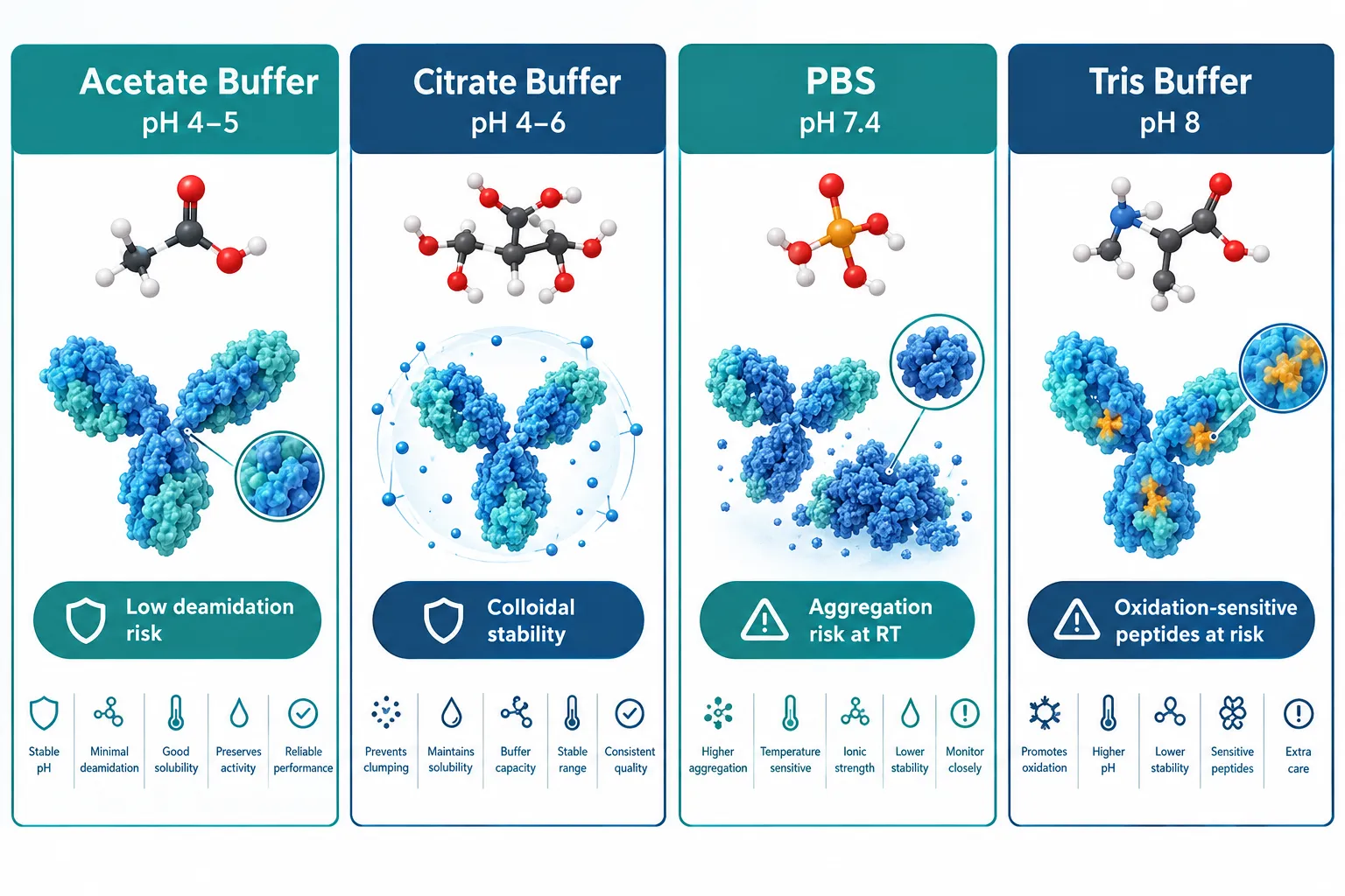
Acetate and citrate buffers at pH 4.0-6.0 have emerged as first-line vehicles for lab reconstitution. Both offer strong buffering capacity in the acidic range where most peptides show the lowest rates of deamidation and backbone hydrolysis. Citrate has the added advantage of mild metal-chelating activity, which is valuable for cysteine-containing and oxidation-sensitive sequences.

PBS (phosphate-buffered saline) at pH 7.4 remains widely used because it matches physiological conditions, but its limitations are now better characterized. At room temperature, PBS accelerates aggregation in hydrophobic peptides and offers no protection against oxidation. For short-term cell-based assays it is acceptable; for storage beyond 24 hours, it is a liability.

Tris and HEPES buffers (pH 7.5-8.0) are common in biochemistry but problematic for peptide stability. The alkaline environment accelerates deamidation of asparagine and glutamine residues and promotes beta-elimination in serine- and threonine-rich sequences.

"Buffer selection is not a formulation footnote, it is the first experimental variable that determines whether a peptide survives long enough to be tested."

Practical buffer selection by peptide class:

Peptide Class Recommended Buffer Target pH Key Risk Mitigated
GLP-class (e.g., GLP-3 analogs) Acetate or citrate 4.0-5.0 Hydrolysis, aggregation
Growth-hormone peptides (e.g., Sermorelin, CJC-1295) Acetate 4.5-5.5 Deamidation
Regenerative/antioxidant (e.g., SS-31, GHK-Cu) Citrate, pH-adjusted water 4.0-5.0 Oxidation, metal coordination
Nasal formulations (e.g., Semax) Citrate-phosphate 5.0-6.5 Mucosal compatibility, stability

Researchers working with SS-31 peptide should pay particular attention to citrate buffers, as the tetrapeptide's aromatic-cationic structure is sensitive to oxidative degradation at neutral or alkaline pH.


pH-Dependent Degradation Pathways: Mapping the Risk Zones

Every peptide has a pH-stability profile, and the degradation chemistry changes depending on where that profile sits.

Below pH 3.0: Acid-catalyzed hydrolysis of Asp-Pro bonds accelerates sharply. Peptides with proline-rich sequences or internal aspartate residues are particularly vulnerable.

pH 3.5-4.5 (optimal zone): For most peptides, this range minimizes both acid-catalyzed hydrolysis and base-catalyzed deamidation simultaneously. Solution purity data consistently show the slowest degradation rates here.

pH 5.0-6.5: Acceptable for many peptides, especially those destined for mucosal delivery. Deamidation begins to increase modestly above pH 5.5 for asparagine-containing sequences.

pH 7.0-8.0: Deamidation of Asn and Gln residues accelerates significantly. Beta-elimination in phosphorylated or glycosylated peptides also increases. Aggregation rates for hydrophobic sequences rise sharply.

Above pH 8.0: Racemization and disulfide scrambling become dominant degradation pathways. Cysteine-containing peptides are at high risk.

Researchers using GLP-3 peptide analogs should note that the long-chain fatty acid modifications common in this class increase hydrophobic aggregation risk at neutral pH, reinforcing the case for acidic reconstitution buffers.


Osmolality, Freeze, Thaw Cycles, and Practical Reconstitution Controls
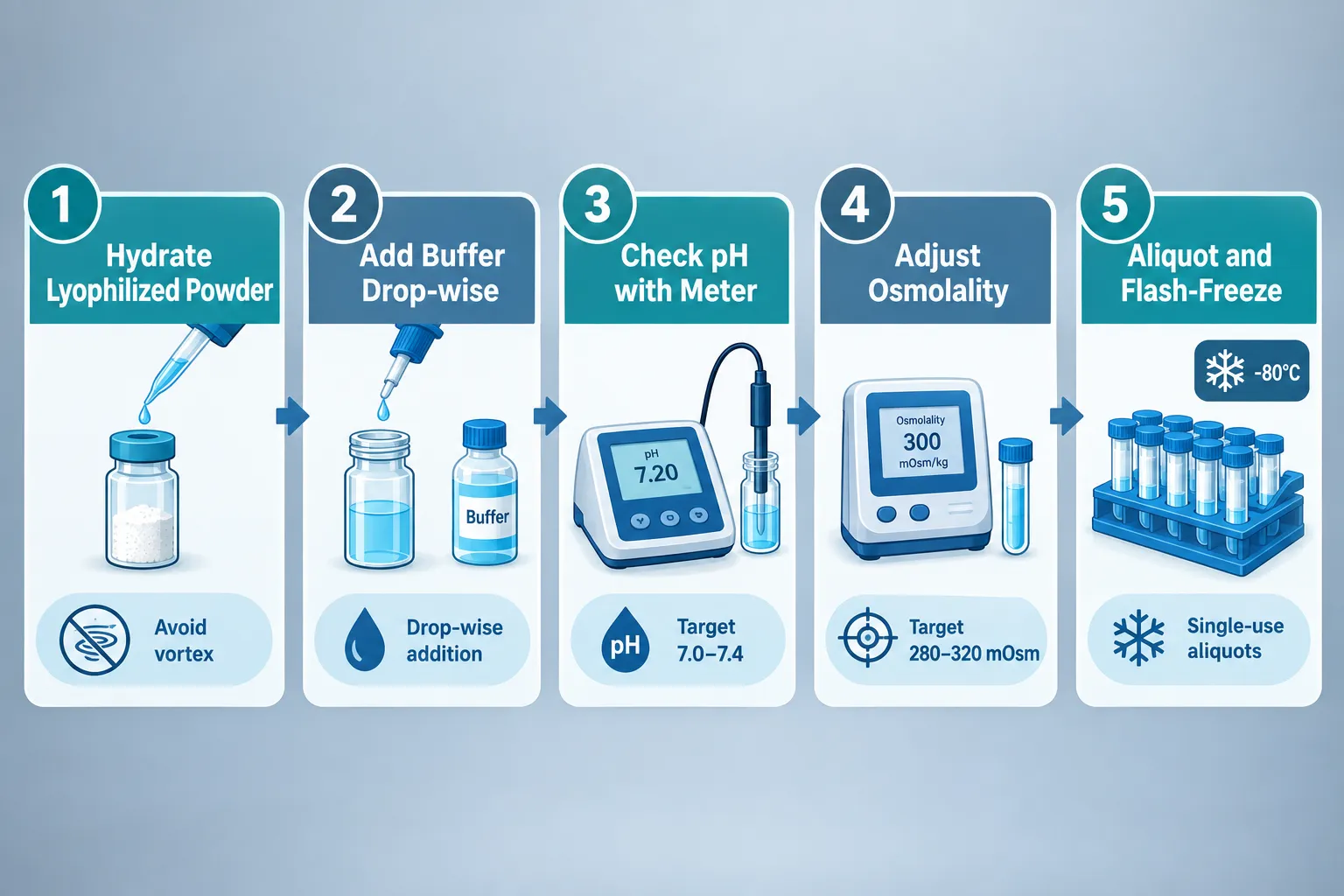
Osmolality, Freeze, Thaw Cycles, and Practical Reconstitution Controls

Osmolality: The Overlooked Variable

Osmolality is frequently treated as relevant only for clinical formulations, but it matters in research too. For intranasal peptides such as Semax peptide formulations, osmolality outside the 270-320 mOsm/kg range can alter mucosal absorption and confound pharmacokinetic data. For parenteral research models, hyperosmolar solutions cause cell stress artifacts that distort results.

Achieving isotonicity in an acidic buffer requires careful addition of sodium chloride or mannitol. Mannitol is preferred when freeze-drying stability is also a concern, as it acts as a lyoprotectant.

Freeze, Thaw Cycles: Quantifying the Damage

Each freeze, thaw cycle introduces two distinct stresses:

  1. Ice crystal formation, mechanical disruption of peptide aggregates and concentration effects at the ice-liquid interface
  2. Cryoconcentration, solutes concentrate in unfrozen microdomains, creating transient local pH extremes and ionic strength spikes

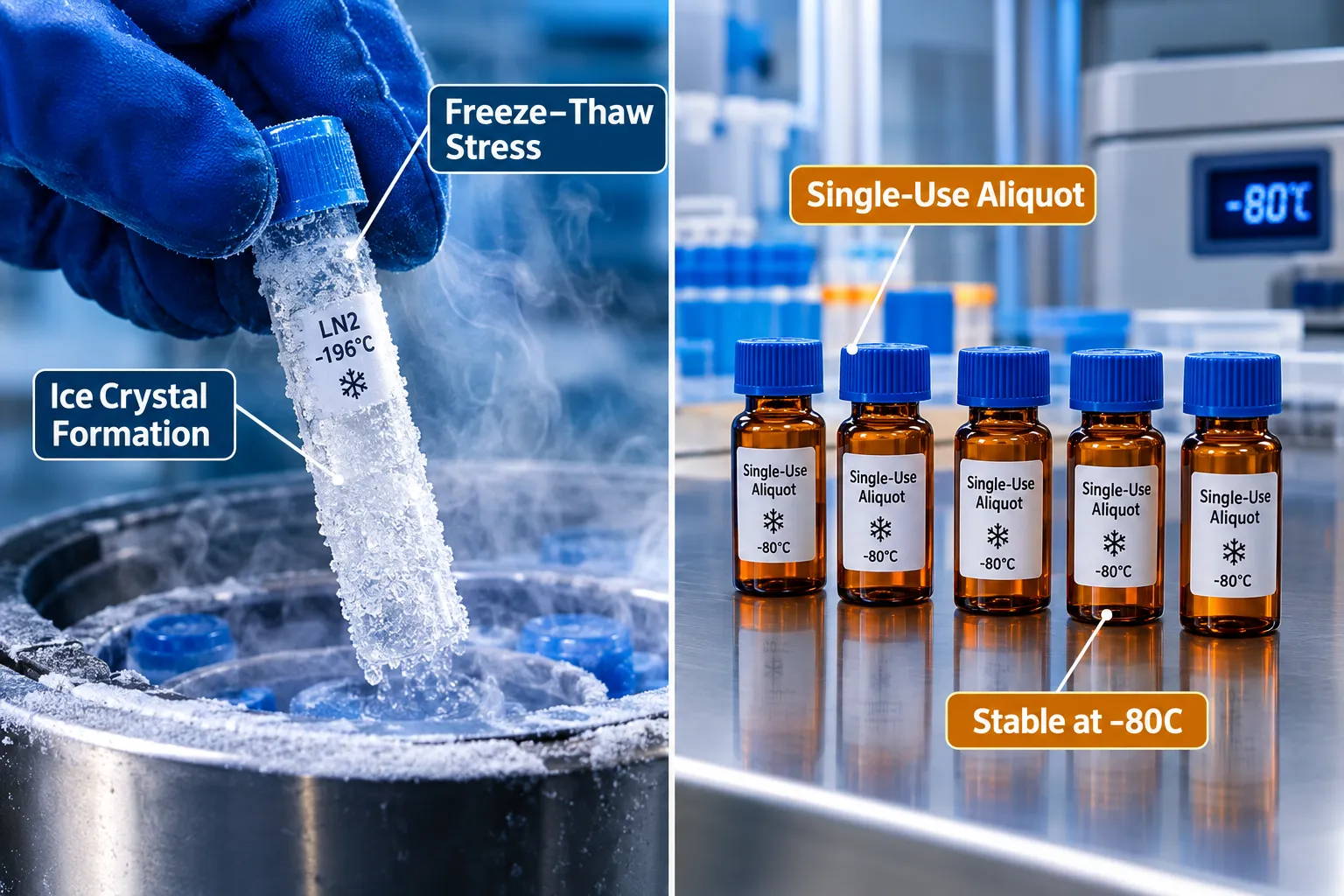
Studies tracking purity over repeated cycles show measurable losses beginning at cycle two for most peptides, with cumulative degradation accelerating nonlinearly by cycle four or five. The practical implication is unambiguous: single-use aliquots are not optional.

Freeze, thaw mitigation checklist:

  • Aliquot into volumes that match a single experiment
  • Use cryoprotectants (5-10% mannitol or trehalose) when lyophilization is not available
  • Flash-freeze in liquid nitrogen rather than slow-freezing at -20°C
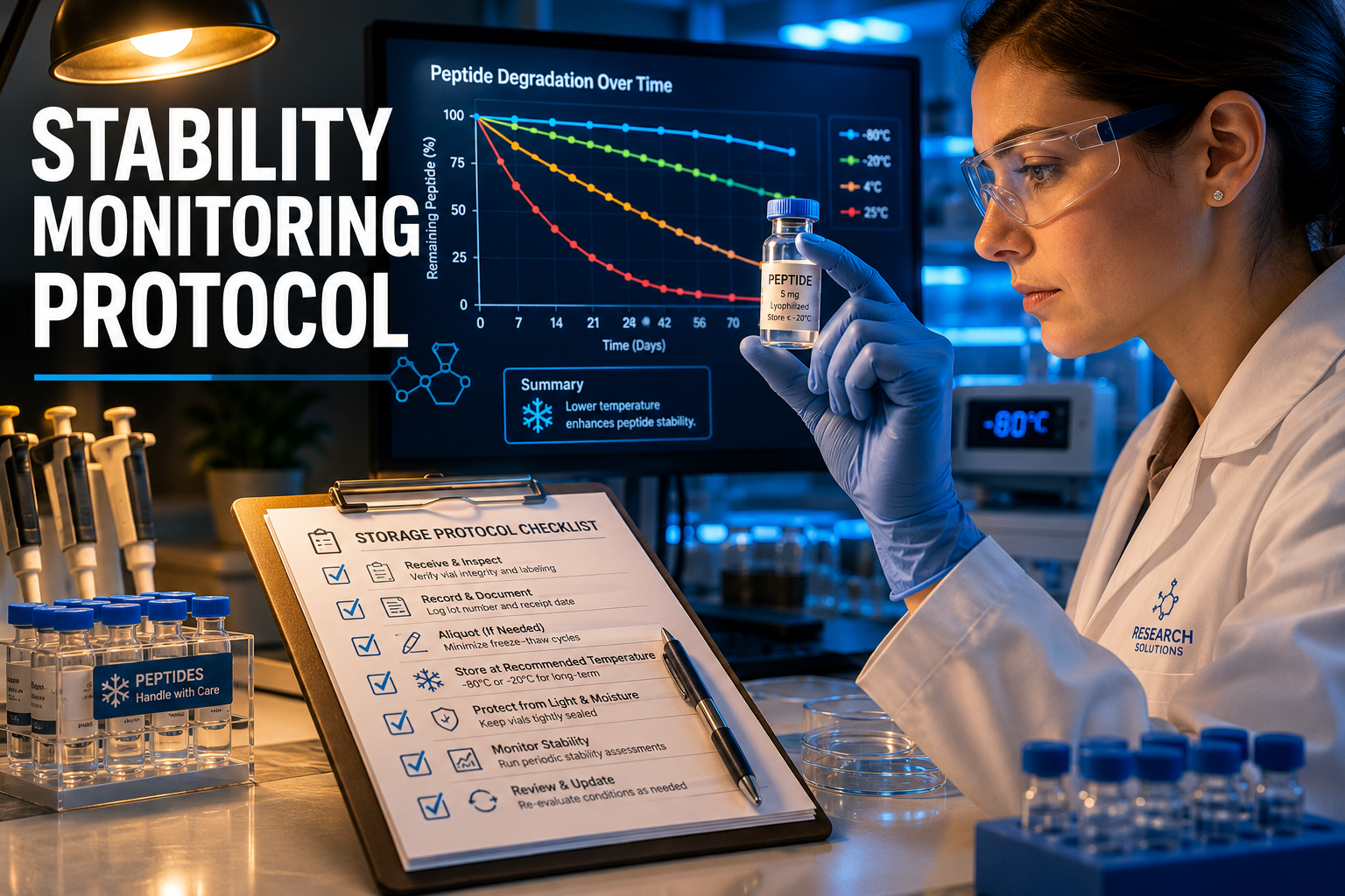
  • Store at -80°C; avoid -20°C for samples held longer than two weeks
  • Never refreeze a thawed aliquot

For complex blends such as Tesamorelin/CJC-1295/Ipamorelin reconstitution, each component has its own freeze, thaw sensitivity, making single-use aliquoting even more critical.

Order of Addition and Mixing

Lab-scale reconstitution protocols now emphasize that order of addition matters. Adding buffer to lyophilized peptide (rather than the reverse) prevents local pH extremes that can occur when a small volume of concentrated peptide contacts a large buffer volume. Gentle swirling, not vortexing, prevents shear-induced aggregation. Sonication is reserved for peptides confirmed to be aggregation-prone, and only in short, controlled pulses.


Stability Matrix Across Peptide Classes
Stability Matrix Across Peptide Classes

The table below consolidates stability variables across four research-relevant peptide classes. It is designed as a practical bench reference.

Variable GLP-Class Analogs Growth-Hormone Peptides Regenerative Peptides (SS-31, GHK-Cu) Nasal Peptides (Semax)
Optimal pH 4.0-5.0 4.5-5.5 4.0-5.0 5.0-6.5
Preferred buffer Acetate Acetate Citrate Citrate-phosphate
Osmolality target 280-310 mOsm/kg 280-310 mOsm/kg 280-310 mOsm/kg 270-320 mOsm/kg
Max freeze, thaw cycles 1-2 2-3 1-2 1
Primary degradation risk Aggregation, hydrolysis Deamidation Oxidation Mucosal pH mismatch
Cryoprotectant recommended Mannitol Mannitol or trehalose Trehalose Mannitol
Stable at -80°C (reconstituted) Up to 3 months Up to 6 months Up to 3 months Up to 4 weeks

Researchers sourcing SS-31 peptide for sale or GHK-Cu peptides should verify that third-party purity certificates reflect post-reconstitution stability data, not just pre-reconstitution synthesis purity.

For GLP-3 peptide research, the fatty acid-modified backbone demands particular attention to aggregation at the reconstitution step. Warming to room temperature before adding buffer, rather than adding cold buffer to a cold vial, reduces nucleation of aggregates.


Conclusion

Peptide reconstitution and stability, how buffer choice, pH, osmolality, and freeze, thaw cycles affect research samples, is a discipline that rewards systematic attention. The actionable steps are clear:

  1. Select buffer based on sequence chemistry, defaulting to acetate at pH 4.0-5.0 for most classes and citrate for oxidation-sensitive peptides.
  2. Verify osmolality before any intranasal or parenteral experiment; adjust with mannitol or NaCl.
  3. Aliquot immediately after reconstitution into single-use volumes and flash-freeze.
  4. Never exceed two freeze, thaw cycles for any peptide; treat cycle one as a warning threshold.
  5. Request purity data at multiple time points and pH values from suppliers, not just a single synthesis certificate.
  6. Match the stability matrix to the peptide class before the experiment begins, not after anomalous results appear.

Implementing these controls does not require expensive equipment. It requires deliberate protocol design and an understanding that every variable in the reconstitution environment is an experimental variable.

https://www.puretestedpeptides.com/wp-content/uploads/2026/09/peptide-reconstitution-and-stability-how-buffer-choice-ph-osmolality-and-freeze.webp 1024 1536 https://www.puretestedpeptides.com/wp-content/uploads/2026/01/buy-peptides-online.jpg 2026-09-23 13:07:532026-09-23 13:07:53Peptide Reconstitution and Stability: How Buffer Choice, pH, Osmolality, and Freeze–Thaw Cycles Affect Research Samples
Magnesium Supplementation and Peptide Reconstitution Stability: Ionic Interactions in Solution Protocols

Magnesium Supplementation and Peptide Reconstitution Stability: Ionic Interactions in Solution Protocols

September 17, 2026/0 Comments/in Uncategorized/by

Fewer than 30% of researchers and formulators who reconstitute research peptides account for the ionic environment of their diluent, yet that single oversight can silently degrade a peptide's tertiary structure before the first dose is ever prepared. Understanding Magnesium Supplementation and Peptide Reconstitution Stability: Ionic Interactions in Solution Protocols is no longer a niche biochemistry concern; it sits at the center of modern formulation science, from injectable research compounds to oral supplement delivery systems.

Key Takeaways

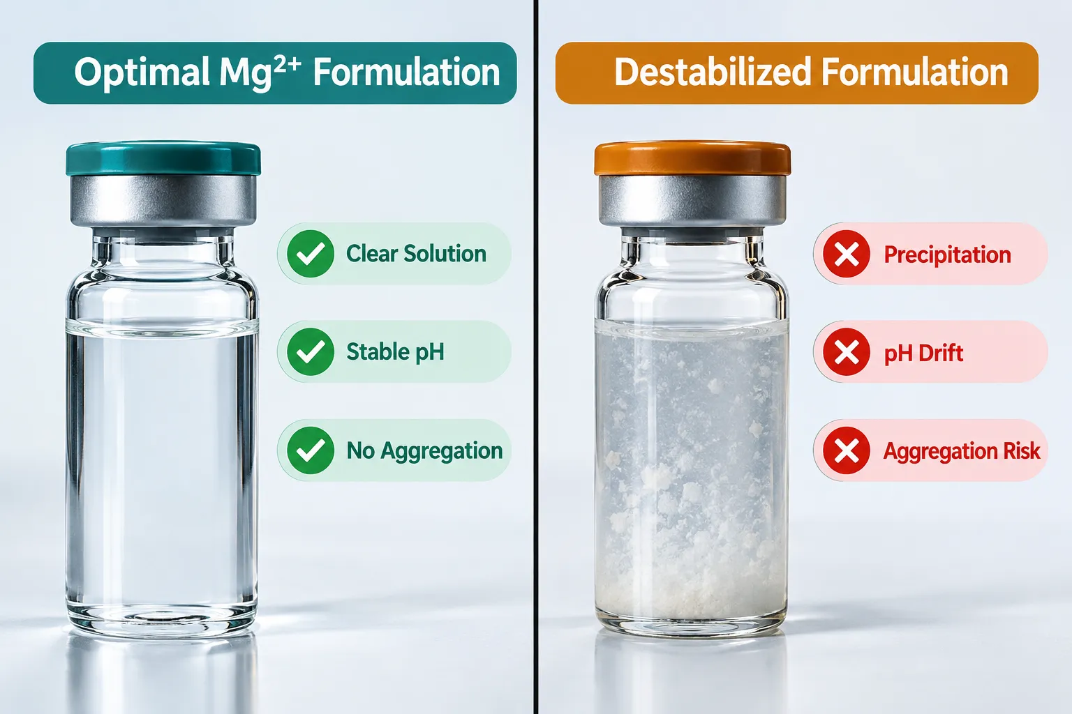
  • Divalent Mg2+ cations directly alter peptide tertiary structure, solubility, and aggregation behavior in liquid storage formats.
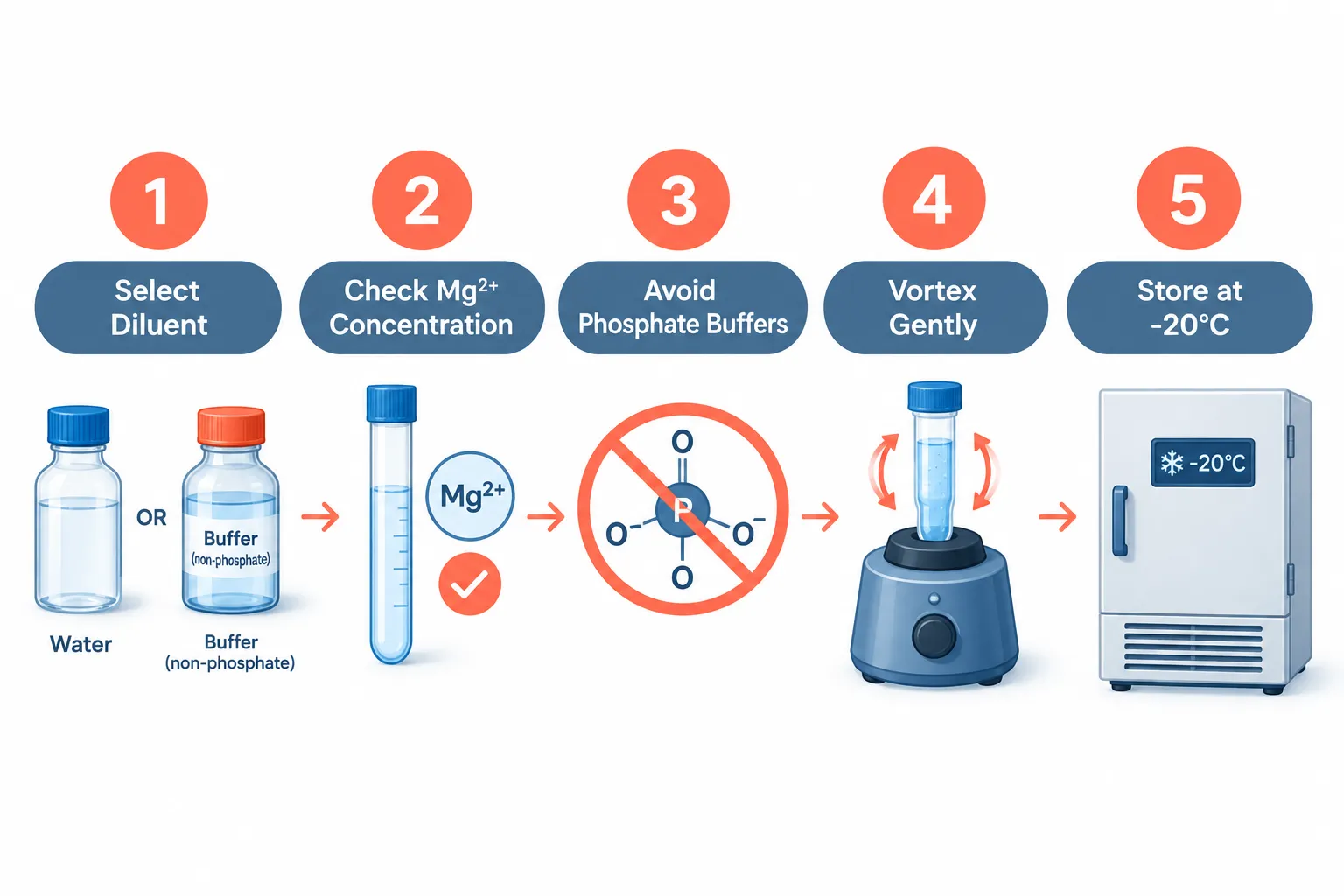
  • Phosphate-buffered solutions must be avoided when magnesium is present, as precipitation is a predictable and preventable outcome.
  • Optimal Mg2+ concentration windows exist, too little provides no stabilizing benefit; too much drives aggregation under contamination conditions.
  • Thermal stability of both cysteine-containing and non-cysteine peptides improves measurably in the presence of controlled magnesium concentrations.
  • Reconstitution protocol design should treat ionic composition as a primary variable, not an afterthought.

How Mg2+ Cations Alter Peptide Tertiary Structure in Solution

How Mg2+ Cations Alter Peptide Tertiary Structure in Solution

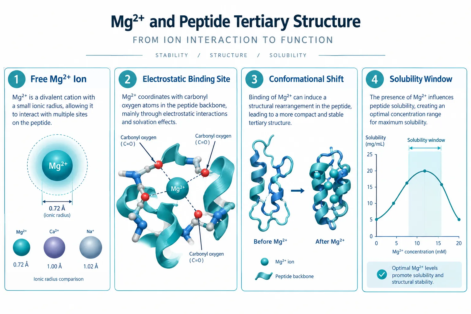
Magnesium exists in solution as a divalent cation (Mg2+), carrying two positive charges that create strong electrostatic fields around the ion. When a peptide is dissolved into a solution containing free Mg2+, those charges interact with negatively charged or polar residues along the peptide backbone, particularly carbonyl oxygens, carboxylate side chains, and certain aromatic groups.

This is not a passive relationship. Mg2+ actively reshapes the electrostatic landscape around a peptide, which in turn influences how the chain folds, how exposed hydrophobic patches become, and whether the molecule remains in stable monomeric form or begins to self-associate.

Recent mechanistic data confirm that Mg2+ enhances thermal stability across both cysteine-containing and non-cysteine peptides. The proposed mechanism involves bridging interactions between Mg2+ and multiple electronegative sites on the same peptide chain, effectively acting as a conformational clamp that resists unfolding under thermal stress. This has direct implications for peptides stored at ambient temperature during shipping or in field-use conditions.

"Ionic composition is not background noise in a reconstitution protocol, it is a primary structural variable."

For peptides with known metal-binding motifs, such as those found in antimicrobial peptide classes, Mg2+ dependence goes further. Conformational activity, the ability of the peptide to adopt its bioactive shape, has been shown to shift meaningfully based on available Mg2+ concentrations. This means that a peptide reconstituted in magnesium-depleted water may behave differently from the same peptide reconstituted in a physiologically relevant ionic environment.

Researchers working with SS-31 peptides for sale or similar mitochondria-targeted sequences should pay particular attention here, as charge-dense peptides are especially sensitive to divalent cation environments.

Reconstitution Protocol Design: Applying Magnesium Supplementation and Peptide Reconstitution Stability Principles

Reconstitution Protocol Design: Applying Magnesium Supplementation and Peptide Reconstitution Stability Principles

Translating ionic chemistry into a practical bench protocol requires clear decision points. The table below summarizes the most critical variables in a magnesium-aware reconstitution workflow.

Variable Recommended Approach Risk if Ignored
Diluent selection Sterile water or acetate buffer Ionic incompatibility
Buffer type Avoid phosphate buffers with Mg2+ Precipitation, loss of peptide
Mg2+ concentration 1-10 mM range for most peptides Aggregation at higher levels
pH 6.5-7.5 for most sequences Charge state shifts
Storage temperature -20C for long-term Hydrolysis, oxidation

Phosphate buffers represent the most commonly cited formulation error when magnesium is present. Mg2+ reacts with phosphate ions to form insoluble magnesium phosphate salts. This precipitation removes both free magnesium from solution and can co-precipitate the peptide itself, causing catastrophic loss of usable material. Current 2026 formulation guidance is unambiguous: phosphate buffers and Mg2+ do not belong in the same reconstitution vial.

Acetate and histidine buffers are preferred alternatives. They maintain pH stability without introducing phosphate anions, preserving the free Mg2+ needed for its stabilizing role.

For those working with complex formulations such as GLP-3R 30mg Peptide GA7 or GLP-3R 30mg Peptide GA9, multi-component ionic environments add another layer of complexity. When multiple peptides or excipients share a single vial, each ionic interaction must be evaluated independently.

Concentration Thresholds and Aggregation Risk

Mg2+ as a formulation excipient follows a non-linear dose-response curve for stability. At low concentrations (below 1 mM), the cation provides minimal structural benefit. Within the 1-10 mM window, stabilizing electrostatic interactions dominate. Above 10 mM, particularly when trace contaminants are present, Mg2+ can paradoxically increase protein and peptide aggregation by screening repulsive charges between molecules, allowing them to cluster.

This aggregation-under-contamination risk is well-documented in biologics data from 2025 to 2026 and represents a practical ceiling for magnesium use in reconstitution protocols.

Ionic Interactions, Hydrogel Stability, and Emerging Formulation Strategies

Ionic Interactions, Hydrogel Stability, and Emerging Formulation Strategies

Beyond simple reconstitution, Magnesium Supplementation and Peptide Reconstitution Stability: Ionic Interactions in Solution Protocols intersects with emerging delivery formats, particularly peptide hydrogels used in wound repair and sustained-release applications.

Divalent cations including Mg2+ markedly alter the gelation behavior and mechanical properties of self-assembling peptide hydrogels. By coordinating with charged residues at gel fiber junctions, Mg2+ can either reinforce or disrupt the crosslink density depending on concentration and peptide sequence. Formulators designing depot-style peptide delivery systems must therefore validate Mg2+ concentration as part of the gelation protocol, not just the reconstitution step.

Researchers exploring wound repair peptides or GHK-Cu peptides for sale, both of which operate in ionic-rich biological environments, benefit from understanding how Mg2+ at wound sites or injection depots will interact with their chosen peptide scaffold.

Mechanistic analogies from nucleic acid aptamer research also inform this space. Mg2+ stabilizes folded aptamer structures through backbone coordination, a process structurally analogous to how it stabilizes folded peptide mimetics. This cross-disciplinary insight supports the use of Mg2+ as a deliberate excipient in DNA-peptide hybrid constructs gaining traction in 2026 research pipelines.

For cognitively active peptides delivered via alternative routes, such as those found in peptides nasal spray formats or Semax peptide preparations, the nasal mucosa presents its own ionic environment. Formulation scientists are beginning to account for endogenous magnesium concentrations in mucosal fluid when designing these delivery systems.

Timing and Synergy in Supplementation Contexts

At the practice level, a magnesium-peptide synergy timing protocol has emerged from expert consensus in 2026. The core principle: when magnesium is used as a co-supplement alongside peptide injection protocols, separating oral magnesium intake from injection timing by at least two hours reduces the likelihood that systemic magnesium fluctuations will interfere with peptide pharmacokinetics in the immediate post-injection window. This is an expert-opinion level recommendation, but it reflects growing awareness that systemic ionic status is not irrelevant to peptide behavior in vivo.

Conclusion

The relationship between divalent magnesium cations and peptide stability in solution is precise, predictable, and actionable. Formulators and researchers who treat ionic composition as a primary protocol variable, rather than background chemistry, will consistently produce more stable, more reproducible results.

Actionable next steps for 2026 practice:

  • Audit existing reconstitution protocols for phosphate buffer use and replace with acetate or histidine alternatives when Mg2+ is present.
  • Target a Mg2+ concentration of 1-10 mM for stabilizing applications, and validate the upper boundary against aggregation assays for each specific peptide.
  • For hydrogel or depot formats, run gelation validation studies across the intended Mg2+ concentration range before finalizing the formulation.
  • When combining oral magnesium supplementation with injectable peptide protocols, apply a minimum two-hour separation window as a precautionary measure.
  • Treat ionic environment as a documented, controlled variable in all stability and storage studies, not an assumption.

Mastering Magnesium Supplementation and Peptide Reconstitution Stability: Ionic Interactions in Solution Protocols is one of the highest-leverage improvements available to any researcher or formulator working with peptide-based compounds in 2026.

https://www.puretestedpeptides.com/wp-content/uploads/2026/09/magnesium-supplementation-and-peptide-reconstitution-stability-ionic-interaction.webp 1024 1536 https://www.puretestedpeptides.com/wp-content/uploads/2026/01/buy-peptides-online.jpg 2026-09-17 13:04:332026-09-17 13:04:33Magnesium Supplementation and Peptide Reconstitution Stability: Ionic Interactions in Solution Protocols
Phosphate Buffered Saline for Peptide Reconstitution: A Complete Research Protocol Guide

Phosphate Buffered Saline for Peptide Reconstitution: A Complete Research Protocol Guide

September 14, 2026/0 Comments/in Uncategorized/by

Roughly 30% of peptide reconstitution failures in research settings trace back to a single, preventable error: choosing the wrong solvent. For researchers working with synthetic peptides, that choice starts with understanding when phosphate buffered saline is the right tool and when it is not. This guide to Phosphate Buffered Saline for Peptide Reconstitution: A Complete Research Protocol Guide covers everything a researcher needs to make that decision confidently, execute the protocol correctly, and store reconstituted stocks safely.

Key Takeaways

  • PBS at pH 7.4 closely mimics physiological conditions, making it ideal for hydrophilic and cell-compatible peptide assays.
  • Phosphate ions can catalyze deamidation and other degradation pathways in susceptible peptides, so PBS is not universally appropriate.
  • Proper concentration calculation, sterile filtration, and cold-chain storage are non-negotiable steps in any PBS reconstitution protocol.
  • Alternatives such as bacteriostatic water, HEPES-buffered saline, and dilute acetic acid each serve specific peptide chemistries better than PBS in certain cases.
  • Sourcing high-purity, third-party-tested peptides is the foundation of any reliable reconstitution outcome.

Why PBS Remains the Default Solvent in Peptide Research

Phosphate buffered saline is a water-based salt solution that maintains a stable pH of 7.4 while matching the osmolarity of human plasma at approximately 300 mOsm/kg. Those two properties explain its dominance in cell culture, biochemical assays, and in vivo research models.

Why PBS Remains the Default Solvent in Peptide Research

For hydrophilic peptides with a net neutral or slightly negative charge at physiological pH, PBS provides an aqueous environment that supports full dissolution without introducing organic solvents that could disrupt downstream cell viability. Researchers running Semax research protocols and similar neuropeptide studies frequently rely on PBS precisely because the buffer does not interfere with receptor-binding assays or neuronal cell lines.

When PBS is the right choice:

  • Hydrophilic peptides that dissolve readily in water
  • Cell-based assays requiring physiological osmolarity
  • In vivo models where isotonicity is critical
  • Short-term stocks used within 24 to 72 hours

When PBS should be avoided:

  • Peptides containing asparagine or glutamine residues prone to deamidation (phosphate accelerates this reaction)
  • Highly hydrophobic sequences that require DMSO or dilute organic acid as a primary solvent
  • Long-term frozen stocks where phosphate precipitation at low temperatures can alter effective concentration

"PBS is not a universal default. It is the best default for a defined subset of peptide chemistries."

Core Protocol Steps for Reconstituting Peptides in PBS

Following a standardized workflow reduces variability and protects peptide integrity from the moment the lyophilized powder is opened.

Core Protocol Steps for Reconstituting Peptides in PBS

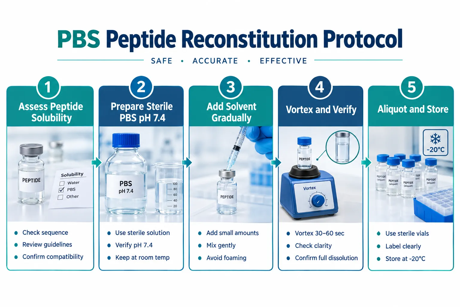
Step 1: Assess Peptide Solubility Before Reconstitution

Review the manufacturer's certificate of analysis and any published solubility data. Peptides with a high proportion of hydrophobic residues (leucine, isoleucine, phenylalanine, valine) will likely require a co-solvent step before PBS dilution. Peptides with multiple charged residues at physiological pH are strong candidates for direct PBS dissolution.

Step 2: Prepare or Verify Sterile PBS

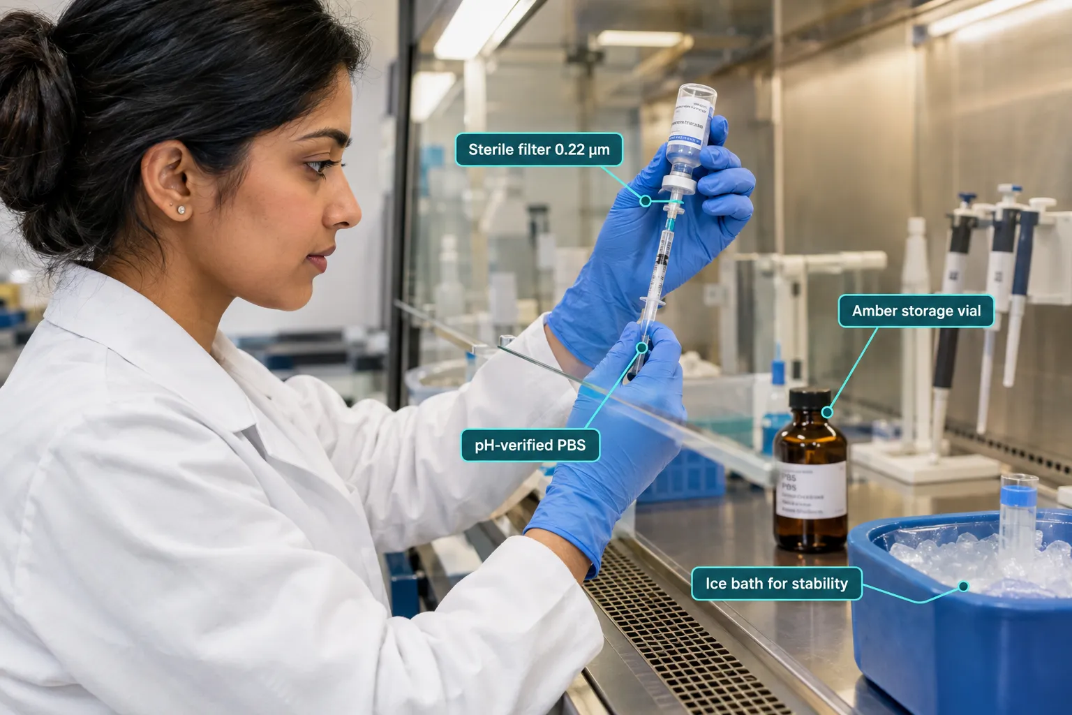
Use sterile, endotoxin-tested PBS at pH 7.4. For in vivo or cell-culture work, confirm the endotoxin level is below 0.1 EU/mL. If preparing PBS in-house, sterile-filter through a 0.22 µm membrane after preparation.

Step 3: Calculate Target Concentration

Use the molecular weight from the certificate of analysis, not a generic database value, since counterion salts affect actual mass.

Target Concentration Peptide Mass (1 mg) PBS Volume Required
1 mg/mL 1 mg 1.0 mL
0.5 mg/mL 1 mg 2.0 mL
0.1 mg/mL 1 mg 10.0 mL

Step 4: Add Solvent Gradually and Mix Gently

Add PBS in small increments to the lyophilized peptide. Avoid vortexing at high speed for extended periods, as mechanical shear can fragment sensitive sequences. Gentle swirling or brief low-speed vortexing for 5 to 10 seconds is sufficient for most hydrophilic peptides.

Step 5: Verify Dissolution and Filter

Inspect the solution visually for particulates. For critical applications, confirm concentration using UV absorbance at 280 nm if the peptide contains aromatic residues, or via HPLC for absolute quantification. Filter through a 0.22 µm syringe filter before aliquoting.

PBS vs. Alternative Solvents: Choosing the Right Buffer

Researchers working with a broad peptide library will encounter situations where PBS is not the optimal first choice. Understanding the alternatives is essential.

PBS vs. Alternative Solvents: Choosing the Right Buffer

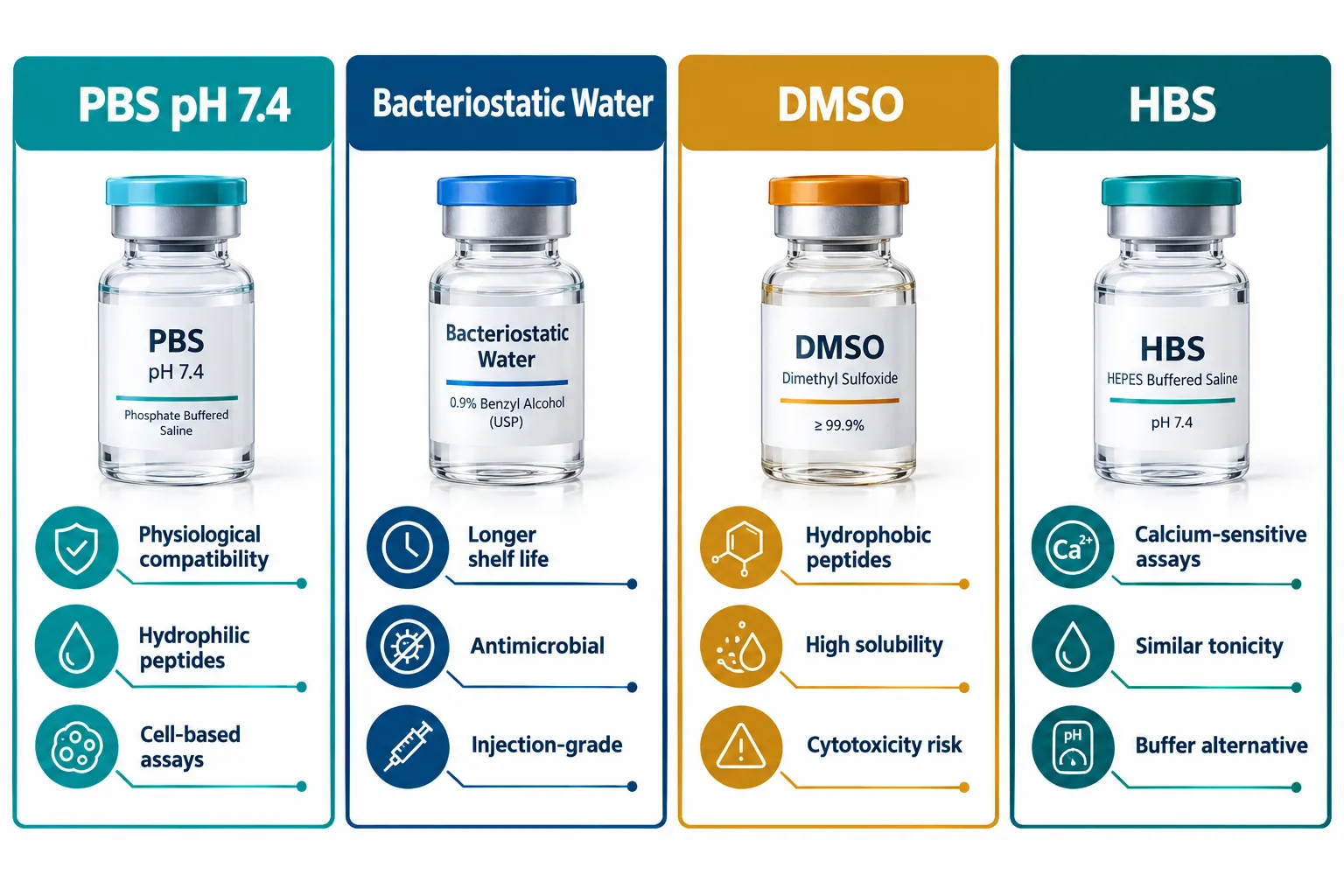
Bacteriostatic water contains 0.9% benzyl alcohol, which inhibits microbial growth and extends the usable life of a reconstituted stock. It is commonly used for peptides intended for repeated withdrawal from the same vial. However, it lacks the buffering capacity of PBS and is not appropriate for pH-sensitive assays. Researchers exploring SS-31 peptide research and related mitochondria-targeted compounds often weigh bacteriostatic water against PBS depending on the assay format.

HEPES-buffered saline (HBS) offers a phosphate-free alternative at physiological pH, making it preferable for calcium-sensitive assays or any protocol where phosphate ions could interfere with signaling pathways. The tradeoff is higher cost and less universal availability.

Dilute acetic acid (0.1% to 1%) is the go-to primary solvent for hydrophobic or aggregation-prone peptides. After initial dissolution in acetic acid, the researcher then dilutes into PBS to reach physiological conditions, keeping the final acetic acid concentration below 0.01%.

DMSO is reserved for extremely hydrophobic sequences. Final DMSO concentration in cell-based assays should remain below 0.1% to avoid cytotoxicity.

For those sourcing peptides for structured research programs, working with a best peptide supplier that provides solubility guidance alongside the certificate of analysis removes much of the guesswork from solvent selection.

Storage and Handling of PBS-Reconstituted Peptide Stocks

Reconstitution is only half the protocol. Improper storage is one of the most common sources of data variability in peptide research.

Recommended storage practices for 2026:

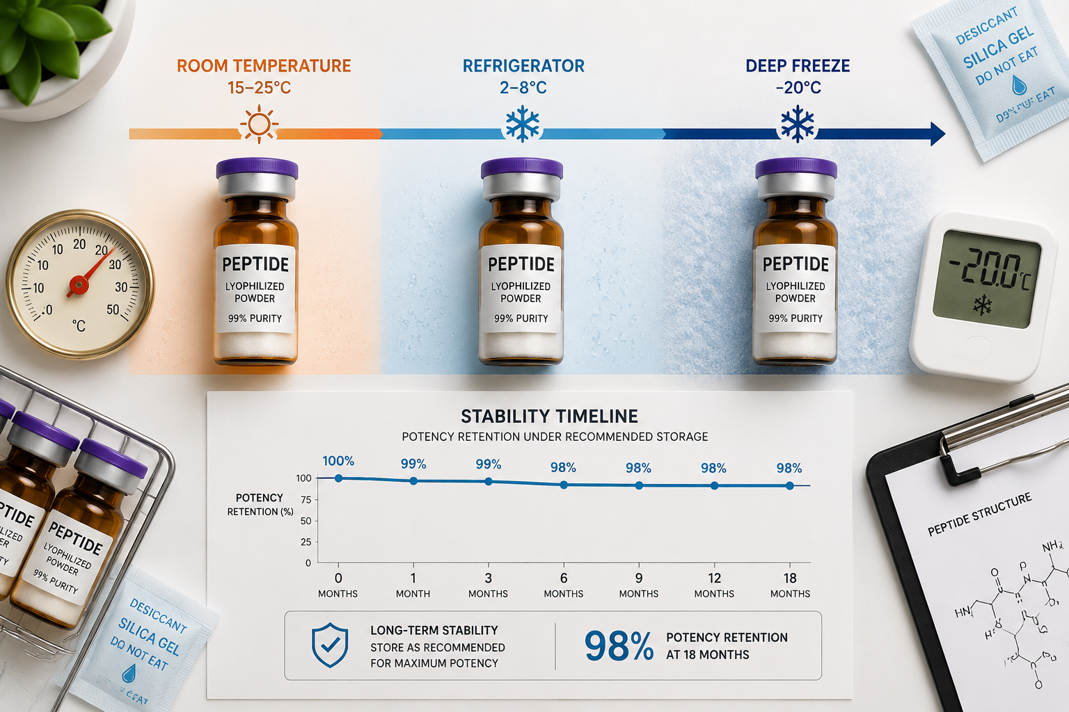
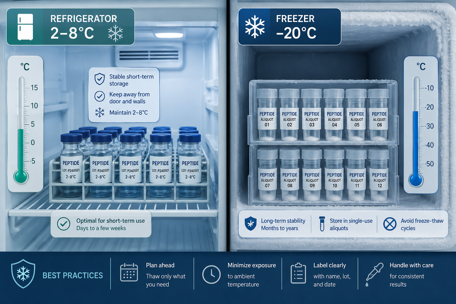
  • Short-term use (less than 72 hours): Store at 2 to 8 degrees Celsius in a sealed, sterile vial. Minimize freeze-thaw cycles.
  • Medium-term storage (up to 4 weeks): Aliquot into single-use volumes and store at -20 degrees Celsius. Label each aliquot with peptide name, concentration, date, and lot number.
  • Long-term storage (beyond 4 weeks): Store at -80 degrees Celsius. Note that phosphate salts can precipitate during freezing; allow complete thaw and gentle mixing before use.
  • Light sensitivity: Many peptides degrade under UV exposure. Use amber vials or wrap clear vials in foil.

Researchers working with compounds such as SS-31 peptide or Mot-C peptide should follow the specific storage guidance provided with each product, as mitochondria-targeted and growth-hormone-related peptides can have unique stability profiles that modify general PBS storage rules.

For broader research contexts such as wound healing peptide studies or metabolic peptide investigations, maintaining a cold chain from reconstitution through assay setup is non-negotiable.

Conclusion

Phosphate Buffered Saline for Peptide Reconstitution: A Complete Research Protocol Guide comes down to three decisions: assess whether PBS suits the peptide's chemistry, execute the reconstitution with sterile technique and accurate concentration math, and store aliquots under conditions that prevent degradation. PBS earns its status as the most common reconstitution solvent because it is physiologically compatible, widely available, and well-characterized, but it is not appropriate for every peptide or every assay.

Actionable next steps for researchers:

  1. Always obtain and review the certificate of analysis before selecting a solvent.
  2. Default to PBS for hydrophilic, charge-bearing peptides destined for cell-based or in vivo work.
  3. Switch to bacteriostatic water, HBS, or acetic acid pre-dissolution when PBS chemistry creates stability or solubility concerns.
  4. Aliquot immediately after reconstitution and label every vial with full traceability information.
  5. Source peptides from a best peptide manufacturer that provides third-party purity testing, so the reconstitution protocol starts with a verified, high-quality substrate.

A disciplined approach to solvent selection and storage transforms peptide reconstitution from a potential failure point into a reliable, reproducible foundation for research.

https://www.puretestedpeptides.com/wp-content/uploads/2026/09/phosphate-buffered-saline-for-peptide-reconstitution-a-complete-research-protoco.webp 1024 1536 https://www.puretestedpeptides.com/wp-content/uploads/2026/01/buy-peptides-online.jpg 2026-09-14 13:05:452026-09-14 13:05:45Phosphate Buffered Saline for Peptide Reconstitution: A Complete Research Protocol Guide
Peptides Calculator for GLP-3, MOTS-c, and BPC-157: How Researchers Estimate Dosing and Concentration Safely

Peptides Calculator for GLP-3, MOTS-c, and BPC-157: How Researchers Estimate Dosing and Concentration Safely

September 2, 2026/0 Comments/in Uncategorized/by

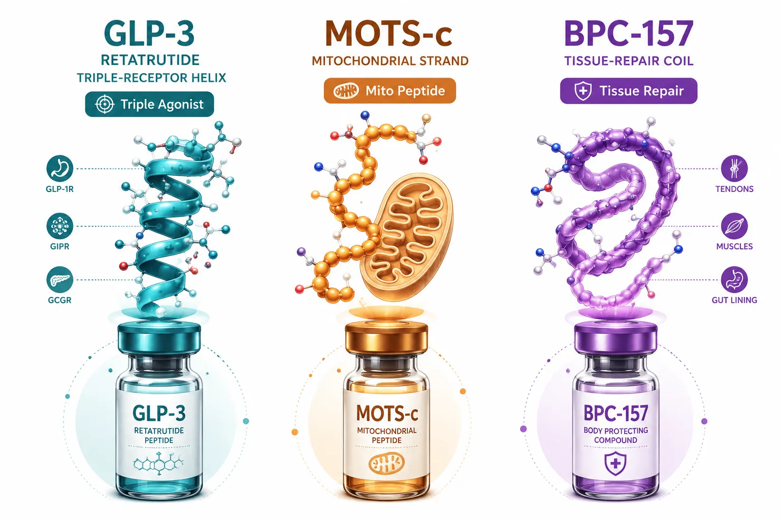
{"cover":"Professional landscape format (1536×1024) hero image with bold text overlay 'Peptides Calculator for GLP-3, MOTS-c' in crisp white modern sans-serif on a deep navy semi-transparent overlay panel, centered with 8% safe margins from every edge, no character touches the edge. Background: bright editorial flat-lay of a laboratory bench with glass vials of peptide solutions, a digital tablet showing a dosing calculator interface, a precision scale, and molecular structure diagrams on white paper. Clean studio lighting, teal and navy palette, pharmaceutical research aesthetic, magazine cover quality.","content":["Four-panel labelled infographic diagram (1536×1024) for peptide calculator core functions: Panel 1 labeled 'Peptide Mass (mg)' shows a vial with callout arrow; Panel 2 labeled 'Reconstitution Volume (mL)' shows a syringe drawing from vial; Panel 3 labeled 'Concentration (mcg/mL)' shows a formula block; Panel 4 labeled 'Dose Volume (mL)' shows a calculator display. Thin callout lines, clean white background, teal-and-navy editorial palette, medical-guide polish, all labels 1-5 words, inside 5% safe margins.","Numbered step-by-step process flow diagram (1536×1024) showing allometric scaling for MOTS-c and BPC-157 research dosing: Step 1 'Animal Study Dose' with mouse icon and callout; Step 2 'Body Surface Area Conversion' with scaling formula block; Step 3 'Species Correction Factor' with human silhouette; Step 4 'Adjusted Research Dose' with vial and syringe. Warm amber and slate-blue palette, clean sans-serif labels 1-5 words each, numbered nodes connected by arrows, editorial scientific illustration style, 5% safe margins throughout.","Split-screen editorial image (1536×1024): left half shows an annotated close-up of a peptide reconstitution setup, bacteriostatic water vial, lyophilized peptide vial, insulin syringe, with three callout labels: 'Lyophilized Powder', 'Bacteriostatic Water', 'Measured Draw Volume'; right half shows a tablet screen mockup of a cross-peptide calculator interface with labeled fields: 'GLP-class Input', 'MOTS-c Protocol', 'BPC-157 Schedule'. High-contrast studio lighting, crisp white and teal palette, research laboratory context, magazine-quality composition."]

Professional landscape hero image () with a reading "Peptides Calculator for GLP-3, MOTS-c". CRITICAL TYPOGRAPHY RULES:

Fewer than 15% of peptide research protocols in preclinical settings include a documented concentration calculation, yet dosing errors at the bench level remain one of the most common sources of unreliable data. A well-structured Peptides Calculator for GLP-3, MOTS-c, and BPC-157: How Researchers Estimate Dosing and Concentration Safely is not a convenience tool; it is a foundational element of rigorous experimental design. This guide breaks down the lab math, scaling logic, and safety checkpoints that researchers rely on when working with GLP-class, mitochondrial, and tissue-repair peptides in 2026.

Key Takeaways

  • A peptide calculator converts lyophilized mass, solvent volume, and target dose into precise draw volumes for each research compound.
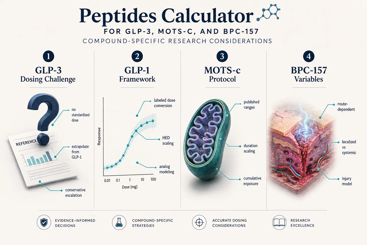
  • GLP-3 lacks a standardized reference dose; researchers currently extrapolate from GLP-1 analog modeling and apply conservative escalation schedules.
  • MOTS-c dosing in animal models varies widely; allometric scaling to human-equivalent doses requires body surface area correction.
  • BPC-157 has no FDA-approved dose, but recent pilot and Phase 2 musculoskeletal data are beginning to anchor practical research ranges.
  • Cross-peptide calculators that handle multiple compound classes in a single interface reduce transcription errors and improve protocol reproducibility.

What a Peptides Calculator Actually Does

What a Peptides Calculator Actually Does

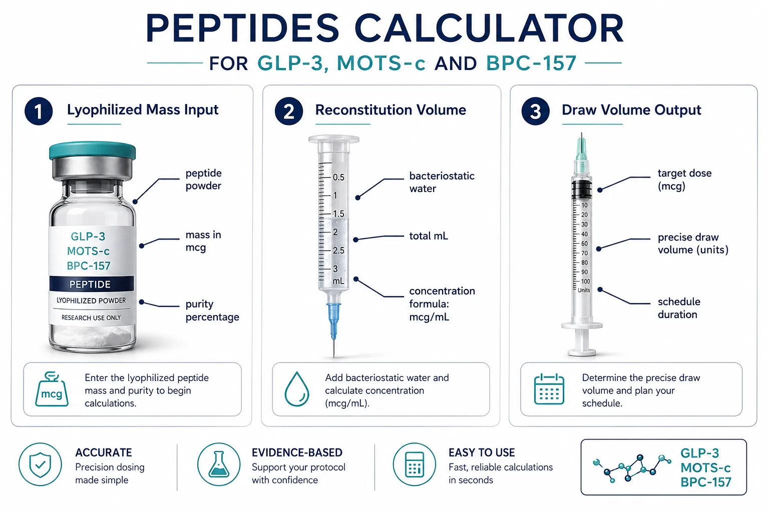
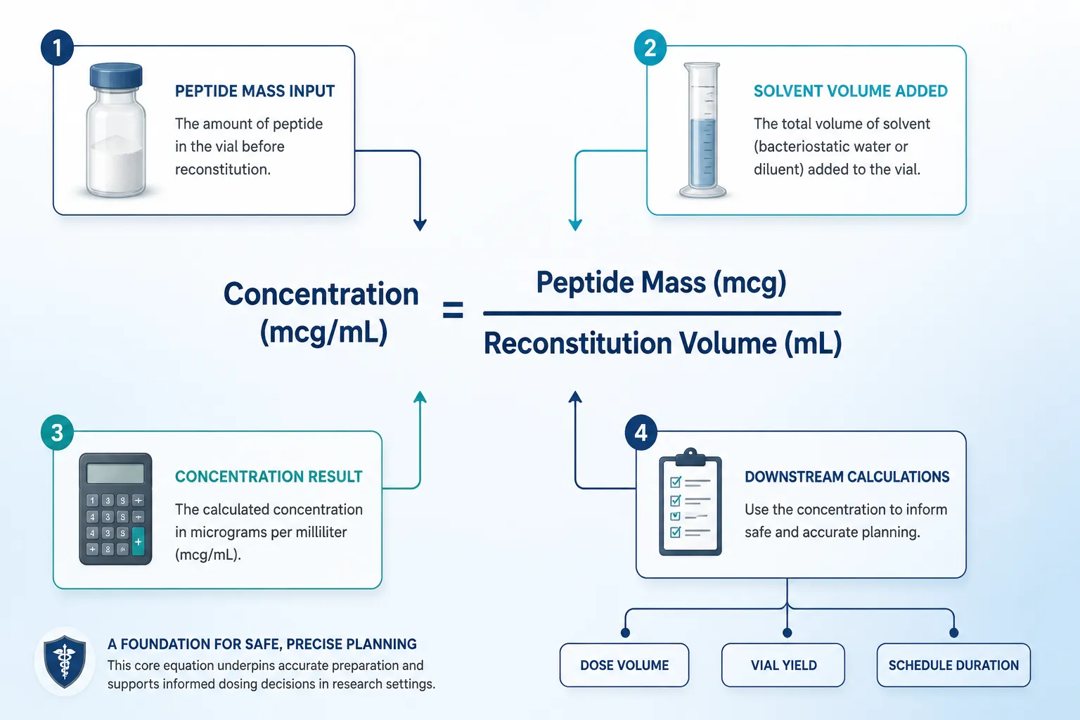
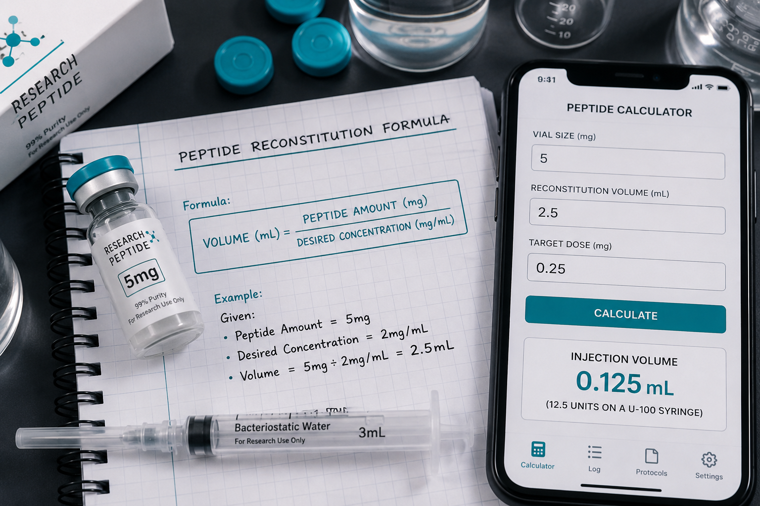
At its core, a peptides calculator solves one equation repeatedly: Concentration (mcg/mL) = Peptide Mass (mcg) / Reconstitution Volume (mL). From that single value, every downstream calculation, dose volume, total vial yield, and schedule duration, flows automatically.

For researchers working with synergistic peptide combinations, the calculator must handle multiple compounds simultaneously without conflating their individual concentration curves. The standard workflow looks like this:

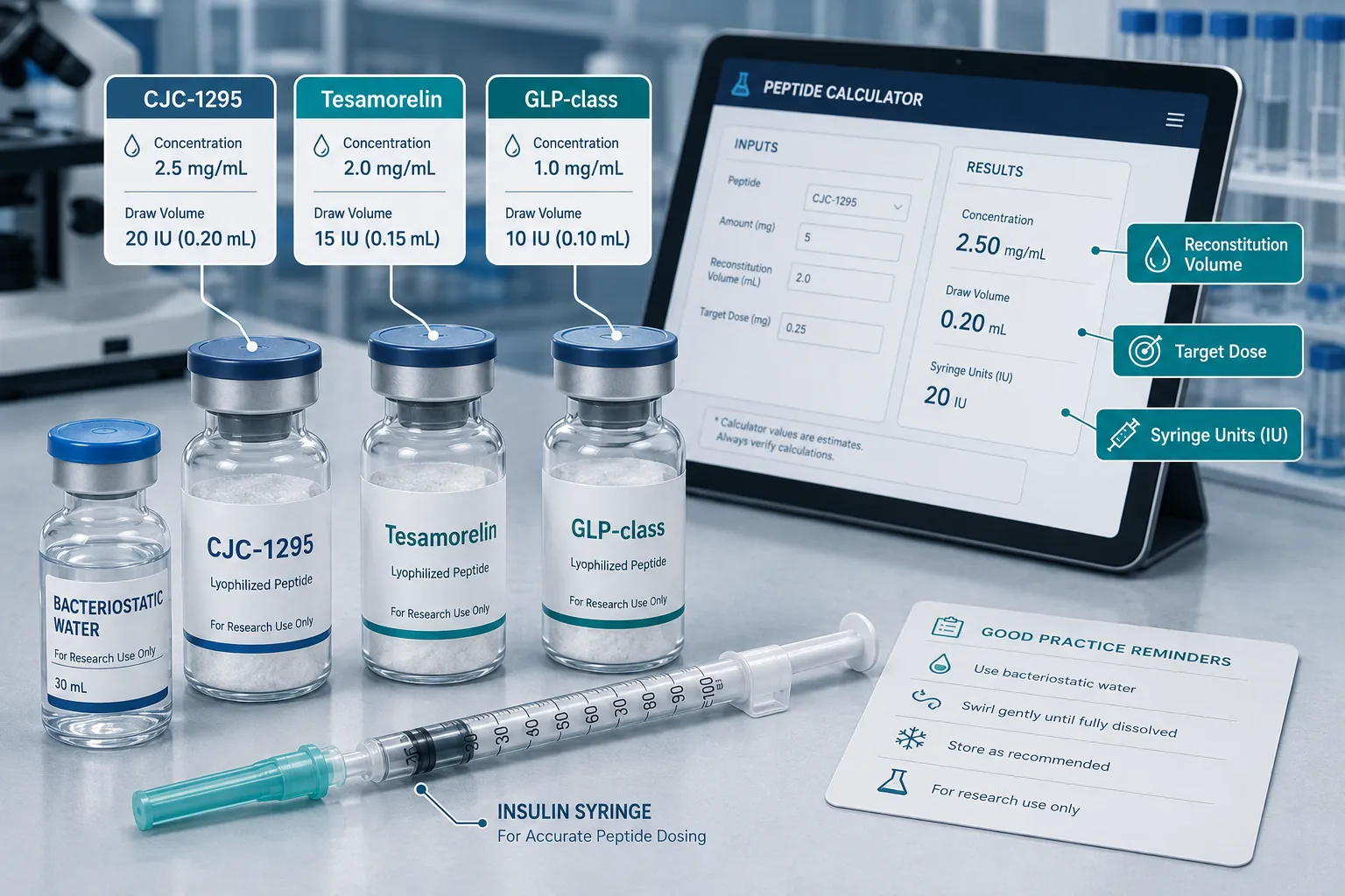
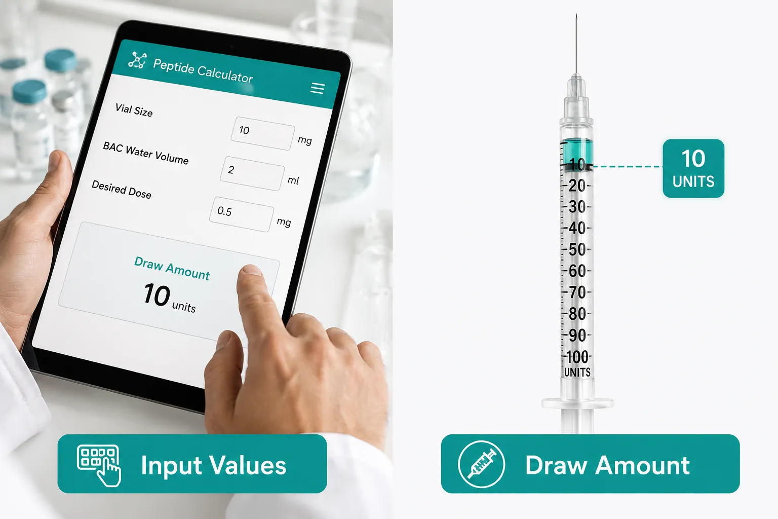
  1. Input lyophilized mass (commonly 5 mg, 10 mg, or custom vial size)
  2. Enter reconstitution solvent volume (bacteriostatic water in mL)
  3. Set target dose in micrograms (mcg) or milligrams (mg)
  4. Read draw volume in mL or units on an insulin syringe

Most modern tools also incorporate half-life modeling, particularly relevant for GLP-1 analogs, and escalation schedule builders that map dose increases across days or weeks. Cross-peptide calculators go further, allowing a researcher to input GLP-class, MOTS-c, and BPC-157 parameters in a single interface, reducing the risk of transcription errors between separate spreadsheets.

"The calculator does not determine whether a dose is appropriate, it determines whether the math behind a chosen dose is internally consistent."

Applying a Peptides Calculator for GLP-3, MOTS-c, and BPC-157: Compound-Specific Considerations

Applying a Peptides Calculator for GLP-3, MOTS-c, and BPC-157: Compound-Specific Considerations

GLP-3 and GLP-Class Peptides

GLP-3 (glucagon-like peptide-3) remains far less characterized than GLP-1 or GLP-2. No standardized reference dose exists in published literature as of 2026. Researchers typically approach GLP-3 by borrowing the labeled dose conversion framework developed for GLP-1 analogs, inputting known receptor affinity ratios and applying a conservative multiplier to the GLP-1 baseline.

Practical steps for GLP-class calculator use:

  • Enter molecular weight to confirm molar concentration
  • Apply half-life correction if modeling sustained-release analogs
  • Build an escalation schedule starting at the lowest published analog equivalent
  • Flag any dose that exceeds the GLP-1 human-equivalent threshold until more GLP-3 data emerges

Researchers interested in small molecule obesity research will find that GLP-class calculators increasingly integrate receptor selectivity filters, though GLP-3 fields remain largely manual in most tools.

MOTS-c

MOTS-c is a mitochondria-derived peptide with highly variable dosing across animal studies, published rodent protocols range from 0.5 mg/kg to 15 mg/kg, a 30-fold spread. This variability makes allometric scaling essential before any human-equivalent estimate can be made.

Allometric scaling formula used in most calculators:

Human Equivalent Dose (HED) = Animal Dose (mg/kg) x (Animal Km / Human Km)

Standard Km factors: mouse = 3, rat = 6, human = 37. A 5 mg/kg mouse dose therefore converts to roughly 0.4 mg/kg HED, a critical reduction that a manual calculation can easily miss.

For those reviewing SS-31 and MOTS-c mitochondrial peptide protocols, pairing allometric scaling with a biomarker monitoring schedule (lactate, ATP markers) is considered standard practice in current translational frameworks.

BPC-157

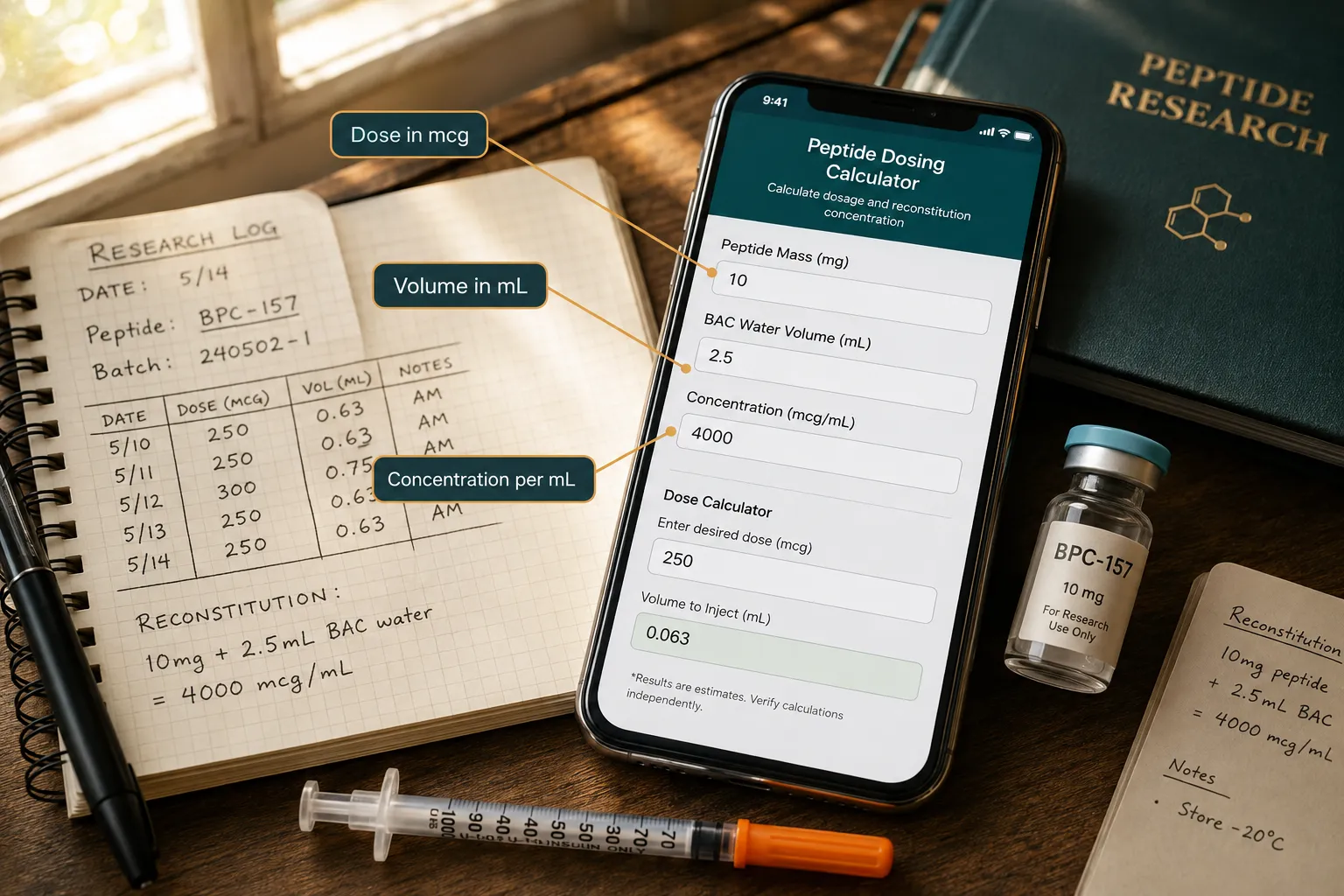
BPC-157 (Body Protection Compound-157) has no FDA-approved dose and limited controlled human data. However, a recent Phase 2 musculoskeletal trial and earlier pilot studies have begun to anchor a practical research range of 200-500 mcg per administration in human-model contexts, administered via subcutaneous or intramuscular routes.

A BPC-157 calculator entry typically includes:

  • Vial size (commonly 5 mg)
  • Reconstitution with 2.5 mL bacteriostatic water = 2,000 mcg/mL
  • Target dose of 250 mcg = 0.125 mL draw volume

Researchers can cross-reference translational research design principles to confirm that their BPC-157 protocol aligns with current Phase 2 reporting standards before finalizing a schedule.

Safety Frameworks and Regulatory Limits When Using Peptide Dosing Calculators

Safety Frameworks and Regulatory Limits When Using Peptide Dosing Calculators

A calculator produces mathematically correct outputs, it does not validate biological safety. Researchers must layer three additional frameworks over any calculator result.

1. Allometric and Duration Scaling
Beyond single-dose HED conversion, cumulative exposure matters. A peptide administered daily for 30 days carries a different risk profile than a single acute dose. Calculators that include duration-adjusted exposure modeling flag when total cumulative dose approaches thresholds seen in toxicology studies.

2. Biomarker Monitoring Checkpoints
Responsible protocols pair dose schedules with defined biomarker checkpoints, liver enzymes, kidney function markers, and peptide-specific indicators (e.g., insulin markers for GLP-class compounds). Some cross-peptide platforms now include monitoring schedule templates alongside the dosing math.

3. Regulatory and Purity Verification
No calculator output is meaningful if the source compound lacks verified purity. Researchers sourcing compounds should confirm certificate of analysis (CoA) data and consider wholesale peptides for sale only from suppliers with third-party tested documentation. Regulatory status in 2026 remains unchanged: BPC-157 and MOTS-c are not approved therapeutic agents in any major jurisdiction, and GLP-3 analogs remain investigational.

A note on future tools: speculative developments suggest that AI-assisted peptide calculators may eventually incorporate real-time biomarker feedback loops, but no validated platform of this type exists commercially as of 2026.

Peptide Common Research Vial Size Typical Reconstitution Resulting Concentration
GLP-class analogs 1-5 mg 1-2 mL BW 500-5,000 mcg/mL
MOTS-c 5-10 mg 2-5 mL BW 1,000-5,000 mcg/mL
BPC-157 5 mg 2.5 mL BW 2,000 mcg/mL

Conclusion

A reliable Peptides Calculator for GLP-3, MOTS-c, and BPC-157: How Researchers Estimate Dosing and Concentration Safely is only as useful as the protocol framework surrounding it. The actionable next steps for any researcher in 2026 are clear:

  • Verify compound purity with a CoA before any calculation has practical meaning.
  • Apply allometric scaling for MOTS-c and any compound where animal data is the primary reference.
  • Use escalation schedule builders for GLP-class peptides rather than starting at maximum estimated doses.
  • Document every calculator input and output as part of the formal research record.
  • Layer biomarker monitoring checkpoints at defined intervals throughout the protocol.

The math is straightforward. The discipline around the math is what separates reproducible research from noise.

https://www.puretestedpeptides.com/wp-content/uploads/2026/09/peptides-calculator-for-glp-3-mots-c-and-bpc-157-how-researchers-estimate-dosing.webp 1024 1536 https://www.puretestedpeptides.com/wp-content/uploads/2026/01/buy-peptides-online.jpg 2026-09-02 13:04:232026-09-02 13:04:23Peptides Calculator for GLP-3, MOTS-c, and BPC-157: How Researchers Estimate Dosing and Concentration Safely
Unpacking the 'Peptides Calculator': Essential Tools and Methods for Accurate Dosing and Reconstitution in Research

Unpacking the ‘Peptides Calculator’: Essential Tools and Methods for Accurate Dosing and Reconstitution in Research

August 22, 2026/0 Comments/in Uncategorized/by

A single decimal-point error during peptide reconstitution can shift an experimental dose by a factor of ten, enough to invalidate months of research data. That reality is precisely why unpacking the 'Peptides Calculator' and its essential tools and methods for accurate dosing and reconstitution in research has become a priority for serious investigators in 2026.

Key Takeaways

  • Peptide calculators standardize the math behind reconstitution, concentration, and dose volume to reduce human error.
  • The core formula, concentration equals mass divided by volume, underpins every reliable calculator on the market.
  • Bacteriostatic water (BAC water) remains the standard diluent for most lyophilized research peptides.
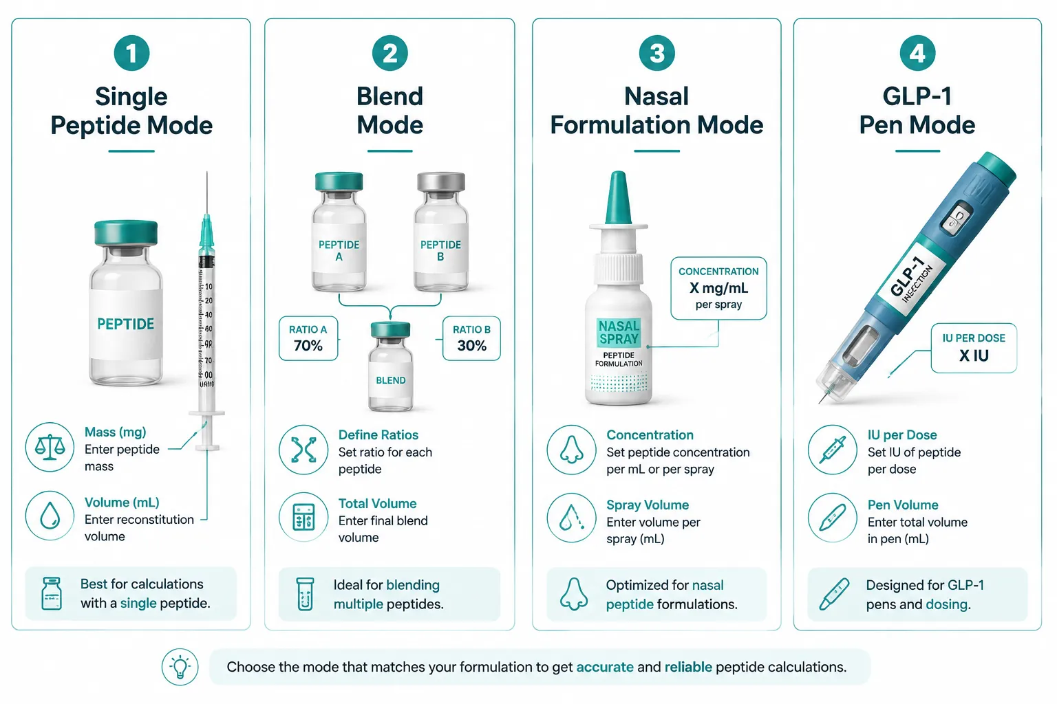
  • Modern tools now support single-peptide, blend, nasal, and GLP-1 pen modes with device-specific syringe guidance.
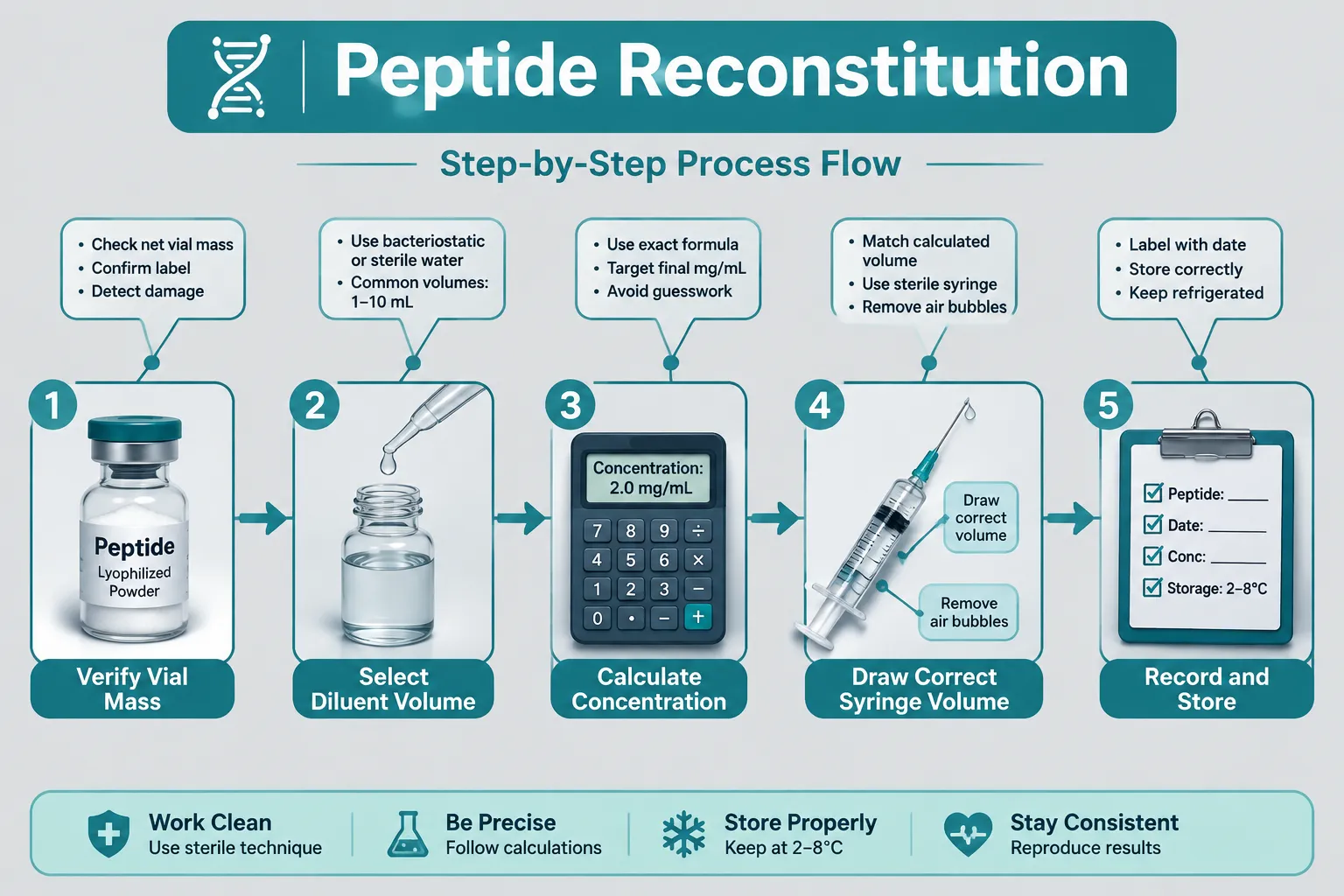
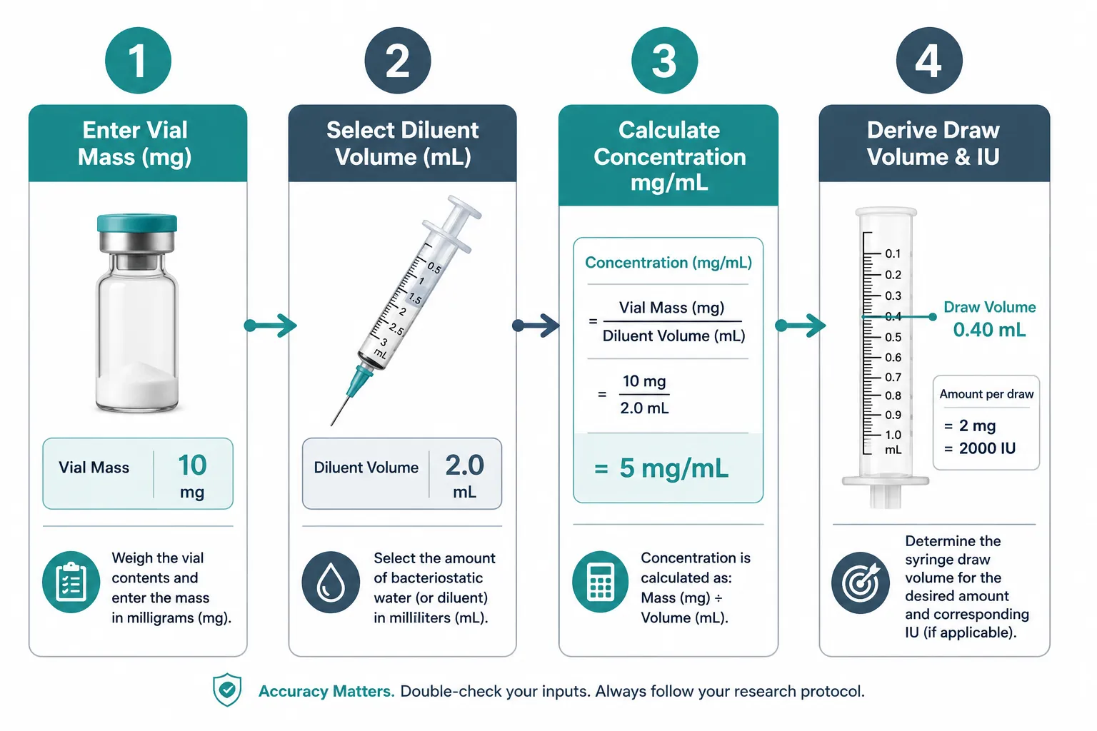
  • Reproducibility depends on consistent workflow: verify vial mass, select diluent volume, calculate concentration, then draw the correct syringe volume.

Why Accurate Peptide Dosing Matters in Research

Lyophilized peptides arrive as a dry powder measured in milligrams or micrograms. Before any research protocol can proceed, that powder must be dissolved in a precise volume of diluent to create a usable liquid concentration. Without a structured calculation method, researchers risk under-dosing (producing no measurable effect) or over-dosing (introducing confounding variables or compromising sample integrity).

Why Accurate Peptide Dosing Matters in Research

The stakes are especially high for sensitive compounds. For example, research into GLP-1 peptide sourcing and generational research concepts highlights how concentration accuracy directly shapes the validity of metabolic outcome data. Similarly, mitochondrial work involving compounds like those covered in SS-31 mitochondrial research themes demands tight dosing windows to produce reproducible results.

The core formula every researcher must internalize:

Concentration (mcg/mL) = Peptide Mass (mcg) / Diluent Volume (mL)

From this single equation, all downstream dose-volume calculations follow.

Standard Reconstitution Protocol with BAC Water

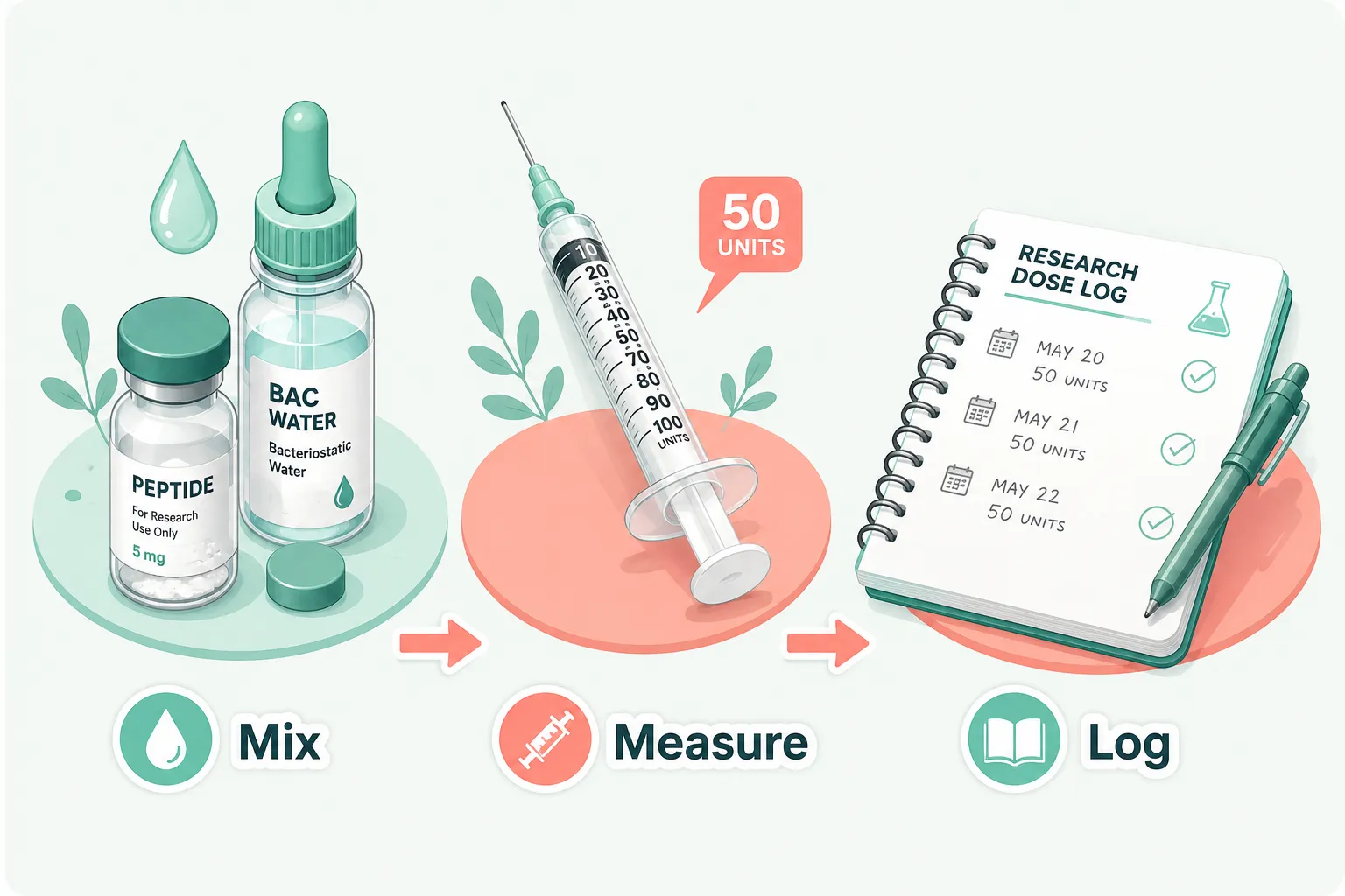
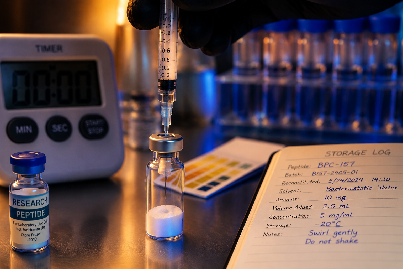
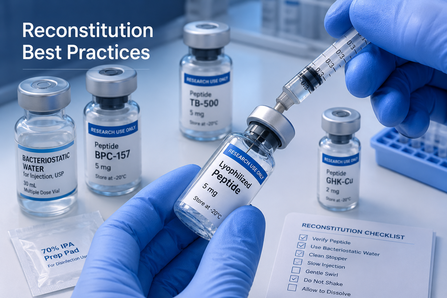
Bacteriostatic water is the preferred diluent for most lyophilized peptides because it contains 0.9% benzyl alcohol, which inhibits microbial growth and extends vial stability. The reconstitution steps below represent the standardized workflow recommended across leading peptide research platforms in 2026:

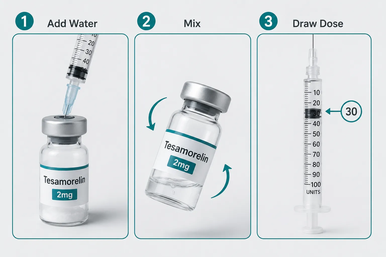
  1. Verify vial mass, confirm the labeled peptide mass (e.g., 5 mg = 5,000 mcg).
  2. Select diluent volume, choose a volume that produces a workable concentration (e.g., 2 mL BAC water for a 5 mg vial yields 2,500 mcg/mL).
  3. Add diluent slowly, inject BAC water along the vial wall; do not shake.
  4. Swirl gently, rotate until the powder fully dissolves.
  5. Calculate dose volume, divide the desired dose (mcg) by the concentration (mcg/mL).

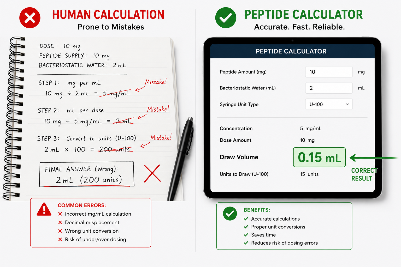
A researcher needing a 250 mcg dose from a 2,500 mcg/mL solution draws exactly 0.1 mL (100 mcL) into an insulin syringe. A peptides calculator automates this final step, eliminating arithmetic errors under lab conditions.

Unpacking the 'Peptides Calculator': Core Features and Input Modes

Modern peptide calculators have expanded well beyond a single-formula widget. Unpacking the 'Peptides Calculator' and its essential tools and methods for accurate dosing and reconstitution in research reveals at least four distinct operational modes now standard across leading platforms.

Unpacking the 'Peptides Calculator': Core Features and Input Modes

Calculator Mode Primary Use Case Key Inputs
Single Peptide Standard vial reconstitution Vial mass, diluent volume, target dose
Blend Mode Multi-peptide stacks Individual masses, shared diluent volume
Nasal Formulation Intranasal delivery research Concentration per spray, spray volume
GLP-1 / Pen Mode Injection pen devices Units per mL, dose in units or mcg

The GLP-1 pen mode deserves particular attention. As research interest in GLP-1 class compounds grows, see the detailed breakdown in Retatrutide Phase 3 and ongoing obesity trial research, calculators must handle "per-IU" concentration reporting alongside standard mcg/mL outputs. This dual-unit capability prevents the unit-conversion errors that historically account for a large share of dosing mistakes.

Enhanced unit conversion features now common in 2026 tools include:

  • Automatic mg-to-mcg conversion on input
  • IU-to-mcg translation for growth hormone-adjacent peptides
  • Syringe-mark visualization (e.g., "draw to the 10-unit line on a U-100 syringe")
  • Mobile-optimized interfaces for field and clinic-adjacent research settings

Researchers sourcing compounds for these protocols should consult resources like where to buy peptides to ensure purity and labeled mass accuracy, both of which are prerequisites for any calculator to produce valid outputs.

Applying the Calculator: Workflow, Reproducibility, and Research Compliance

Unpacking the 'Peptides Calculator' and its essential tools and methods for accurate dosing and reconstitution in research is only half the task. The other half is embedding the tool into a reproducible, documented workflow.

Applying the Calculator: Workflow, Reproducibility, and Research Compliance

Recommended documentation checklist for each reconstitution event:

  • Record the peptide name, lot number, and labeled mass.
  • Log the diluent type, volume added, and date of reconstitution.
  • Calculate and record the resulting concentration.
  • Note storage conditions (temperature, light exposure).
  • Document each dose drawn: target dose, calculated volume, and actual syringe reading.

This level of documentation supports reproducibility, the cornerstone of credible research. It also aligns with the quality-control principles discussed in resources like PT-141 research context, QA, and controls and the reference standard benchmarks explored in Bachem and reference standards for peptide benchmarks.

A critical compliance note: All peptide calculator tools and the research protocols they support are intended strictly for laboratory and investigational use. Regulatory frameworks in most jurisdictions classify research peptides as not approved for human administration outside of licensed clinical trials. Every workflow built around these tools must reflect that framing clearly.

Avoiding the Most Common Calculation Errors

  • Unit mismatch: Entering mass in mg but volume in mL without converting produces a 1,000-fold concentration error.
  • Assuming full vial mass: Overfill or underfill from the manufacturer means the labeled mass may differ slightly from actual mass; always use a calibrated scale when precision is critical.
  • Ignoring dead volume: Syringes retain a small volume in the needle hub; account for this in high-precision protocols.

Conclusion

Accurate dosing and reconstitution are not optional refinements, they are foundational to any research protocol that expects reproducible, interpretable results. The rapid evolution of peptide calculator tools in 2026 has made it easier than ever to perform these calculations correctly, but the tools only work when researchers understand the underlying math and commit to a disciplined workflow.

Actionable next steps for researchers:

  1. Select a calculator that supports the specific mode required (single peptide, blend, nasal, or pen-based).
  2. Verify vial mass with a calibrated scale before every reconstitution.
  3. Document every reconstitution event and dose draw in a dedicated lab log.
  4. Cross-check unit conversions manually at least once per new peptide or protocol.
  5. Source peptides from suppliers who provide verified purity data, ensuring the labeled mass is reliable input for any calculation.

Applying these steps consistently transforms a peptides calculator from a convenience tool into a genuine instrument of scientific rigor.

https://www.puretestedpeptides.com/wp-content/uploads/2026/08/unpacking-the-peptides-calculator-essential-tools-and-methods-for-accurate-dosin.webp 1024 1536 https://www.puretestedpeptides.com/wp-content/uploads/2026/01/buy-peptides-online.jpg 2026-08-22 13:06:062026-08-22 13:06:06Unpacking the ‘Peptides Calculator’: Essential Tools and Methods for Accurate Dosing and Reconstitution in Research
Peptide Calculators in Research: How Labs Estimate Dosing, Concentration, and Reconstitution

Peptide Calculators in Research: How Labs Estimate Dosing, Concentration, and Reconstitution

August 15, 2026/0 Comments/in Uncategorized/by

A single miscalculation in peptide reconstitution can render an entire experiment invalid, yet the math behind it is straightforward once researchers understand the core formulas. Peptide calculators in research: how labs estimate dosing, concentration, and reconstitution has become a central workflow topic in 2026, with multiple platforms releasing or updating dedicated calculation tools to support bench scientists working with compounds such as CJC-1295, Tesamorelin, and GLP-class molecules. This article is a technical tutorial for research use only, covering the underlying math, practical workflows, and common error-checking steps that modern peptide calculators are built around.

Key Takeaways

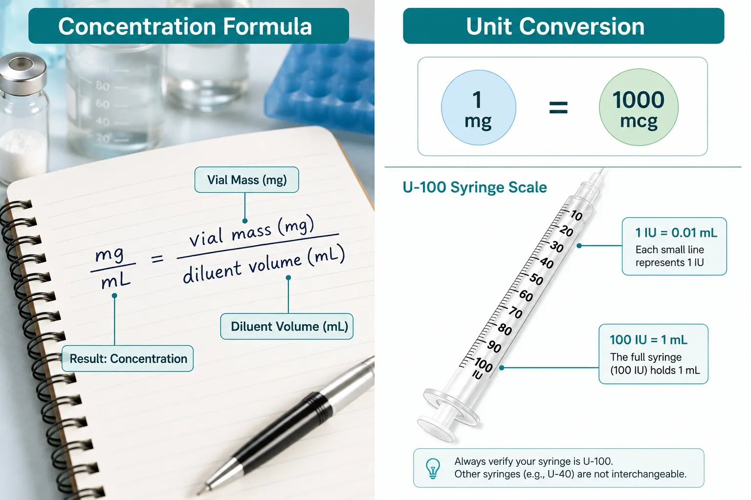
  • The core concentration formula is: Concentration (mg/mL) = Vial Mass (mg) / Diluent Volume (mL)
  • Converting between mg and mcg (1 mg = 1,000 mcg) is the most common source of calculation error
  • U-100 insulin syringes require an additional unit conversion: 1 IU = 0.01 mL
  • Modern peptide calculators use 3-4 step workflows and include error-checking to validate syringe capacity
  • All calculations assume uniform dissolution of the peptide in bacteriostatic water

The Core Math Behind Peptide Concentration Calculations

The Core Math Behind Peptide Concentration Calculations

Every peptide calculator in research begins with one foundational equation. Once a lyophilized peptide is reconstituted in bacteriostatic water (BW), the resulting solution is assumed to be uniformly dissolved throughout the vial. This assumption makes the concentration formula clean and reliable:

Concentration (mg/mL) = Vial Mass (mg) / Reconstitution Volume (mL)

For example, a 5 mg vial of CJC-1295 reconstituted with 2.5 mL of bacteriostatic water yields a concentration of 2 mg/mL. If a researcher needs outputs in micrograms per milliliter, common in cell culture and assay protocols, the formula adjusts:

Concentration (mcg/mL) = (Vial Mass in mg × 1,000) / Diluent Volume (mL)

Using the same example: (5 × 1,000) / 2.5 = 2,000 mcg/mL.

The relationship 1 mg = 1,000 mcg is highlighted repeatedly in calculator documentation because it is the most frequent source of dosing errors. Researchers working with compounds like Tesamorelin and Ipamorelin combination protocols must pay particular attention to this conversion, as both compounds are often dosed in the low-microgram range.

Deriving Injection Volume from Concentration

Once concentration is established, the draw volume for a target dose follows directly:

Draw Volume (mL) = Target Dose (mg or mcg) / Concentration (mg/mL or mcg/mL)

If the target research dose is 1 mg and the concentration is 2 mg/mL, the draw volume is 0.5 mL. This simple division is the backbone of every peptide dosing calculator available in 2026.

Syringe Unit Conversion: Translating mL Into IU on a U-100 Scale

Syringe Unit Conversion: Translating mL Into IU on a U-100 Scale

Most research labs use U-100 insulin syringes for subcutaneous peptide administration in animal models. These syringes are calibrated in International Units (IU), not milliliters, which introduces a conversion step that peptide calculators in research consistently address.

The key relationship is:

Measurement Equivalent
1 mL 100 IU
1 IU 0.01 mL
0.5 mL 50 IU
0.1 mL 10 IU

To convert a draw volume in mL to syringe units:

Syringe Units (IU) = Draw Volume (mL) × 100

Using the earlier example: 0.5 mL × 100 = 50 IU on the syringe scale.

Modern calculators present this as part of a guided 4-step workflow:

  1. Enter vial mass (mg)
  2. Enter bacteriostatic water volume (mL)
  3. Enter target dose (mg or mcg)
  4. Receive draw volume in mL and IU

Some tools include visual syringe meters that animate exactly where to stop drawing on the scale, a practical feature for labs running high-throughput assays with compounds like those explored in SS-31 mechanism and research.

Error-Checking and Capacity Validation

A notable feature in 2026 calculator updates is capacity validation, the tool checks whether the calculated draw volume exceeds the selected syringe's maximum capacity. If a researcher selects a 0.3 mL syringe but the calculation returns 0.45 mL, the calculator flags the mismatch before any solution is drawn. Back-check logic also allows users to confirm that concentration, dose, and syringe units are internally consistent, reducing the risk of compounding errors across multi-compound protocols.

Applying Peptide Calculators to GLP-Class and Advanced Research Compounds

Applying Peptide Calculators to GLP-Class and Advanced Research Compounds

The same mg/mL concentration logic that governs classic research peptides applies directly to GLP-1 agonists, GLP-3 class molecules, and adjunct compounds like NAD+. This consistency has driven broad adoption of standardized peptide calculators across metabolic and longevity research programs.

For a GLP-class compound supplied as a 10 mg vial, reconstituted with 5 mL of bacteriostatic water:

  • Concentration = 10 / 5 = 2 mg/mL (or 2,000 mcg/mL)
  • Target dose of 0.5 mg = draw volume of 0.25 mL (25 IU on a U-100 syringe)

Researchers studying compounds such as those reviewed in Selank peptide research benefits and dosing concepts or MOTS-c mitochondrial signaling and metabolic research apply identical formulas, adjusting only the vial mass and target dose inputs.

For in-vitro protocols, such as cell culture or enzyme assays, calculators offer fields labeled "research amount" and "volume of reconstituted solution used in your experiment," returning outputs in mg/mL, mcg/mL, and IU. This makes the same tool useful across both in-vivo animal model work and bench-based assay preparation.

"The uniformity assumption, that a reconstituted peptide is evenly dissolved throughout the vial, is what makes the mg/mL formula reliable and repeatable across research contexts."

Labs sourcing compounds like GHK-Cu copper peptides or PT-141 in research context QA and controls benefit from pairing supplier documentation with a validated calculator workflow to ensure concentration consistency across experimental batches.

Quick Reference: Common Reconstitution Scenarios

Vial Size BW Added Concentration 0.5 mg Dose Draw
2 mg 1 mL 2 mg/mL 0.25 mL / 25 IU
5 mg 2.5 mL 2 mg/mL 0.25 mL / 25 IU
10 mg 5 mL 2 mg/mL 0.25 mL / 25 IU
5 mg 5 mL 1 mg/mL 0.5 mL / 50 IU

Conclusion

Peptide calculators in research: how labs estimate dosing, concentration, and reconstitution comes down to three sequential calculations, concentration from vial mass and diluent volume, draw volume from concentration and target dose, and syringe units from draw volume and the U-100 scale. The math is accessible, but the consequences of skipping steps or mishandling unit conversions are significant in a research context.

Actionable next steps for research teams:

  • Standardize on a single reconstitution volume per compound to keep concentration consistent across experimental runs
  • Always verify the mg-to-mcg conversion before entering values into any calculator
  • Use a calculator with capacity validation to confirm syringe selection before drawing
  • Document concentration, draw volume, and IU for every batch in the lab notebook
  • Cross-reference calculator outputs against the underlying formula manually at least once per new compound

For labs working with a broad compound library, pairing a validated peptide calculator with high-purity, lab-tested peptides and reliable supplier documentation is the most effective way to maintain experimental integrity across studies.

https://www.puretestedpeptides.com/wp-content/uploads/2026/08/peptide-calculators-in-research-how-labs-estimate-dosing-concentration-and-recon.webp 1024 1536 https://www.puretestedpeptides.com/wp-content/uploads/2026/01/buy-peptides-online.jpg 2026-08-15 13:04:532026-08-15 13:04:53Peptide Calculators in Research: How Labs Estimate Dosing, Concentration, and Reconstitution
PT-141 Peptide: Mechanism of Action, Research Applications, and Protocol Questions

PT-141 Peptide: Mechanism of Action, Research Applications, and Protocol Questions

August 14, 2026/0 Comments/in Uncategorized/by

Only one FDA-approved compound targets sexual desire by acting directly on the brain rather than on blood vessels, and that compound is bremelanotide, better known in research settings as PT-141. Understanding the PT-141 peptide: mechanism of action, research applications, and protocol questions requires moving past surface-level descriptions and into the melanocortin pathway itself, where the real scientific interest lies.

Key Takeaways

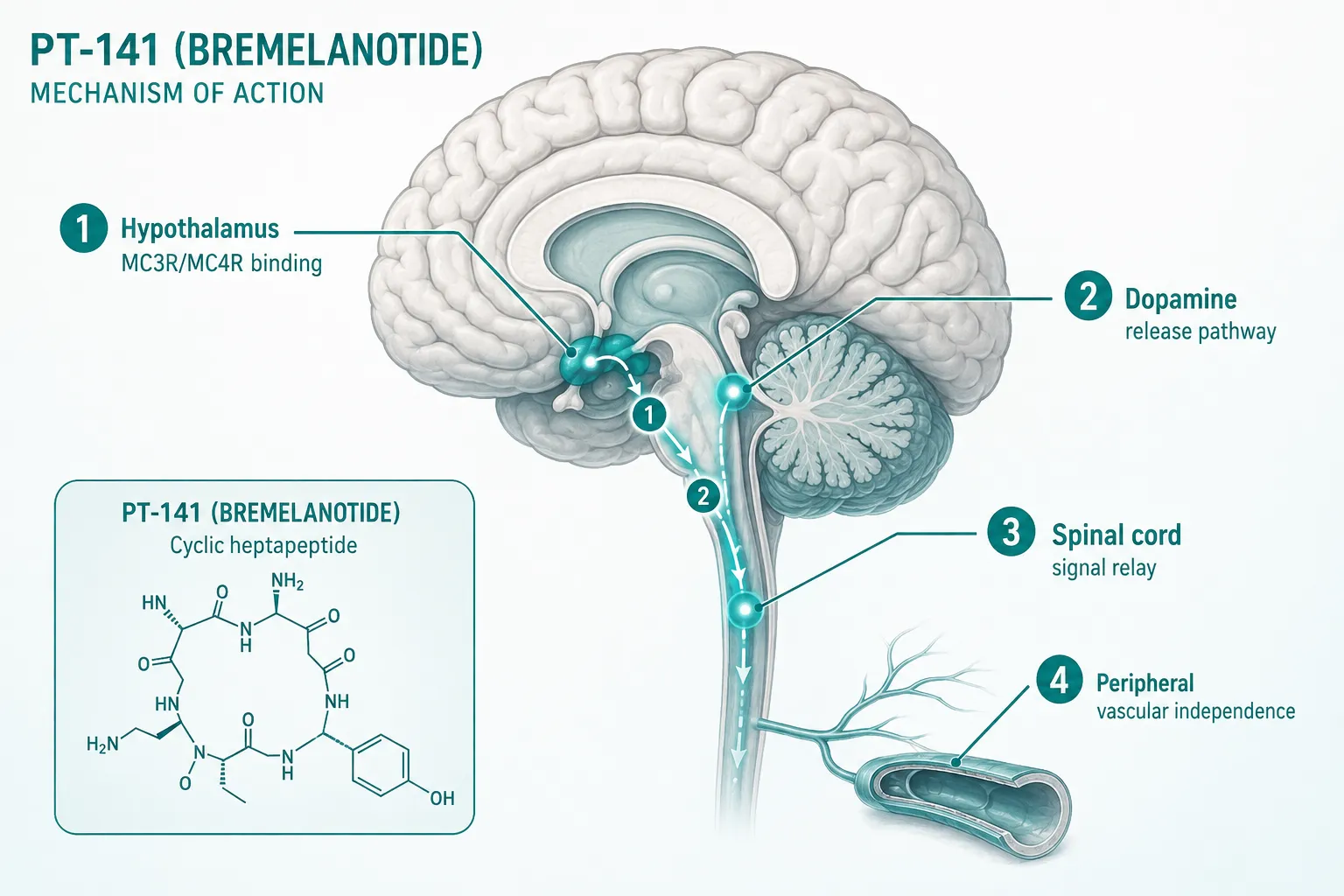
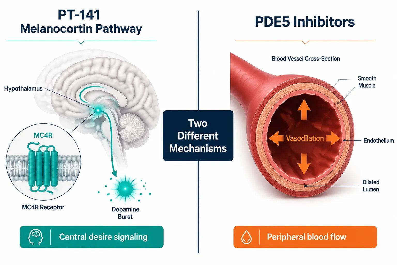
  • PT-141 (bremelanotide) acts centrally through melanocortin receptors MC3R and MC4R, triggering dopamine release rather than peripheral vasodilation.
  • It is the only FDA-approved agent for hypoactive sexual desire disorder (HSDD) in premenopausal women that works via a CNS mechanism.
  • Research interest extends beyond its approved indication to male populations, CNS desire pathways, and multi-peptide experimental stacks.
  • PT-141 is structurally distinct from PDE5 inhibitors, making it a complementary rather than competing research target.
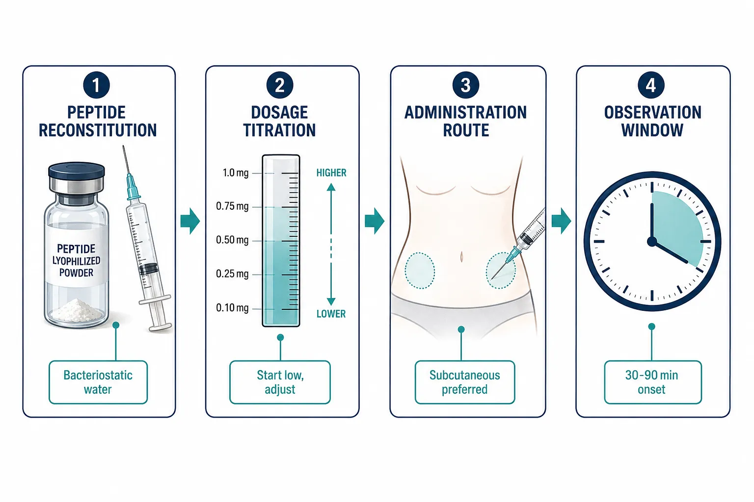
  • Protocol questions in research settings center on reconstitution, dosing titration, and observation windows rather than on cardiovascular endpoints.

The Melanocortin Pathway: Core Mechanism of Action

The Melanocortin Pathway: Core Mechanism of Action

PT-141 is a synthetic cyclic heptapeptide derived from alpha-melanocyte-stimulating hormone (alpha-MSH). When researchers study the PT-141 peptide: mechanism of action, research applications, and protocol questions, the starting point is always the melanocortin system, a family of G-protein-coupled receptors distributed throughout the central nervous system.

How the pathway works:

  • PT-141 binds with high affinity to MC3R and MC4R receptors, primarily in the hypothalamus and limbic system.
  • Receptor activation triggers downstream dopamine release in mesolimbic circuits.
  • The resulting signal is interpreted as increased sexual motivation or desire, a centrally mediated effect.
  • Crucially, this mechanism does not rely on nitric oxide signaling or penile/vaginal smooth muscle relaxation.

This last point is what separates PT-141 from the entire class of phosphodiesterase-5 (PDE5) inhibitors. Sildenafil and its relatives address the mechanical capacity for arousal; PT-141 addresses the motivational component. In research models, this distinction allows investigators to study desire and arousal as separable constructs.

"PT-141 offers a rare window into centrally mediated desire, a target that PDE5 inhibitors simply do not touch."

For researchers interested in how different peptide classes engage distinct receptor families, the broader overview at Peptides Mechanism 101: From GLP-3 Retatrutide to CJC-1295 and MOTS-c provides useful comparative context.

Research Applications: What PT-141 Is Actually Used to Study

Research Applications: What PT-141 Is Actually Used to Study

The approved clinical indication for bremelanotide is HSDD in premenopausal women, supported by the Phase 3 RECONNECT trial program. However, the research community has consistently explored a wider set of questions around this compound.

Female HSDD and the RECONNECT Data

The RECONNECT studies demonstrated statistically significant improvements in satisfying sexual events and reductions in distress scores compared to placebo. These outcomes established bremelanotide as the first on-demand pharmacological option for HSDD, distinguishing it from the daily-dosing requirement of flibanserin.

Key outcomes observed in Phase 3 data:

Endpoint Direction of Effect
Satisfying sexual events per month Increased vs. placebo
Female Sexual Distress Scale score Decreased vs. placebo
Desire domain scores Improved vs. placebo
Nausea (most common adverse effect) Present; dose-dependent

Male Population Research

Off-label and preclinical research has explored PT-141 in males with erectile dysfunction who show inadequate response to PDE5 inhibitors. The hypothesis is that some cases of ED have a significant central desire component that peripheral vasodilators cannot address. Early-phase human data showed meaningful erectile response signals, though this application remains outside the approved label.

Multi-Compound Research Stacks

In 2026, a growing segment of research interest involves pairing PT-141 with other peptides to probe synergistic CNS effects. Researchers studying hormonal and desire pathways sometimes combine PT-141 with growth hormone secretagogues or other CNS-active compounds. For context on how stacking strategies are designed, the IPA Sermorelin Stack Research resource outlines how multi-peptide protocols are structured in research settings.

Those sourcing research-grade material can review available PT-141 10mg peptide for sale options, or explore the PT141 peptide for sale catalog for purity specifications relevant to lab use.

Safety Profile Considerations

The adverse effect profile in clinical trials was dominated by:

  • Nausea (most frequent, dose-related)
  • Flushing and transient facial redness
  • Transient blood pressure increases (typically small, short-lived)
  • Injection site reactions

Cardiovascular monitoring is recommended in protocols involving subjects with hypertension risk, given the documented transient blood pressure signal.

Protocol Questions in Research Settings

Protocol Questions in Research Settings

When researchers engage with the PT-141 peptide: mechanism of action, research applications, and protocol questions in a practical lab context, the most frequent questions cluster around preparation and timing rather than pharmacodynamics.

Reconstitution and Storage

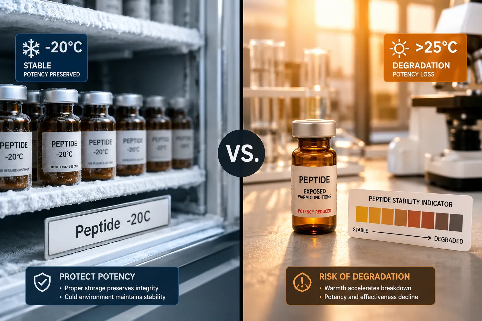
PT-141 is supplied as a lyophilized powder. Standard reconstitution uses bacteriostatic water. Once reconstituted, storage at 2-8°C is appropriate for short-term use, with lyophilized stock maintained at -20°C for longer periods.

Dosing Considerations in Research Protocols

The approved clinical dose for bremelanotide is 1.75 mg subcutaneous, administered approximately 45 minutes before anticipated activity. Research protocols often begin at lower titration points to characterize dose-response relationships.

Common protocol structure:

  1. Baseline observation period, establish pre-dose behavioral or physiological measures
  2. Low-dose administration, subcutaneous preferred for consistent absorption
  3. Observation window, 30 to 90 minutes post-administration for peak effect window
  4. Washout period, minimum 24 hours between doses in clinical data; research protocols vary

Delivery Route Considerations

Subcutaneous injection remains the best-characterized route. Intranasal delivery was explored in early development (the original PT-141 formulation was intranasal) but was not pursued to approval due to bioavailability variability. Researchers interested in nasal delivery formats for other peptides can review Nasal Spray Peptides: Delivery Methods, Bioavailability, and Research Advantages for a broader comparison of routes.

For researchers building out a more complete peptide research library, understanding structural classifications is foundational. The article Polypeptide Peptides Explained: Structure, Function, and Research Applications provides that structural grounding. Additionally, those exploring the regulatory and quality criteria for sourcing compounds can reference Where to Buy Research-Grade MOTS-c and 5-Amino-1MQ: Quality Criteria for vendor evaluation frameworks applicable across peptide categories.

Researchers who want the full product specification for bremelanotide can review the Buy PT-141 Peptide (Bremelanotide) 10mg | 99% Pure | Melanocortin Agonist listing for purity and certificate of analysis details.

Conclusion

The PT-141 peptide: mechanism of action, research applications, and protocol questions represent one of the more well-defined areas in CNS-active peptide research. The compound's selectivity for MC3R and MC4R, its dopamine-mediated desire signaling, and its structural independence from the PDE5 pathway give it a genuinely distinct research profile.

Actionable next steps for researchers in 2026:

  • Confirm purity documentation (minimum 99%) before incorporating PT-141 into any protocol.
  • Design observation windows around the 30-to-90-minute peak effect period documented in clinical data.
  • When exploring multi-compound stacks, map each compound's receptor targets to avoid overlapping CNS stimulation.
  • Review the RECONNECT Phase 3 data as the most rigorous human-subject dataset available for dose-response benchmarking.
  • Treat the transient blood pressure signal as a monitoring checkpoint, not a disqualifying factor, when designing subject selection criteria.

The melanocortin pathway remains an underexplored frontier in CNS pharmacology. PT-141 is currently the most research-accessible tool for probing it.

https://www.puretestedpeptides.com/wp-content/uploads/2026/08/pt-141-peptide-mechanism-of-action-research-applications-and-protocol-questions.webp 1024 1536 https://www.puretestedpeptides.com/wp-content/uploads/2026/01/buy-peptides-online.jpg 2026-08-14 13:07:032026-08-14 13:07:03PT-141 Peptide: Mechanism of Action, Research Applications, and Protocol Questions
GHK-Cu Peptide: Copper Complex Chemistry, Research Stability, and Lab Use Considerations

GHK-Cu Peptide: Copper Complex Chemistry, Research Stability, and Lab Use Considerations

August 8, 2026/0 Comments/in Uncategorized/by

Fewer than 10% of commercially sold research peptides are independently verified for metal-chelation integrity, and for GHK-Cu, that gap matters more than with almost any other compound. Unlike single-chain peptides, GHK-Cu is a coordination complex. Its biological activity depends not just on peptide purity, but on the precise stoichiometric relationship between the tripeptide glycyl-L-histidyl-L-lysine (GHK) and its bound copper(II) ion. Understanding the chemistry behind that bond is the first step toward reliable, reproducible research.

This article focuses on the chemistry, stability, and practical handling of GHK-Cu Peptide: Copper Complex Chemistry, Research Stability, and Lab Use Considerations, giving researchers the technical foundation needed to evaluate product quality and design sound experimental protocols in 2026.

Key Takeaways

  • GHK-Cu is a copper(II) coordination complex, not a simple peptide, its activity depends on intact metal chelation.
  • The histidine imidazole nitrogen is the primary copper-binding site; disruption of this bond compromises the compound's function.
  • Reconstituted GHK-Cu solutions degrade faster than lyophilized powder and require careful pH and temperature control.
  • Purity certificates should confirm both peptide sequence identity and copper content via ICP-MS or equivalent methods.
  • Contamination, repeated freeze-thaw cycles, and oxidative conditions are the leading causes of GHK-Cu degradation in lab settings.

The Copper Coordination Chemistry of GHK-Cu

The Copper Coordination Chemistry of GHK-Cu

The tripeptide GHK (Gly-His-Lys) forms a square-planar coordination complex with copper(II) through three nitrogen donor atoms. The binding sites are:

  • The alpha-amino group of glycine
  • The deprotonated amide nitrogen of the glycine-histidine peptide bond
  • The imidazole nitrogen (N3) of histidine

This 3N coordination geometry is sometimes called an ATCUN (amino terminal copper and nickel) motif. It is highly specific and produces a stable complex at physiological pH. The lysine residue at the C-terminus does not directly coordinate copper but contributes to solubility and cellular uptake behavior.

"The integrity of the Cu(II) coordination sphere is inseparable from GHK-Cu's reported biological activity. A peptide sold without confirmed copper content is, chemically speaking, just GHK."

Why this matters for researchers: Products labeled "GHK-Cu" that lack verified copper loading are effectively dechelated peptide. The free GHK tripeptide and the copper complex are distinct chemical entities with different physical properties and likely different biological profiles. Researchers sourcing material should request certificates of analysis that include elemental copper quantification, not just HPLC purity of the peptide backbone.

For context on how rigorous reference standards apply to peptide research more broadly, see this overview of Bachem and reference standards for building robust peptide benchmarks.

Research Stability: What Degrades GHK-Cu and How Fast

Research Stability: What Degrades GHK-Cu and How Fast

Understanding degradation pathways is central to GHK-Cu Peptide: Copper Complex Chemistry, Research Stability, and Lab Use Considerations in any serious lab context. GHK-Cu faces three primary degradation threats:

Oxidative Degradation

Copper(II) is a redox-active metal. In solution, it can catalyze the oxidation of the histidine imidazole ring, the very residue responsible for coordination. Dissolved oxygen accelerates this process significantly. Researchers should prepare solutions under inert gas where possible and use low-oxygen water.

pH Sensitivity

The ATCUN coordination geometry is pH-dependent. At pH below 5.0, protonation of the amide nitrogen weakens the complex. At pH above 8.5, competing hydroxide ligands can displace the peptide. The optimal stability window is pH 6.5-7.4, closely matching physiological conditions.

Condition Effect on GHK-Cu Stability
pH < 5.0 Copper dissociation, complex breakdown
pH 6.5-7.4 Optimal coordination, maximum stability
pH > 8.5 Hydroxide competition, partial dechelation
Temperature > 37°C Accelerated oxidation and peptide hydrolysis
Freeze-thaw cycling (>3x) Aggregation, loss of copper coordination

Temperature and Freeze-Thaw Stress

Lyophilized GHK-Cu powder is stable at -20°C for extended periods when stored desiccated and away from light. Reconstituted solutions, however, should be aliquoted immediately and used within 24-48 hours at 4°C. Repeated freeze-thaw cycles promote aggregation and copper dissociation.

This storage discipline parallels best practices described for other sensitive research peptides, such as those outlined in AOD-9604 sale research method notes on storage and traceability and SS-31 10mg research peptide considerations.

Lab Use Considerations for GHK-Cu Research

Lab Use Considerations for GHK-Cu Research

Translating chemistry knowledge into sound lab practice is the practical core of GHK-Cu Peptide: Copper Complex Chemistry, Research Stability, and Lab Use Considerations. The following protocols reduce experimental variability.

Reconstitution Best Practices

  • Use sterile water for injection or phosphate-buffered saline at pH 7.0-7.2.
  • Avoid DMSO as a primary solvent, it can disrupt metal coordination at higher concentrations.
  • Prepare working concentrations fresh; do not store diluted solutions overnight.
  • Use amber or opaque vials to minimize photodegradation.

Purity and Identity Verification

Researchers should request certificates that include:

  1. HPLC purity (peptide backbone, >98% preferred)
  2. Mass spectrometry confirmation of molecular weight (GHK-Cu: ~340 Da for the complex)
  3. ICP-MS or atomic absorption spectroscopy for copper content verification
  4. Endotoxin testing for cell-based assays

Experimental Controls

Because free copper ions are biologically active on their own, every GHK-Cu experiment should include:

  • A free CuSO4 control at equivalent copper concentration
  • A free GHK peptide control (dechelated)
  • A vehicle-only control

This three-arm control design isolates the effect of the intact complex from its individual components, a distinction that is frequently overlooked in published literature.

For researchers working with other structurally complex peptides, the documentation practices described in the BPC-157 core peptides documentation-first research guide offer transferable methodology. Similarly, researchers comparing peptide classes may find value in reviewing TB-500 peptide handling and research notes.

Conclusion

GHK-Cu is one of the most chemically nuanced compounds in the research peptide space. Its activity is inseparable from the integrity of its copper coordination complex, meaning that sourcing, storage, and experimental design all carry higher stakes than with standard single-chain peptides. Researchers should prioritize suppliers who provide elemental copper verification alongside peptide purity data, prepare solutions at controlled pH within the 6.5-7.4 window, limit reconstituted solution storage to 48 hours, and include both free-copper and dechelated-peptide controls in every assay.

Actionable next steps:

  • Request ICP-MS copper content data from any GHK-Cu supplier before purchasing.
  • Review current peptide research products available and confirm COA documentation standards before ordering.
  • Establish a dedicated aliquoting protocol to eliminate freeze-thaw degradation from your workflow.
  • Design three-arm controls (intact complex, free Cu, free GHK) as a standard operating procedure for all GHK-Cu experiments.

Rigorous attention to these chemistry and handling details is what separates reproducible data from ambiguous results.

https://www.puretestedpeptides.com/wp-content/uploads/2026/08/ghk-cu-peptide-copper-complex-chemistry-research-stability-and-lab-use-considera.webp 1024 1536 https://www.puretestedpeptides.com/wp-content/uploads/2026/01/buy-peptides-online.jpg 2026-08-08 13:03:252026-08-08 13:03:25GHK-Cu Peptide: Copper Complex Chemistry, Research Stability, and Lab Use Considerations
Peptide Calculators for Tesamorelin and Ipamorelin: Optimizing Reconstitution and Dosing Accuracy

Peptide Calculators for Tesamorelin and Ipamorelin: Optimizing Reconstitution and Dosing Accuracy

July 31, 2026/0 Comments/in Uncategorized/by

"

Professional () hero image with (≤42 chars): 'Peptide Calculators: Tesamorelin' in crisp white on a deep navy

A dosing error as small as 0.1 mL can translate to a 50% deviation from the intended peptide amount, a margin that renders research data unreliable before the experiment even begins. For researchers working with growth hormone secretagogues, precision is not optional. Using peptide calculators for Tesamorelin and Ipamorelin: optimizing reconstitution and dosing accuracy is one of the most practical steps any researcher can take to eliminate preventable errors and produce consistent, reproducible outcomes.

This guide walks through the mechanics of peptide calculators, explains why reconstitution ratios matter, and provides a clear framework for applying these tools to Tesamorelin and Ipamorelin research protocols.

Key Takeaways

  • Peptide calculators convert vial concentration and desired dose into exact injection volumes, removing guesswork from the process.
  • The amount of bacteriostatic water (BAC water) added during reconstitution directly determines the concentration of every subsequent dose.
  • Tesamorelin and Ipamorelin have different molecular weights and standard research dosing ranges, requiring separate calculations.
  • Small syringe selection errors compound over time and can significantly skew cumulative dosing across a research cycle.
  • Verifying purity and peptide mass through third-party-tested sources is a prerequisite for any calculation to be meaningful.

Key Takeaways

Understanding the Core Math Behind Peptide Calculators

Before any syringe is filled, a researcher must establish one foundational number: concentration, expressed in micrograms per milliliter (mcg/mL). Every downstream calculation depends on it.

The formula is straightforward:

Concentration (mcg/mL) = Total peptide mass (mcg) / Volume of BAC water added (mL)

For example, a 2 mg (2,000 mcg) vial of Tesamorelin reconstituted with 2 mL of BAC water yields a concentration of 1,000 mcg/mL. If the target research dose is 500 mcg, the required injection volume is exactly 0.5 mL.

Why BAC Water Volume Is the Critical Variable

Many researchers focus on dose size but overlook that the volume of BAC water added is the variable that controls everything else. Adding more water lowers concentration and increases injection volume per dose. Adding less water raises concentration and shrinks injection volume, which can make accurate measurement on a standard insulin syringe harder.

A practical rule: aim for a reconstitution volume that places the target dose between 0.1 mL and 0.5 mL on a 1 mL insulin syringe. This range offers the best balance of measurement accuracy and manageable injection volume.

"The most common reconstitution mistake is not calculating the dose wrong, it is adding an unmeasured amount of BAC water and then trying to back-calculate afterward."

Researchers exploring Tesamorelin dosage protocols should establish their BAC water volume before reconstitution, not after.

Applying Peptide Calculators for Tesamorelin and Ipamorelin: Optimizing Reconstitution and Dosing Accuracy

Applying Peptide Calculators for Tesamorelin and Ipamorelin: Optimizing Reconstitution and Dosing Accuracy

Tesamorelin and Ipamorelin are frequently used together in research settings, but they have distinct properties that affect how calculations are performed.

Tesamorelin Calculation Example

Tesamorelin is a synthetic analogue of growth hormone-releasing hormone (GHRH). Common research vial sizes are 2 mg and 5 mg. For a Tesamorelin research peptide vial of 5 mg (5,000 mcg):

Reconstitution Volume Concentration Volume for 1,000 mcg dose
2.5 mL BAC water 2,000 mcg/mL 0.50 mL
5.0 mL BAC water 1,000 mcg/mL 1.00 mL
1.0 mL BAC water 5,000 mcg/mL 0.20 mL

The 2.5 mL option is often preferred because the 0.50 mL draw is easy to read on a standard U-100 insulin syringe.

Researchers comparing growth hormone secretagogue options may also find the Sermorelin vs Tesamorelin breakdown useful for contextualizing dosing differences.

Ipamorelin Calculation Example

Ipamorelin is a selective growth hormone secretagogue receptor agonist. Vials are commonly available at 2 mg and 5 mg. For a 2 mg (2,000 mcg) vial:

Reconstitution Volume Concentration Volume for 200 mcg dose
2.0 mL BAC water 1,000 mcg/mL 0.20 mL
1.0 mL BAC water 2,000 mcg/mL 0.10 mL

Researchers using combination products should note that blend vials, such as those in Tesamorelin/CJC-1295/Ipamorelin 12 mg blends, require the calculator to account for the total mass of all peptides combined, not just one component.

For those comparing secretagogue combinations, the Ipamorelin vs Sermorelin vs Hexarelin comparison provides relevant research context.

Avoiding Common Errors: Practical Tips for Dosing Accuracy

Avoiding Common Errors: Practical Tips for Dosing Accuracy

Even with a calculator, errors occur. The following checklist addresses the most frequent failure points in peptide reconstitution and dosing workflows.

Before Reconstitution

  • Confirm vial mass matches the label (third-party COA verification is essential, see quality peptides sourcing guidance)
  • Use a calibrated, sterile BAC water syringe for adding diluent
  • Record the exact volume of BAC water added immediately

During Dosing

  • Use a U-100 insulin syringe for doses under 1 mL
  • Read the syringe at eye level to avoid parallax error
  • Never estimate, if the dose falls between graduation marks, recalculate the reconstitution

Storage and Stability

  • Reconstituted peptides should be stored at 2-8°C and used within the manufacturer's recommended window
  • Avoid repeated freeze-thaw cycles, which degrade peptide integrity and alter effective concentration

Researchers working with multi-peptide protocols, for instance, those incorporating CJC-1295/Ipamorelin assay planning, should maintain a separate calculation log for each peptide in the stack.

For fat-loss focused research designs, the Tesamorelin dosage for fat loss resource offers protocol-specific dosing context that complements calculator outputs.

Conclusion

Accurate research outcomes with Tesamorelin and Ipamorelin depend on a simple but non-negotiable chain: verified peptide mass, precise BAC water volume, correct concentration calculation, and accurate syringe measurement. Peptide calculators for Tesamorelin and Ipamorelin: optimizing reconstitution and dosing accuracy are not a shortcut, they are the standard operating procedure for any researcher who wants data they can trust.

Actionable next steps:

  1. Before reconstituting any vial, calculate your target concentration and write it down.
  2. Select a BAC water volume that places your dose in the 0.1-0.5 mL range on a U-100 syringe.
  3. Source peptides only from suppliers with third-party purity verification to ensure the labeled mass is accurate.
  4. Keep a dosing log for every session, recording concentration, draw volume, and administration time.
  5. Revisit your calculations if you switch vial sizes, suppliers, or reconstitution volumes mid-protocol.

Precision at the preparation stage is the single highest-leverage action a researcher can take before any experiment begins.

https://www.puretestedpeptides.com/wp-content/uploads/2026/07/peptide-calculators-for-tesa-and-ipamorelin-optimizing-reconstitution-and.webp 1024 1536 https://www.puretestedpeptides.com/wp-content/uploads/2026/01/buy-peptides-online.jpg 2026-07-31 13:03:552026-07-31 13:03:55Peptide Calculators for Tesamorelin and Ipamorelin: Optimizing Reconstitution and Dosing Accuracy

Tag Archive for: peptide reconstitution

Peptide Calculator Use Cases Beyond Growth Hormone: Working Through GLP‑3 Retatrutide, MOTS‑c, and BPC‑157 Research Dosing

Peptide Calculator Use Cases Beyond Growth Hormone: Working Through GLP‑3 Retatrutide, MOTS‑c, and BPC‑157 Research Dosing

July 18, 2026/0 Comments/by Pure Tested

Most researchers reach for a peptide calculator when reconstituting a growth hormone secretagogue blend, then stop there. Yet the same arithmetic logic that converts a Tesamorelin vial into syringe units applies equally to metabolic triple agonists, mitochondrial peptides, and tissue-repair compounds. Peptide Calculator Use Cases Beyond Growth Hormone: Working Through GLP-3 Retatrutide, MOTS-c, and BPC-157 Research Dosing is a topic that deserves its own worked framework, because each compound carries unique concentration targets, titration schedules, and reconstitution constraints that a generic calculator must accommodate.

Bright infographic-style editorial landscape (): isometric illustration of three distinct peptide molecules — GLP-3

Key Takeaways

  • Peptide calculators are not limited to GH secretagogues, they handle any lyophilized compound requiring reconstitution math.
  • Retatrutide (GLP-3) uses slow titration schedules that demand week-by-week dose recalculation.
  • MOTS-c reconstitution targets are typically low-volume and require precise unit conversion.
  • BPC-157 research often involves both injectable and oral formats, each with different concentration logic.
  • GHK-Cu and other repair peptides follow the same calculator inputs: vial mass, diluent volume, and desired dose.

Why Peptide Calculator Use Cases Extend Well Beyond Growth Hormone

Growth hormone peptides like Ipamorelin and CJC-1295 popularized the reconstitution calculator because their dosing windows are narrow and their blends are common. A Tesamorelin dosage calculator works on the same three-input model every other peptide uses:

Input Example Value
Vial mass (mg) 5 mg
Diluent added (mL) 2 mL bacteriostatic water
Desired dose (mcg) 250 mcg

Result: concentration = 2,500 mcg/mL; draw = 0.10 mL (10 units on a U-100 syringe).

That formula is universal. The only variable is the peptide itself, and that is where researchers working with newer metabolic and tissue-repair compounds need a more expanded mental model.


Worked Examples: Peptide Calculator Use Cases Beyond Growth Hormone for Retatrutide, MOTS-c, and BPC-157

Retatrutide (GLP-3): Titration Math Week by Week

Retatrutide is a triple receptor agonist targeting GLP-1, GIP, and glucagon receptors simultaneously. It remains investigational, with Phase 3 trials ongoing as of 2026. Because it uses a slow titration schedule, commonly starting at 2 mg per week and stepping up over several weeks, the calculator must be re-run at each dose change.

Example scenario:

  • Vial: 10 mg retatrutide
  • BAC water added: 2 mL
  • Concentration: 5,000 mcg/mL (5 mg/mL)
  • Week 1 dose: 2 mg = draw 0.40 mL (40 units)
  • Week 4 dose: 4 mg = draw 0.80 mL (80 units)

Tools like PeptiTools and PeptideDeck provide live syringe diagrams that update as the dose field changes, which is especially useful for multi-week titration. For background on the incretin research context, see the GLP-3 retatrutide incretin research themes overview, and for a broader generational comparison, the generations of GLP-1 differences resource is instructive.

"The arithmetic never changes, only the target dose does. Running the calculator fresh at each titration step prevents cumulative dosing errors."

MOTS-c: Low-Volume Precision

MOTS-c, the mitochondrial peptide, is typically studied at doses in the 5-10 mg range. Because vials are often supplied at 5 mg, researchers frequently add only 1 mL of BAC water to achieve a 5 mg/mL concentration, meaning a 5 mg dose draws a full 1 mL, while a 2.5 mg dose draws 0.50 mL (50 units).

Key consideration: At low diluent volumes, measurement error is amplified. A 0.02 mL miscalculation at 5 mg/mL equals a 100 mcg dosing error. Dedicated MOTS-c calculators, such as those offered by MOTS-c Research, handle the unit conversion explicitly, displaying results in both mL and U-100 syringe units side by side. Researchers interested in MOTS-c metabolic stress applications can explore the MOTS-c metabolic stress research page for additional context.

BPC-157: Injectable vs. Oral Concentration Logic

BPC-157 is unique because it appears in both injectable and oral research formats. For injectable use, a common reconstitution is 5 mg into 2.5 mL BAC water, yielding 2 mg/mL. A 250 mcg research dose then draws 0.125 mL (12.5 units).

For oral BPC-157 formats, concentration logic shifts entirely, volume is less relevant than total mass per capsule or solution. Platforms like PeptideCalcs allow researchers to toggle between injectable and oral modes, keeping the math format-appropriate. The BPC-157 10mg vial research themes page provides additional reconstitution reference points.

BPC-157: Injectable vs. Oral Concentration Logic


Applying the Same Framework to GHK-Cu and Multi-Peptide Protocols

The calculator framework extends cleanly to copper peptides and combination protocols. GHK-Cu longevity research typically involves doses of 1-2 mg, often reconstituted in 1 mL of sterile water for a 1-2 mg/mL concentration. At 1 mg/mL, a 1 mg dose draws exactly 1 mL, straightforward, but only if the researcher has confirmed the vial mass and diluent volume before calculating.

Multi-peptide protocols, for example, combining BPC-157 with a mitochondrial support compound like SS-31 elamipretide, require running the calculator independently for each compound. Shared syringes or combined vials change the concentration of both peptides and invalidate pre-calculated draw volumes. Each compound must retain its own reconstitution record.

Best practices for multi-peptide calculator use:

  • Label each vial with concentration (mg/mL) and reconstitution date.
  • Store calculator outputs alongside vial records, not just in memory.
  • Re-run calculations if a vial is partially used and diluent volume has changed.
  • Use platforms that support custom vial inputs rather than locked preset values.

Platforms such as PeptiTools, PepExact, and Blackwell BioLabs offer free, no-signup calculators that accommodate custom inputs across a wide range of peptides, including Retatrutide, BPC-157, MOTS-c, and GHK-Cu, making them practical choices for researchers managing diverse compound libraries.

Applying the Same Framework to GHK-Cu and Multi-Peptide Protocols


Conclusion

Peptide Calculator Use Cases Beyond Growth Hormone: Working Through GLP-3 Retatrutide, MOTS-c, and BPC-157 Research Dosing confirms a straightforward principle: the reconstitution formula is universal, but each peptide demands context-specific inputs and awareness of format, titration schedule, and concentration sensitivity.

Actionable next steps for researchers in 2026:

  1. Identify the vial mass and intended diluent volume for each compound before touching a syringe.
  2. Use a calculator that displays results in both mL and U-100 syringe units simultaneously.
  3. For titrating compounds like Retatrutide, bookmark the calculator and re-run it at each dose step.
  4. Maintain a written reconstitution log per vial, do not rely on memory for concentration values.
  5. Consult a qualified clinician before applying any calculated dose in a research context.

The math is accessible. The discipline around it is what separates reliable research from avoidable error.

https://www.puretestedpeptides.com/wp-content/uploads/2026/07/peptide-calculator-use-cases-beyond-growth-hormone-working-through-glp-3-retatru.webp 1024 1536 Pure Tested https://www.puretestedpeptides.com/wp-content/uploads/2026/01/buy-peptides-online.jpg Pure Tested2026-07-18 13:04:442026-07-20 14:59:49Peptide Calculator Use Cases Beyond Growth Hormone: Working Through GLP‑3 Retatrutide, MOTS‑c, and BPC‑157 Research Dosing
Peptides Calculator for Advanced Blends: Worked Examples for Tesamorelin, CJC‑1295, and Ipamorelin Stacks

Peptides Calculator for Advanced Blends: Worked Examples for Tesamorelin, CJC‑1295, and Ipamorelin Stacks

July 17, 2026/0 Comments/by Pure Tested

A miscalculated peptide dose, even by a single decimal place, can mean delivering ten times the intended amount. For researchers working with multi-peptide blends, precision is not optional. This guide applies a practical peptides calculator for advanced blends: worked examples for Tesamorelin, CJC-1295, and Ipamorelin stacks to walk through real reconstitution math, dose conversions, and error-prevention strategies that protect both data quality and research integrity.

Key Takeaways

  • Combining CJC-1295 (a GHRH analog) with Ipamorelin (a GHRP) stimulates growth hormone release through two complementary pathways, producing a stronger GH pulse than either peptide alone.
  • Accurate peptide calculator math starts with knowing vial mass (mcg), diluent volume (mL), and target dose (mcg) before drawing any syringe.
  • Tesamorelin, CJC-1295, and Ipamorelin can be stacked in a single blend or dosed separately; each approach requires its own reconstitution calculation.
  • Timing injections on an empty stomach, ideally 90 minutes after the last meal or before sleep, aligns with natural GH secretion rhythms.
  • Cycling protocols (commonly 8 weeks on, 12 weeks off) help maintain receptor sensitivity over time.

Why Stack Tesamorelin, CJC-1295, and Ipamorelin

Why Stack Tesamorelin, CJC-1295, and Ipamorelin

Growth hormone secretion is governed by two main signals: growth hormone-releasing hormone (GHRH) and growth hormone-releasing peptides (GHRPs). Tesamorelin and CJC-1295 are both GHRH analogs, while Ipamorelin is a selective GHRP. When a GHRH analog and a GHRP are administered together, they act on different receptors simultaneously, producing a synergistic GH pulse that exceeds what either compound generates alone.

Tesamorelin is an FDA-approved GHRH analog with a well-characterized mechanism. CJC-1295 (without DAC, also called Mod GRF 1-29) offers a shorter half-life that mimics a natural pulsatile release. Ipamorelin is favored in research for its selectivity, it stimulates GH release with minimal effect on cortisol or prolactin. For a deeper look at how these mechanisms compare, see this Ipamorelin vs Tesamorelin research overview.

Researchers also explore triple-component blends. The Tesamorelin, CJC-1295, and Ipamorelin 12mg blend combines all three peptides in a single vial, simplifying logistics while maintaining the synergistic rationale. Safety considerations for combining these compounds are covered in this guide on combining Tesamorelin with CJC and Ipamorelin.


Using a Peptides Calculator for Advanced Blends: Worked Examples for Tesamorelin, CJC-1295, and Ipamorelin Stacks

Using a Peptides Calculator for Advanced Blends: Worked Examples for Tesamorelin, CJC-1295, and Ipamorelin Stacks

The core peptide calculator formula is straightforward:

Injection volume (mL) = Target dose (mcg) / Concentration (mcg/mL)

Concentration is determined during reconstitution:

Concentration (mcg/mL) = Vial mass (mcg) / Diluent volume (mL)

Worked Example 1: Separate Vials

A researcher has three separate 5 mg (5,000 mcg) vials, one each of Tesamorelin, CJC-1295, and Ipamorelin, and adds 2 mL of bacteriostatic water to each.

Peptide Vial Mass Diluent Concentration
Tesamorelin 5,000 mcg 2 mL 2,500 mcg/mL
CJC-1295 5,000 mcg 2 mL 2,500 mcg/mL
Ipamorelin 5,000 mcg 2 mL 2,500 mcg/mL

Target doses per injection: Tesamorelin 500 mcg, CJC-1295 100 mcg, Ipamorelin 100 mcg.

  • Tesamorelin: 500 / 2,500 = 0.20 mL (20 units on a 100-unit insulin syringe)
  • CJC-1295: 100 / 2,500 = 0.04 mL (4 units)
  • Ipamorelin: 100 / 2,500 = 0.04 mL (4 units)

For protocol-specific dosage guidance, the Tesamorelin dosage calculator provides additional reference values.

Worked Example 2: Pre-Mixed 12mg Blend

Using a 12mg blend vial dosed at 140 mcg with 2 mL bacteriostatic water added:

  • Total vial mass: 12,000 mcg
  • Concentration: 12,000 / 2 = 6,000 mcg/mL
  • Target dose: 140 mcg
  • Injection volume: 140 / 6,000 = 0.023 mL (~2.3 units)

For lower-dose protocols, the 90 mcg dosing variant follows the same formula with a smaller draw.

Error-Prevention Checklist

  • Confirm vial label units (mg vs. mcg) before calculating
  • Use a fresh insulin syringe for each draw
  • Never shake vials, roll gently to mix
  • Administer subcutaneously, at least 90 minutes after the last meal
  • Log every reconstitution date; discard after 28 days refrigerated

Cycle Protocols and Timing Strategies

Cycle Protocols and Timing Strategies

Standard research protocols for CJC-1295 and Ipamorelin use 100-200 mcg per peptide per injection, administered 2-3 times daily. Tesamorelin is commonly studied at 500-2,000 mcg per day depending on the research objective. The Tesamorelin dosage for fat loss page outlines dose ranges used in published research.

Injection timing matters. Administering doses before sleep aligns with the body's natural nocturnal GH surge, potentially amplifying the peptide-induced pulse. A widely used research cycle runs 8 weeks on, followed by 12 weeks off to preserve receptor sensitivity and avoid desensitization.

For researchers exploring related secretagogue combinations, the Sermorelin, Ipamorelin, and CJC-1295 stack overview provides a useful point of comparison. Staying current with evolving protocols is also supported by resources like what is new in peptide research.


Conclusion

Accurate dose math is the foundation of credible peptide research. By applying the peptides calculator for advanced blends: worked examples for Tesamorelin, CJC-1295, and Ipamorelin stacks shown above, researchers can eliminate the most common reconstitution errors before they occur.

Actionable next steps:

  1. Identify your vial mass and choose a diluent volume that yields a workable concentration for your target dose.
  2. Use the formula (dose / concentration = volume) before every draw, never estimate.
  3. Follow a documented cycle protocol (8 weeks on, 12 weeks off) and log biomarker data throughout.
  4. Cross-reference dose ranges with established resources such as the Tesamorelin dosage reference guide before finalizing any research protocol.

Precision, documentation, and consistent timing transform a promising peptide stack into reproducible, trustworthy research data.

https://www.puretestedpeptides.com/wp-content/uploads/2026/07/peptides-calculator-for-advanced-blends-worked-examples-for-tesa-cjc-1295.webp 1024 1536 Pure Tested https://www.puretestedpeptides.com/wp-content/uploads/2026/01/buy-peptides-online.jpg Pure Tested2026-07-17 13:06:202026-07-20 14:59:50Peptides Calculator for Advanced Blends: Worked Examples for Tesamorelin, CJC‑1295, and Ipamorelin Stacks

Using a Peptides Calculator for Accurate Reconstitution: Worked Examples for CJC‑1295, Ipamorelin, PT‑141, and BPC‑157

July 13, 2026/0 Comments/by Pure Tested

Cover Image

A dosing error as small as 0.05 mL can mean the difference between delivering 100 mcg and 250 mcg of a research peptide, a 150% overshoot from a single misread syringe line. Using a peptides calculator for accurate reconstitution: worked examples for CJC‑1295, Ipamorelin, PT‑141, and BPC‑157 removes that guesswork entirely by converting vial weight, bacteriostatic water (BAC water) volume, and target dose into a precise syringe unit every time.

Peptide reconstitution step-flow infographic showing vial, water, and syringe

Key Takeaways

  • A peptides calculator requires only three inputs: vial size (mg), BAC water volume (mL), and desired dose (mcg).
  • The core formula is: Draw volume (mL) = Desired dose (mcg) / Concentration (mcg/mL).
  • Concentration is set entirely by how much BAC water is added, not by the peptide itself.
  • Standard U-100 insulin syringes read in units; 1 unit = 0.01 mL, so 10 units = 0.10 mL.
  • Running the same math through a dedicated calculator tool eliminates manual arithmetic errors.

The Core Formula Behind Every Peptides Calculator

Before working through individual compounds, it helps to lock in the two-step math that every peptides calculator runs automatically.

Step 1, Calculate concentration:

Concentration (mcg/mL) = Vial size (mcg) / BAC water added (mL)

Step 2, Calculate draw volume:

Draw volume (mL) = Desired dose (mcg) / Concentration (mcg/mL)

Step 3, Convert mL to insulin syringe units (U-100):

Syringe units = Draw volume (mL) × 100

That is the entire engine. Tools such as PeptiTools, DrawDose, and VialDex automate these three steps and add a visual syringe guide so researchers can verify the correct fill line at a glance.


Using a Peptides Calculator for Accurate Reconstitution: Worked Examples for CJC‑1295, Ipamorelin, PT‑141, and BPC‑157

CJC‑1295 (No DAC), 2 mg Vial, 100 mcg Dose

CJC‑1295 without DAC is a short-acting growth hormone-releasing hormone analogue commonly studied at doses between 100-300 mcg. For CJC‑1295 no-DAC research applications, a 2 mg vial reconstituted with 2 mL BAC water is a practical starting point.

Variable Value
Vial size 2,000 mcg
BAC water added 2.0 mL
Concentration 1,000 mcg/mL
Target dose 100 mcg
Draw volume 0.10 mL
Syringe units (U-100) 10 units

Ipamorelin, 5 mg Vial, 200 mcg Dose

Ipamorelin is a selective ghrelin mimetic often paired with CJC‑1295. Research on the CJC‑1295 plus Ipamorelin combination typically targets 200-300 mcg of Ipamorelin per injection.

Variable Value
Vial size 5,000 mcg
BAC water added 2.5 mL
Concentration 2,000 mcg/mL
Target dose 200 mcg
Draw volume 0.10 mL
Syringe units (U-100) 10 units

Note how a higher concentration still produces the same draw volume, a counterintuitive result that a peptides calculator makes immediately clear.

PT‑141 (Bremelanotide), 10 mg Vial, 1 mg Dose

PT‑141 is a melanocortin receptor agonist. For PT‑141 research contexts, doses are typically expressed in milligrams rather than micrograms, so the unit conversion is slightly different.

Variable Value
Vial size 10,000 mcg
BAC water added 2.0 mL
Concentration 5,000 mcg/mL
Target dose 1,000 mcg (1 mg)
Draw volume 0.20 mL
Syringe units (U-100) 20 units

BPC‑157-5 mg Vial, 250 mcg Dose

BPC‑157 is a synthetic pentadecapeptide studied for tissue repair and angiogenesis. Researchers exploring BPC‑157 angiogenesis and tendon research often work in the 250-500 mcg range.

Variable Value
Vial size 5,000 mcg
BAC water added 2.0 mL
Concentration 2,500 mcg/mL
Target dose 250 mcg
Draw volume 0.10 mL
Syringe units (U-100) 10 units

Four research peptide vials with syringe and handwritten calculations


Practical Tips for Reducing Reconstitution Errors

Even with a calculator, lab technique matters. The following practices reduce error at the bench:

  • Always use bacteriostatic water, not sterile water, for multi-use vials. BAC water contains 0.9% benzyl alcohol, which inhibits microbial growth.
  • Inject BAC water slowly down the vial wall, never directly onto the lyophilized cake, to preserve peptide structure.
  • Swirl gently; never vortex. Aggressive agitation can degrade fragile peptide bonds.
  • Store reconstituted vials at 2-8°C and use within the manufacturer's recommended window.
  • Double-check units vs. mL. The most common syringe error is confusing "units" on an insulin syringe with milliliters. On a U-100 syringe, 10 units = 0.10 mL, always.

For researchers working with multi-peptide protocols, tools like VialDex and PepPal support blend calculations, which is especially useful when studying Tesamorelin/CJC‑1295/Ipamorelin 12 mg blends where each component has a different concentration within the same vial.

Researchers interested in broader peptide categories can also explore the full peptide blends research catalog for additional compound options, or review the GH-axis product line overview for context on growth hormone secretagogue research.

Those studying recovery-focused compounds should also reference the recovery and tissue biology overview for dosing context alongside BPC‑157 reconstitution work.

Scientist using peptide calculator tablet beside reconstitution vials


Conclusion

Using a peptides calculator for accurate reconstitution, with worked examples for CJC‑1295, Ipamorelin, PT‑141, and BPC‑157, reduces the three-step math to a reliable, repeatable process. The formula never changes: divide vial micrograms by BAC water volume to get concentration, then divide target dose by concentration to get draw volume, and multiply by 100 to read off syringe units.

Actionable next steps for 2026 research protocols:

  1. Select a dedicated calculator tool (PeptiTools, DrawDose, VialDex, or EZ PepCalc) and bookmark it before any reconstitution session.
  2. Record every reconstitution in a lab notebook: vial lot, BAC water volume, date, and resulting concentration.
  3. Cross-reference calculated draw volumes against a visual syringe guide before each draw.
  4. Review compound-specific dosing literature, such as the Sermorelin/Ipamorelin/CJC‑1295 dosage reference, to confirm that target doses fall within studied research ranges.

Precision at the reconstitution stage is the foundation of reproducible peptide research. A calculator does not replace scientific judgment, but it does eliminate the arithmetic errors that undermine it.

https://www.puretestedpeptides.com/wp-content/uploads/2026/01/buy-peptides-online.jpg 0 0 Pure Tested https://www.puretestedpeptides.com/wp-content/uploads/2026/01/buy-peptides-online.jpg Pure Tested2026-07-13 13:18:112026-07-20 15:00:13Using a Peptides Calculator for Accurate Reconstitution: Worked Examples for CJC‑1295, Ipamorelin, PT‑141, and BPC‑157
Safety, stability, and storage of research‑grade retatrutide/“GLP‑3” solutions

Safety, stability, and storage of research‑grade retatrutide/“GLP‑3” solutions

July 4, 2026/0 Comments/by Pure Tested

Lyophilized retatrutide stored at −20 °C retains approximately 98% of its potency after 12 months, yet a significant share of research buyers still keep peptide vials at room temperature, a practice that can destroy bioactivity within days. As new stability data emerges and interest in this triple-receptor agonist grows, understanding the safety, stability, and storage of research-grade retatrutide/"GLP-3" solutions has become essential knowledge for any serious laboratory.

Key Takeaways

  • Retatrutide is an investigational research peptide only, not approved for human use.
  • Lyophilized (freeze-dried) powder is far more stable than reconstituted solution and can last up to 48 months at −20 °C.
  • Reconstituted solutions should be refrigerated at 2-8 °C and used within 4 weeks.
  • Proper PPE, biological safety cabinets, and biohazardous waste disposal are required for safe handling.
  • Light, heat, and repeated freeze-thaw cycles are the primary causes of peptide degradation.

Key Takeaways

Safe Handling of Research-Grade Retatrutide/"GLP-3" Solutions

Retatrutide, often labeled GLP-3 RT by vendors, is sold strictly as a research chemical. Safety Data Sheet (SDS) documentation classifies it as a laboratory chemical with health hazards typical of peptide and protein compounds, including potential for skin irritation and allergenic responses.

Required PPE for safe handling:

  • Nitrile gloves (minimum)
  • Lab coat or protective gown
  • Safety glasses or goggles
  • Work within a biological safety cabinet when handling powders

Researchers must avoid inhalation of lyophilized powder, ingestion, and direct skin or eye contact. Any spill should be absorbed with inert material and disposed of as biohazardous waste following local regulations.

"Research peptides like retatrutide must be treated with the same rigor as any uncharacterized bioactive compound, controlled environment, documented handling, and proper disposal."

For researchers exploring other peptides with similar handling requirements, guidance on safe peptide combinations and research protocols provides a useful reference point. Similarly, those working with mitochondria-targeted compounds can consult SS-31 research peptide handling considerations for parallel best practices.


Safe Handling of Research-Grade Retatrutide/"GLP-3" Solutions

Stability of Research-Grade Retatrutide/"GLP-3" Solutions: What the Data Shows

Peptide stability depends on three core variables: temperature, moisture, and light exposure. Retatrutide is no exception.

Lyophilized Powder Stability

Storage Condition Estimated Shelf Life Notes
−20 °C or below (frozen) 24-48 months Gold standard; ~98% potency at 12 months
2-8 °C (refrigerated) 12-24 months Acceptable for shorter-term storage
Room temperature Days to weeks Not recommended; rapid degradation risk

Reconstituted Solution Stability

Once reconstituted with bacteriostatic water, retatrutide solutions are considerably more vulnerable. Key guidelines include:

  • Store reconstituted vials at 2-8 °C (standard refrigerator)
  • Use within 4 weeks of reconstitution
  • Never freeze a reconstituted solution, ice crystal formation disrupts peptide structure
  • Protect from light by wrapping vials in foil or storing in opaque containers

The primary degradation pathways are oxidation, hydrolysis, and aggregation, all of which accelerate with heat and UV exposure. Researchers working with other sensitive peptides such as MOTS-c and Elamipretide will recognize these same degradation risks.


Reconstituted Solution Stability

Storage Best Practices for Research-Grade Retatrutide/"GLP-3" Solutions

Consistent, documented storage protocols protect both sample integrity and research validity.

Practical storage checklist:

  • Store lyophilized vials at −20 °C in a dedicated laboratory freezer
  • Include a desiccant packet in the storage container to control moisture
  • Label each vial with the date of receipt and reconstitution date
  • Minimize the number of times a vial is opened to reduce contamination risk
  • Avoid storing near freezer doors where temperature fluctuates

Researchers sourcing retatrutide should verify that suppliers provide Certificates of Analysis (CoA) confirming purity and identity. Reviewing a supplier's CoA documentation standards is a critical step before beginning any protocol. For those evaluating the retatrutide GLP-3 research peptide directly, verified purity data should accompany every order.

Researchers comparing peptide classes may also find value in reviewing how related compounds like ipamorelin and sermorelin stacks are handled, as overlapping storage principles apply across many research-grade peptides.


Conclusion

The safety, stability, and storage of research-grade retatrutide/"GLP-3" solutions demand the same disciplined approach applied to any high-value investigational compound. Three actionable priorities stand out:

  1. Handle with full PPE in a controlled environment and dispose of waste as biohazardous material.
  2. Store lyophilized powder at −20 °C to maximize shelf life up to 48 months; refrigerate reconstituted solutions and use within four weeks.
  3. Source from verified suppliers that provide independent CoA documentation confirming peptide identity and purity before beginning any research protocol.

Following these standards protects both the integrity of the research and the safety of everyone in the laboratory.

https://www.puretestedpeptides.com/wp-content/uploads/2026/07/Safety-stability-and-storage-of-research‑grade-retatrutideGLP‑3-solutions.png 1024 1536 Pure Tested https://www.puretestedpeptides.com/wp-content/uploads/2026/01/buy-peptides-online.jpg Pure Tested2026-07-04 13:02:462026-07-20 15:01:10Safety, stability, and storage of research‑grade retatrutide/“GLP‑3” solutions
How Peptide Calculator Tools Aid in Accurate Research Dosing and Reconstitution

How Peptide Calculator Tools Aid in Accurate Research Dosing and Reconstitution

July 1, 2026/0 Comments/by Pure Tested

A single decimal point error during peptide reconstitution can render an entire research protocol meaningless. As peptide research expands in 2026, digital calculator tools have moved from optional convenience to essential infrastructure. Understanding how peptide calculator tools aid in accurate research dosing and reconstitution is now a foundational skill for any serious researcher working with lyophilized compounds.

() close-up overhead flat-lay of a research lab workspace showing a peptide vial labeled '5mg', a 3mL bacteriostatic water

Key Takeaways

  • Peptide calculator tools automate the three-variable reconstitution formula, eliminating common unit conversion errors.
  • A standardized calculation approach converts vial size, reconstitution volume, and target dose into a precise draw volume in milliliters.
  • Digital platforms now offer integrated research suites combining dosing calculators with protocol planners and stack compatibility tools.
  • As of mid-2026, leading peptide calculator apps have logged over one million dose events, confirming widespread real-world adoption.
  • Accurate reconstitution math is especially critical for multi-compound protocols and blended peptide formulations.

The Core Math Behind Peptide Reconstitution

Every reconstitution calculation relies on three variables:

  1. Vial size (total peptide content, expressed in mg)
  2. Reconstitution volume (amount of bacteriostatic water added, in mL)
  3. Target research dose (desired dose per administration, in mcg or mg)

The formula is straightforward:

Draw volume (mL) = (Target dose / Total vial content) x Reconstitution volume

A practical example makes this concrete. A 5 mg vial reconstituted with 3 mL of bacteriostatic water, with a target dose of 250 mcg, produces a draw volume of 0.15 mL, which corresponds to 15 units on a standard insulin syringe.

Without a calculator, researchers must manually convert mg to mcg, divide, and then translate mL into syringe units. Each step introduces potential error. Calculator tools codify this formula, embed unit toggles between mcg and mg, and include vial-size presets, removing the most common failure points.

This matters enormously for complex compounds. Researchers working with a Tesamorelin/CJC-1295/Ipamorelin blend face a higher-mg vial requiring precise dilution math to avoid under- or over-dosing any single peptide component.


How Peptide Calculator Tools Aid in Accurate Research Dosing and Reconstitution Across Platforms

How Peptide Calculator Tools Aid in Accurate Research Dosing and Reconstitution Across Platforms

The landscape of available tools has expanded significantly. As of March 2026, platforms like Peptide Protocol Wiki launched 18 free interactive research tools, including dosing calculators, protocol planners, stack compatibility checkers, and evidence explorers. This shift reflects a broader trend: calculators are no longer standalone utilities but components of integrated research suites tied directly to published literature.

Key features researchers should look for in a quality peptide calculator:

Feature Why It Matters
Unit toggle (mcg/mg) Prevents the most common conversion error
Vial size presets Speeds input for standard commercial vials
Reconstitution volume input Accounts for researcher-defined dilution ratios
Draw volume in syringe units Translates mL into practical insulin syringe markings
Protocol logging Tracks dose consistency over time

For researchers using compounds like GHK-Cu or CJC-1295, where dosing windows are relatively narrow, these features directly support protocol integrity.


Longitudinal Tracking and the Future of Research Dosing Tools

How peptide calculator tools aid in accurate research dosing and reconstitution extends beyond single-dose math. The Peptides Calculator iOS and Apple Watch app surpassed 50,000 users and logged over one million recorded dose events by June 2026. This scale of data demonstrates that researchers are using these tools for longitudinal protocol tracking, not just one-time calculations.

Consistent dose logging enables researchers to:

  • Identify administration timing patterns across a protocol window
  • Confirm dose-to-dose reproducibility
  • Flag deviations that could confound results

This is particularly relevant for multi-peptide research programs. Protocols involving compounds like PT-141 or GLP-1 pathway agents often span weeks, making consistent dosing records a research quality control asset.

Researchers exploring blended formulations, such as the Klow Blend multi-pathway protocol, benefit especially from tools that handle multiple compounds simultaneously rather than requiring separate calculations for each.

Longitudinal Tracking and the Future of Research Dosing Tools

Pairing a reliable calculator with a verified peptide supplier and a well-documented tesa dosage reference creates a complete accuracy framework from sourcing through administration.


Conclusion

Peptide calculator tools are not a luxury for researchers who value precision. They are a practical safeguard against the arithmetic errors that undermine reproducibility. The actionable steps are clear: adopt a calculator that handles unit conversion, vial presets, and draw volume output in syringe units; use longitudinal logging features to maintain dose consistency across a full protocol; and integrate dosing tools with evidence-based stack compatibility resources. As research compounds grow more complex and protocols longer, the role of these tools in maintaining data integrity will only grow.

https://www.puretestedpeptides.com/wp-content/uploads/2026/07/How-Peptide-Calculator-Tools-Aid-in-Accurate-Research-Dosing-and-Reconstitution.png 1024 1536 Pure Tested https://www.puretestedpeptides.com/wp-content/uploads/2026/01/buy-peptides-online.jpg Pure Tested2026-07-01 13:04:382026-07-20 15:01:17How Peptide Calculator Tools Aid in Accurate Research Dosing and Reconstitution
Peptide Reconstitution, Storage, and Stability: A Complete Research Protocol Guide

Peptide Reconstitution, Storage, and Stability: A Complete Research Protocol Guide

June 22, 2026/0 Comments/by Pure Tested

Roughly 30% of research setbacks involving peptide compounds trace back not to flawed experimental design, but to improper handling before the experiment even begins. For researchers working with sensitive biological molecules in 2026, mastering the fundamentals of this Peptide Reconstitution, Storage, and Stability: A Complete Research Protocol Guide is not optional — it is the foundation of reproducible, reliable results.

Key Takeaways

  • Lyophilized peptides remain stable at 2-8 degrees Celsius for 12-24 months; long-term storage requires -20 degrees Celsius.
  • Always use bacteriostatic water for reconstitution to extend solution stability to 4-6 weeks under refrigeration.
  • Reconstituted peptides should be used within approximately 28 days and never left at room temperature for more than a few hours.
  • Divide reconstituted solutions into single-use aliquots to avoid damaging freeze-thaw cycles.
  • Visual inspection alone cannot confirm peptide integrity — degraded peptides often look identical to intact ones.

Key Takeaways

Reconstitution Best Practices for Research-Grade Peptides

Proper reconstitution is the first critical step in any peptide research protocol. Done incorrectly, it can denature the compound before a single experiment runs.

Choosing the right diluent matters enormously. Bacteriostatic water — containing 0.9% benzyl alcohol — is the preferred choice for most research peptides. The benzyl alcohol inhibits microbial growth, extending the stability of the reconstituted solution to 4-6 weeks under refrigeration. Sterile water is an acceptable alternative but offers no antimicrobial protection, shortening the usable window significantly.

Reconstitution technique:

  1. Allow the lyophilized vial to reach room temperature before opening to reduce condensation risk.
  2. Draw the appropriate volume of diluent into a clean syringe.
  3. Inject the diluent slowly along the inner glass wall of the vial — never directly onto the peptide powder.
  4. Gently swirl (do not shake) until the peptide fully dissolves.
  5. Avoid foaming, which can cause denaturation and compromise yield.

This slow-wall technique is especially important for fragile sequences. Researchers exploring compounds like GHK-Cu or TB-500 and BPC-157 blends should pay particular attention to gentle handling during this step, as both are sensitive to mechanical agitation.

For those working with multi-peptide formulations, the Tesamorelin/CJC-1295/Ipamorelin blend reconstitution guide provides compound-specific volume and diluent recommendations.


Reconstitution Best Practices for Research-Grade Peptides

Storage Protocols: Temperature, Location, and Aliquoting

Following this Peptide Reconstitution, Storage, and Stability: A Complete Research Protocol Guide means understanding that storage is not a passive step — it is an active variable that determines outcome quality.

Lyophilized (Unreconstituted) Peptides

Storage Condition Temperature Stability Window
Short-term / Room Temp 15-25 degrees Celsius Days to weeks
Refrigerated 2-8 degrees Celsius 12-24 months
Frozen (long-term) -20 degrees Celsius Beyond 12 months

Keep lyophilized vials sealed, dry, and away from light. Moisture is the primary enemy at this stage.

Reconstituted Peptide Solutions

Once reconstituted, the stability window narrows considerably:

  • Refrigerate immediately at 2-8 degrees Celsius after reconstitution.
  • Use within 28 days under standard refrigerated conditions.
  • Never store at room temperature for more than a few hours — degradation accelerates sharply above 10 degrees Celsius.
  • Store vials in the main body of the refrigerator, not the door, to avoid temperature swings from repeated opening.

"Consistent temperature is not a convenience — it is a research variable. Fluctuations above 10 degrees Celsius can accelerate peptide degradation in ways that are invisible to the naked eye."

Aliquoting to Prevent Freeze-Thaw Damage

Repeated freeze-thaw cycles are one of the most common causes of peptide degradation in research settings. The solution is straightforward: divide reconstituted solutions into single-use aliquots immediately after reconstitution. Thaw each portion only once when needed, then discard any unused volume.
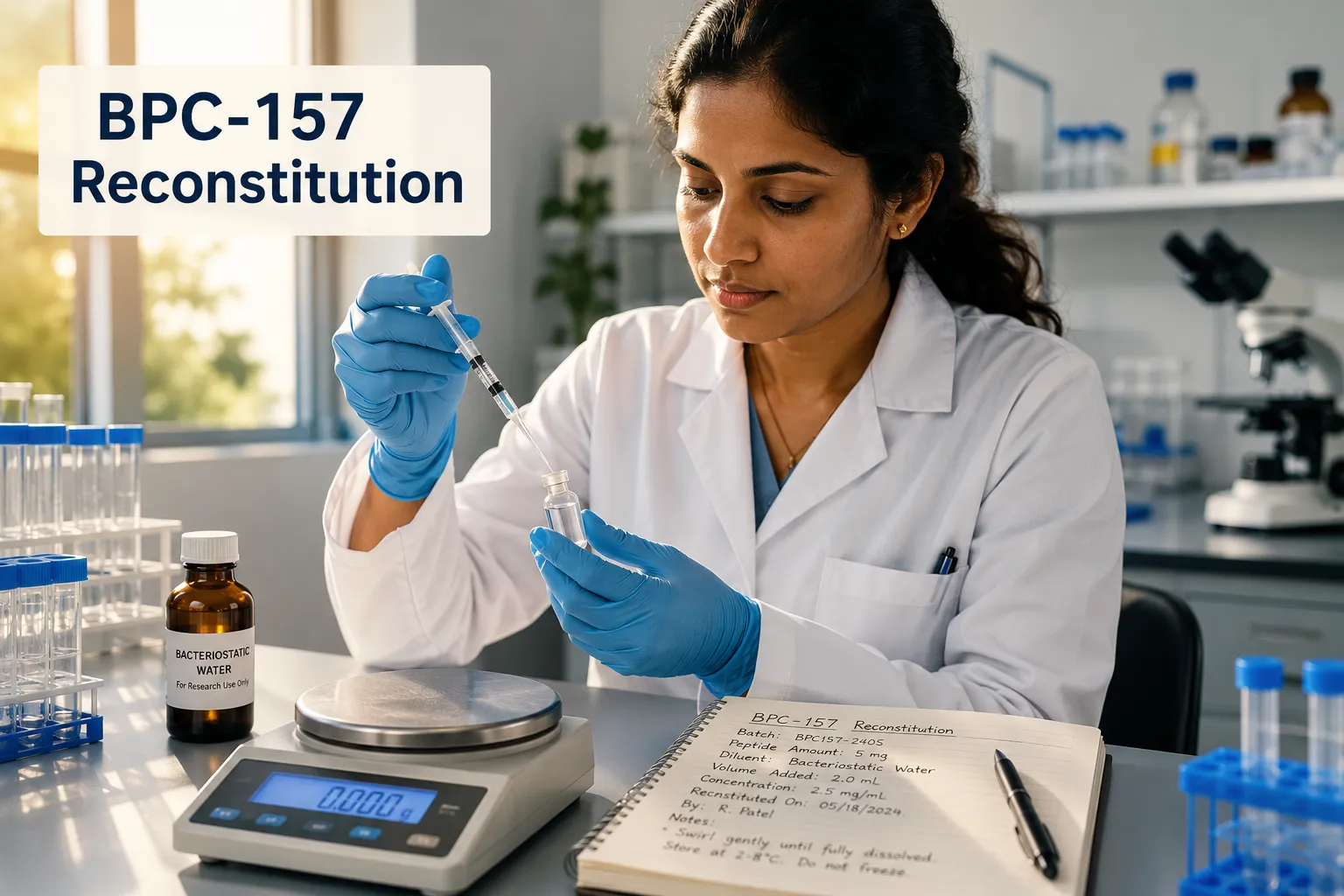
This practice is particularly relevant for longer research cycles involving compounds studied through resources like the longevity peptide research overview or MOTS-C metabolic flexibility research, where consistency across multiple sessions is essential.


Aliquoting to Prevent Freeze-Thaw Damage

Stability Monitoring and Quality Assurance in Peptide Research

This section of the Peptide Reconstitution, Storage, and Stability: A Complete Research Protocol Guide addresses a widely misunderstood risk: assuming a peptide is still viable based on appearance alone.

Degraded peptides often look identical to intact ones. Clarity, color, and consistency do not confirm biological activity. Researchers must rely on documented storage timelines, proper labeling, and sourcing from suppliers with verified quality testing protocols.

Practical stability checklist:

  • Label every vial with reconstitution date and diluent used.
  • Track cumulative freeze-thaw events per aliquot.
  • Discard any solution stored beyond its recommended window, regardless of appearance.
  • Source peptides from suppliers who provide third-party purity verification.

For researchers sourcing compounds such as AOD-9604 for metabolic research or GLP-1 peptides, purity documentation at the point of purchase directly affects downstream stability outcomes.


Conclusion

Applying the principles outlined in this Peptide Reconstitution, Storage, and Stability: A Complete Research Protocol Guide protects both the integrity of the research and the investment in high-quality compounds. The actionable next steps are clear: use bacteriostatic water for reconstitution, store reconstituted solutions at 2-8 degrees Celsius in the main refrigerator body, aliquot immediately to avoid freeze-thaw damage, and never rely on visual inspection as a stability indicator. Source peptides from suppliers who provide transparent purity testing, label every vial with date and diluent, and adhere strictly to the 28-day reconstituted use window. Rigorous handling at every stage is what separates reproducible research from wasted resources.

https://www.puretestedpeptides.com/wp-content/uploads/2026/06/Peptide-Reconstitution-Storage-and-Stability-A-Complete-Research-Protocol-Guide.png 1024 1536 Pure Tested https://www.puretestedpeptides.com/wp-content/uploads/2026/01/buy-peptides-online.jpg Pure Tested2026-06-22 13:03:092026-07-20 15:02:36Peptide Reconstitution, Storage, and Stability: A Complete Research Protocol Guide
×

Helpful Links

  • My account
  • Cart
  • Checkout
  • Refund and Returns Policy
  • Privacy Policy
  • SMS Privacy Policy
  • Login
  • My Account
  • Logout

USA Made Lab Tested Peptides

All products are sold for research, laboratory, or analytical purposes only, and are not for human consumption


Pure Tested Peptides is a chemical supplier. Pure Tested Peptides is not a compounding / chemical compounding facility as defined under 503A of the Federal Food, Drug, and Cosmetic act. Pure Tested Peptides is not an outsourcing facility as defined under 503B of the Federal Food, Drug, and Cosmetic act.

The statements made within this website have not been evaluated by the US Food and Drug Administration. The products we offer are not intended to diagnose, treat, cure or prevent any disease.

Human/Animal Consumption Prohibited. Laboratory/In-Vitro Experimental Use Only

Scroll to top Scroll to top Scroll to top