Epithalon Peptide Research: Telomere-Length Measurement Pitfalls and Better Senescence Controls
Fewer than a dozen completed human trials have ever prospectively measured telomere length before and after any peptide intervention, and epitalon is not yet among them. That gap between compelling cell-culture data and verified human outcomes sits at the heart of the current debate in Epithalon Peptide Research: Telomere-Length Measurement Pitfalls and Better Senescence Controls, where the quality of the assay used to measure telomeres can determine whether a study's conclusion holds up or quietly collapses under replication pressure.
Key Takeaways
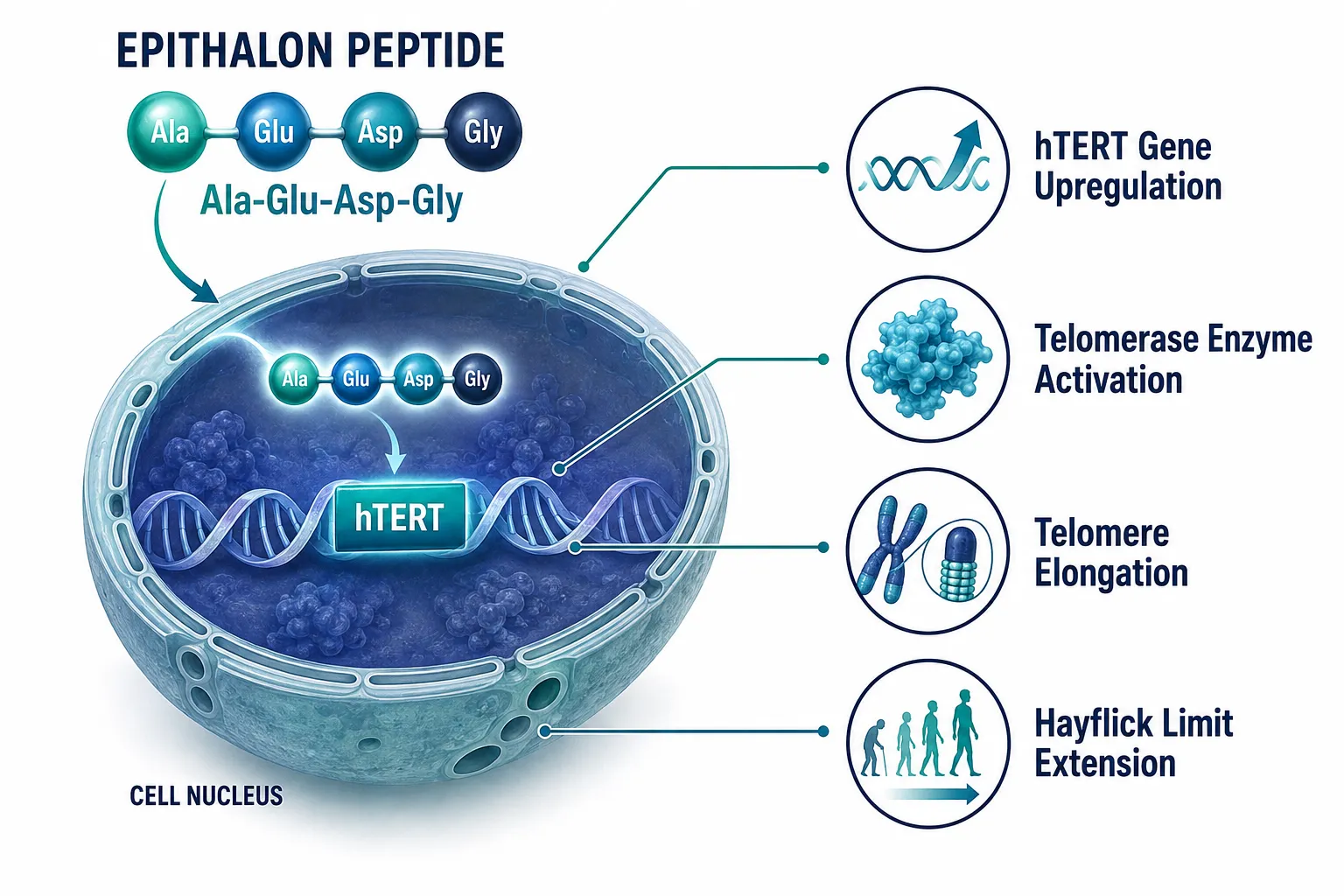
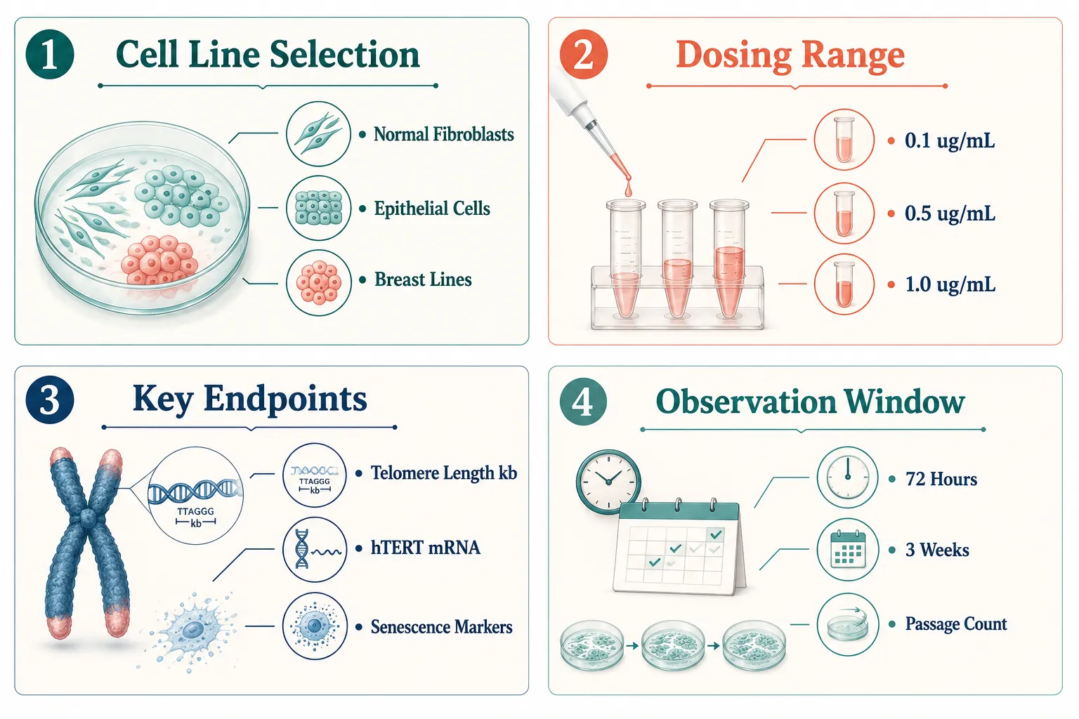
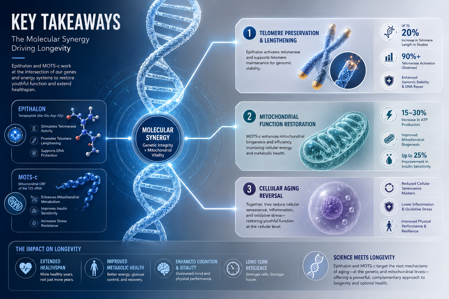
- Epitalon has demonstrated dose-dependent telomere elongation and hTERT upregulation in normal human fibroblasts and mammary epithelial cells in controlled culture studies, with some lines showing roughly 12-fold increases in hTERT expression at 1 ug/ml.
- The same peptide elongated telomeres in breast cancer cell lines through an Alternative Lengthening of Telomeres (ALT) mechanism, raising important questions about oncogenic risk in telomerase-targeting research.
- qPCR, the most common telomere assay in epitalon studies, reports only average telomere length and suffers from significant inter-laboratory variability, making it insufficient as a standalone measure.
- No completed, well-controlled human clinical trial has yet linked epitalon administration to measurable telomere extension in living people.
- Robust epitalon research requires multi-method telomere validation paired with functional senescence markers, not telomere length data alone.
What the Cell-Culture Evidence Actually Shows
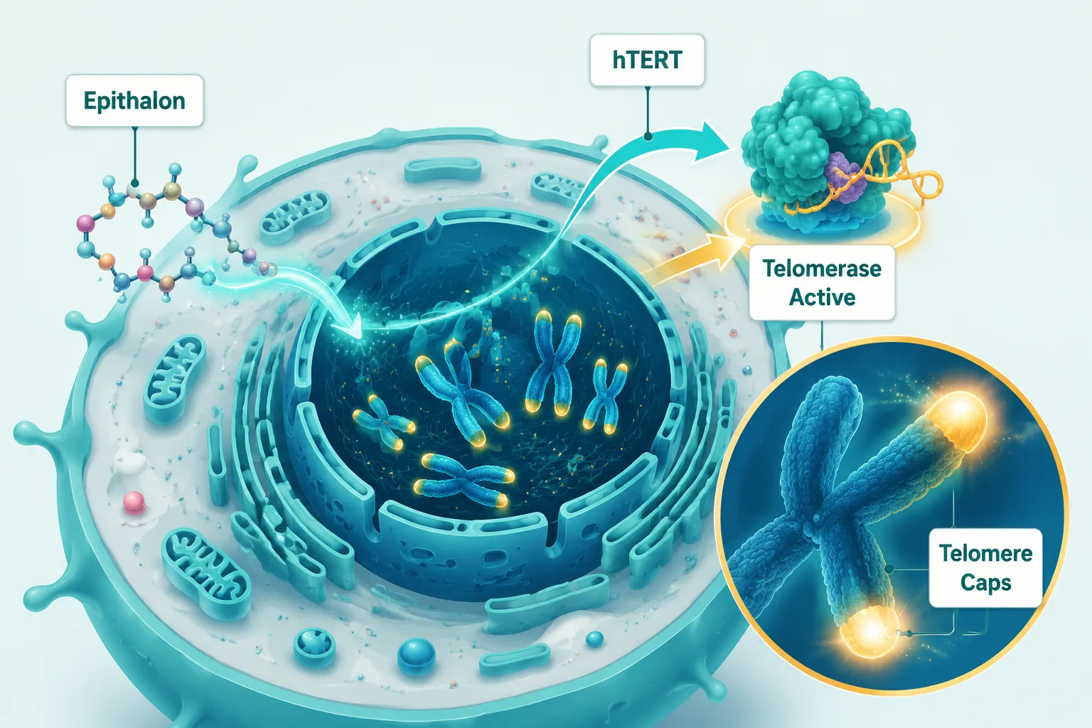
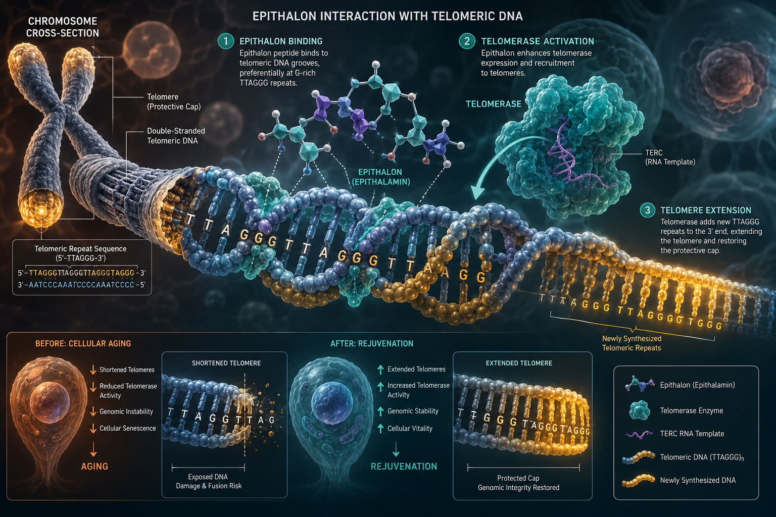
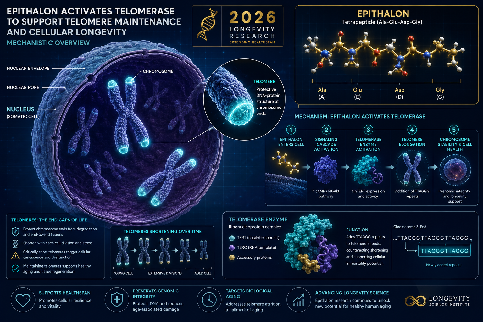
A 2025 study from Brunel University London published in Biogerontology produced some of the most detailed quantitative data on epitalon's telomere effects to date. Using both qPCR and telomere immunofluorescence, researchers documented telomere elongation in normal human fibroblasts and mammary epithelial cells that was both dose-dependent and time-dependent. In certain normal cell lines, 1 ug/ml epitalon drove approximately 12-fold upregulation of hTERT, the catalytic subunit of telomerase, while 0.5 ug/ml produced roughly 5-fold upregulation. Overall telomere length in some cultures reached about 2.4 times the baseline measurement.
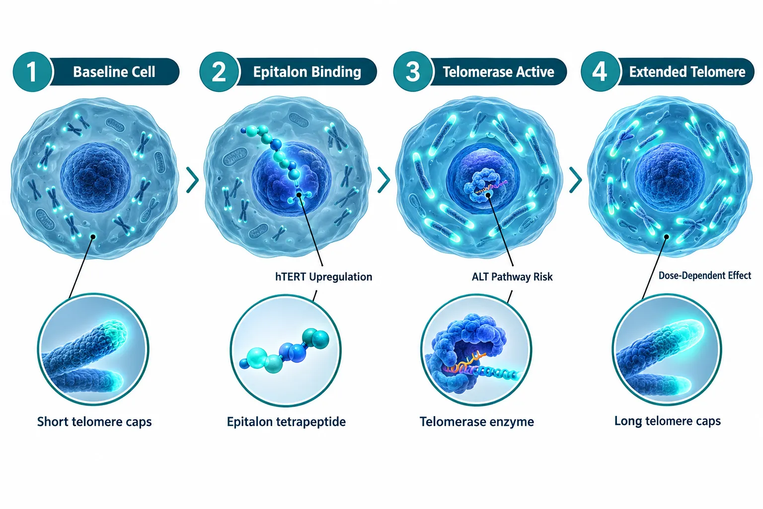
A correction published later in 2025 refined the mechanistic interpretation of that study without overturning the core quantitative findings. The key clarification involved distinguishing between telomerase-driven elongation in normal cells and ALT-mediated elongation in cancer cell lines. In breast cancer lines (21NT and BT474), epitalon did extend telomeres, but via ALT rather than canonical telomerase activation. That distinction matters enormously for safety evaluation.
The takeaway is not that epitalon fails in cancer cells, it is that the mechanism differs, and the implications for oncogenic risk require separate, careful assessment.
A 2026 review on therapeutic peptides in gerontology confirmed that epitalon's telomerase effects in normal somatic cells have been replicated across independent laboratories, strengthening the mechanistic case. Additional 2025 experimental work extended these findings to bovine cumulus cells and cumulus-oocyte complexes, suggesting the peptide's influence on telomerase is not limited to a single cell type.
For researchers sourcing compounds for controlled studies, understanding purity and batch consistency is critical. Resources on lab tested peptides and guidance on choosing a best peptide manufacturer are relevant starting points for experimental design.
Telomere-Length Measurement Pitfalls in Epithalon Peptide Research
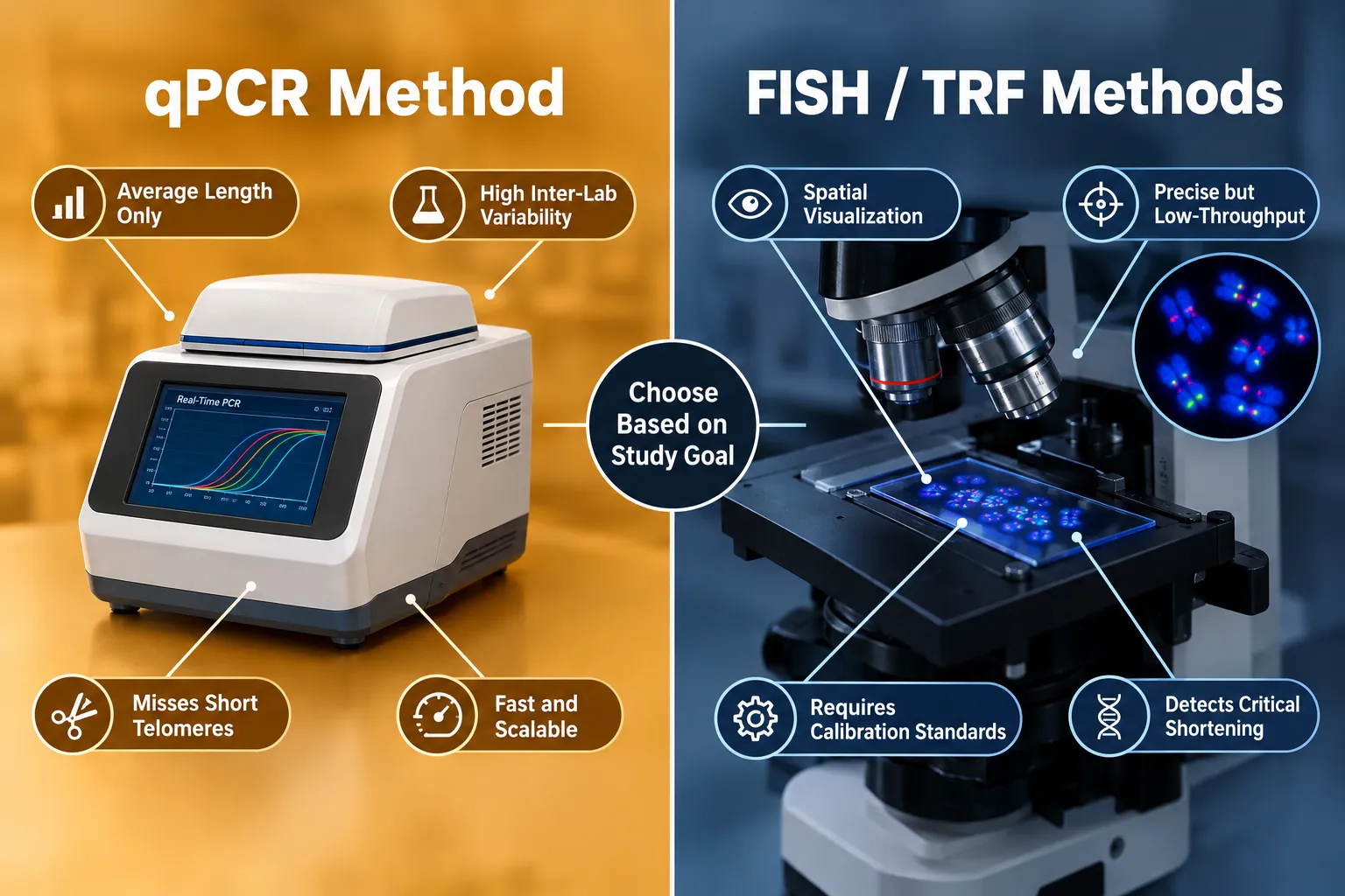
The most significant methodological vulnerability in current Epithalon Peptide Research: Telomere-Length Measurement Pitfalls and Better Senescence Controls literature is over-reliance on a single assay type, particularly qPCR.
Why qPCR Alone Is Not Enough
qPCR-based telomere measurement is fast and scalable, which explains its dominance. However, it carries serious limitations:
| Limitation | Why It Matters for Epitalon Studies |
|---|---|
| Reports average telomere length only | Misses critically short telomeres that drive senescence |
| High inter-laboratory variability | Results may not replicate across research groups |
| Sensitive to DNA quality and normalization | Small technical errors produce large apparent differences |
| Cannot resolve telomere length distributions | A few very long telomeres can mask many short ones |
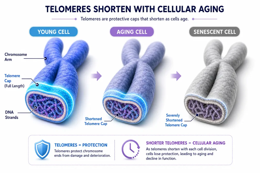
Critically short telomeres, not average telomere length, are the primary drivers of genomic instability and replicative senescence. A treatment that lengthens average telomeres while leaving critically short ones intact may show impressive qPCR numbers without meaningfully reducing senescence burden.
Alternative Methods and Their Trade-Offs
Fluorescence in situ hybridization (FISH) and quantitative FISH (Q-FISH) allow spatial visualization of individual telomeres and can detect length distributions rather than averages. However, they require meticulous calibration with standards of known telomeric repeat content and are low throughput. Terminal Restriction Fragment (TRF) analysis offers greater precision but is labor-intensive and difficult to scale.
The expert consensus from 2025-2026 methodology reviews is clear: multi-method validation combining qPCR with FISH/Q-FISH or TRF is the minimum standard for interventional telomere research. Cross-laboratory comparisons without harmonized protocols should be avoided.
Research teams exploring synergistic peptide combinations, such as those examining Epithalon and MOTS-c synergy, face compounded assay complexity, since each compound may affect telomere biology through distinct pathways that interact unpredictably in mixed-assay conditions.
Better Senescence Controls: Moving Beyond Telomere Length
The most important shift recommended by 2026 gerontology and methodology reviews is reframing the research question. Telomere elongation is a molecular event. Cellular senescence is a functional state. The two are related but not identical, and Epithalon Peptide Research: Telomere-Length Measurement Pitfalls and Better Senescence Controls must address both to produce actionable conclusions.
Recommended Senescence Control Strategies
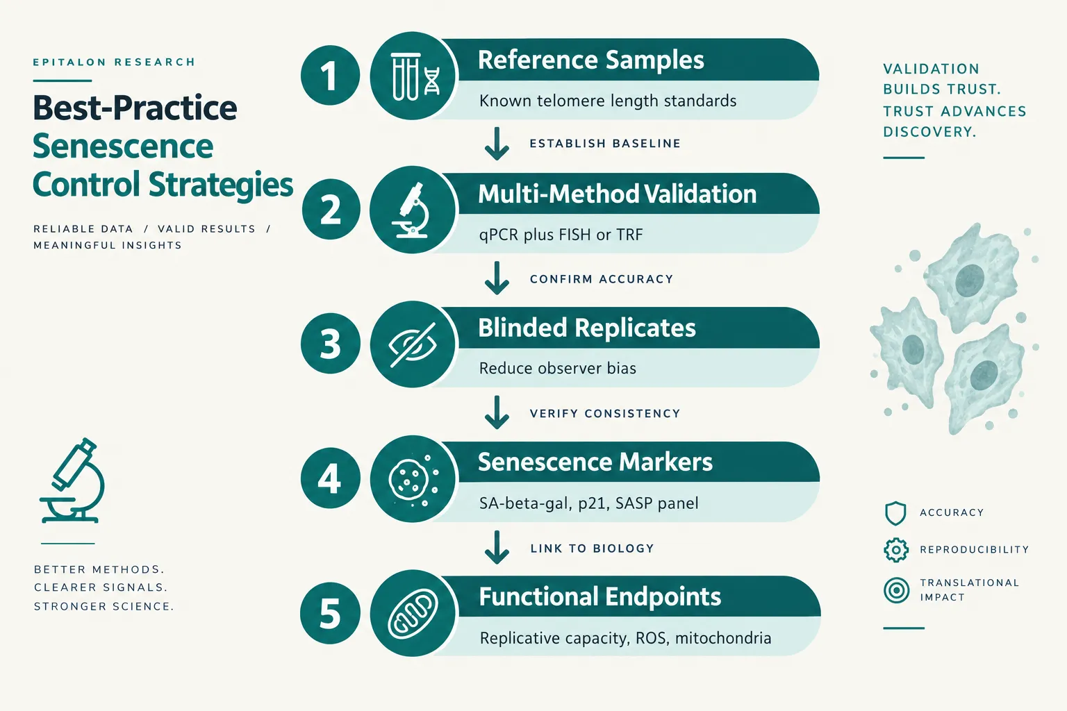
Leading experts propose the following framework for future epitalon studies:
- Include reference samples with independently verified telomere lengths in every assay run
- Report assay variability explicitly, including coefficient of variation across replicates
- Use blinded replicates to reduce observer bias in both measurement and interpretation
- Characterize telomere length distributions, not just means, to identify critically short telomere populations
- Add functional senescence markers: senescence-associated beta-galactosidase (SA-beta-gal), p21 expression, DNA damage foci (gamma-H2AX), and senescence-associated secretory phenotype (SASP) components
- Measure replicative capacity: does epitalon-treated cell culture actually divide more times before reaching the Hayflick limit?
- Assess mitochondrial function and reactive oxygen species as independent indicators of cellular health
This multi-endpoint approach transforms a telomere chemistry study into a genuine senescence biology study. Researchers interested in comparing mitochondrial-targeting peptides alongside epitalon may find the MOTS-c peptide dosage calculator and information on buying MOTS-c peptide useful for designing comparative experimental arms.
The Human Translation Gap
Despite strong in vitro data, no completed, well-controlled human clinical trial has prospectively linked epitalon administration to telomere length changes in blood or tissue samples. Existing human data, primarily from older Khavinson-era studies, are limited in sample size and methodologically heterogeneous. This gap does not invalidate the cell-culture findings, but it does mean that claims about epitalon extending human lifespan or reversing human aging remain scientifically unsupported as of 2026.
Telomerase activation also carries a theoretical oncogenic risk. The ALT-pathway findings in cancer cell lines are not evidence that epitalon causes cancer, but they are a clear signal that telomerase-modulating interventions require rigorous long-term safety monitoring in any future human studies.
For researchers building out broader peptide research programs, resources on wholesale peptides and buying bulk peptide can support the scale needed for properly powered, multi-endpoint studies.
Conclusion
The cell-culture evidence for epitalon's telomere-extending and telomerase-activating effects is reproducible and quantitatively significant. However, the field's current reliance on average telomere length measured by qPCR alone is a structural weakness that undermines the translational value of many published findings.
Actionable next steps for researchers:
- Adopt multi-method telomere measurement protocols (qPCR plus Q-FISH or TRF) as a minimum standard for any new epitalon study.
- Add functional senescence endpoints, SA-beta-gal, p21, SASP markers, and replicative capacity, to every telomere-focused experimental design.
- Distinguish clearly between telomerase-driven and ALT-mediated elongation, especially when working with any cancer-derived cell lines.
- Prioritize harmonized protocols before conducting cross-laboratory comparisons.
- Treat the human translation gap as an open research question requiring prospective, controlled clinical study design, not an assumption that can be bridged by extrapolating cell-culture data.
The science of epitalon is genuinely promising. Making that promise actionable requires the field to hold itself to the same measurement standards it would demand of any other longevity intervention.