Call or Text 727-513-9780
  • Shopping Cart Shopping Cart
    0Shopping Cart
Pure Tested Peptides | America's most trusted Peptides for sale online
  • Peptides for sale
    • Oral Peptides for sale
      • Peptide Capsules for sale
      • BPC 157 Capsules 1000mcg
      • SLU-PP-332 Capsules | 1000 mcg
      • 5-Amino-1MQ 50mg Capsules
      • Tesofensine 500mcg
    • All Peptides for sale
    • Peptide Sprays
      • BPC 157 Nasal Spray Kit
      • BPC-157 TB500 Nasal Spray Kit
      • Semax Nasal Spray 10mg
      • Selank – Nasal Spray Kit – 10mg
      • Epithalon 50MG Nasal Spray Kit
      • Ipamorelin 10mg Nasal Spray
      • Klow Nasal Spray (BPC-157 + TB-500 + GHK-Cu + KPV) | 80mg
      • Hulk Nasal Spray Tesa / Ipa Blend 6/3 MG
      • Klow Nasal Spray
      • NAD + 500 mg Nasal Spray
      • PT-141 Nasal Spray Kit
    • GHRH Peptides
      • Ipa Peptides
      • CJC-1295 Peptides
        • CJC-1295 with DAC 5 mg
        • CJC-1295 without DAC 5 mg
        • CJC-1295 Ipa 10mg
      • Tesa Peptides
        • Tesa Peptide
        • Tesa 20 mg
    • GHK-Cu Peptides
      • All GHK-Cu Peptides
      • GHK-Cu 100mg
      • KLOW Peptide Blend – Buy KLOW blend online
    • BPC Peptides
      • All BPC Peptides
      • BPC-157
      • BPC-157 TB-500
      • BPC 157 capsules 1000mcg
    • SLU-PP-332 Peptides
      • All SLU-PP-332 Peptides
      • SLU-PP-332 5mg
    • GLP3 Peptides
    • PT-141 Peptides
      • PT-141 Peptides for sale
      • PT-141 10mg
      • PT-141 Nasal Spray
    • CAG Peptides
      • Lipo-C Peptide Blend
      • CAG 5mg
      • CAG 10mg
    • MOTS-C Peptides
      • MOTS-C Peptides for sale
      • MOTS-c peptide
      • MOTS-c 10mg *6 pack*
    • 5 Amino 1MQ Peptides
      • 5 Amino 1MQ Peptides for sale
      • 5-Amino-1MQ 50mg Capsules
      • 5-Amino-1MQ 5mg
    • Epithalon Peptides
      • Epithalon Peptides for sale
      • Epithalon 10mg
      • Epithalon 50mg
  • Shop
    • GLPs
      • 5-Amino-1MQ 50mg Capsules
      • 5-Amino-1MQ 5mg
      • L-Carnitine 500mg/ml
      • Tesofensine 500mcg
      • SLU-PP-332 5mg
      • MOTS-c 10mg *6 pack*
    • Epithalon & BPC Peptides
      • Epithalon 10mg
      • Epithalon 50mg
      • BPC-157
      • BPC 157 capsules 1000mcg
      • BPC-157 TB-500
      • BPC-157 TB500 Nasal Spray Kit
      • BPC 157 Nasal Spray Kit
    • BPC TB-500 & NAD+ Peptides
      • NAD+ 500 mg
      • KLOW Peptide Blend – Buy KLOW blend online
      • GLOW Peptide Blend
      • TB 500 5mg
      • BPC 157 capsules 1000mcg – Supplement
      • BPC 157 Nasal Spray Kit
      • BPC-157
      • BPC-157 TB500 Nasal Spray Kit
      • BPC-157 TB-500
      • BPC 157 capsules 1000mcg
    • LL-37 Peptide
      • LL-37 10 mg
    • MOTS-C & Selank
      • MOTS-c peptide
      • Selank 10mg
    • GHK Peptides
      • GHK-Cu 100mg
      • GLOW Peptide Blend
      • KLOW Peptide Blend – Buy KLOW blend online
  • COAs
  • Wholesale
    • Wholesale Peptides for sale
  • PTP FAQ
  • Affiliates
    • Selank Peptide: Advanced Pharmacological Mechanisms Underlying Its Anxiolytic and Nootropic Effects in Research
      • 5-Amino-1MQ Peptide: Detailed Mechanisms of NNMT Inhibition and Its Impact on Cellular Metabolism Research
        • MOTS-C Peptide: Unraveling Its Role in Mitochondrial Dynamics and Energy Metabolism Research
          • Epithalon Peptide and Telomerase Regulation: Investigating Its Impact on Cellular Senescence and Lifespan Research Models
            • GLP2-T vs GLP2 Tirz Peptide: Understanding the Naming, Mechanistic Nuances, and Research Implications for Gut Health
      • Peptides and Polypeptides in Modern Pharmacology: What Research on Metoprolol, Prednisone, and Amlodipine Reveals
        • Polypeptide Peptides vs NSAIDs: What Naproxen and Diclofenac Teach Tissue-Repair Researchers About BPC‑157 and TB‑500
          • Peptides and Polypeptides in Endocrine Pharmacology: How GLP-1, GLP-2, and GLP-3 Retatrutide Differ From Classic Drugs Like Prednisone and Amlodipine
          • 5‑Amino‑1MQ Peptide and NNMT Inhibition: How It Compares With Statins Like Atorvastatin in Adipose and Lipid Metabolism Research
      • Enclomiphene, Estrogen Receptor Signaling, and Luteinizing Phase Biology: What Hormone Researchers Should Measure
        • Mesenchymal Stem Cells, BPC‑157, and GHK‑Cu: How Tissue Repair Peptides Compare With Classic NSAIDs Like Naproxen in Injury Models
          • Peptide Calculator Use Cases Beyond Growth Hormone: Working Through GLP‑3 Retatrutide, MOTS‑c, and BPC‑157 Research Dosing
        • Polypeptide Peptides in Endocrine and Metabolic Pharmacology: Lessons From Amlodipine, Prednisone, and Metoprolol
        • Peptides Calculator for Advanced Blends: Worked Examples for Tesamorelin, CJC‑1295, and Ipamorelin Stacks
          • Peptides and Polypeptides in Human Physiology: How Molecular Size Shapes Research Applications
            • Selank Peptide: Advanced Pharmacological Mechanisms Underlying Its Anxiolytic and Nootropic Effects in Research
          • 5-Amino-1MQ Peptide: Detailed Mechanisms of NNMT Inhibition and Its Impact on Cellular Metabolism Research
            • MOTS-C Peptide: Unraveling Its Role in Mitochondrial Dynamics and Energy Metabolism Research
              • Epithalon Peptide and Telomerase Regulation: Investigating Its Impact on Cellular Senescence and Lifespan Research Models
                • GLP2-T vs GLP2 Tirz Peptide: Understanding the Naming, Mechanistic Nuances, and Research Implications for Gut Health
                  • Peptides and Polypeptides in Endocrine Pharmacology: How GLP-1, GLP-2, and GLP-3 Retatrutide Differ From Classic Drugs Like Prednisone and Amlodipine
                    • Polypeptide Peptides vs NSAIDs: What Naproxen and Diclofenac Teach Tissue-Repair Researchers About BPC‑157 and TB‑500
                      • Peptides and Polypeptides in Modern Pharmacology: What Research on Metoprolol, Prednisone, and Amlodipine Reveals
                        • 5‑Amino‑1MQ Peptide and NNMT Inhibition: How It Compares With Statins Like Atorvastatin in Adipose and Lipid Metabolism Research
                        • Enclomiphene, Estrogen Receptor Signaling, and Luteinizing Phase Biology: What Hormone Researchers Should Measure
                        • Mesenchymal Stem Cells, BPC‑157, and GHK‑Cu: How Tissue Repair Peptides Compare With Classic NSAIDs Like Naproxen in Injury Models
                        • Peptide Calculator Use Cases Beyond Growth Hormone: Working Through GLP‑3 Retatrutide, MOTS‑c, and BPC‑157 Research Dosing
                        • Polypeptide Peptides in Endocrine and Metabolic Pharmacology: Lessons From Amlodipine, Prednisone, and Metoprolol
                        • Peptides Calculator for Advanced Blends: Worked Examples for Tesamorelin, CJC‑1295, and Ipamorelin Stacks
                        • Peptides and Polypeptides in Human Physiology: How Molecular Size Shapes Research Applications
                        • Selank Peptide: Advanced Pharmacological Mechanisms Underlying Its Anxiolytic and Nootropic Effects in Research
                        • MOTS-C Peptide: Unraveling Its Role in Mitochondrial Dynamics and Energy Metabolism Research
                        • Peptides and Polypeptides in Modern Pharmacology: What Research on Metoprolol, Prednisone, and Amlodipine Reveals
                        • Peptides and Polypeptides in Endocrine Pharmacology: How GLP-1, GLP-2, and GLP-3 Retatrutide Differ From Classic Drugs Like Prednisone and Amlodipine
                        • 5-Amino-1MQ Peptide: Detailed Mechanisms of NNMT Inhibition and Its Impact on Cellular Metabolism Research
                        • Mesenchymal Stem Cells, BPC‑157, and GHK‑Cu: How Tissue Repair Peptides Compare With Classic NSAIDs Like Naproxen in Injury Models
                        • Peptide Calculator Use Cases Beyond Growth Hormone: Working Through GLP‑3 Retatrutide, MOTS‑c, and BPC‑157 Research Dosing
                        • GLP2-T vs GLP2 Tirz Peptide: Understanding the Naming, Mechanistic Nuances, and Research Implications for Gut Health
                        • Polypeptide Peptides in Endocrine and Metabolic Pharmacology: Lessons From Amlodipine, Prednisone, and Metoprolol
                        • Polypeptide Peptides vs NSAIDs: What Naproxen and Diclofenac Teach Tissue-Repair Researchers About BPC‑157 and TB‑500
                        • Peptides and Polypeptides in Human Physiology: How Molecular Size Shapes Research Applications
                        • Peptides Calculator for Advanced Blends: Worked Examples for Tesamorelin, CJC‑1295, and Ipamorelin Stacks
                        • 5‑Amino‑1MQ Peptide and NNMT Inhibition: How It Compares With Statins Like Atorvastatin in Adipose and Lipid Metabolism Research
                        • Enclomiphene, Estrogen Receptor Signaling, and Luteinizing Phase Biology: What Hormone Researchers Should Measure
                        • Epithalon Peptide and Telomerase Regulation: Investigating Its Impact on Cellular Senescence and Lifespan Research Models
                        • Best research protocol Klow blend
                        • best time to take BPC-157
                        • best time to take DSIP (Delta Sleep Inducing Peptide)
                        • best time to take CJC-1295
                        • best time to take AOD-9604
                        • best time to take Follistatin 344
                        • best time to take Ipamorelin
                        • best time to take MK-677 (Ibutamoren)
                        • best time to take Ligandrol (LGD-4033) — research compound
                        • best time to take Ostarine (MK-2866) — research compound
                        • best time to take GHK-CU
                        • best time to take TB-500
                        • best time to take MOTS-c
                        • best time to take Semax
                        • best time to take RAD-140 (Testolone) — research compound
                        • best time to take Thymosin Alpha-1
                        • best time to take PEG-MGF
                        • Biolife Plasma, Octapharma Plasma, and Research Peptides: How Plasma Donation Labs Differ From Peptide Suppliers
                        • best time to take YK-11 — research compound
                        • best time to take PT-141 (Bremelanotide)
                        • Best research protocol Klow blend
                        • 5-Amino-1MQ and MOTS-C Synergy: Metabolic Signaling, Mitochondria, and Research Design
                        • BPC-157 and TB-500: Investigating Their Combined Effects on Angiogenesis and Cellular Migration in Tissue Repair Models
                        • BPC-157 Peptide: Gut Barrier Function, Inflammation, and Tissue-Recovery Research
                        • 5‑Amino‑1MQ and MOTS‑c Synergy in Metabolic Research: Designing NNMT and Mitochondrial Biogenesis Stacks
                        • CJC-1295 with DAC vs. Without DAC: Half-Life, Release Kinetics, and Research Implications
                        • CJC‑1295 with DAC vs. Without DAC: Expanding on Half‑Life Differences Using Tesamorelin and Ipamorelin Blend Case Studies
                        • Collagen Biology and Copper‑Binding Peptides: How GHK‑Cu, Glow Blend, and Klow Blend Interact with Skin and Connective Tissue
                        • Collagen Biology and Regenerative Peptides: How GHK‑Cu, Glow Blend, and Klow Blend Affect Extracellular Matrix Research
                    • DNA, Telomeres, and Longevity Peptides: Positioning Epithalon and MOTS‑c in Genetic Aging Research
                      • Enclomiphene Citrate: serm Mechanism, Testosterone Research, and Stack Compatibility
                        • Enclomiphene vs Enclomiphene Citrate: Formulation, Bioavailability, and Research Distinctions
                        • Epithalon Peptide Research: Telomerase Activation, Aging, and Pineal Gland Function
                        • Estrogen Receptor Signaling and Enclomiphene: How Selective Modulators Compare with Classic Polypeptide Hormones
                        • GHK-Cu Peptide: Advanced Mechanisms in Extracellular Matrix Remodeling and Wound Healing Research
                        • GHK-Cu Peptide: Collagen Synthesis, Wound Repair, and Skin-Barrier Research Models
                        • GLP-1 vs GLP-3 vs GLP-2: Peptide Classification and Research Applications
                        • GLP-2 Peptide Research Guide: Gut Barrier Function, Nutrient Absorption, and Intestinal Recovery Models
                        • GLP-3 Retatrutide vs. GLP-1 Drugs: What Triple-Agonist Biology Changes in Research Models
                        • Ipamorelin and Tesamorelin Combination: Synergistic GH Secretagogue Research and Dosing Protocols
                        • GLP2 Tirz Peptide: What It Is, Why the Name Exists, and How Researchers Should Interpret It
                        • Klow Blend Peptide Nasal Spray: What the Formulation Is Trying to Do in Cognitive Research
                        • Mitochondria, NNMT Inhibition, and Peptide Modulators: Where MOTS‑c and 5‑Amino‑1MQ Fit in Cellular Energy Research
                        • MOTS-c Peptide: Mitochondrial Function, Energy Metabolism, and What Researchers Measure
                        • MOTS-c vs. 5-Amino-1MQ: Which Metabolic Research Questions Each Compound Actually Answers
                        • Nasal Spray Peptides: Bioavailability, Administration, and Semax/Selank Research Applications
                        • PT-141 Peptide Research: Mechanism of Action and Melanocortin Receptor Signaling
                        • Retatrutide for Research: Mechanism, Structure, and GLP-1/GLP-3 Dual Action
                        • Retatrutide for Obesity and Type 2 Diabetes: What the Latest Trial Data Suggest
                        • Tesofensine Peptide Research: Mechanism, Appetite Suppression, and Neuropeptide Y Pathways
  • Contact
    • Contact Customer Service
    • Text Customer Support
  • About US
  • Shop all peptides
  • Affiliate Program
    • Affiliate Signup
  • Login / Register Login / Register Page Link Login / Register Page Link
  • Click to open the search input field Click to open the search input field Search
  • Menu Menu

Tag Archive for: glp-1 peptide research

Prednisone and Metabolic Peptides: A Glucocorticoid-Control Framework for Glucose, Appetite, and Inflammation Experiments

Prednisone and Metabolic Peptides: A Glucocorticoid-Control Framework for Glucose, Appetite, and Inflammation Experiments

October 13, 2026/0 Comments/in Uncategorized/by

}

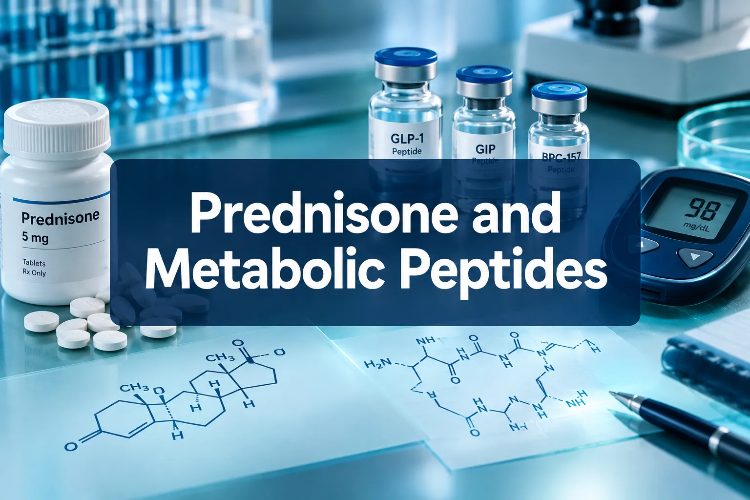
Professional () hero image with 'Prednisone and Metabolic Peptides' in crisp white on a deep navy semi-transparent panel,

Glucocorticoids cause measurable hyperglycemia in roughly 50 percent of hospitalized patients who receive them, yet most experimental designs still treat prednisone as a simple background variable rather than an active metabolic driver. That gap matters enormously when researchers are trying to isolate peptide activity from steroid noise. Building a rigorous Prednisone and Metabolic Peptides: A Glucocorticoid-Control Framework for Glucose, Appetite, and Inflammation Experiments requires separating three overlapping effects, glucose dysregulation, appetite amplification, and inflammatory signaling, and assigning each a mechanistic control arm that can be tested independently.

Key Takeaways

  • Prednisone activates glucocorticoid receptors in multiple organs simultaneously, producing coordinated changes in glucose, appetite peptides, and inflammatory markers.
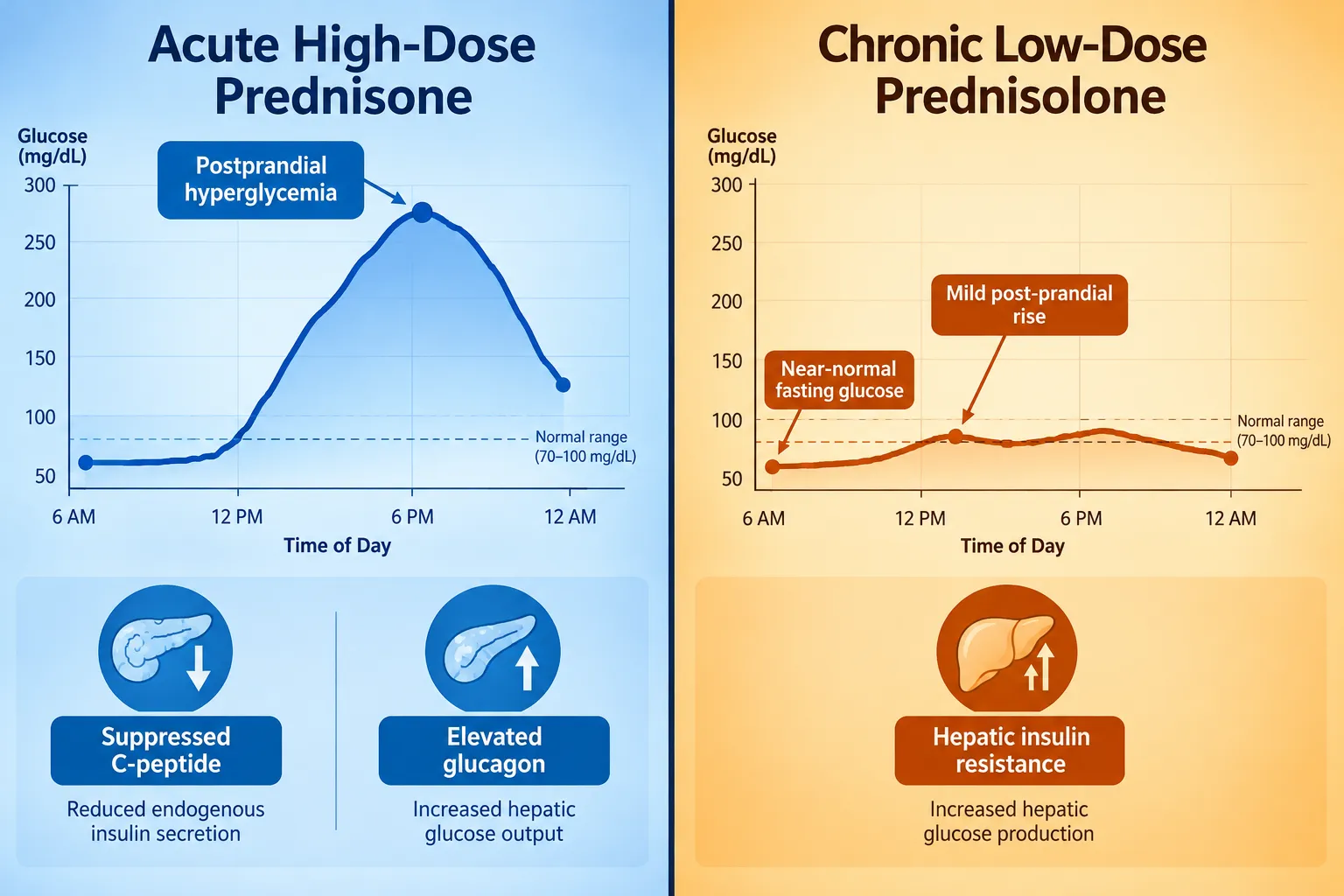
  • Acute medium-dose exposure creates a predictable midday-to-midnight postprandial glucose and peptide profile that can anchor time-structured experimental sampling.
  • Chronic low-dose exposure produces a distinct "mild steroid diabetes" phenotype dominated by hepatic insulin resistance rather than sustained fasting hyperglycemia.
  • Adipokines such as leptin and resistin serve as measurable intermediaries linking glucocorticoid exposure to both appetite and inflammation endpoints.
  • GLP-1 receptor agonists are the leading peptide class for countering glucocorticoid metabolic toxicity and represent a strong experimental comparator arm.

How Prednisone Disrupts the Glucose-Peptide Axis
How Prednisone Disrupts the Glucose-Peptide Axis

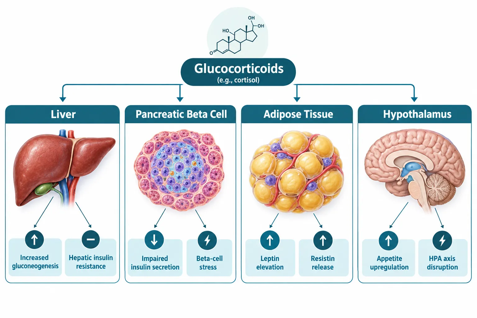
Prednisone is converted to prednisolone in the liver, where it binds glucocorticoid receptors and triggers a cascade that spans at least five organ systems. Understanding each node is essential before selecting experimental endpoints.

The four primary disruption sites are:

Organ Glucocorticoid Effect Measurable Peptide Change
Liver Increased gluconeogenesis Elevated fasting glucose, altered C-peptide
Pancreatic beta cells Impaired insulin secretion Reduced insulin, suppressed C-peptide
Adipose tissue Leptin and resistin release Elevated adipokines, insulin resistance
Hypothalamus Appetite upregulation Altered hunger signaling, increased caloric intake

Acute medium-dose prednisone in controlled studies produces marked postprandial hyperglycemia from midday through midnight. In individuals with type 2 diabetes, insulin and C-peptide secretion are suppressed during this window, while at-risk subjects show altered C-peptide patterns even without overt diabetes. This predictable 24-hour glucose and peptide profile is highly useful as a temporal anchor for sampling schedules.

Short-term moderate doses in healthy volunteers significantly increase circulating glucose, insulin, C-peptide, glucagon, and IGF-I, while simultaneously lowering IGF-II and specific IGF-binding proteins. Notably, these changes occur without immediate impairment of whole-body muscle protein metabolism, which means acute experiments can focus on the glucose-peptide axis without confounding from skeletal muscle wasting in the early window.

"Prednisone does not act on one pathway, it acts on an inter-organ control system, and experiments that ignore that architecture will produce uninterpretable peptide data."


Designing the Glucocorticoid-Control Framework for Glucose, Appetite, and Inflammation Experiments
Designing the Glucocorticoid-Control Framework for Glucose, Appetite, and Inflammation Experiments

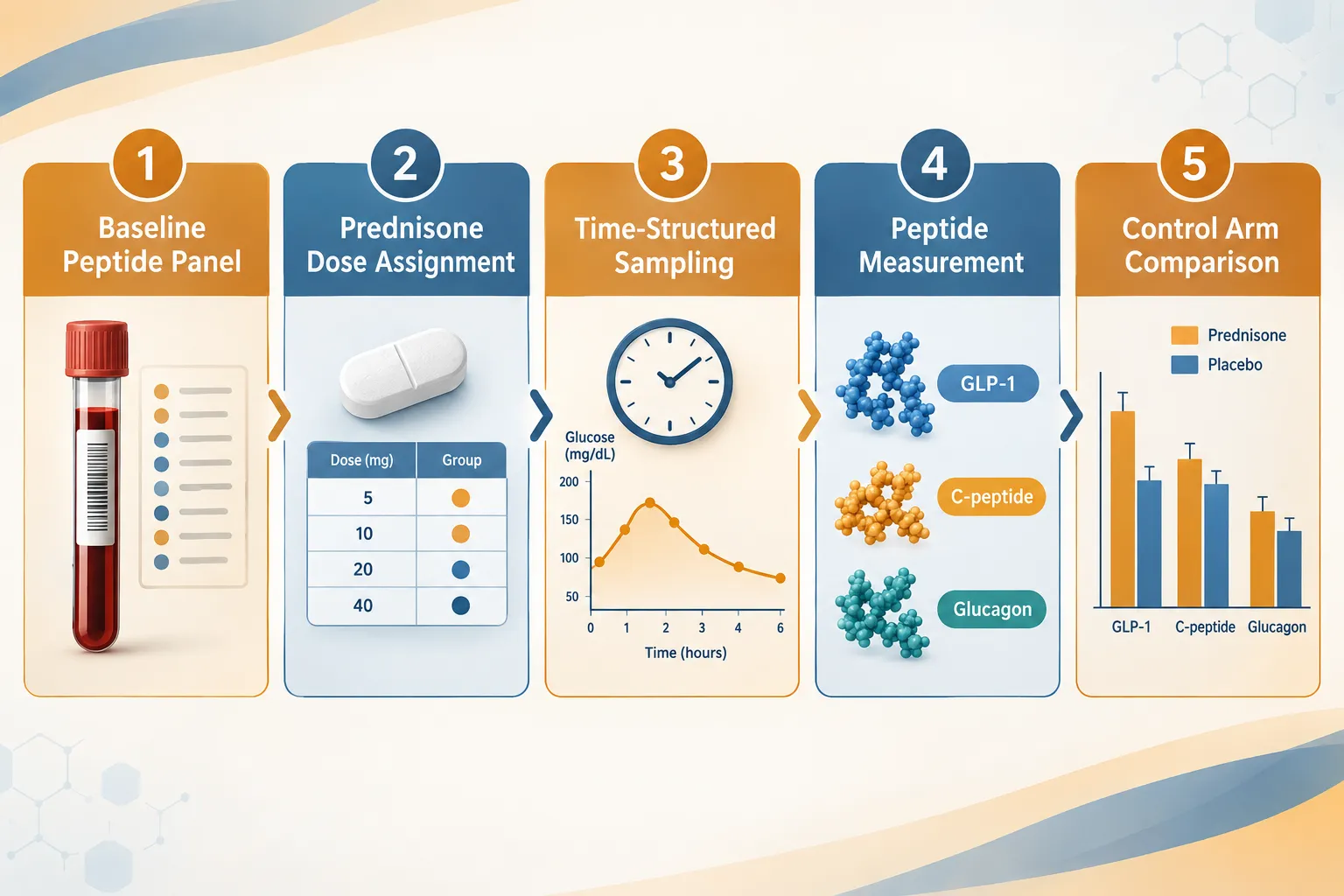
A well-structured Prednisone and Metabolic Peptides: A Glucocorticoid-Control Framework for Glucose, Appetite, and Inflammation Experiments separates dose-dependent effects into three distinct experimental arms.

Arm 1, Acute High-Dose Glucose and Peptide Profiling

Acute high-dose exposure is characterized by rapid fluid retention, appetite stimulation, and a sharp postprandial glucose rise. The recommended peptide panel for this arm includes:

  • Insulin and C-peptide (sampled hourly from 0800 to 2400)
  • Glucagon (to capture counter-regulatory response)
  • IGF-I and IGF-II (to detect early anabolic-catabolic shift)
  • Leptin and resistin (adipokine intermediaries linking appetite and inflammation)

High-dose prednisolone increases both leptin and resistin, making these adipokines reliable biomarkers of glucocorticoid intensity. Incorporating them into the panel connects glucose dysregulation to appetite and inflammatory signaling within a single measurement framework.

For researchers working with lab tested peptides in this context, purity verification is a non-negotiable prerequisite to avoid confounding the peptide signal with degradation products.

Arm 2, Chronic Low-Dose Steroid Diabetes Control

Chronic low-dose prednisolone at 10 mg/day or less produces a distinct phenotype: hepatic insulin resistance and reduced peripheral non-oxidative glucose disposal, resulting in mild postprandial hyperglycemia while fasting glucose and HbA1c remain near baseline. This "mild steroid diabetes" profile is easily missed by fasting-only sampling protocols.

Critical design points for this arm:

  • Use postprandial glucose sampling rather than fasting-only measures
  • Include hepatic insulin sensitivity indices alongside peripheral measures
  • Track weight trajectory and fluid balance as appetite endpoints
  • Sample adipokines at baseline and at four-week intervals

Arm 3, GLP-1 Peptide Comparator

GLP-1 receptor agonists are now the leading peptide class for countering glucocorticoid-related hyperglycemia and weight gain. Early evidence positions them as central candidates in experimental designs examining peptide-based control of glucocorticoid metabolic toxicity. Researchers exploring GLP-1 peptide sourcing and research concepts will find that this comparator arm requires careful dose-timing relative to prednisone administration to avoid masking the steroid's glucose signature.

Related incretin-based research can also draw on GLP-3 peptide formulations and cagrilintide peptide combinations, which target both appetite suppression and glucose regulation through complementary receptor pathways.


Separating Appetite Effects from Glucose Effects in Prednisone Research
Separating Appetite Effects from Glucose Effects in Prednisone Research

One of the most common errors in glucocorticoid research is conflating appetite-driven weight gain with glucose-driven metabolic change. The Prednisone and Metabolic Peptides: A Glucocorticoid-Control Framework for Glucose, Appetite, and Inflammation Experiments addresses this by assigning independent measurement tracks to each pathway.

Appetite track endpoints:

  • Daily caloric intake logs (validated recall instruments)
  • Leptin and resistin at fasting and two hours postprandial
  • Body weight and waist circumference at weekly intervals
  • Fluid balance (to separate fluid-related weight gain from fat mass gain)

Glucose track endpoints:

  • Continuous glucose monitoring or hourly postprandial sampling
  • Fasting insulin, C-peptide, and HOMA-IR
  • Urinary 3-methylhistidine (early muscle wasting marker from metabolomic profiling)
  • DHEA-S (decreases under glucocorticoid load, useful as an exposure marker)

Metabolomic studies of prednisolone in healthy subjects show increased urinary and serum proteinogenic amino acids alongside elevated glucose and carnitine-related metabolites. These profiles offer rich readouts that can integrate glucocorticoid dose with downstream inflammation or muscle outcomes when added to a standard peptide panel.

For mitochondrial and oxidative stress dimensions of glucocorticoid exposure, SS-31 peptide research offers a mechanistically distinct comparator, given SS-31's role in cardiolipin stabilization and mitochondrial membrane protection under metabolic stress conditions.

Researchers interested in cellular energy metabolism alongside glucocorticoid effects may also find value in reviewing NAD peptides as a complementary readout for mitochondrial function during steroid exposure.


Conclusion

A rigorous glucocorticoid-control framework does not treat prednisone as background noise, it treats it as an active experimental variable with predictable, dose-dependent effects on glucose, appetite peptides, and inflammatory markers. The actionable next steps for researchers are clear:

  1. Select dose tier first. Acute high-dose and chronic low-dose regimens produce mechanistically distinct phenotypes that require different sampling schedules and peptide panels.
  2. Build a multi-peptide panel. Insulin, C-peptide, glucagon, IGF-I, leptin, and resistin together cover the glucose, appetite, and inflammation axes simultaneously.
  3. Assign a GLP-1 comparator arm. GLP-1 receptor agonists are the best-evidenced peptide class for countering glucocorticoid metabolic toxicity and provide a clean experimental contrast.
  4. Separate appetite from glucose tracking. Use independent measurement instruments for caloric intake and fluid balance so that weight gain data can be correctly attributed.
  5. Verify peptide purity. Every peptide used in the experimental panel should come from a verified, lab-tested source to ensure signal integrity across the full measurement window.

Applying these principles transforms prednisone from a confounding variable into a well-characterized experimental tool, and makes every downstream peptide measurement more interpretable.

https://www.puretestedpeptides.com/wp-content/uploads/2026/10/prednisone-and-metabolic-peptides-a-glucocorticoid-control-framework-for-glucose.webp 1024 1536 https://www.puretestedpeptides.com/wp-content/uploads/2026/01/buy-peptides-online.jpg 2026-10-13 13:05:542026-10-13 13:05:54Prednisone and Metabolic Peptides: A Glucocorticoid-Control Framework for Glucose, Appetite, and Inflammation Experiments
What Does MCHC Mean in Peptide Research? Interpreting Hemoglobin Indices When Evaluating GLP-Class and Tissue-Repair Models

What Does MCHC Mean in Peptide Research? Interpreting Hemoglobin Indices When Evaluating GLP-Class and Tissue-Repair Models

October 11, 2026/0 Comments/in Uncategorized/by

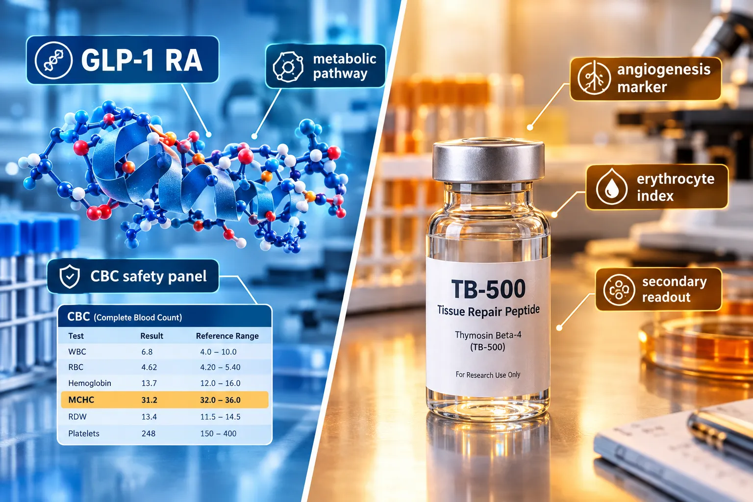
Fewer than 15% of published preclinical peptide studies list MCHC as a primary endpoint, yet researchers routinely flag unexpected shifts in this index and struggle to interpret what they mean. Understanding what does MCHC mean in peptide research, and how to interpret hemoglobin indices when evaluating GLP-class and tissue-repair models, is a practical skill that separates rigorous data review from guesswork.

Key Takeaways

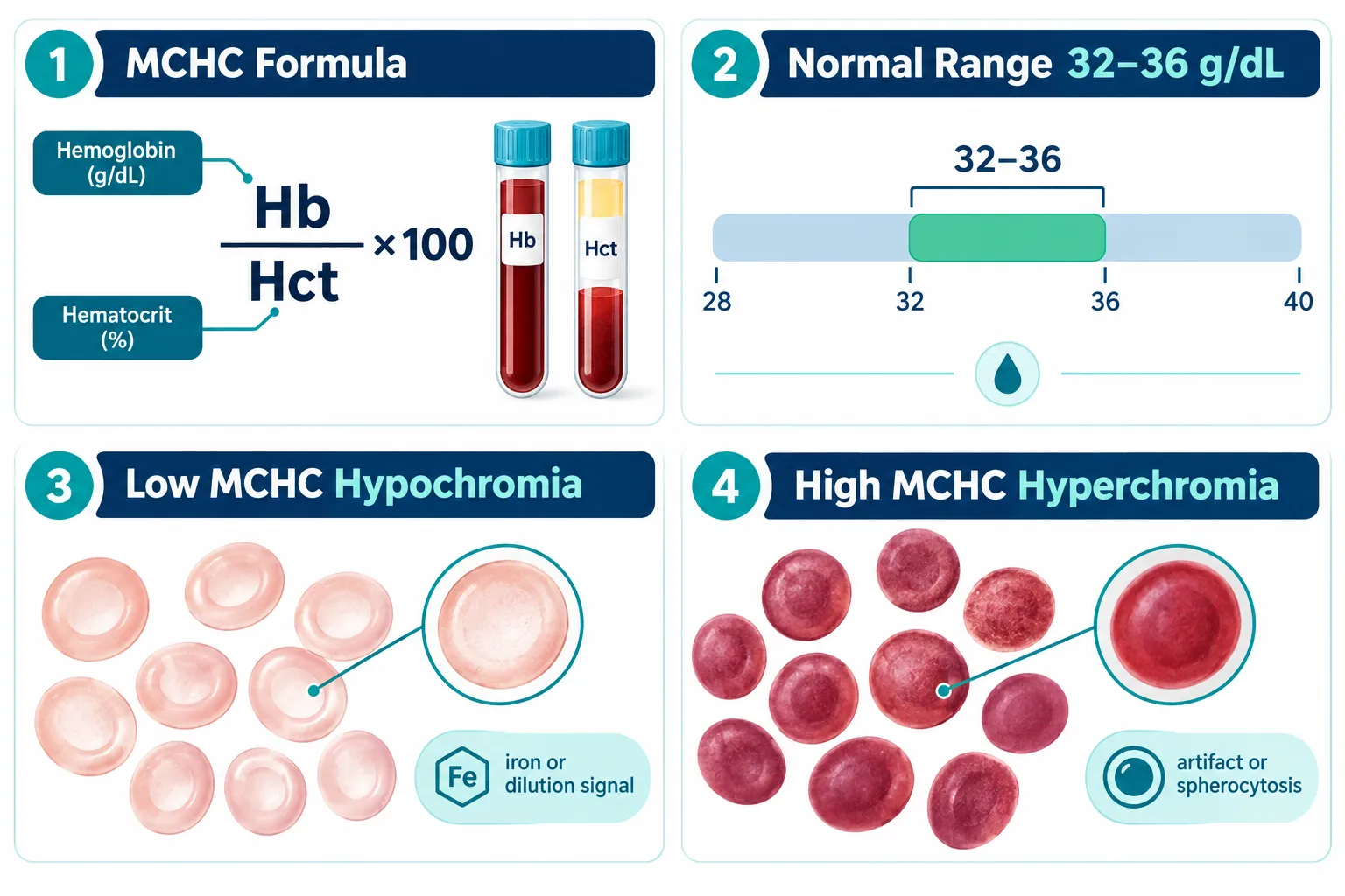
  • MCHC (Mean Corpuscular Hemoglobin Concentration) measures hemoglobin density inside red blood cells and is calculated as hemoglobin divided by hematocrit, expressed in g/dL.
  • In peptide research, MCHC functions as a secondary safety and physiology marker, not a primary efficacy endpoint.
  • Low MCHC (hypochromia) most often signals iron deficiency, chronic inflammation, or sample dilution rather than a direct peptide effect.
  • High MCHC (hyperchromia) in preclinical panels frequently reflects laboratory artifact, particularly hemolysis from poor sample handling.
  • MCHC should always be interpreted alongside MCV, RDW, and full study context before attributing changes to any peptide compound.

Defining MCHC: Calculation, Normal Ranges, and the Erythrocyte Index Hierarchy

MCHC stands for Mean Corpuscular Hemoglobin Concentration. It quantifies how much hemoglobin is packed into a given volume of red blood cells. The formula is straightforward:

MCHC (g/dL) = (Hemoglobin / Hematocrit) x 100

In most mammalian species used in preclinical research, the reference range sits between 32 and 36 g/dL. Values below this threshold indicate hypochromia (pale, hemoglobin-poor cells). Values above suggest hyperchromia, though true biological hyperchromia is rare.

Defining MCHC: Calculation, Normal Ranges, and the Erythrocyte Index Hierarchy

Within the broader erythrocyte index hierarchy, MCHC ranks below MCV (Mean Corpuscular Volume) and RDW (Red Cell Distribution Width) in diagnostic weight. Here is how the indices compare:

Index What It Measures Research Priority
MCV Average red cell size High
RDW Size variation across cells High
MCH Average hemoglobin per cell Moderate
MCHC Hemoglobin concentration per cell volume Supportive

MCHC is most useful when read as part of this full panel, not in isolation. Researchers sourcing compounds through platforms that emphasize building robust peptide benchmarks will recognize that baseline CBC panels, including MCHC, are part of responsible quality-control workflows.


Why MCHC Matters When Interpreting Hemoglobin Indices in GLP-Class Models

GLP-1 receptor agonist (GLP-RA) compounds have become central to metabolic research. When reviewing CBC safety data from GLP-class studies, MCHC appears as a secondary safety lab rather than a target biomarker. Its role is to confirm that metabolic changes driven by energy restriction, body composition shifts, or altered nutrient absorption are not producing clinically meaningful hematologic stress.

Researchers working with GLP-1 peptide sourcing and generational research concepts should note that caloric restriction models, common in GLP-RA efficacy studies, can mildly suppress MCHC through reduced dietary iron intake. This is a model-design artifact, not a compound toxicity signal.

Key considerations for GLP-class CBC interpretation:

  • Weight loss velocity can transiently alter plasma volume, diluting hemoglobin concentration and lowering apparent MCHC.
  • Visceral fat mobilization studies, such as those explored in visceral fat research models, may produce mild hemodilution as fluid compartments shift.
  • GLP-2-class compounds, which target intestinal repair and nutrient absorption, carry a more direct theoretical link to iron and B12 uptake, making MCHC monitoring more relevant in those specific models.

For researchers evaluating next-generation triple-agonist compounds, resources on GLP-3 for sale and triple agonist research planning provide useful context on how expanded receptor profiles may affect systemic physiology, including hematologic readouts.

Why MCHC Matters When Interpreting Hemoglobin Indices in GLP-Class Models


MCHC in Tissue-Repair Peptide Models: A Practical Interpretation Framework

Tissue-repair peptides, including TB-500 analogs and mitochondria-targeting compounds, introduce different considerations for hemoglobin index interpretation. Researchers following TB-500 controlled experimental models and QC workflow already understand that angiogenesis-promoting compounds can alter red cell dynamics through vascular remodeling rather than direct erythropoietic effects.

For mitochondria-targeted peptides, the relationship between cellular energy metabolism and red cell lifespan is an active area of interest. Work in SS-31 mitochondrial research and related SS-31 kidney health research highlights how oxidative stress reduction may indirectly support erythrocyte membrane integrity, a variable that influences MCHC readings over longer study durations.

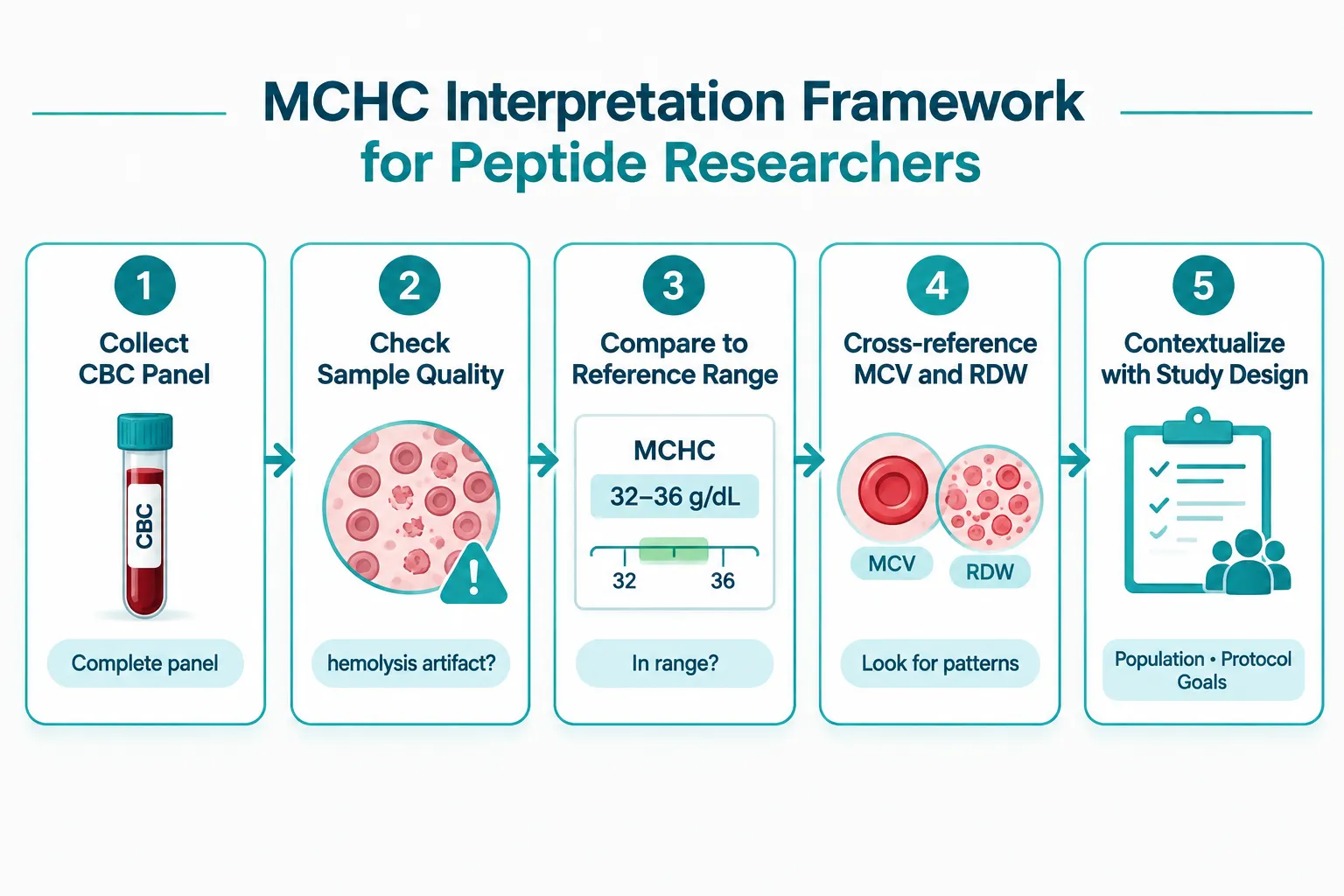
A practical five-step MCHC interpretation framework for peptide researchers:

  1. Verify sample quality first. Hemolysis from poor venipuncture or freeze-thaw cycling is the single most common cause of falsely elevated MCHC. Confirm sample integrity before drawing conclusions.
  2. Check the full CBC panel. An isolated MCHC shift without corresponding MCV or RDW changes is more likely an artifact than a biological signal.
  3. Compare against species-specific reference ranges. Murine, rat, and canine reference ranges differ meaningfully from human norms.
  4. Cross-reference study design variables. Diet, hydration status, and blood collection timing all affect MCHC independently of any peptide compound.
  5. Assign interpretive weight proportionally. MCHC is a supporting character in the CBC story, not the lead.

MCHC in Tissue-Repair Peptide Models: A Practical Interpretation Framework

As of late 2026, the published literature contains very few peptide-specific MCHC studies. Most available data comes from broader hematology safety panels embedded in metabolic or regenerative medicine trials. This gap means researchers must apply general hematology principles carefully rather than relying on peptide-specific MCHC benchmarks.


Conclusion

Understanding what does MCHC mean in peptide research, and how to interpret hemoglobin indices when evaluating GLP-class and tissue-repair models, ultimately comes down to context. MCHC is a reliable supporting marker when used correctly, but it is frequently misread when treated as a standalone signal.

Actionable next steps for peptide researchers in 2026:

  • Always collect a full CBC panel, not individual indices, as part of baseline and endpoint safety monitoring.
  • Document sample collection conditions meticulously to rule out hemolysis artifacts before flagging MCHC shifts.
  • When MCHC deviates from reference range, cross-check MCV, RDW, and reticulocyte count before attributing the change to a peptide compound.
  • For GLP-class models, account for caloric restriction and fluid redistribution as confounding variables in hematologic data.
  • For tissue-repair and mitochondria-targeting models, monitor MCHC trends over time rather than at single time points to distinguish transient from sustained changes.

Rigorous interpretation of hemoglobin indices strengthens the overall quality of peptide research data and supports more reliable conclusions across all model types.

https://www.puretestedpeptides.com/wp-content/uploads/2026/10/what-does-mchc-mean-in-peptide-research-interpreting-hemoglobin-indices-when-eva.webp 1024 1536 https://www.puretestedpeptides.com/wp-content/uploads/2026/01/buy-peptides-online.jpg 2026-10-11 13:04:122026-10-11 13:04:12What Does MCHC Mean in Peptide Research? Interpreting Hemoglobin Indices When Evaluating GLP-Class and Tissue-Repair Models
Electrolytes in Peptide Studies: Designing Sodium, Potassium, and Fluid-Balance Controls for GLP and Growth-Hormone Models

Electrolytes in Peptide Studies: Designing Sodium, Potassium, and Fluid-Balance Controls for GLP and Growth-Hormone Models

October 9, 2026/0 Comments/in Uncategorized/by

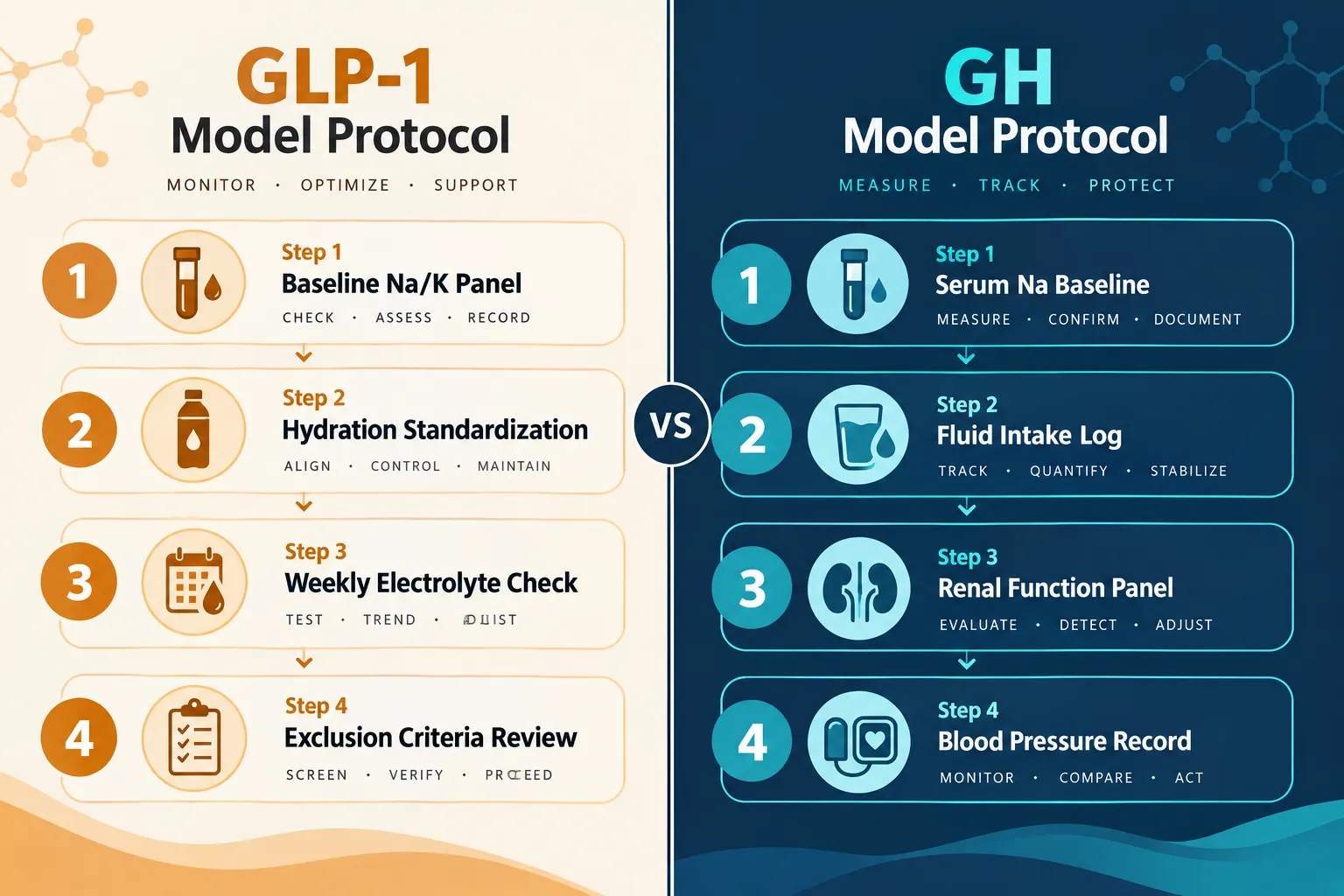
Fluid retention of just 1-2 liters can add more than 1 kg to a subject's body weight overnight, a shift large enough to mask or mimic a meaningful treatment effect in a peptide study. When the compounds under investigation are GLP-1 agonists or growth-hormone analogues, both of which directly alter renal sodium handling and total body water, uncontrolled electrolyte variables are not a minor nuisance. They are a primary source of confounding. Designing rigorous electrolyte controls is therefore central to valid endpoint measurement in these models, and the field of electrolytes in peptide studies: designing sodium, potassium, and fluid-balance controls for GLP and growth-hormone models has matured considerably in 2026.

Key Takeaways

  • GLP-1 agonists suppress thirst and reduce fluid intake, creating real dehydration and hyponatremia risk that must be controlled prospectively.
  • Growth hormone is anti-natriuretic: it promotes sodium and water retention through direct renal mechanisms, altering body weight and blood pressure readouts.
  • Serum sodium and potassium can appear stable even when total-body balances are significantly altered, making urine and fluid-intake logs essential.
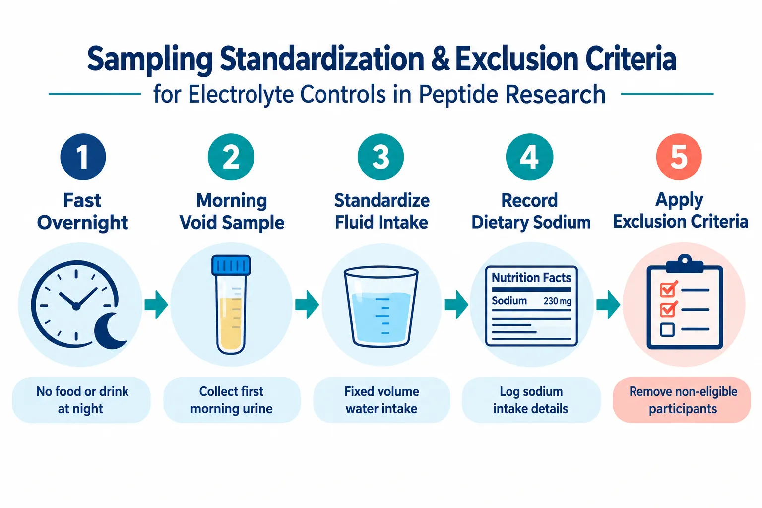
  • Sampling standardization, fixed fasting state, fixed hydration window, morning collection, is non-negotiable for reproducible electrolyte endpoints.
  • Exclusion criteria for baseline electrolyte abnormalities protect data integrity and reduce the risk of confounded renal and cardiovascular readouts.

Why Electrolyte Physiology Matters in GLP and GH Peptide Research

Both GLP-1 receptor agonists and growth-hormone-related peptides interact with the kidney in ways that directly alter sodium excretion, potassium distribution, and extracellular fluid volume. Understanding these mechanisms is the first step toward building controls that actually work.

Why Electrolyte Physiology Matters in GLP and GH Peptide Research

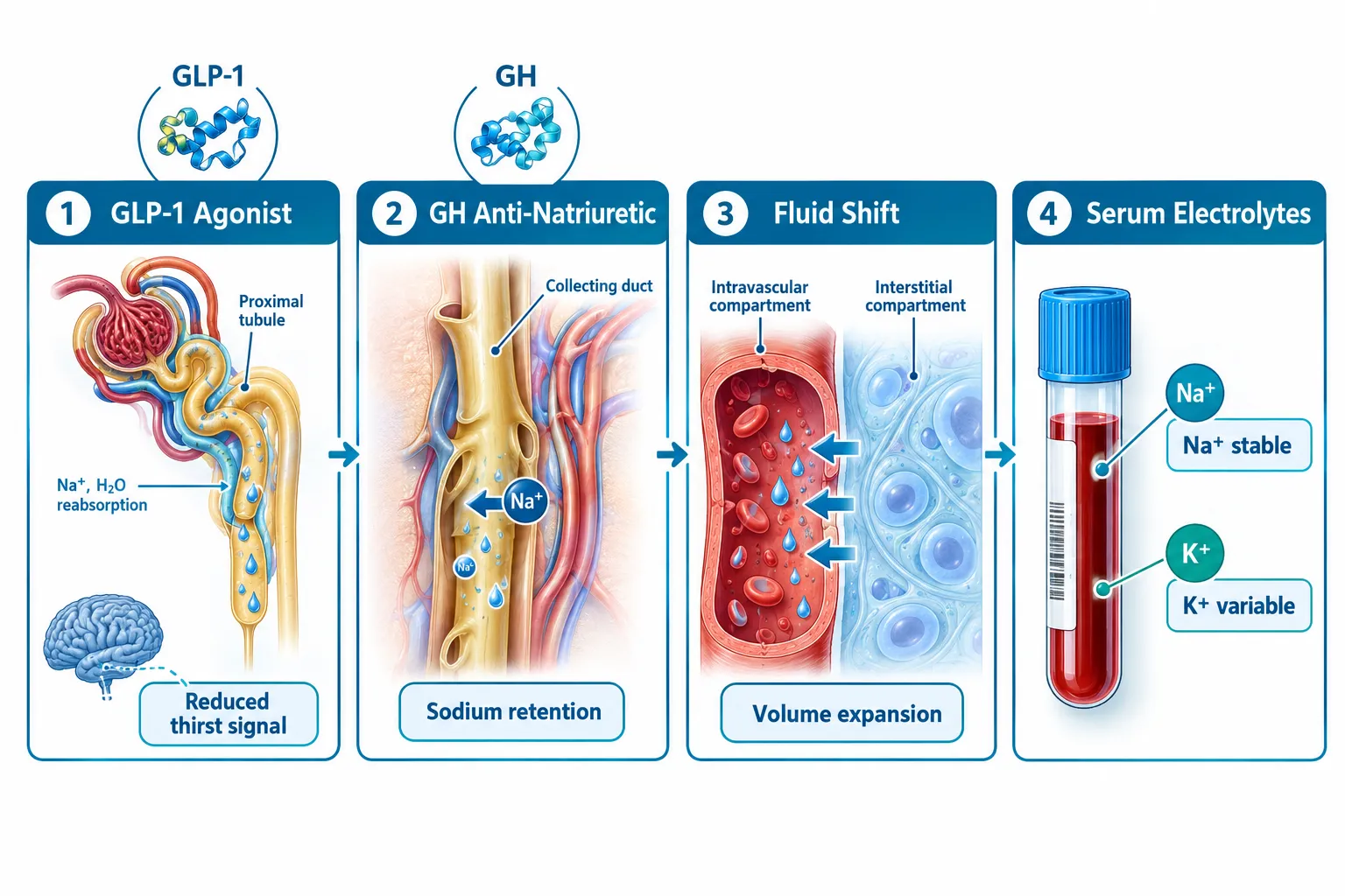
GLP-1 agonists reduce appetite and caloric intake, but they also suppress thirst, a phenomenon now described as hypodipsia. Subjects in GLP-1 models frequently show reduced spontaneous fluid consumption, raising the risk of mild-to-moderate dehydration. This matters because dehydration concentrates serum sodium, elevates hematocrit, and reduces glomerular filtration rate (GFR). If these changes are not tracked, a researcher may misattribute them to the peptide's metabolic action rather than to simple fluid deficit. Researchers working with GLP-3 R Peptide GA3 or GLP-3 R Peptide GA4 should treat hydration monitoring as a core protocol element, not an afterthought.

Growth hormone operates through a different but equally important mechanism. GH is anti-natriuretic: it increases renal tubular sodium reabsorption, expands extracellular fluid volume, and raises total body water. In GH-deficient subjects receiving replacement therapy, serum sodium and potassium often remain within normal reference ranges even as total-body sodium and water increase measurably. This dissociation between serum values and actual balance is a critical insight. Relying solely on a serum electrolyte panel will miss the fluid-volume shifts that inflate body weight and raise blood pressure, two of the most common endpoints in GH analogue studies.


Designing GLP-1 Models: Sodium, Potassium, and Hydration Controls

A well-designed GLP-1 model requires prospective hydration targets and a formalized electrolyte monitoring schedule built into the protocol from day one.

Hydration targets should be set at a defined minimum daily fluid intake, typically 30-35 mL per kilogram of body weight, and documented through daily fluid-intake logs. Subjects who fall below threshold on two or more consecutive days should trigger a protocol review. Urine-specific gravity measured at each morning sample collection provides a rapid, low-cost hydration proxy.

Sodium monitoring in GLP-1 models should include:

  • Baseline serum sodium before compound administration
  • Weekly serum sodium during active dosing
  • Urine sodium-to-creatinine ratio at baseline and at key timepoints
  • Dietary sodium standardization (target: 2,000-2,300 mg/day) or at minimum, a validated dietary recall at each visit

Potassium deserves separate attention. Rapid weight loss, which GLP-1 agonists can accelerate, increases the risk of hypokalemia through reduced dietary intake and increased renal losses. A serum potassium below 3.5 mEq/L should be a pre-specified action threshold: halt dose escalation, initiate dietary supplementation, and re-test within 72 hours.

"The most common electrolyte error in GLP-1 peptide models is treating hydration as a background variable rather than a controlled input."

For researchers sourcing compounds like Cagrilintide peptide or Retatrutide peptide, agents that combine GLP-1 and other receptor activity, the electrolyte burden is compounded, and monitoring frequency should increase accordingly.


Designing GH Models: Fluid Volume, Renal Endpoints, and Sampling Standards

Growth-hormone models present a different design challenge. Because GH expands extracellular fluid without reliably shifting serum sodium outside normal ranges, standard chemistry panels alone are insufficient as electrolyte controls.

Designing GH Models: Fluid Volume, Renal Endpoints, and Sampling Standards

Recommended control variables for GH models include:

Variable Measurement Method Frequency
Serum sodium Venous blood draw Baseline, weeks 2, 4, 8
Total body water Bioimpedance or deuterium dilution Baseline and end-of-study
Blood pressure Standardized seated measurement Every visit
Urine sodium Spot urine Na/Cr ratio Baseline and monthly
Body weight Morning, post-void, fasted Every visit

The key principle here is morning, post-void, fasted body weight. Because GH-driven fluid retention fluctuates with activity, posture, and dietary sodium intake, weight measurements taken at inconsistent times introduce noise that can easily exceed the magnitude of the treatment effect being studied.

Researchers using GH analogue peptides or PEG-MGF peptide should also pre-specify renal endpoints. GH's anti-natriuretic effect is mediated partly through IGF-1-dependent increases in GFR and partly through direct tubular action. Both pathways can alter creatinine clearance, so eGFR should be tracked alongside electrolytes rather than treated as a separate safety variable.


Sampling Standardization and Exclusion Criteria

Reproducible electrolyte data requires standardized collection conditions. Variability in fasting duration, hydration state, or posture at blood draw can shift serum sodium by 2-4 mEq/L and potassium by 0.3-0.5 mEq/L, differences large enough to cross clinical action thresholds.

Sampling Standardization and Exclusion Criteria

Standardization checklist for every electrolyte sample:

  1. Overnight fast of at least 8 hours
  2. Morning collection (6:00-9:00 AM window)
  3. Seated rest for 10 minutes before venipuncture
  4. No strenuous exercise in the prior 24 hours
  5. Standardized fluid intake the evening before (250-500 mL water, no electrolyte beverages)
  6. Document any diuretic, laxative, or supplement use within 48 hours

Exclusion criteria should be applied at screening to protect endpoint validity. Subjects with baseline serum sodium below 135 or above 145 mEq/L, potassium below 3.5 or above 5.0 mEq/L, or eGFR below 60 mL/min/1.73 m² should be excluded from electrolyte-sensitive peptide models. These thresholds reduce the risk that a pre-existing imbalance will be misread as a compound effect.

For models involving MOTS-C peptide, which influences mitochondrial and metabolic function, or BPC-157 / TB-500 peptide in wound healing models, electrolyte controls serve a secondary but still meaningful role in ensuring that inflammatory and fluid shifts do not confound tissue-repair endpoints.


Conclusion

The integrity of GLP and growth-hormone peptide research depends on treating electrolytes and fluid balance as primary controlled variables, not secondary safety checks. Actionable next steps for researchers in 2026 include:

  • Build electrolyte monitoring into the protocol at study design, not as an amendment after data collection begins.
  • Set pre-specified action thresholds for sodium, potassium, and hydration markers with clear decision rules for dose adjustment or subject review.
  • Standardize all sampling conditions, fasting state, time of day, posture, and prior-evening fluid intake, and document deviations as protocol deviations, not minor notes.
  • Apply strict exclusion criteria at screening to remove subjects with baseline electrolyte abnormalities that would confound interpretation.
  • Separate serum values from total-body balance in GH models by adding bioimpedance or urine electrolyte measures alongside standard chemistry panels.

Electrolytes in peptide studies, specifically designing sodium, potassium, and fluid-balance controls for GLP and growth-hormone models, is no longer an optional refinement. It is a prerequisite for data that can withstand scrutiny and support meaningful conclusions.

https://www.puretestedpeptides.com/wp-content/uploads/2026/10/electrolytes-in-peptide-studies-designing-sodium-potassium-and-fluid-balance-con.webp 1024 1536 https://www.puretestedpeptides.com/wp-content/uploads/2026/01/buy-peptides-online.jpg 2026-10-09 13:05:152026-10-09 13:05:15Electrolytes in Peptide Studies: Designing Sodium, Potassium, and Fluid-Balance Controls for GLP and Growth-Hormone Models
Unpacking the 'Peptides Calculator': Essential Tools and Methods for Accurate Dosing and Reconstitution in Research

Unpacking the ‘Peptides Calculator’: Essential Tools and Methods for Accurate Dosing and Reconstitution in Research

August 22, 2026/0 Comments/in Uncategorized/by

A single decimal-point error during peptide reconstitution can shift an experimental dose by a factor of ten, enough to invalidate months of research data. That reality is precisely why unpacking the 'Peptides Calculator' and its essential tools and methods for accurate dosing and reconstitution in research has become a priority for serious investigators in 2026.

Key Takeaways

  • Peptide calculators standardize the math behind reconstitution, concentration, and dose volume to reduce human error.
  • The core formula, concentration equals mass divided by volume, underpins every reliable calculator on the market.
  • Bacteriostatic water (BAC water) remains the standard diluent for most lyophilized research peptides.
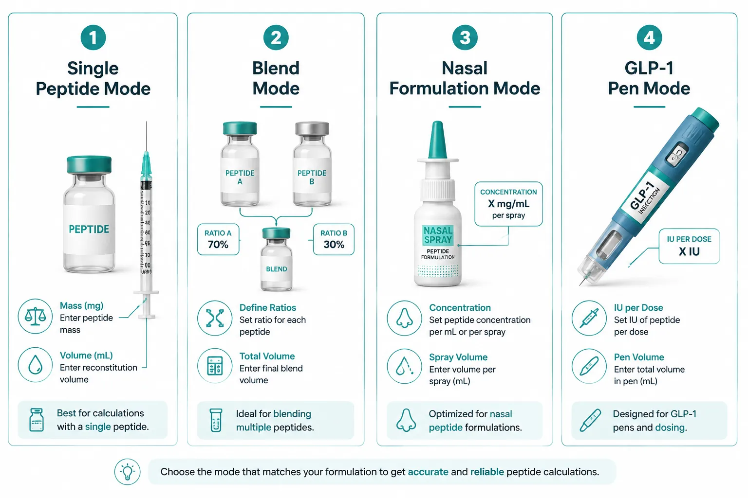
  • Modern tools now support single-peptide, blend, nasal, and GLP-1 pen modes with device-specific syringe guidance.
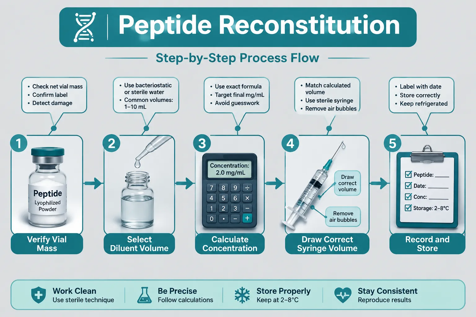
  • Reproducibility depends on consistent workflow: verify vial mass, select diluent volume, calculate concentration, then draw the correct syringe volume.

Why Accurate Peptide Dosing Matters in Research

Lyophilized peptides arrive as a dry powder measured in milligrams or micrograms. Before any research protocol can proceed, that powder must be dissolved in a precise volume of diluent to create a usable liquid concentration. Without a structured calculation method, researchers risk under-dosing (producing no measurable effect) or over-dosing (introducing confounding variables or compromising sample integrity).

Why Accurate Peptide Dosing Matters in Research

The stakes are especially high for sensitive compounds. For example, research into GLP-1 peptide sourcing and generational research concepts highlights how concentration accuracy directly shapes the validity of metabolic outcome data. Similarly, mitochondrial work involving compounds like those covered in SS-31 mitochondrial research themes demands tight dosing windows to produce reproducible results.

The core formula every researcher must internalize:

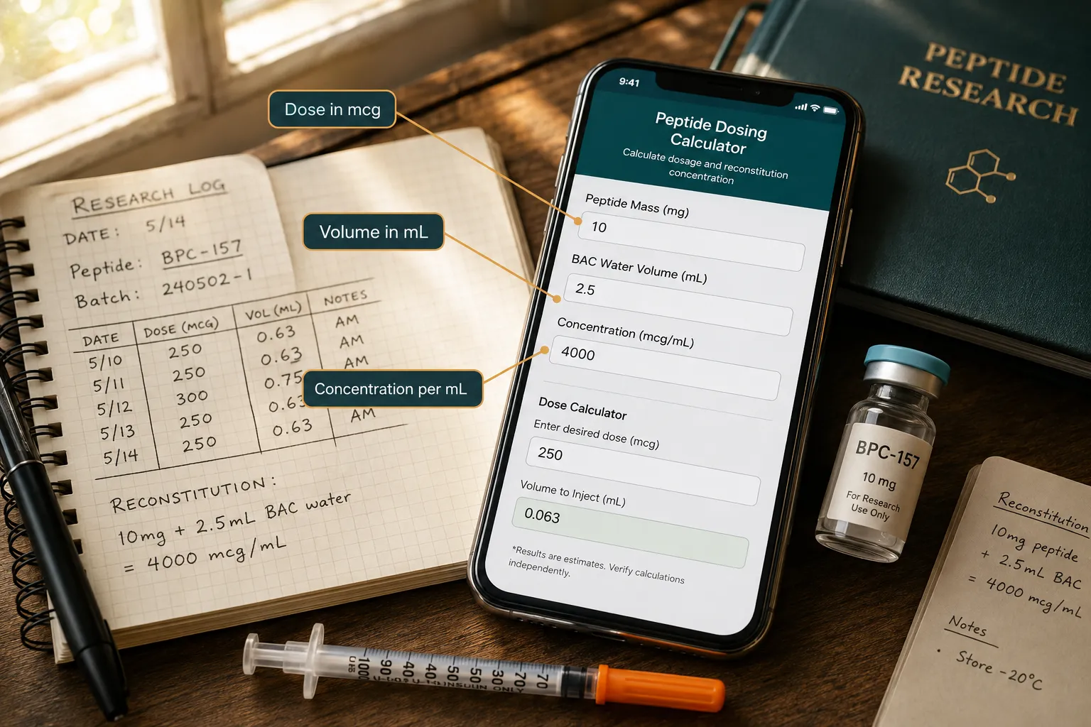
Concentration (mcg/mL) = Peptide Mass (mcg) / Diluent Volume (mL)

From this single equation, all downstream dose-volume calculations follow.

Standard Reconstitution Protocol with BAC Water

Bacteriostatic water is the preferred diluent for most lyophilized peptides because it contains 0.9% benzyl alcohol, which inhibits microbial growth and extends vial stability. The reconstitution steps below represent the standardized workflow recommended across leading peptide research platforms in 2026:

  1. Verify vial mass, confirm the labeled peptide mass (e.g., 5 mg = 5,000 mcg).
  2. Select diluent volume, choose a volume that produces a workable concentration (e.g., 2 mL BAC water for a 5 mg vial yields 2,500 mcg/mL).
  3. Add diluent slowly, inject BAC water along the vial wall; do not shake.
  4. Swirl gently, rotate until the powder fully dissolves.
  5. Calculate dose volume, divide the desired dose (mcg) by the concentration (mcg/mL).

A researcher needing a 250 mcg dose from a 2,500 mcg/mL solution draws exactly 0.1 mL (100 mcL) into an insulin syringe. A peptides calculator automates this final step, eliminating arithmetic errors under lab conditions.

Unpacking the 'Peptides Calculator': Core Features and Input Modes

Modern peptide calculators have expanded well beyond a single-formula widget. Unpacking the 'Peptides Calculator' and its essential tools and methods for accurate dosing and reconstitution in research reveals at least four distinct operational modes now standard across leading platforms.

Unpacking the 'Peptides Calculator': Core Features and Input Modes

Calculator Mode Primary Use Case Key Inputs
Single Peptide Standard vial reconstitution Vial mass, diluent volume, target dose
Blend Mode Multi-peptide stacks Individual masses, shared diluent volume
Nasal Formulation Intranasal delivery research Concentration per spray, spray volume
GLP-1 / Pen Mode Injection pen devices Units per mL, dose in units or mcg

The GLP-1 pen mode deserves particular attention. As research interest in GLP-1 class compounds grows, see the detailed breakdown in Retatrutide Phase 3 and ongoing obesity trial research, calculators must handle "per-IU" concentration reporting alongside standard mcg/mL outputs. This dual-unit capability prevents the unit-conversion errors that historically account for a large share of dosing mistakes.

Enhanced unit conversion features now common in 2026 tools include:

  • Automatic mg-to-mcg conversion on input
  • IU-to-mcg translation for growth hormone-adjacent peptides
  • Syringe-mark visualization (e.g., "draw to the 10-unit line on a U-100 syringe")
  • Mobile-optimized interfaces for field and clinic-adjacent research settings

Researchers sourcing compounds for these protocols should consult resources like where to buy peptides to ensure purity and labeled mass accuracy, both of which are prerequisites for any calculator to produce valid outputs.

Applying the Calculator: Workflow, Reproducibility, and Research Compliance

Unpacking the 'Peptides Calculator' and its essential tools and methods for accurate dosing and reconstitution in research is only half the task. The other half is embedding the tool into a reproducible, documented workflow.

Applying the Calculator: Workflow, Reproducibility, and Research Compliance

Recommended documentation checklist for each reconstitution event:

  • Record the peptide name, lot number, and labeled mass.
  • Log the diluent type, volume added, and date of reconstitution.
  • Calculate and record the resulting concentration.
  • Note storage conditions (temperature, light exposure).
  • Document each dose drawn: target dose, calculated volume, and actual syringe reading.

This level of documentation supports reproducibility, the cornerstone of credible research. It also aligns with the quality-control principles discussed in resources like PT-141 research context, QA, and controls and the reference standard benchmarks explored in Bachem and reference standards for peptide benchmarks.

A critical compliance note: All peptide calculator tools and the research protocols they support are intended strictly for laboratory and investigational use. Regulatory frameworks in most jurisdictions classify research peptides as not approved for human administration outside of licensed clinical trials. Every workflow built around these tools must reflect that framing clearly.

Avoiding the Most Common Calculation Errors

  • Unit mismatch: Entering mass in mg but volume in mL without converting produces a 1,000-fold concentration error.
  • Assuming full vial mass: Overfill or underfill from the manufacturer means the labeled mass may differ slightly from actual mass; always use a calibrated scale when precision is critical.
  • Ignoring dead volume: Syringes retain a small volume in the needle hub; account for this in high-precision protocols.

Conclusion

Accurate dosing and reconstitution are not optional refinements, they are foundational to any research protocol that expects reproducible, interpretable results. The rapid evolution of peptide calculator tools in 2026 has made it easier than ever to perform these calculations correctly, but the tools only work when researchers understand the underlying math and commit to a disciplined workflow.

Actionable next steps for researchers:

  1. Select a calculator that supports the specific mode required (single peptide, blend, nasal, or pen-based).
  2. Verify vial mass with a calibrated scale before every reconstitution.
  3. Document every reconstitution event and dose draw in a dedicated lab log.
  4. Cross-check unit conversions manually at least once per new peptide or protocol.
  5. Source peptides from suppliers who provide verified purity data, ensuring the labeled mass is reliable input for any calculation.

Applying these steps consistently transforms a peptides calculator from a convenience tool into a genuine instrument of scientific rigor.

https://www.puretestedpeptides.com/wp-content/uploads/2026/08/unpacking-the-peptides-calculator-essential-tools-and-methods-for-accurate-dosin.webp 1024 1536 https://www.puretestedpeptides.com/wp-content/uploads/2026/01/buy-peptides-online.jpg 2026-08-22 13:06:062026-08-22 13:06:06Unpacking the ‘Peptides Calculator’: Essential Tools and Methods for Accurate Dosing and Reconstitution in Research
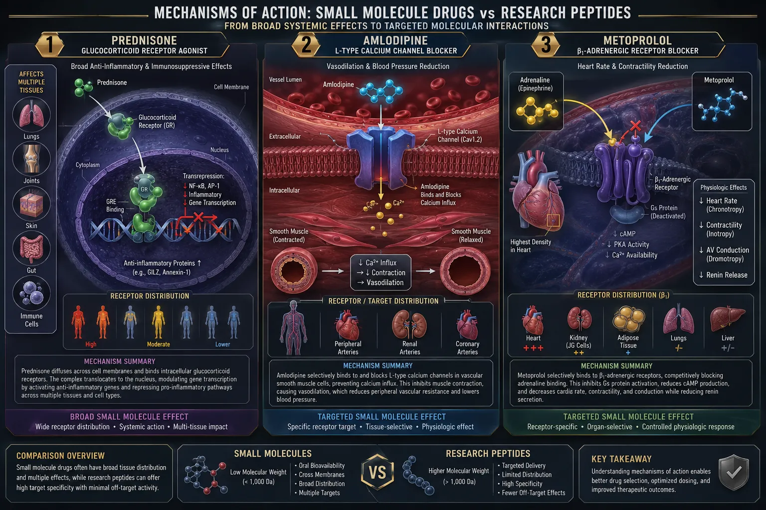
Peptides vs Classic Small-Molecule Drugs: How Compounds Like Prednisone, Amlodipine, and Metoprolol Differ From Modern Research-Use Peptides

Peptides vs Classic Small-Molecule Drugs: How Compounds Like Prednisone, Amlodipine, and Metoprolol Differ From Modern Research-Use Peptides

August 4, 2026/0 Comments/in Uncategorized/by

{"cover":"Professional landscape format (1536×1024) hero image with bold text overlay (≤42 chars): 'Peptides vs Classic Small-Molecule Drugs' in crisp white bold sans-serif centered on a deep navy semi-transparent panel, 8% safe margins from every edge, no text touching edges. Background: split editorial scene, left side shows a clinical pharmacy shelf with amber pill bottles (prednisone, amlodipine labels), right side shows a modern research lab with peptide vials on ice and molecular chain diagrams on a lightboard. Cool blue and white color palette, magazine-quality editorial lighting, high contrast, pharmaceutical science aesthetic.","content":["Bright editorial infographic-style landscape (1536×1024): two large molecular structure illustrations side by side, left shows a compact steroid ring structure labeled 'Small Molecule' in bold 4-word label, right shows an elongated amino acid chain labeled 'Peptide Chain', both rendered as glowing 3D models on a clean white laboratory background with soft blue accent lighting. Scientific diagram aesthetic, vivid teal and orange contrast palette, no tables, no pricing grids, save-worthy Pinterest science illustration style.","Split-screen editorial photograph landscape (1536×1024): left half shows a close-up of white prednisone tablets and amlodipine capsules arranged on a stainless steel lab tray under bright clinical white lighting; right half shows research-use peptide vials (BPC-157, MOTS-c labeled in 3-word max text) on crushed ice in a modern biochemistry lab with blue LED ambient lighting. High-contrast pharmaceutical editorial photography, cool clinical palette of white, steel-blue, and amber, sharp product focus, magazine quality.","Symbolic conceptual illustration landscape (1536×1024): isometric flat-vector scene of a research laboratory interior, a female South Asian scientist in a white coat examines a glowing peptide chain hologram floating above a lab bench, while in the background a wall display shows simplified receptor-binding diagrams with short labels 'Receptor Target' and 'Signal Path'. Bright daylight through large windows, clean pastel palette of mint green, soft yellow, and white, modern scientific illustration style, no tables, no grids, Pinterest-quality editorial."]

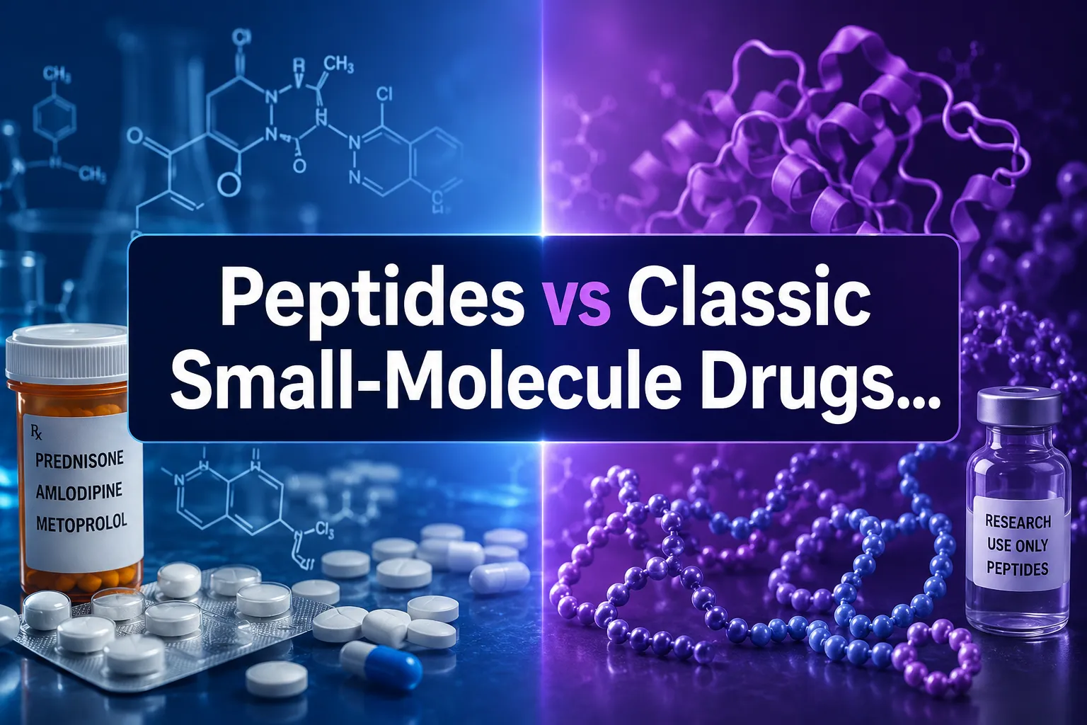
Professional landscape hero image () with a reading "Peptides vs Classic Small-Molecule Drugs…". CRITICAL TYPOGRAPHY RULES:

More than 90% of all approved drugs on the market today are small molecules, yet the fastest-growing segment of pharmaceutical research now centers on peptides. This shift is not accidental. As researchers probe the limits of traditional pharmacology, the structural and mechanistic gap between classic drugs like prednisone, amlodipine, and metoprolol and modern research-use peptides has become one of the most important distinctions in biochemistry. Understanding peptides vs classic small-molecule drugs clarifies why compounds like BPC-157, MOTS-c, and GLP-3 occupy a fundamentally different category from the drugs most people take daily.

Key Takeaways

  • Small-molecule drugs are compact, chemically synthesized compounds that typically act on a single receptor or enzyme target.
  • Peptides are short chains of amino acids that mimic or modulate the body's own signaling molecules, enabling more targeted biological interactions.
  • Classic drugs like prednisone, amlodipine, and metoprolol have well-established clinical profiles; research-use peptides are studied under controlled laboratory conditions and are not approved for human therapeutic use.
  • Peptides generally have higher target specificity but lower oral bioavailability than small molecules.
  • The regulatory and research frameworks governing peptides differ substantially from those governing licensed pharmaceuticals.

Key Takeaways

Structural Foundations: What Separates Small Molecules From Peptides

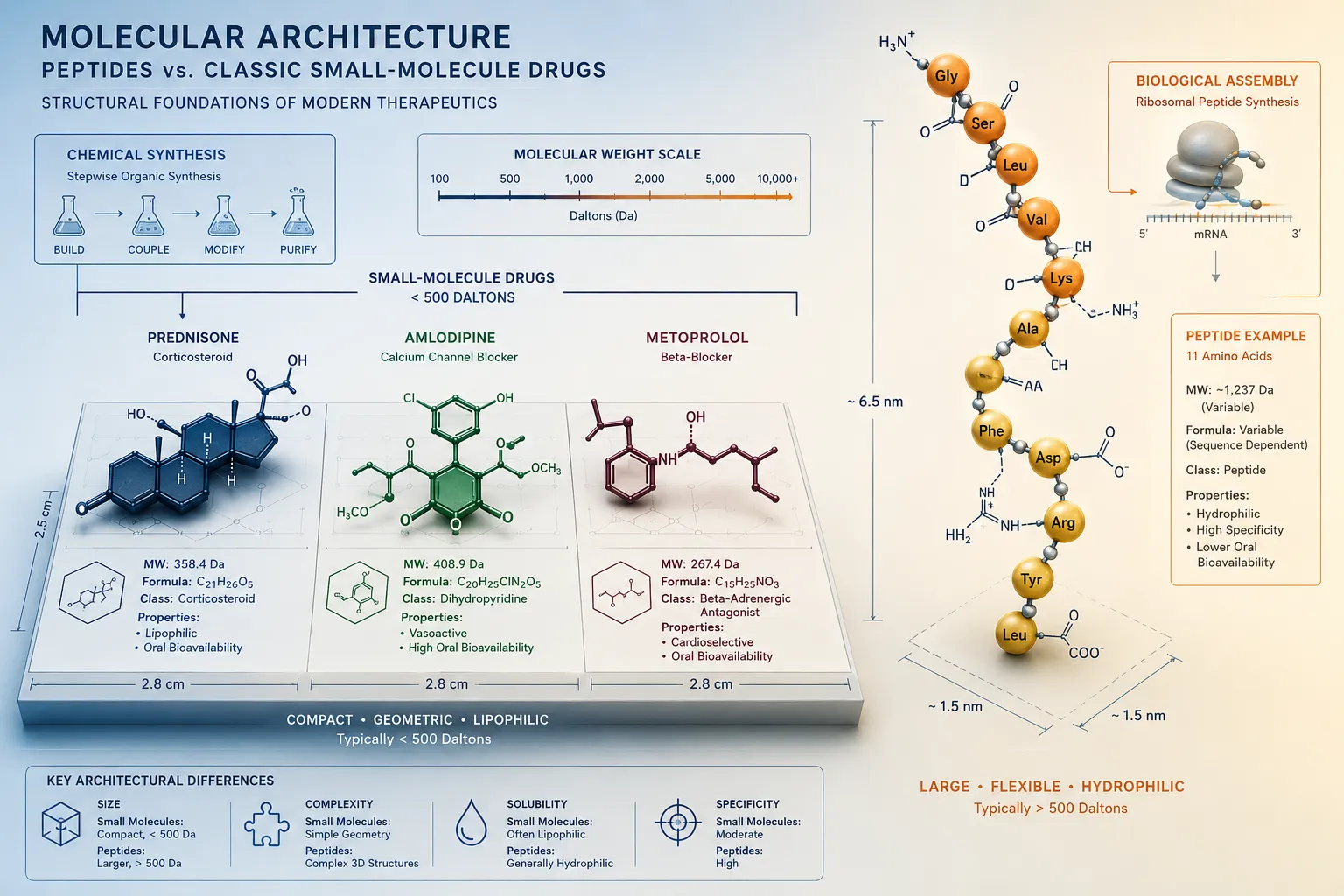
The most fundamental difference in peptides vs classic small-molecule drugs is molecular architecture.

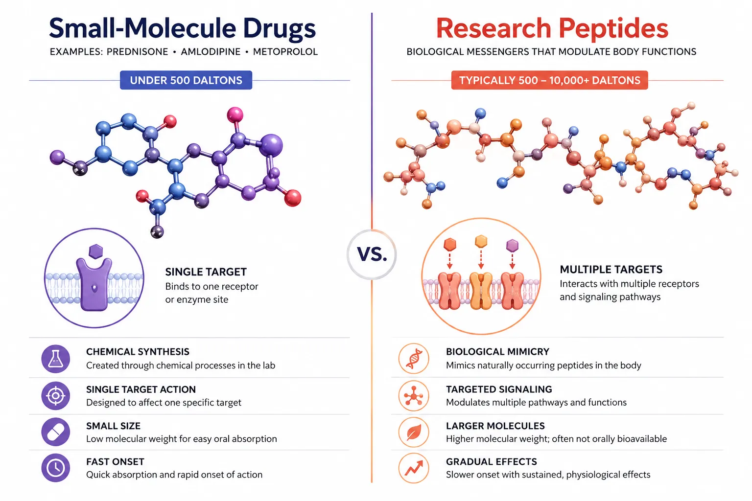
Small molecules, including prednisone, amlodipine, and metoprolol, are low-molecular-weight organic compounds, typically under 500 daltons. They are built through chemical synthesis, not biological processes, and their compact size allows them to cross cell membranes, enter the bloodstream via oral administration, and bind to specific receptor pockets.

Feature Small-Molecule Drugs Research-Use Peptides
Molecular weight Under 500 Da 500-5,000+ Da
Composition Synthetic organic chemistry Amino acid chains
Oral bioavailability Generally high Generally low
Synthesis route Chemical Chemical or biosynthetic
Target specificity Moderate to high High

Peptides, by contrast, are short chains of amino acids, typically 2 to 50 residues, that mimic or modulate the body's endogenous signaling molecules. Their larger size and more complex three-dimensional shape allow them to interact with biological targets in ways small molecules cannot, but this same size makes them vulnerable to digestive enzymes, which is why many research-use peptides require parenteral administration.

"The structural complexity of a peptide is both its greatest advantage and its primary delivery challenge."

Compounds like TB-500 or the BPC-157 and TB-500 combination illustrate this point well, their amino acid sequences enable highly specific tissue interactions that a small steroid molecule like prednisone simply cannot replicate.

Mechanisms of Action: How Prednisone, Amlodipine, and Metoprolol Work vs Research Peptides

Mechanisms of Action: How Prednisone, Amlodipine, and Metoprolol Work vs Research Peptides

Classic small-molecule drugs each act through well-characterized, narrow mechanisms:

  • Prednisone is a synthetic corticosteroid. It binds glucocorticoid receptors inside cells, suppressing inflammatory gene transcription broadly across multiple tissue types. Its wide receptor distribution explains both its therapeutic power and its side-effect profile (blood sugar changes, bone density loss, immune suppression).
  • Amlodipine is a calcium channel blocker. It binds L-type calcium channels in vascular smooth muscle, reducing calcium influx and causing vasodilation. The mechanism is highly localized to one channel subtype.
  • Metoprolol is a beta-1 selective adrenergic blocker. It competes with catecholamines at beta-1 receptors in cardiac tissue, slowing heart rate and reducing myocardial oxygen demand.

Each of these drugs acts on a defined, single-class receptor. Their mechanisms are predictable, well-studied, and the basis for decades of clinical data.

Research-use peptides operate differently. Rather than blocking or activating a single receptor, many peptides act as signaling modulators, they interact with receptor complexes, growth factor pathways, or intracellular signaling cascades in a more context-dependent way.

For example:

  • BPC-157 is studied for its interactions with growth hormone receptor pathways and nitric oxide systems, with research endpoints focused on tissue repair models.
  • MOTS-c is a mitochondria-derived peptide investigated for its role in metabolic regulation and cellular stress responses. Research on MOTS-c and mitochondrial function explores mechanisms that have no equivalent in classic pharmacology.
  • GLP-1 and GLP-3 class peptides act on incretin receptors involved in insulin secretion and gut motility, a mechanism that bridges peptide biology and metabolic research.

The SS-31 peptide's mitochondrial research themes demonstrate another dimension: peptides can localize to specific organelles, something small molecules rarely achieve with the same precision.

Research Context, Regulatory Status, and Practical Differences

Research Context, Regulatory Status, and Practical Differences

Understanding peptides vs classic small-molecule drugs also requires clarity on their regulatory and research contexts.

Prednisone, amlodipine, and metoprolol are FDA-approved pharmaceuticals. They have completed clinical trials, carry established dosing guidelines, and are prescribed by licensed clinicians for defined indications. Their safety and efficacy data span millions of patient-years.

Research-use peptides occupy a different category entirely. Compounds like AOD-9604 or Epithalon are sold strictly for laboratory and preclinical research purposes. They are not approved for human therapeutic use, and their research endpoints are studied in controlled in vitro and animal model settings.

Key practical distinctions include:

  • Stability: Small molecules are generally shelf-stable at room temperature. Most research peptides require refrigeration or lyophilization to maintain structural integrity.
  • Administration route: Classic drugs are predominantly oral. Research peptides are typically reconstituted and administered via injection in research settings.
  • Selectivity: Peptides often show higher target selectivity, which is why combinations like LL-37 and SS-31 are studied for their complementary, non-overlapping mechanisms.
  • Research endpoints: Small-molecule research focuses on receptor occupancy and clinical outcomes. Peptide research often examines upstream signaling, gene expression changes, and cellular repair processes.

Researchers exploring BDNF-related peptide pathways or Selank's neurological research profile encounter a level of mechanistic specificity that classic pharmacology rarely achieves.

Conclusion

The comparison of peptides vs classic small-molecule drugs is not a question of which category is superior, it is a question of purpose, mechanism, and context. Prednisone, amlodipine, and metoprolol are proven therapeutic tools with decades of clinical validation. Research-use peptides like BPC-157, MOTS-c, and GLP-3 represent a different scientific frontier: larger, more structurally complex molecules that interact with biological systems in ways that mirror the body's own signaling language.

Actionable next steps for researchers and informed readers:

  1. Review primary literature on specific peptide mechanisms before drawing comparisons to approved drugs.
  2. Source research-use peptides only from verified suppliers with documented purity testing.
  3. Consult the growing body of preclinical data on mitochondrial peptides, incretin analogs, and tissue-repair compounds to understand where the science currently stands.
  4. Recognize that regulatory status is not a proxy for scientific interest, many of the most actively studied peptides are pre-clinical compounds with significant research momentum.

The structural and mechanistic divide between small molecules and peptides will continue to shape pharmacology research well into the future.

https://www.puretestedpeptides.com/wp-content/uploads/2026/08/peptides-vs-classic-small-molecule-drugs-how-compounds-like-prednisone-amlodipin.webp 1024 1536 https://www.puretestedpeptides.com/wp-content/uploads/2026/01/buy-peptides-online.jpg 2026-08-04 13:06:152026-08-04 13:06:15Peptides vs Classic Small-Molecule Drugs: How Compounds Like Prednisone, Amlodipine, and Metoprolol Differ From Modern Research-Use Peptides
GLP‑3 Retatrutide in Phase 3 Trials: How Triple Agonism Is Reshaping Obesity and MASLD Research Endpoints

GLP‑3 Retatrutide in Phase 3 Trials: How Triple Agonism Is Reshaping Obesity and MASLD Research Endpoints

July 31, 2026/0 Comments/in Uncategorized/by

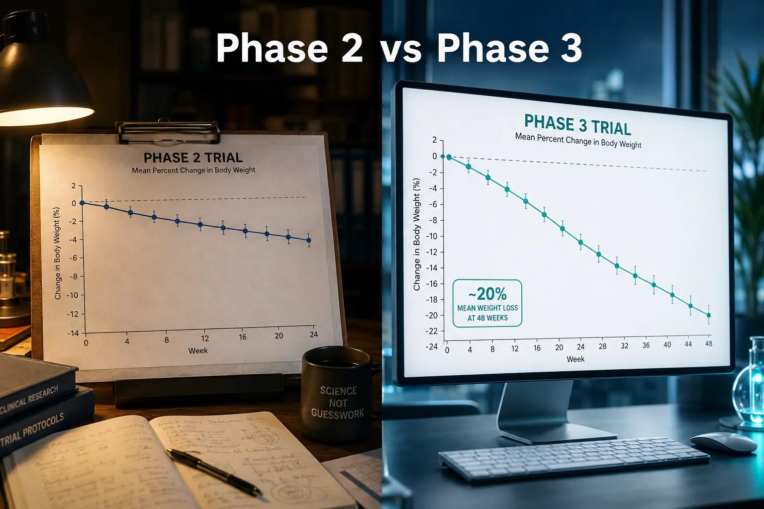
Participants in the retatrutide Phase 2 trial lost up to 24.2% of body weight over 48 weeks — a figure that outpaced every approved GLP-1 therapy on record at the time. That single data point accelerated Eli Lilly's decision to move retatrutide into Phase 3 development, and it fundamentally changed how researchers are designing metabolic endpoints for obesity and liver disease trials in 2026.

This article examines what GLP-3 retatrutide in Phase 3 trials means for obesity and MASLD research, how triple receptor agonism differs mechanistically from classic GLP-1 approaches, and what endpoint design shifts are emerging as a result.

Key Takeaways

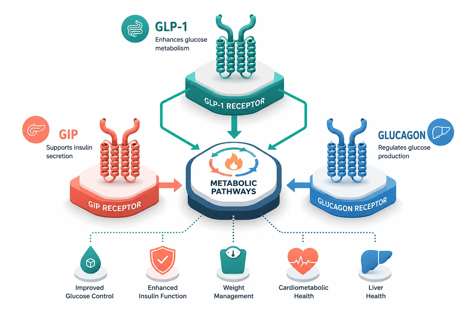
  • Retatrutide simultaneously activates GLP-1, GIP, and glucagon receptors, producing greater weight loss than dual or single agonists in early trials.
  • Phase 3 programs are now incorporating liver-specific endpoints such as fibrosis resolution and MASLD Activity Score changes, not just body weight.
  • Triple agonism introduces unique metabolic signals — particularly through glucagon receptor activation — that require researchers to monitor hepatic and cardiovascular markers differently.
  • Comparing retatrutide to classic GLP-1 peptides reveals meaningful differences in energy expenditure, lipid clearance, and tolerability profiles.
  • Endpoint design for MASLD trials is evolving to capture histological, biomarker, and imaging outcomes simultaneously.

Key Takeaways

What Is Triple Agonism and Why Does It Matter for Metabolic Research

Classic GLP-1 receptor agonists like semaglutide act on a single receptor pathway to reduce appetite and slow gastric emptying. Dual agonists such as tirzepatide added GIP receptor co-activation, improving insulin sensitivity and amplifying weight loss. Retatrutide goes one step further by adding glucagon receptor (GCGR) agonism to the GLP-1 and GIP combination.

This triple mechanism matters for several reasons:

  • GLP-1 receptor activation reduces appetite and slows gastric emptying
  • GIP receptor activation enhances insulin secretion and improves adipose tissue metabolism
  • Glucagon receptor activation increases hepatic glucose output, raises energy expenditure, and promotes fat oxidation in the liver

The glucagon component is particularly relevant for MASLD research. Glucagon signaling directly reduces hepatic lipid accumulation, a core driver of metabolic dysfunction-associated steatotic liver disease. For researchers studying GLP-1 peptide mechanisms and sourcing, retatrutide represents a meaningful evolution beyond single-pathway tools.

"Triple agonism does not simply add effects — it creates synergistic metabolic signals that single or dual agonists cannot replicate."

This synergy is precisely why GLP-3 retatrutide in Phase 3 trials is reshaping obesity and MASLD research endpoints: the compound forces investigators to measure outcomes that single-receptor drugs rarely moved.

Phase 3 Trial Design: How Retatrutide Is Changing Research Endpoints

Phase 3 Trial Design: How Retatrutide Is Changing Research Endpoints

Eli Lilly's TRIUMPH Phase 3 program covers obesity, type 2 diabetes, and MASLD (metabolic dysfunction-associated steatotic liver disease, formerly NAFLD/NASH). Each arm introduces endpoint complexity that reflects the drug's multi-receptor biology.

Obesity Endpoints

Traditional obesity trials used percent body weight change as the primary endpoint. Phase 3 retatrutide trials now layer in:

Endpoint Category Specific Measures
Body composition MRI-based visceral adipose tissue volume
Cardiometabolic LDL-C, triglycerides, blood pressure
Functional 6-minute walk test, patient-reported outcomes
Safety Glucagon-related hepatic markers, bone density

The inclusion of visceral fat imaging reflects the glucagon receptor's targeted effect on hepatic and visceral lipid stores — a signal that waist circumference alone cannot capture.

MASLD-Specific Endpoints

This is where GLP-3 retatrutide in Phase 3 trials is most dramatically reshaping obesity and MASLD research endpoints. Liver trials now require:

  • Histological resolution of steatohepatitis without worsening fibrosis (FDA-aligned primary endpoint)
  • Fibrosis stage improvement by at least one stage on the METAVIR scale
  • MRI-PDFF (proton density fat fraction) as a non-invasive imaging biomarker
  • Liver stiffness measurement via FibroScan or MRE
  • Serum ALT normalization as a secondary biochemical marker

These layered endpoints are more demanding than what GLP-1-only trials required, but they are appropriate given retatrutide's direct hepatic signaling. Researchers interested in metabolic peptide tools for liver-focused protocols may also find value in reviewing research-only peptides used in complementary preclinical models.

Comparing Retatrutide to Classic GLP-1 Agents

The table below summarizes key mechanistic and endpoint differences:

Feature GLP-1 Agonist Dual Agonist (GIP+GLP-1) Retatrutide (Triple)
Weight loss (approx.) 10-15% 15-22% Up to 24%+
Hepatic fat reduction Moderate Moderate-High High
Energy expenditure Minimal increase Moderate Significant
MASLD endpoint utility Limited Moderate High

For researchers already tracking GLP-2 receptor biology or GLP-1 peptide product categories, the triple agonist framework offers a useful comparative reference point.

MASLD Research Design Implications in 2026

MASLD Research Design Implications in 2026

The shift toward composite histological endpoints in MASLD trials is not unique to retatrutide, but the drug's glucagon component has accelerated it. Researchers designing MASLD protocols in 2026 are now expected to pre-specify:

  1. Biopsy timing aligned with expected fibrosis response windows (typically 48-72 weeks)
  2. Non-invasive biomarker panels including Enhanced Liver Fibrosis (ELF) score and FIB-4
  3. Imaging sub-studies using MRI-PDFF at baseline, 24 weeks, and end of treatment
  4. Cardiovascular safety monitoring given glucagon's effects on heart rate and blood pressure

This multi-modal design philosophy is influencing adjacent research areas. Investigators studying metabolic peptides with hepatic or mitochondrial relevance — such as those reviewing SS-31 mitochondrial research themes or tesa dosage protocols for fat loss — are adopting similar composite endpoint frameworks.

The MASLD field has also begun distinguishing between steatosis resolution and fibrosis regression as separate but related outcomes. Retatrutide's Phase 3 design treats these as co-primary endpoints in the liver arm, a precedent that other investigational agents are now following.

Researchers working with research blog resources on peptide science will find the retatrutide endpoint framework a useful template for designing metabolic intervention studies across multiple tissue targets.

Conclusion

GLP-3 retatrutide in Phase 3 trials is doing more than testing a new weight-loss drug — it is redefining what rigorous metabolic research endpoints look like for both obesity and MASLD. The triple agonist mechanism forces investigators to measure visceral fat, hepatic histology, fibrosis staging, and cardiometabolic markers simultaneously, raising the bar for the entire field.

Actionable next steps for researchers and protocol designers:

  • Adopt composite endpoints that include both imaging (MRI-PDFF) and histological measures for any MASLD-adjacent study
  • Monitor glucagon receptor-related safety signals (heart rate, hepatic glucose output) when designing triple agonist or multi-receptor protocols
  • Use retatrutide Phase 3 endpoint frameworks as a reference template when designing studies with GLP-1-class or metabolic peptide tools
  • Stay current with TRIUMPH trial interim data releases, which are expected to report through 2026-2027
  • Review GLP-1 peptide research concepts to understand how single-receptor baselines compare to triple agonist benchmarks

The triple agonism era is not a refinement of existing metabolic research — it is a structural shift in how endpoints are conceived, measured, and interpreted.

https://www.puretestedpeptides.com/wp-content/uploads/2026/07/glp-3-retatrutide-in-phase-3-trials-how-triple-agonism-is-reshaping-obesity-and.webp 1024 1536 https://www.puretestedpeptides.com/wp-content/uploads/2026/01/buy-peptides-online.jpg 2026-07-31 13:04:112026-07-31 13:04:11GLP‑3 Retatrutide in Phase 3 Trials: How Triple Agonism Is Reshaping Obesity and MASLD Research Endpoints

Tag Archive for: glp-1 peptide research

GLP-3 Retatrutide: Latest Research on Its Impact on Liver Fat Reduction and MASLD Management

GLP-3 Retatrutide: Latest Research on Its Impact on Liver Fat Reduction and MASLD Management

July 3, 2026/0 Comments/by Pure Tested

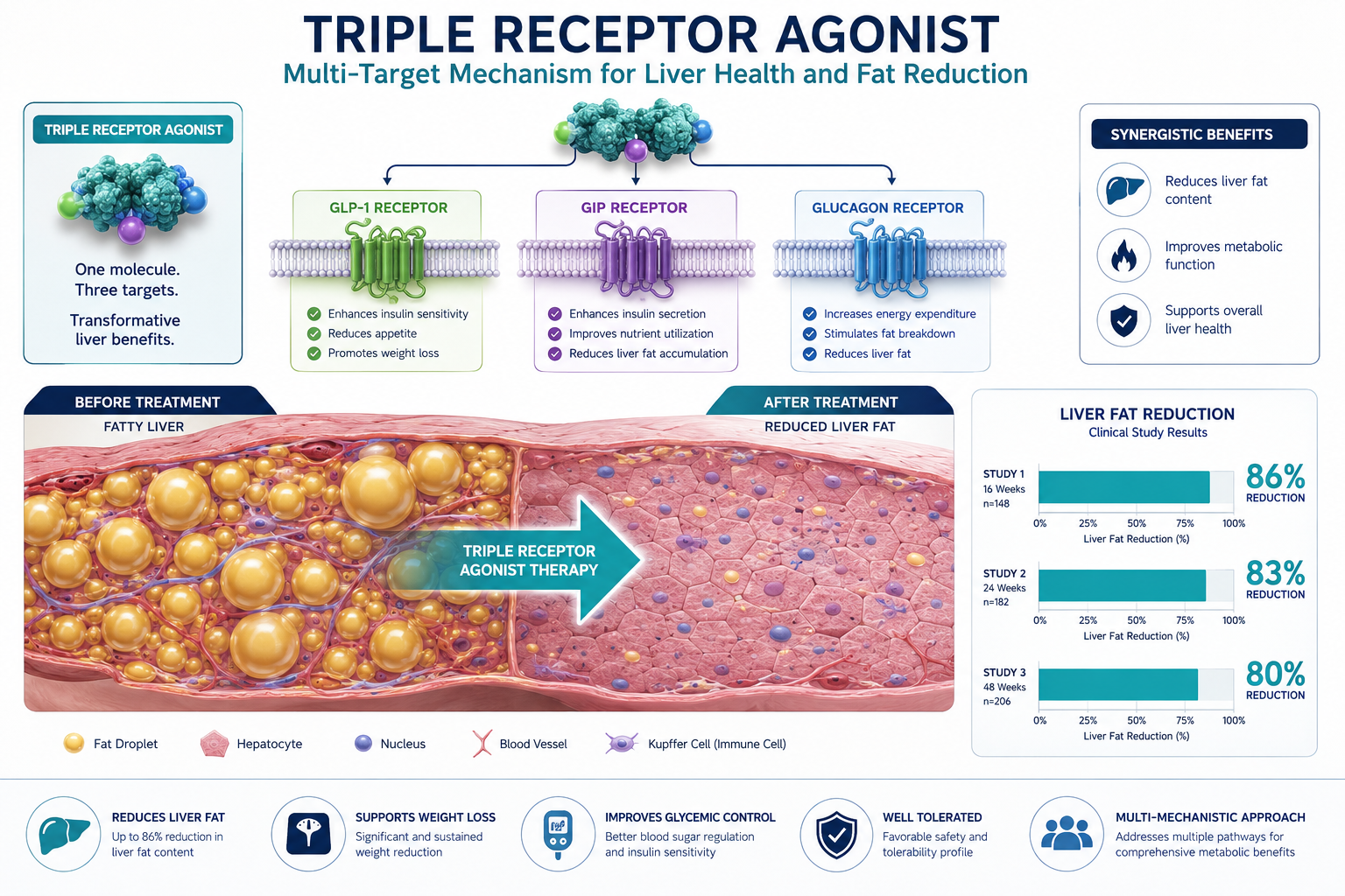
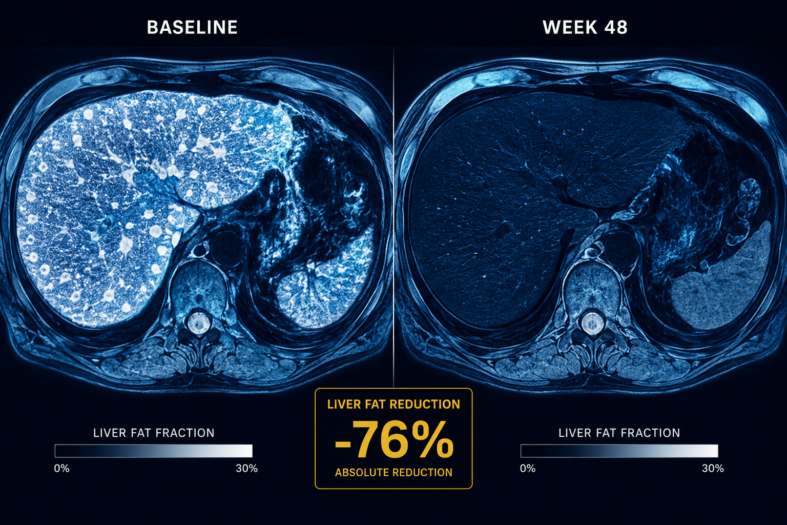
More than 80% of participants with fatty liver disease who received retatrutide in a phase 2 trial had their liver fat completely normalized by week 48, a result researchers described as among the largest liver-fat reductions ever reported in an obesity or MASLD trial. That single data point has reshaped how the research community thinks about triple receptor agonists and metabolic liver disease.

This article examines what the most current evidence says about GLP-3 Retatrutide: Latest Research on Its Impact on Liver Fat Reduction and MASLD Management, who may benefit most, and what questions still need answering.

Key Takeaways

  • Retatrutide is a triple agonist targeting GLP-1, GIP, and glucagon receptors simultaneously.
  • Phase 2 data show mean relative liver fat reductions exceeding 80% at 48 weeks.
  • More than 90% of participants on the 12 mg dose achieved liver fat normalization below the 5% MRI threshold.
  • Weight loss of nearly 24-26% accompanied the liver fat improvements, suggesting dual metabolic benefit.
  • The safety profile mirrors other incretin-based therapies, with no new hepatotoxicity signal identified.

Key Takeaways

What Is Retatrutide and Why Does It Matter for MASLD

Metabolic Dysfunction-Associated Steatotic Liver Disease (MASLD), formerly called NAFLD, affects an estimated 25% of the global adult population. It ranges from simple fat accumulation in liver cells to progressive inflammation, fibrosis, and cirrhosis. Until recently, no pharmacological agent had demonstrated the ability to reliably normalize liver fat across a broad patient population.

Retatrutide changes that conversation. Unlike semaglutide or tirzepatide, which act on one or two receptors, retatrutide simultaneously activates three receptors:

Receptor Primary Role
GLP-1 Appetite suppression, insulin secretion
GIP Energy metabolism, fat storage regulation
Glucagon Hepatic fat oxidation, energy expenditure

The glucagon component is particularly relevant for liver fat. Glucagon receptor activation directly stimulates hepatic fat burning, meaning retatrutide works on the liver through a mechanism that single or dual agonists do not fully replicate. Researchers interested in the broader landscape of GLP-1 peptide research will recognize this as a meaningful mechanistic step forward.


Phase 2 Trial Data: Retatrutide and Liver Fat Reduction

Phase 2 Trial Data: Retatrutide and Liver Fat Reduction

The most compelling evidence comes from a pre-specified MASLD sub-study within the obesity phase 2 trial. Participants with confirmed hepatic steatosis received weekly injections of either 8 mg or 12 mg retatrutide for 48 weeks, with liver fat measured by MRI-PDFF, the gold-standard imaging method.

The headline results:

  • Mean relative liver fat reduction exceeded 80% in both dose groups
  • More than 80% of participants on either dose achieved at least a 70% relative reduction in liver fat
  • Hepatic steatosis resolved in over 85% of participants on 8 mg
  • Over 90% achieved liver fat normalization (below the 5% MRI threshold) on 12 mg

A Virginia Commonwealth University-led analysis of the same sub-study reported that 81.7% relative liver fat reduction occurred with 8 mg and 86% with 12 mg. Average body weight fell by 23.8% and 25.9% respectively, underscoring that retatrutide delivers simultaneous, substantial benefits to both body weight and liver health.

"These are not incremental improvements. Resolving fatty liver in more than 9 out of 10 participants represents a potential paradigm shift in MASLD pharmacotherapy."

For context on how peptide-based approaches compare in metabolic research, the MOTS-c metabolic flexibility research page offers useful background on mitochondrial and metabolic mechanisms.


2026 Research Updates and Remaining Questions

2026 Research Updates and Remaining Questions

A 2026 ENDO meeting presentation reviewing phase 2 data confirmed weight reductions up to 24.2%, HbA1c reductions up to 2.16%, and liver fat normalization in up to 86% of MASLD participants. The safety profile remained consistent with other incretin-based therapies, primarily dose-dependent gastrointestinal side effects, with no new hepatotoxicity signal.

However, critical gaps remain:

  • No liver biopsy data, histological confirmation of fibrosis regression is still pending from phase 3
  • Long-term durability beyond 48 weeks has not been established
  • Head-to-head comparisons with tirzepatide or semaglutide in MASLD-specific populations are lacking

Phase 3 trials are underway in 2026, and the field is watching closely for histological endpoints that would confirm whether the dramatic MRI improvements translate to reduced fibrosis and cirrhosis risk.

Those following the evolution of retatrutide peptide research will find the upcoming phase 3 data particularly significant. Related metabolic research on compounds like tesa for fat loss and AOD-9604 provides additional context for how peptide science is advancing metabolic health broadly. Researchers also tracking longevity peptide research themes may find retatrutide's hepatic effects relevant to long-term metabolic aging.


Conclusion

The evidence on GLP-3 Retatrutide: Latest Research on Its Impact on Liver Fat Reduction and MASLD Management is, by any measure, striking. Phase 2 data consistently show liver fat normalization rates above 85-90%, weight loss approaching 25%, and a safety profile that does not introduce new hepatic risk. The triple-receptor mechanism, particularly glucagon receptor activation, appears to be the key driver of effects that surpass what single or dual agonists have achieved.

Actionable next steps for researchers and clinicians:

  1. Monitor phase 3 trial readouts for histological fibrosis data, which will determine whether MRI improvements predict long-term liver health outcomes.
  2. Review the GLP-1 Retatrutide product research page for the latest compound specifications and purity standards relevant to preclinical study design.
  3. Consider how retatrutide's metabolic profile compares to other peptides in your research stack by exploring the full peptide catalog.
  4. Stay current with ENDO and EASL 2026 conference updates, where phase 3 interim data are expected to be presented.

The next 12-18 months will determine whether retatrutide becomes the first agent to achieve broad regulatory approval specifically for MASLD, a milestone the field has been working toward for decades.

https://www.puretestedpeptides.com/wp-content/uploads/2026/07/GLP-3-Retatrutide-Latest-Research-on-Its-Impact-on-Liver-Fat-Reduction-and-MASLD-Management.png 1024 1536 Pure Tested https://www.puretestedpeptides.com/wp-content/uploads/2026/01/buy-peptides-online.jpg Pure Tested2026-07-03 13:03:342026-07-20 15:01:13GLP-3 Retatrutide: Latest Research on Its Impact on Liver Fat Reduction and MASLD Management
×

Helpful Links

  • My account
  • Cart
  • Checkout
  • Refund and Returns Policy
  • Privacy Policy
  • SMS Privacy Policy
  • Login
  • My Account
  • Logout

USA Made Lab Tested Peptides

All products are sold for research, laboratory, or analytical purposes only, and are not for human consumption


Pure Tested Peptides is a chemical supplier. Pure Tested Peptides is not a compounding / chemical compounding facility as defined under 503A of the Federal Food, Drug, and Cosmetic act. Pure Tested Peptides is not an outsourcing facility as defined under 503B of the Federal Food, Drug, and Cosmetic act.

The statements made within this website have not been evaluated by the US Food and Drug Administration. The products we offer are not intended to diagnose, treat, cure or prevent any disease.

Human/Animal Consumption Prohibited. Laboratory/In-Vitro Experimental Use Only

Scroll to top Scroll to top Scroll to top