Call or Text 727-513-9780
  • Shopping Cart Shopping Cart
    0Shopping Cart
Pure Tested Peptides | America's most trusted Peptides for sale online
  • Peptides for sale
    • Oral Peptides for sale
      • Peptide Capsules for sale
      • BPC 157 Capsules 1000mcg
      • SLU-PP-332 Capsules | 1000 mcg
      • 5-Amino-1MQ 50mg Capsules
      • Tesofensine 500mcg
    • All Peptides for sale
    • Peptide Sprays
      • BPC 157 Nasal Spray Kit
      • BPC-157 TB500 Nasal Spray Kit
      • Semax Nasal Spray 10mg
      • Selank – Nasal Spray Kit – 10mg
      • Epithalon 50MG Nasal Spray Kit
      • Ipamorelin 10mg Nasal Spray
      • Klow Nasal Spray (BPC-157 + TB-500 + GHK-Cu + KPV) | 80mg
      • Hulk Nasal Spray Tesa / Ipa Blend 6/3 MG
      • Klow Nasal Spray
      • NAD + 500 mg Nasal Spray
      • PT-141 Nasal Spray Kit
    • GHRH Peptides
      • Ipa Peptides
      • CJC-1295 Peptides
        • CJC-1295 with DAC 5 mg
        • CJC-1295 without DAC 5 mg
        • CJC-1295 Ipa 10mg
      • Tesa Peptides
        • Tesa Peptide
        • Tesa 20 mg
    • GHK-Cu Peptides
      • All GHK-Cu Peptides
      • GHK-Cu 100mg
      • KLOW Peptide Blend – Buy KLOW blend online
    • BPC Peptides
      • All BPC Peptides
      • BPC-157
      • BPC-157 TB-500
      • BPC 157 capsules 1000mcg
    • SLU-PP-332 Peptides
      • All SLU-PP-332 Peptides
      • SLU-PP-332 5mg
    • GLP3 Peptides
      • GLP3-R
      • GLP3-R CAG 10mg
      • GLP3-R 20mg
    • PT-141 Peptides
      • PT-141 Peptides for sale
      • PT-141 10mg
      • PT-141 Nasal Spray
    • CAG Peptides
      • Lipo-C Peptide Blend
      • CAG 5mg
      • CAG 10mg
    • MOTS-C Peptides
      • MOTS-C Peptides for sale
      • MOTS-c peptide
      • MOTS-c 10mg *6 pack*
    • 5 Amino 1MQ Peptides
      • 5 Amino 1MQ Peptides for sale
      • 5-Amino-1MQ 50mg Capsules
      • 5-Amino-1MQ 5mg
    • Epithalon Peptides
      • Epithalon Peptides for sale
      • Epithalon 10mg
      • Epithalon 50mg
  • Shop
    • GLPs
      • 5-Amino-1MQ 50mg Capsules
      • 5-Amino-1MQ 5mg
      • GLP3-Reta
      • L-Carnitine 500mg/ml
      • Tesofensine 500mcg
      • SLU-PP-332 5mg
      • MOTS-c 10mg *6 pack*
    • Epithalon & BPC Peptides
      • Epithalon 10mg
      • Epithalon 50mg
      • BPC-157
      • BPC 157 capsules 1000mcg
      • BPC-157 TB-500
      • BPC-157 TB500 Nasal Spray Kit
      • BPC 157 Nasal Spray Kit
    • BPC TB-500 & NAD+ Peptides
      • NAD+ 500 mg
      • KLOW Peptide Blend – Buy KLOW blend online
      • GLOW Peptide Blend
      • TB 500 5mg
      • BPC 157 capsules 1000mcg – Supplement
      • BPC 157 Nasal Spray Kit
      • BPC-157
      • BPC-157 TB500 Nasal Spray Kit
      • BPC-157 TB-500
      • BPC 157 capsules 1000mcg
    • LL-37 Peptide
      • LL-37 10 mg
    • MOTS-C & Selank
      • MOTS-c peptide
      • Selank 10mg
    • GHK Peptides
      • GHK-Cu 100mg
      • GLOW Peptide Blend
      • KLOW Peptide Blend – Buy KLOW blend online
  • COAs
  • Wholesale
    • Wholesale Peptides for sale
  • PTP FAQ
  • Affiliates
    • Selank Peptide: Advanced Pharmacological Mechanisms Underlying Its Anxiolytic and Nootropic Effects in Research
      • 5-Amino-1MQ Peptide: Detailed Mechanisms of NNMT Inhibition and Its Impact on Cellular Metabolism Research
        • MOTS-C Peptide: Unraveling Its Role in Mitochondrial Dynamics and Energy Metabolism Research
          • Epithalon Peptide and Telomerase Regulation: Investigating Its Impact on Cellular Senescence and Lifespan Research Models
            • GLP2-T vs GLP2 Tirz Peptide: Understanding the Naming, Mechanistic Nuances, and Research Implications for Gut Health
      • Peptides and Polypeptides in Modern Pharmacology: What Research on Metoprolol, Prednisone, and Amlodipine Reveals
        • Polypeptide Peptides vs NSAIDs: What Naproxen and Diclofenac Teach Tissue-Repair Researchers About BPC‑157 and TB‑500
          • Peptides and Polypeptides in Endocrine Pharmacology: How GLP-1, GLP-2, and GLP-3 Retatrutide Differ From Classic Drugs Like Prednisone and Amlodipine
          • 5‑Amino‑1MQ Peptide and NNMT Inhibition: How It Compares With Statins Like Atorvastatin in Adipose and Lipid Metabolism Research
      • Enclomiphene, Estrogen Receptor Signaling, and Luteinizing Phase Biology: What Hormone Researchers Should Measure
        • Mesenchymal Stem Cells, BPC‑157, and GHK‑Cu: How Tissue Repair Peptides Compare With Classic NSAIDs Like Naproxen in Injury Models
          • Peptide Calculator Use Cases Beyond Growth Hormone: Working Through GLP‑3 Retatrutide, MOTS‑c, and BPC‑157 Research Dosing
        • Polypeptide Peptides in Endocrine and Metabolic Pharmacology: Lessons From Amlodipine, Prednisone, and Metoprolol
        • Peptides Calculator for Advanced Blends: Worked Examples for Tesamorelin, CJC‑1295, and Ipamorelin Stacks
          • Peptides and Polypeptides in Human Physiology: How Molecular Size Shapes Research Applications
            • Selank Peptide: Advanced Pharmacological Mechanisms Underlying Its Anxiolytic and Nootropic Effects in Research
          • 5-Amino-1MQ Peptide: Detailed Mechanisms of NNMT Inhibition and Its Impact on Cellular Metabolism Research
            • MOTS-C Peptide: Unraveling Its Role in Mitochondrial Dynamics and Energy Metabolism Research
              • Epithalon Peptide and Telomerase Regulation: Investigating Its Impact on Cellular Senescence and Lifespan Research Models
                • GLP2-T vs GLP2 Tirz Peptide: Understanding the Naming, Mechanistic Nuances, and Research Implications for Gut Health
                  • Peptides and Polypeptides in Endocrine Pharmacology: How GLP-1, GLP-2, and GLP-3 Retatrutide Differ From Classic Drugs Like Prednisone and Amlodipine
                    • Polypeptide Peptides vs NSAIDs: What Naproxen and Diclofenac Teach Tissue-Repair Researchers About BPC‑157 and TB‑500
                      • Peptides and Polypeptides in Modern Pharmacology: What Research on Metoprolol, Prednisone, and Amlodipine Reveals
                        • 5‑Amino‑1MQ Peptide and NNMT Inhibition: How It Compares With Statins Like Atorvastatin in Adipose and Lipid Metabolism Research
                        • Enclomiphene, Estrogen Receptor Signaling, and Luteinizing Phase Biology: What Hormone Researchers Should Measure
                        • Mesenchymal Stem Cells, BPC‑157, and GHK‑Cu: How Tissue Repair Peptides Compare With Classic NSAIDs Like Naproxen in Injury Models
                        • Peptide Calculator Use Cases Beyond Growth Hormone: Working Through GLP‑3 Retatrutide, MOTS‑c, and BPC‑157 Research Dosing
                        • Polypeptide Peptides in Endocrine and Metabolic Pharmacology: Lessons From Amlodipine, Prednisone, and Metoprolol
                        • Peptides Calculator for Advanced Blends: Worked Examples for Tesamorelin, CJC‑1295, and Ipamorelin Stacks
                        • Peptides and Polypeptides in Human Physiology: How Molecular Size Shapes Research Applications
                        • Selank Peptide: Advanced Pharmacological Mechanisms Underlying Its Anxiolytic and Nootropic Effects in Research
                        • MOTS-C Peptide: Unraveling Its Role in Mitochondrial Dynamics and Energy Metabolism Research
                        • Peptides and Polypeptides in Modern Pharmacology: What Research on Metoprolol, Prednisone, and Amlodipine Reveals
                        • Peptides and Polypeptides in Endocrine Pharmacology: How GLP-1, GLP-2, and GLP-3 Retatrutide Differ From Classic Drugs Like Prednisone and Amlodipine
                        • 5-Amino-1MQ Peptide: Detailed Mechanisms of NNMT Inhibition and Its Impact on Cellular Metabolism Research
                        • Mesenchymal Stem Cells, BPC‑157, and GHK‑Cu: How Tissue Repair Peptides Compare With Classic NSAIDs Like Naproxen in Injury Models
                        • Peptide Calculator Use Cases Beyond Growth Hormone: Working Through GLP‑3 Retatrutide, MOTS‑c, and BPC‑157 Research Dosing
                        • GLP2-T vs GLP2 Tirz Peptide: Understanding the Naming, Mechanistic Nuances, and Research Implications for Gut Health
                        • Polypeptide Peptides in Endocrine and Metabolic Pharmacology: Lessons From Amlodipine, Prednisone, and Metoprolol
                        • Polypeptide Peptides vs NSAIDs: What Naproxen and Diclofenac Teach Tissue-Repair Researchers About BPC‑157 and TB‑500
                        • Peptides and Polypeptides in Human Physiology: How Molecular Size Shapes Research Applications
                        • Peptides Calculator for Advanced Blends: Worked Examples for Tesamorelin, CJC‑1295, and Ipamorelin Stacks
                        • 5‑Amino‑1MQ Peptide and NNMT Inhibition: How It Compares With Statins Like Atorvastatin in Adipose and Lipid Metabolism Research
                        • Enclomiphene, Estrogen Receptor Signaling, and Luteinizing Phase Biology: What Hormone Researchers Should Measure
                        • Epithalon Peptide and Telomerase Regulation: Investigating Its Impact on Cellular Senescence and Lifespan Research Models
                        • Best research protocol Klow blend
                        • best time to take BPC-157
                        • best time to take DSIP (Delta Sleep Inducing Peptide)
                        • best time to take CJC-1295
                        • best time to take AOD-9604
                        • best time to take Follistatin 344
                        • best time to take Ipamorelin
                        • best time to take MK-677 (Ibutamoren)
                        • best time to take Ligandrol (LGD-4033) — research compound
                        • best time to take Ostarine (MK-2866) — research compound
                        • best time to take GHK-CU
                        • best time to take TB-500
                        • best time to take MOTS-c
                        • best time to take Semax
                        • best time to take RAD-140 (Testolone) — research compound
                        • best time to take Thymosin Alpha-1
                        • best time to take PEG-MGF
                        • Biolife Plasma, Octapharma Plasma, and Research Peptides: How Plasma Donation Labs Differ From Peptide Suppliers
                        • best time to take YK-11 — research compound
                        • best time to take PT-141 (Bremelanotide)
                        • Best research protocol Klow blend
  • Contact
    • Contact Customer Service
    • Text Customer Support
  • About US
  • Shop all peptides
  • Affiliate Program
    • Affiliate Signup
  • Login / Register Login / Register Page Link Login / Register Page Link
  • Click to open the search input field Click to open the search input field Search
  • Menu Menu

Tag Archive for: glp-1 receptor

GLP-2-T and GLP2 Tirz Peptides: Naming Confusion, Mechanistic Differences, and Research Use Cases

GLP-2-T and GLP2 Tirz Peptides: Naming Confusion, Mechanistic Differences, and Research Use Cases

July 13, 2026/0 Comments/by Pure Tested

GLP-2-T vs GLP2 Tirz Peptides cover image

Researchers searching for "GLP-2 Tirz" in 2026 frequently land on content about tirzepatide, a dual incretin agonist, when they actually need information about GLP-2-T, a modified analog of glucagon-like peptide-2 studied for gut barrier biology. That single naming overlap can derail an entire literature review. Understanding GLP-2-T and GLP2 Tirz Peptides: Naming Confusion, Mechanistic Differences, and Research Use Cases is therefore not just an academic exercise; it directly shapes which experimental model a researcher selects and which receptor pathways they target.

Key Takeaways

  • "GLP-2 Tirz" is an informal, technically inaccurate label for tirzepatide, a GLP-1/GIP dual agonist with no direct GLP-2 pathway activity.
  • GLP-2-T is a research-grade, stability-enhanced analog of the endogenous peptide GLP-2, focused on intestinal mucosal biology.
  • The two compounds act on completely different receptors and serve distinct research purposes.
  • Informal generational numbering (GLP-2, GLP-3) for incretin drugs creates systematic confusion in the research community.
  • Selecting the correct compound requires understanding both receptor targets and the biological systems under study.

Where the Naming Confusion Originates

Split diagram comparing GLP-2-T and Tirzepatide molecular pathways

The confusion around GLP-2-T and GLP2 Tirz Peptides stems from an informal numbering convention that circulates in research blogs, supplement forums, and even some vendor catalogs. In this system, semaglutide is called "GLP-1," tirzepatide is called "GLP-2," and retatrutide is called "GLP-3." The logic follows the number of receptor targets each drug engages.

The problem: these numbers already belong to real, endogenous peptides.

  • GLP-1 (glucagon-like peptide-1): a well-characterized incretin hormone.
  • GLP-2 (glucagon-like peptide-2): a 33-amino acid hormone secreted by intestinal L-cells, primarily involved in gut mucosal growth and barrier function.
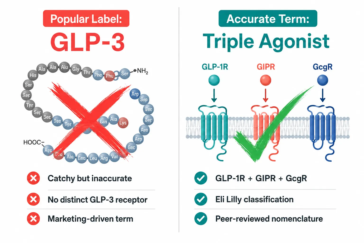
  • GLP-3: not a recognized endogenous hormone; "retatrutide" is its informal nickname, targeting GLP-1, GIP, and glucagon receptors.

The World Health Organization's International Nonproprietary Names system designates the generic name tirzepatide, with the stem "-tirz-" signaling its dual incretin activity. Calling tirzepatide "GLP-2 Tirz" blends an endogenous peptide name with a drug suffix, producing a label that implies receptor overlap where none exists.

For researchers exploring incretin-based metabolic research, the GLP-1-T incretin research themes page provides a useful parallel on how GLP-1 analogs are properly categorized. Similarly, the GLP-3 Reta research page illustrates how the triple-agonist space is being studied without conflating it with endogenous peptide families.


Mechanistic Differences: Two Compounds, Two Entirely Different Systems

Researcher's lab bench with peptide vials and pathway research cards

The core issue in the GLP-2-T and GLP2 Tirz Peptides naming confusion is that these compounds act through fundamentally separate biological systems.

How GLP-2 and GLP-2-T Work

GLP-2 is co-released with GLP-1 from enteroendocrine L-cells after nutrient intake. Its primary roles include:

  • Promoting intestinal mucosal growth and villus elongation
  • Supporting tight junction regulation and gut barrier integrity
  • Modulating enteric nervous system signaling

Critically, the GLP-2 receptor is expressed in the enteric nervous system rather than directly on intestinal epithelial cells, which means GLP-2 acts through an indirect mechanism involving neural intermediaries.

GLP-2-T is a modified, stability-enhanced analog of this endogenous peptide. Its structural modifications extend its half-life, allowing researchers to study longer-lasting gut mucosal effects without repeated peptide dosing in experimental setups. This makes it a practical tool for intestinal barrier and villus growth models.

How Tirzepatide (Informally "GLP-2 Tirz") Works

Tirzepatide is a dual agonist at the GLP-1 receptor and the glucose-dependent insulinotropic polypeptide (GIP) receptor. Its research-relevant actions include:

  • Stimulating glucose-dependent insulin secretion
  • Suppressing appetite via central GLP-1 receptor pathways
  • Modulating fat metabolism through GIP receptor activity

Tirzepatide has no direct activity at the GLP-2 receptor. Placing it under a "GLP-2" label is therefore mechanistically misleading. Researchers interested in dual incretin signaling may also find value in reviewing cagrilintide synergy with GLP-1 to understand how complementary peptide combinations are studied in metabolic contexts.

Feature GLP-2-T Tirzepatide ("GLP-2 Tirz")
Receptor target GLP-2 receptor GLP-1 + GIP receptors
Primary system Intestinal/gut mucosal Metabolic/pancreatic
Research focus Gut barrier, villi growth Insulin secretion, appetite
Endogenous basis GLP-2 analog Synthetic dual agonist

Research Use Cases: Selecting the Right Compound

GLP-2-T research use cases infographic with four key application icons

Understanding GLP-2-T and GLP2 Tirz Peptides: Naming Confusion, Mechanistic Differences, and Research Use Cases becomes most practical when deciding which compound belongs in a specific experimental design.

GLP-2-T Research Applications

GLP-2-T is primarily examined in preclinical gut biology models for:

  1. Intestinal villi growth and maintenance, studying how mucosal architecture responds to GLP-2 receptor stimulation
  2. Gut barrier permeability models, examining tight junction proteins and paracellular transport
  3. Enteric nervous system signaling, probing how GLP-2 receptor activation translates into epithelial responses via neural intermediaries
  4. Metabolic gut hub research, because the gut functions as a metabolic signaling organ, GLP-2-T is increasingly discussed alongside metabolic peptides

Recent research directions have also explored long-acting GLP-2 analogs through lipidation strategies, which enhance half-life and gut-tropic efficacy in rodent models, a design principle that informs GLP-2-T's structural modifications.

For researchers building multi-peptide protocols, longevity peptide research and MOTS-C mechanism and research offer context on how gut-metabolic signaling intersects with broader longevity pathways.

Tirzepatide Research Applications

Tirzepatide is studied for:

  • Glucose homeostasis and beta-cell function models
  • Adipose tissue metabolism via GIP receptor pathways
  • Appetite regulation through central GLP-1 receptor mechanisms

These are entirely separate research domains from GLP-2-T's intestinal focus. Researchers who require verified, lab-tested compounds for either pathway should consult resources on peptide purity testing to ensure compound integrity before experimental use.

Key distinction: If the research question involves gut mucosal biology, tight junctions, or intestinal villi, GLP-2-T is the relevant compound. If the question involves insulin secretion, appetite, or dual incretin signaling, tirzepatide is the appropriate subject, and it should be referred to by its correct INN name.


Conclusion

The naming overlap between GLP-2-T and "GLP-2 Tirz" (tirzepatide) is not a minor stylistic issue, it represents a mechanistic mismatch that can send researchers down the wrong experimental path. GLP-2-T targets the GLP-2 receptor and serves gut mucosal biology research. Tirzepatide targets GLP-1 and GIP receptors and belongs to metabolic and incretin research. They share no receptor overlap, no shared biological system, and no interchangeable research applications.

Actionable next steps for researchers:

  • Use the WHO-designated INN name "tirzepatide" in all literature and protocols, not the informal "GLP-2 Tirz" label.
  • Confirm receptor targets before selecting a compound for any experimental model.
  • Cross-reference vendor catalogs against peer-reviewed receptor pharmacology data.
  • Explore the all peptides for sale resource for context on how research-grade peptides are classified and combined.
  • Review innovative peptide delivery systems for updates on stability-enhancing modifications relevant to GLP-2-T analog design.

Precise nomenclature is the foundation of reproducible science. Resolving this naming confusion is the first step toward cleaner experimental design and more reliable results.

https://www.puretestedpeptides.com/wp-content/uploads/2026/07/glp-2-t-and-glp2-tirz-peptides-naming-confusion-mechanistic-differences-and-rese-1.png 1024 1536 Pure Tested https://www.puretestedpeptides.com/wp-content/uploads/2026/01/buy-peptides-online.jpg Pure Tested2026-07-13 13:18:122026-07-20 15:00:12GLP-2-T and GLP2 Tirz Peptides: Naming Confusion, Mechanistic Differences, and Research Use Cases
Retatrutide Structural Mechanism: What Cryo-EM Reveals About Triple-Receptor Agonism

Retatrutide Structural Mechanism: What Cryo-EM Reveals About Triple-Receptor Agonism

July 10, 2026/0 Comments/by Pure Tested

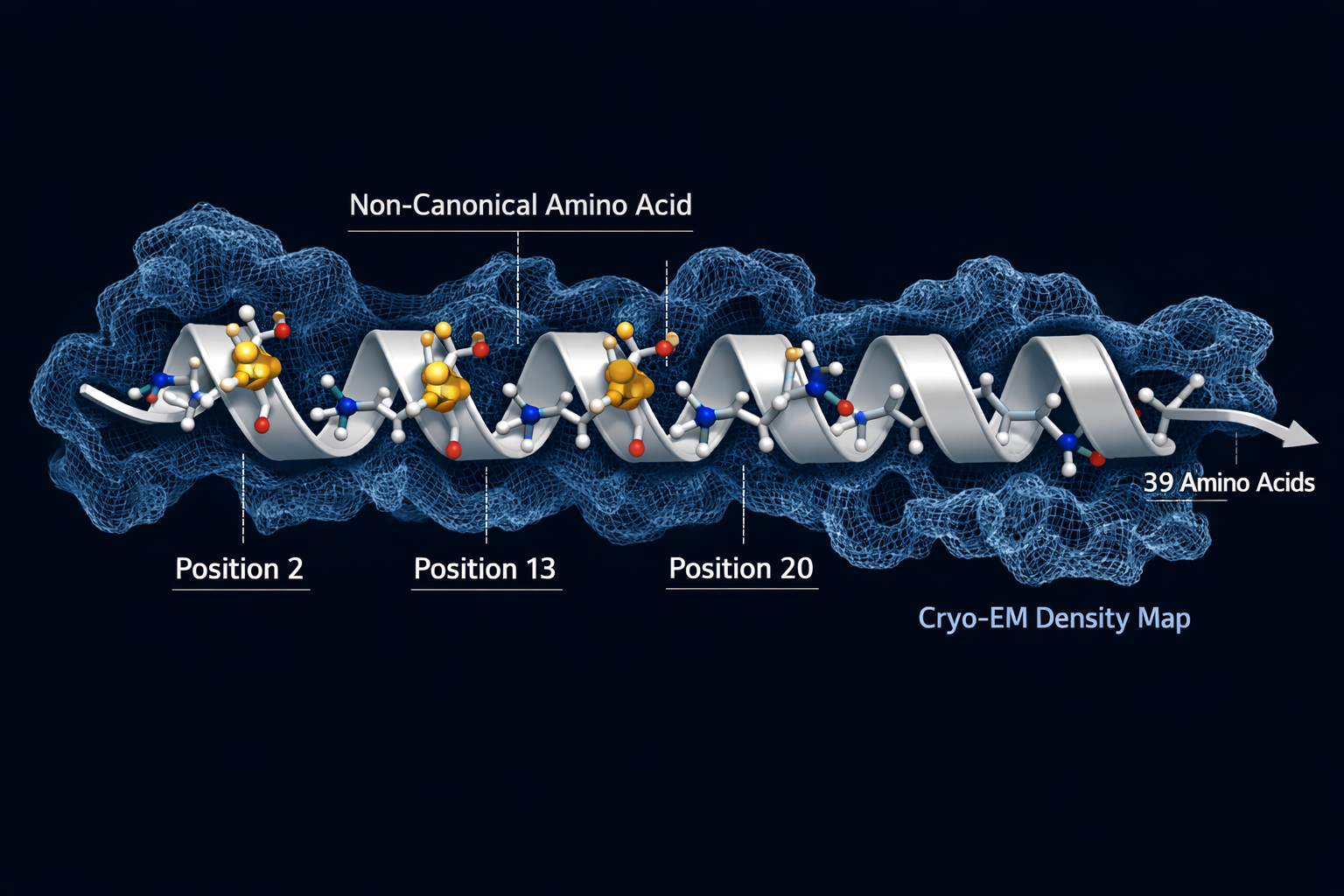
A single peptide that fits three different receptor locks simultaneously, that is the central engineering feat behind retatrutide. Understanding the Retatrutide Structural Mechanism: What Cryo-EM Reveals About Triple-Receptor Agonism requires stepping inside the molecular architecture of a 39-amino acid chain and asking a precise question: how does one molecule activate the GLP-1 receptor, the GIP receptor, and the glucagon receptor at the same time without losing potency at any of them? Cryo-electron microscopy (cryo-EM) has now provided detailed answers, and those answers explain why retatrutide behaves so differently from earlier incretin-based therapies.

Key Takeaways

  • Retatrutide adopts a single continuous alpha-helix conformation when binding to all three target receptors, a structural uniformity confirmed by cryo-EM.
  • Non-canonical amino acids at specific positions protect the peptide from enzymatic degradation and fine-tune receptor selectivity.
  • The N-terminal segment drives receptor activation by penetrating the transmembrane core, while the C-terminal segment governs selectivity through extracellular interactions.
  • Retatrutide is roughly 8.9 times more potent at the GIP receptor than native GIP, while its glucagon receptor activity is intentionally moderated to limit hyperglycemia risk.
  • A fatty acid side chain enables albumin binding, extending the half-life to approximately six days and supporting once-weekly dosing.

Key Takeaways

The Alpha-Helix Architecture Behind Triple-Receptor Binding

The most striking finding from cryo-EM studies is structural simplicity at the core. Despite engaging three pharmacologically distinct receptors, GLP-1R, GIPR, and GCGR, retatrutide maintains a single continuous alpha-helix conformation across all three binding events. This is not a trivial achievement. Most peptide ligands adopt slightly different conformations depending on the receptor environment they encounter. Retatrutide's rigid helical backbone allows it to slot into each receptor's binding pocket without requiring a structural reset.

This conformational consistency is not accidental. The peptide's sequence was engineered to include non-canonical amino acids that lock the helix in place:

  • Alpha-aminoisobutyric acid (Aib) at positions 2 and 20, resists degradation by dipeptidyl peptidase-4 (DPP-4), the enzyme that rapidly breaks down native GLP-1.
  • Alpha-methyl-L-leucine at position 13, supports GIP receptor activity and contributes to helical stability.

These modifications are part of what separates retatrutide from earlier GLP-1 peptide generations that lacked this level of structural engineering.

"The rigid alpha-helical backbone of retatrutide is not a byproduct of its design, it is the design."

The peptide also carries a fatty acid side chain that binds albumin in circulation, extending its half-life to roughly six days. This pharmacokinetic feature, combined with its enzymatic resistance, supports a once-weekly dosing schedule, a significant practical advantage over shorter-acting compounds.


The Alpha-Helix Architecture Behind Triple-Receptor Binding

How Cryo-EM Maps the Retatrutide Structural Mechanism Across Three Receptors

Cryo-EM resolved the bound structures of retatrutide at each of its three target receptors, revealing a consistent two-part binding strategy:

Segment Residues Primary Interaction
N-terminal 1 to 13 Penetrates transmembrane domain core
C-terminal 14 to 30 Engages extracellular regions

The N-terminal segment is the activation trigger. It inserts into the hydrophobic core of each receptor's transmembrane bundle, initiating the conformational change that signals downstream G-protein coupling. The C-terminal segment is the selectivity filter, making contact with extracellular loops that differ between receptor subtypes.

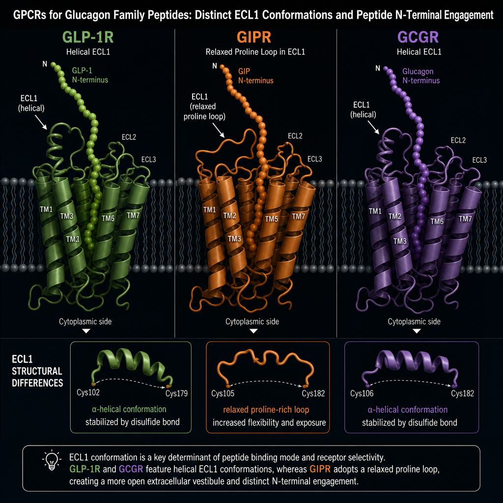
One notable receptor-specific difference involves extracellular loop 1 (ECL1). In GLP-1R and GCGR, ECL1 adopts a helical structure. In GIPR, ECL1 takes a relaxed loop conformation because of proline residues in that region. Retatrutide accommodates this difference without altering its core helical shape, a testament to the design flexibility built into its sequence.

For researchers exploring dual receptor agonism mechanisms, this structural data illustrates precisely why adding a third receptor target requires more than simply extending a peptide chain.


Potency Profile and Metabolic Consequences of Triple-Receptor Agonism

Understanding the Retatrutide Structural Mechanism: What Cryo-EM Reveals About Triple-Receptor Agonism is incomplete without examining what each receptor activation actually does metabolically:

  • GLP-1R activation, suppresses appetite and slows gastric emptying, reducing caloric intake.
  • GIPR activation, enhances glucose-dependent insulin secretion and influences adipose tissue metabolism.
  • GCGR activation, increases energy expenditure through hepatic lipid oxidation and thermogenesis.

Retatrutide's potency is deliberately asymmetric. It is approximately 8.9 times more potent at GIPR than native GIP, amplifying the insulin-sensitizing and fat-mobilizing effects of that receptor. At GCGR and GLP-1R, it operates at roughly 0.3 to 0.4 times the potency of endogenous glucagon and GLP-1, respectively. This deliberate moderation at GCGR limits the hyperglycemia risk that full glucagon activation would otherwise carry.

This potency calibration helps explain why clinical data show retatrutide producing 4 to 8 percent more weight loss than dual GLP-1/GIP agonists at comparable doses. The added glucagon receptor contribution raises resting energy expenditure in ways that appetite suppression alone cannot achieve.

Researchers interested in how incretin-based peptides compare across generations can explore GLP-1 incretin research themes for broader context. Those examining metabolic peptide research may also find value in reviewing body composition research themes related to tesa, which targets a different but metabolically relevant pathway. For a direct look at the compound itself, the GLP-3 retatrutide research product page provides additional sourcing context. Researchers comparing peptide purity standards should also consult resources on Bachem reference standards and peptide benchmarks when evaluating research-grade materials.


Conclusion

The Retatrutide Structural Mechanism: What Cryo-EM Reveals About Triple-Receptor Agonism comes down to a single engineered alpha-helix that speaks three receptor languages simultaneously. Cryo-EM has made it possible to see exactly how the peptide's N-terminal segment activates each receptor's transmembrane core while its C-terminal end navigates receptor-specific extracellular differences. Non-canonical amino acids provide enzymatic stability and receptor selectivity, while the fatty acid side chain extends circulating half-life to a clinically practical range.

For researchers working in this space, the actionable steps are clear: examine the structural data to understand why potency ratios were calibrated the way they were, compare retatrutide's binding architecture against earlier single and dual agonists, and track Phase 3 trial outcomes that will test whether structural advantages translate into durable clinical benefit. The cryo-EM data already provides a compelling molecular rationale for the efficacy signals observed so far.

https://www.puretestedpeptides.com/wp-content/uploads/2026/07/Retatrutide-Structural-Mechanism-What-Cryo-EM-Reveals-About-Triple-Receptor-Agonism.webp 1024 1536 Pure Tested https://www.puretestedpeptides.com/wp-content/uploads/2026/01/buy-peptides-online.jpg Pure Tested2026-07-10 13:18:392026-07-20 15:00:29Retatrutide Structural Mechanism: What Cryo-EM Reveals About Triple-Receptor Agonism
Retatrutide Mechanism of Action: How Triple Agonism Changes Metabolic Signaling in Research Models

Retatrutide Mechanism of Action: How Triple Agonism Changes Metabolic Signaling in Research Models

June 24, 2026/0 Comments/by Pure Tested

Activating three distinct metabolic receptors with a single molecule is not a theoretical concept — retatrutide does exactly that, and the downstream signaling consequences are reshaping how researchers think about obesity, glycemic control, and liver health. Understanding the Retatrutide Mechanism of Action: How Triple Agonism Changes Metabolic Signaling in Research Models is essential for anyone tracking the frontier of incretin-based research in 2026.

Key Takeaways

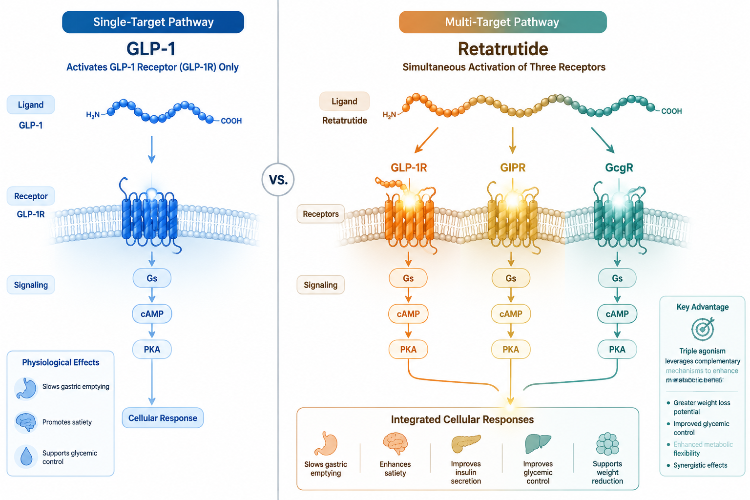
  • Retatrutide simultaneously activates GLP-1, GIP, and glucagon receptors, producing broader metabolic effects than single or dual agonists
  • Its highest receptor potency is at the GIP receptor (EC50 = 0.0643 nM), followed by GLP-1 and glucagon
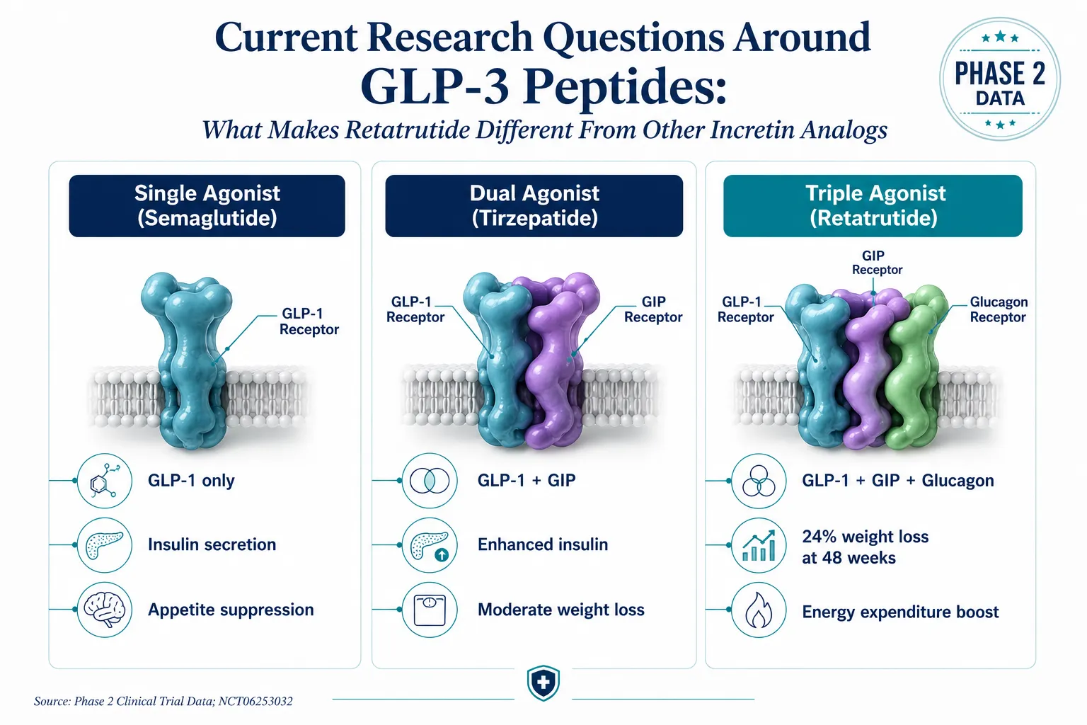
  • Phase 2 data showed a 24.2% reduction in total body weight over 48 weeks at the 12-mg dose
  • Hepatic fat was reduced by 82.4% relative, with 86% of subjects achieving liver fat normalization
  • Triple agonism integrates appetite suppression, insulin secretion, and energy expenditure into one coordinated signal

How Triple Receptor Activation Defines the Retatrutide Mechanism of Action

GLP-1 GIP glucagon receptor binding molecular diagram

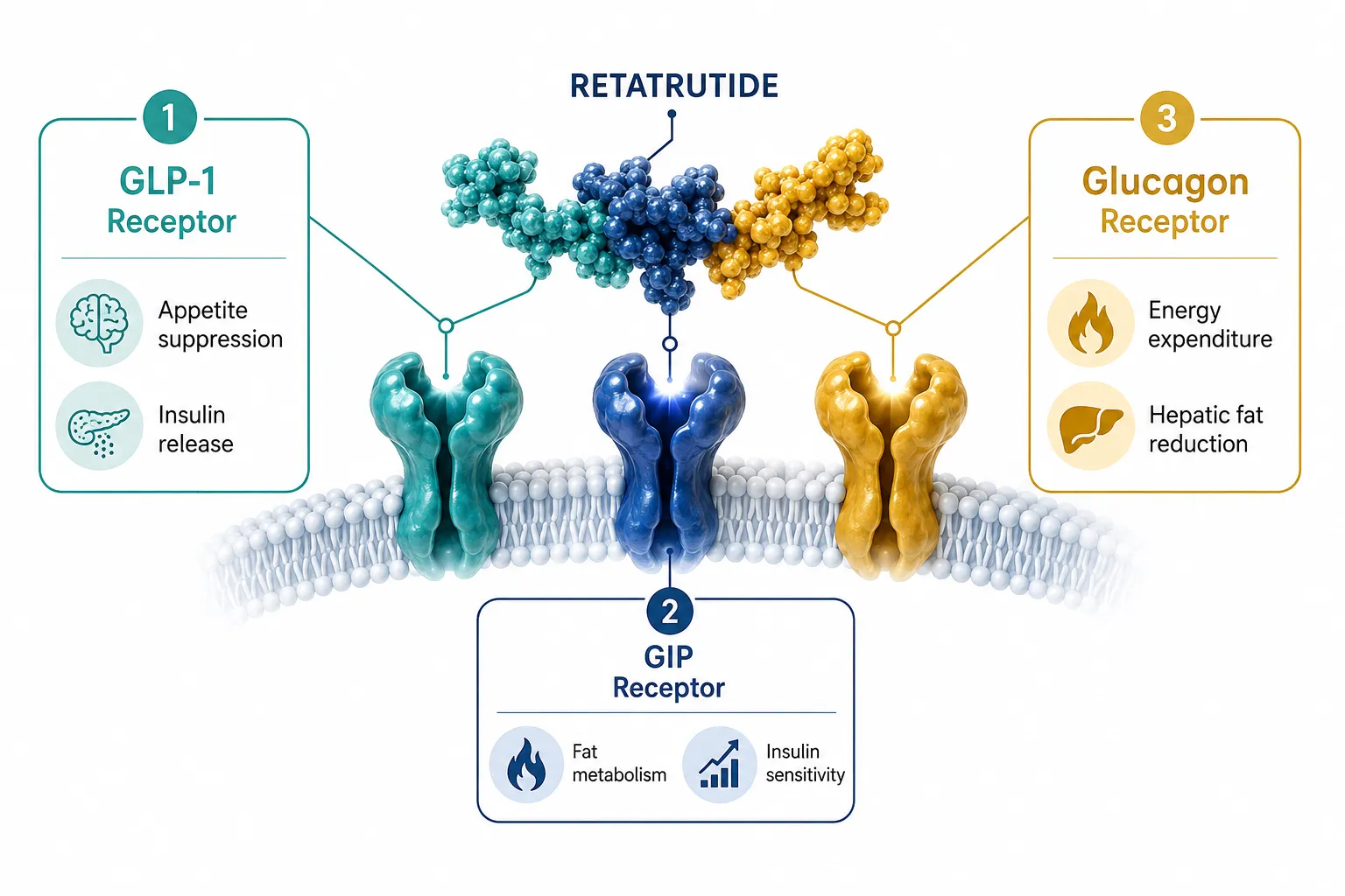
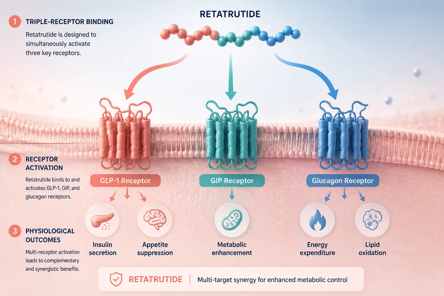
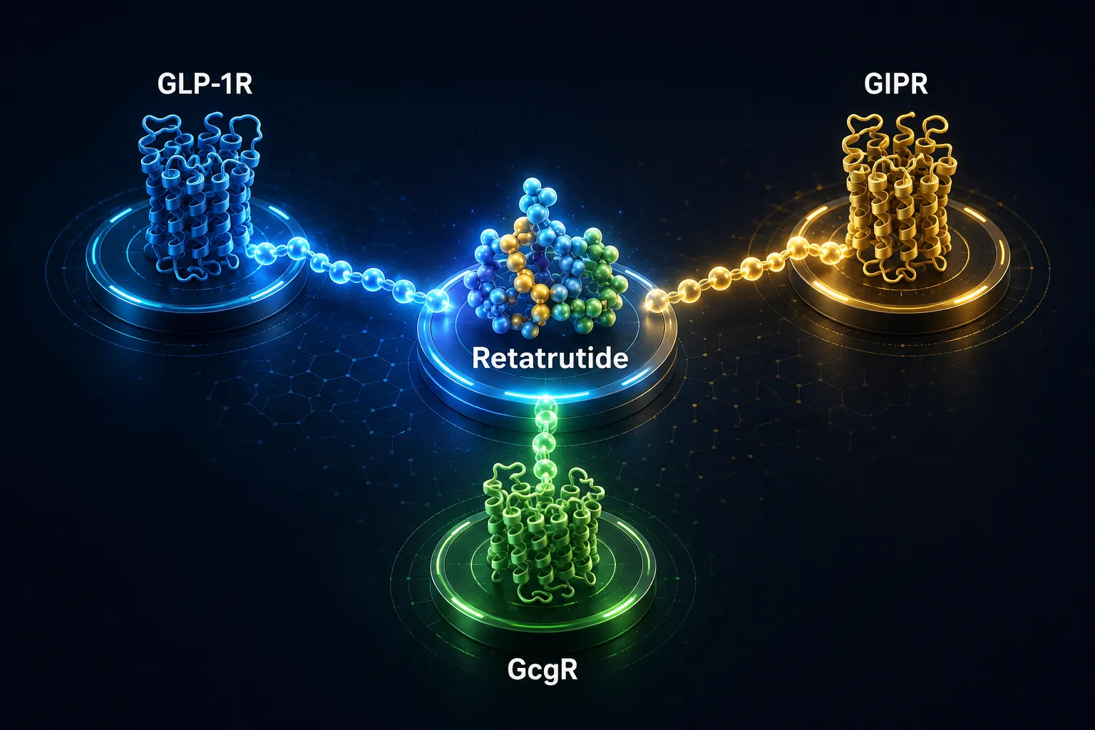
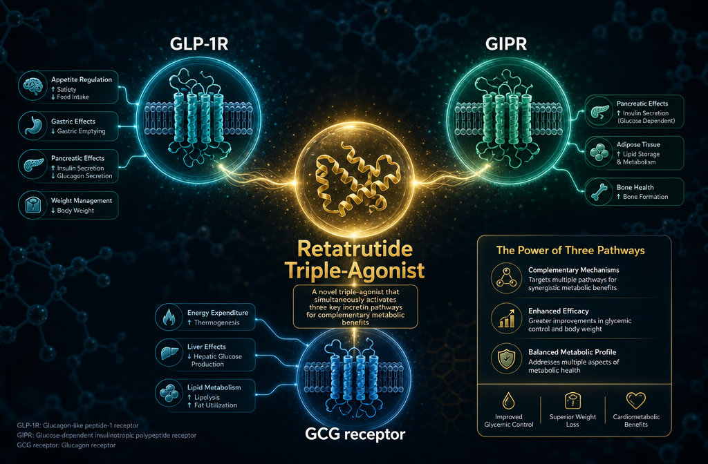
Retatrutide is a synthetic peptide engineered to bind three G-protein-coupled receptors: the glucagon-like peptide-1 (GLP-1) receptor, the glucose-dependent insulinotropic polypeptide (GIP) receptor, and the glucagon receptor (GCGR). Each receptor contributes a distinct layer of metabolic regulation.

Receptor Primary Metabolic Role EC50 (Potency)
GIP Insulin secretion, fat metabolism 0.0643 nM
GLP-1 Appetite suppression, insulin release 0.775 nM
Glucagon Energy expenditure, hepatic glucose output 5.79 nM

Retatrutide shows the strongest binding affinity at the GIP receptor, making GIP activity a dominant driver of its early metabolic effects. GLP-1 receptor activation adds appetite suppression and slows gastric emptying, which reduces caloric intake. Glucagon receptor co-activation increases thermogenesis and promotes hepatic fat oxidation — a mechanism largely absent from GLP-1-only therapies.

For context on how GIP receptor biology fits into the broader incretin landscape, the GIP receptor and its importance overview provides useful background on why this target matters.

This triple-pathway engagement is also explored in the GLP-3 triple agonist research overview, which compares receptor-targeting strategies across next-generation incretin compounds.


Metabolic Signaling Outcomes Observed in Research Models

Metabolic pathway downstream signaling liver fat weight loss data

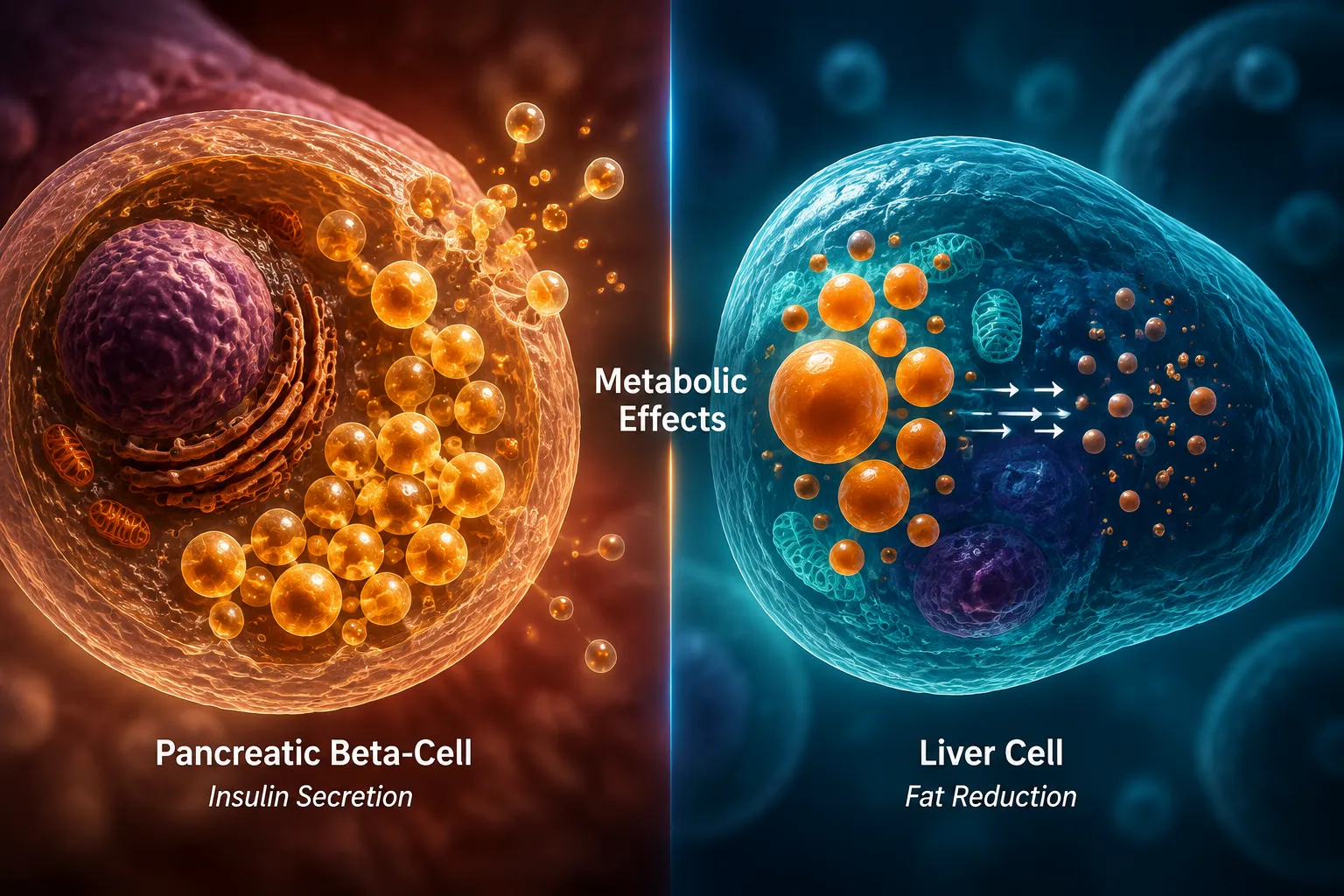
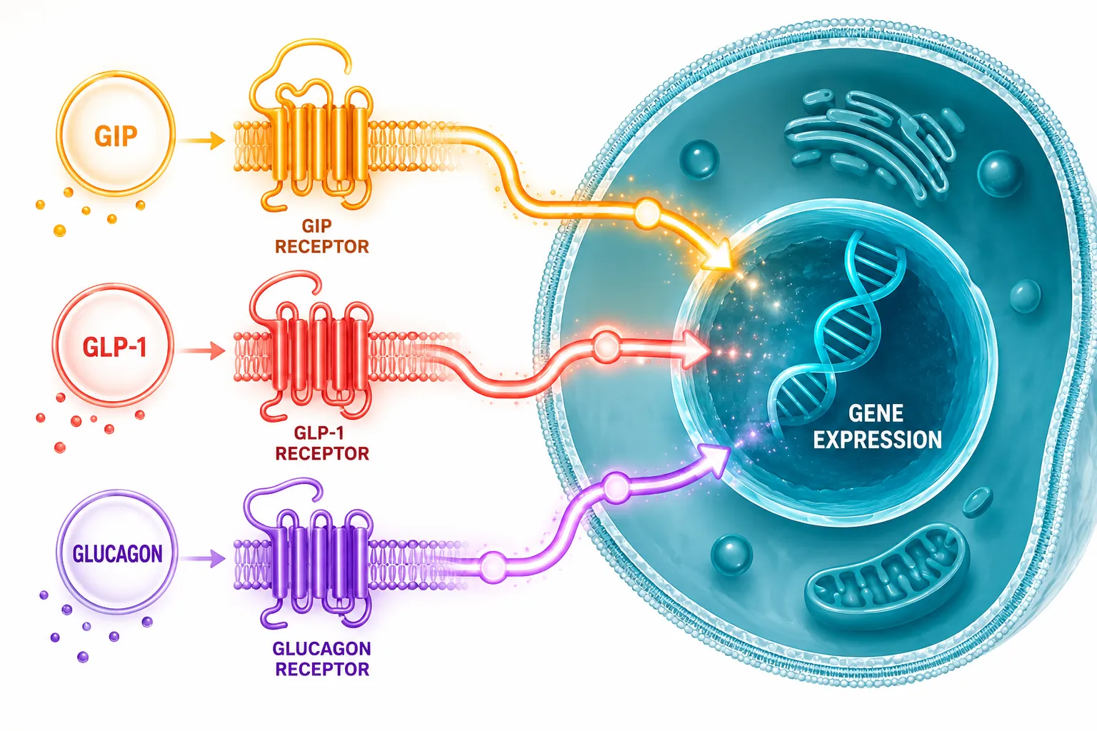
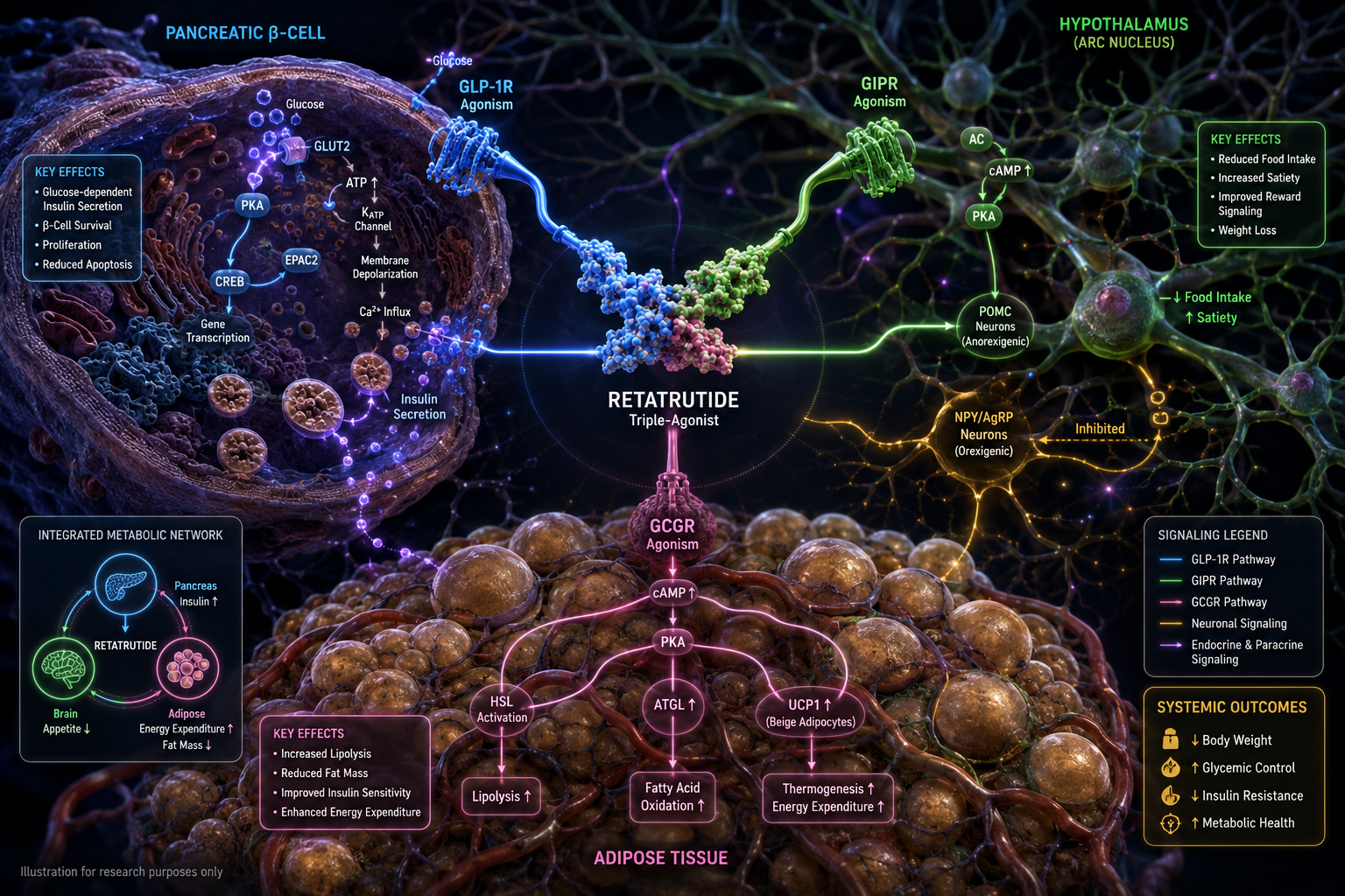
The Retatrutide Mechanism of Action: How Triple Agonism Changes Metabolic Signaling in Research Models becomes most apparent when examining what happens downstream of receptor binding. Each activated receptor triggers intracellular cAMP elevation, which cascades into tissue-specific effects:

  • Pancreatic beta cells: Enhanced glucose-stimulated insulin secretion via GLP-1 and GIP pathways
  • Hypothalamus: Appetite-suppressing signals that reduce total caloric intake
  • Adipose tissue: Increased lipolysis and thermogenic activation via glucagon receptor
  • Liver: Reduced de novo lipogenesis and accelerated fatty acid oxidation

These coordinated signals produced striking outcomes in Phase 2 research. At the 12-mg weekly dose over 48 weeks, subjects achieved a mean 24.2% reduction in total body weight, with 63% reaching at least 20% weight loss. Glycemic improvements were equally notable — an absolute HbA1c reduction of 2.02%, with 27% of diabetic participants reaching normoglycemia (HbA1c below 5.7%).

Liver outcomes were particularly compelling. Retatrutide produced an 82.4% relative reduction in hepatic fat, normalizing liver fat levels in 86% of participants — a finding with direct implications for metabolic dysfunction-associated steatotic liver disease research.

Researchers studying complementary metabolic pathways may find value in reviewing MOTS-c and metabolic flexibility research, which examines mitochondrial-level energy regulation as a parallel axis of metabolic control.

For those tracking incretin-based approaches more broadly, the GLP-1 incretin research themes page contextualizes where retatrutide sits within the evolving GLP receptor pharmacology space.


Comparative Advantage and the Broader Research Context

Comparative bar chart triple agonist vs single dual agonist outcomes

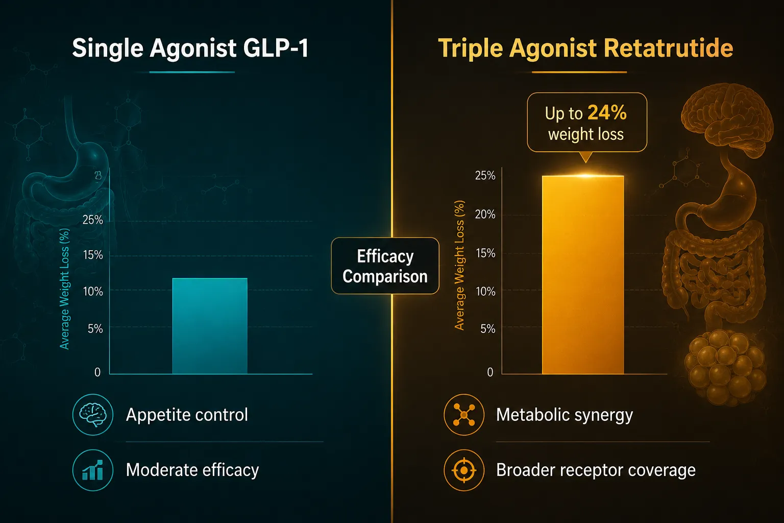
The Retatrutide Mechanism of Action: How Triple Agonism Changes Metabolic Signaling in Research Models stands apart from earlier incretin therapies precisely because it does not rely on a single signaling axis. Single GLP-1 agonists suppress appetite effectively but offer limited thermogenic benefit. Dual GLP-1/GIP agonists add insulin sensitization but leave glucagon-driven energy expenditure largely untouched.

Retatrutide closes that gap. The glucagon receptor component raises resting energy expenditure without triggering hyperglycemia — a balance made possible because GLP-1 and GIP co-activation simultaneously stimulates insulin secretion to offset glucagon's glucose-raising effect.

"Triple agonism represents a significant advancement in addressing complex metabolic disorders," noted lead Phase 2 investigator Dr. Ania M. Jastreboff — a statement supported by the breadth of endpoints improved in the trial data.

The safety profile observed in research settings was consistent with other incretin-based therapies, with gastrointestinal adverse events being the most commonly reported and generally non-severe.

Researchers exploring adjacent peptide mechanisms may also find the cagrilintide and GLP-1 synergy research article relevant, as it examines how amylin-pathway co-targeting compares to incretin stacking strategies.

For those interested in the specific retatrutide compound used in research settings, the GLP-3 Retatrutide product page provides purity and specification details relevant to preclinical study design.

Additional context on the evolving peptide research landscape is available through the what is new in peptide research resource.


Conclusion

The Retatrutide Mechanism of Action: How Triple Agonism Changes Metabolic Signaling in Research Models represents a meaningful step forward in metabolic pharmacology. By engaging GLP-1, GIP, and glucagon receptors simultaneously, retatrutide produces coordinated effects on appetite, insulin secretion, thermogenesis, and hepatic fat that no single-axis therapy can replicate.

Actionable next steps for researchers:

  • Review Phase 2 endpoint data across weight, glycemic, and hepatic fat outcomes to identify which research models align with your study design
  • Compare retatrutide's receptor potency profile against dual agonists to define the incremental contribution of glucagon receptor activation
  • Assess preclinical model selection criteria based on the compound's dominant GIP receptor affinity
  • Explore complementary metabolic peptides such as MOTS-c or cagrilintide to understand synergistic or additive signaling possibilities

As triple agonism moves through later-stage research phases in 2026, its mechanistic profile offers a detailed map for designing studies that capture the full breadth of metabolic signaling it engages.

https://www.puretestedpeptides.com/wp-content/uploads/2026/06/Retatrutide-Mechanism-of-Action-How-Triple-Agonism-Changes-Metabolic-Signaling-in-Research-Models.png 1024 1536 Pure Tested https://www.puretestedpeptides.com/wp-content/uploads/2026/01/buy-peptides-online.jpg Pure Tested2026-06-24 13:20:112026-07-20 15:02:19Retatrutide Mechanism of Action: How Triple Agonism Changes Metabolic Signaling in Research Models
GLP-3 Retatrutide vs. Polypeptide Peptides: A Comparative Research Guide to Metabolic Signaling Pathways

GLP-3 Retatrutide vs. Polypeptide Peptides: A Comparative Research Guide to Metabolic Signaling Pathways

June 19, 2026/0 Comments/by Pure Tested

Metabolic peptide research has shifted dramatically — where single-receptor agents once dominated laboratory inquiry, a new class of multi-target molecules is redefining what researchers expect from incretin-based signaling. This guide to GLP-3 Retatrutide vs. Polypeptide Peptides: A Comparative Research Guide to Metabolic Signaling Pathways examines how retatrutide's triple-receptor mechanism compares to conventional polypeptide agents, giving researchers a clear framework for understanding the underlying biology.

Key Takeaways

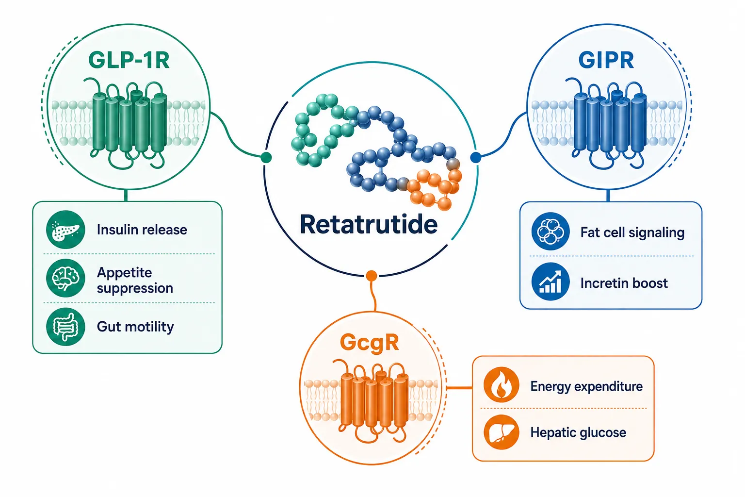
  • Retatrutide simultaneously activates three metabolic receptors: GLP-1R, GIPR, and the glucagon receptor (GcgR).
  • Conventional polypeptide peptides typically act on one or two receptor targets, producing narrower metabolic effects.
  • Triple agonism reshapes energy balance through complementary, overlapping signaling pathways.
  • Understanding receptor-level distinctions helps researchers design more targeted metabolic studies.
  • The term "GLP-3" is an informal research label — retatrutide's formal classification reflects its triple-agonist pharmacology.

Key Takeaways

Understanding the GLP-3 Label and Retatrutide's Classification

The label "GLP-3" circulates in research communities as shorthand for retatrutide, but it requires clarification. Retatrutide is not a third member of the glucagon-like peptide family in the classical sense. It is a synthetic triple agonist engineered to activate three distinct G-protein-coupled receptors simultaneously.

Conventional polypeptide peptides — including native GLP-1, GIP, and glucagon analogs — are typically single-receptor or, at most, dual-receptor agents. Their signaling is more contained. Retatrutide's design deliberately crosses those boundaries, which is why researchers studying GLP-3 Retatrutide incretin research themes often need a broader mechanistic framework than standard incretin models provide.

For context on how incretin generations have evolved, the overview of GLP-1 generations and their differences provides useful background on the progression from first-generation GLP-1 analogs to today's multi-agonist compounds.


Receptor-Level Mechanisms: How Retatrutide Differs from Conventional Polypeptide Peptides

This section of the GLP-3 Retatrutide vs. Polypeptide Peptides: A Comparative Research Guide to Metabolic Signaling Pathways focuses on what happens at the receptor level — the core distinction between retatrutide and standard polypeptide agents.

Receptor-Level Mechanisms: How Retatrutide Differs from Conventional Polypeptide Peptides

GLP-1 Receptor Activation

GLP-1R activation is shared by both retatrutide and conventional GLP-1 analogs. This pathway drives glucose-dependent insulin secretion, slows gastric emptying, and reduces appetite through both central nervous system and vagal nerve signaling. Single-agonist GLP-1 peptides operate primarily through this mechanism alone.

GIP Receptor Activation

GIPR activation adds a second layer. GIP further potentiates insulin release and modulates adipose tissue metabolism. Emerging research also suggests GIPR signaling may influence reward-related feeding behavior. Most traditional polypeptide peptides do not engage this receptor.

Glucagon Receptor Activation

GcgR activation is where retatrutide most clearly separates itself. Glucagon receptor signaling increases hepatic glucose output and, critically for metabolic research, raises resting energy expenditure. This thermogenic component is largely absent from conventional incretin peptides.

Receptor Retatrutide GLP-1 Analogs GIP Analogs
GLP-1R Yes Yes No
GIPR Yes No Yes
GcgR Yes No No
Thermogenic effect Yes Minimal Minimal

Researchers exploring complementary metabolic peptides such as MOTS-C, the mitochondrial peptide, will recognize that energy expenditure modulation is a recurring theme across multiple research-stage compounds — though the mechanisms differ significantly.


Metabolic Signaling Pathways: Triple Agonism vs. Conventional Peptide Approaches

The practical research value of the GLP-3 Retatrutide vs. Polypeptide Peptides: A Comparative Research Guide to Metabolic Signaling Pathways comparison lies in understanding how these mechanisms interact at the systems level.

Metabolic Signaling Pathways: Triple Agonism vs. Conventional Peptide Approaches

Triple agonism creates overlapping, reinforcing signals across three metabolic axes:

  • Insulin axis — amplified through both GLP-1R and GIPR co-activation
  • Appetite axis — suppressed via central GLP-1R pathways and potentially GIPR reward modulation
  • Energy expenditure axis — elevated through GcgR-driven thermogenesis

Conventional polypeptide peptides typically address one or two of these axes. Researchers studying body composition agents like Tesamorelin and its metabolic effects or AOD-9604 research methodology will note that each compound targets a narrower physiological window.

"Multi-receptor engagement is not simply additive — the convergence of three distinct signaling pathways creates metabolic effects that single-agonist models cannot fully replicate."

For researchers building broader metabolic panels, understanding cagrilintide's synergy with GLP-1 pathways also illustrates how combination approaches are increasingly central to advanced metabolic research design.

Those sourcing research-grade material can review GLP-3 Retatrutide product details for specification and traceability information.


Conclusion

The distinction between retatrutide and conventional polypeptide peptides is not merely a matter of degree — it reflects a fundamentally different approach to metabolic receptor engagement. Where single or dual-agonist peptides offer focused, well-characterized signaling, retatrutide's triple-agonist profile introduces a more complex, multi-axis mechanism that researchers must account for in study design.

Actionable next steps for researchers:

  1. Map which receptor pathways are relevant to your specific metabolic research question before selecting a peptide agent.
  2. Review the GLP-1 generations overview to contextualize retatrutide within the broader incretin research landscape.
  3. Cross-reference thermogenic and energy expenditure data when comparing triple-agonist results against single-receptor peptide benchmarks.
  4. Consult available innovative peptide delivery systems research to ensure study protocols reflect current best practices.

Understanding these mechanistic foundations is the starting point for rigorous, reproducible metabolic peptide research in 2026.

https://www.puretestedpeptides.com/wp-content/uploads/2026/06/GLP-3-Retatrutide-vs.-Polypeptide-Peptides-A-Comparative-Research-Guide-to-Metabolic-Signaling-Pathways.png 1024 1024 Pure Tested https://www.puretestedpeptides.com/wp-content/uploads/2026/01/buy-peptides-online.jpg Pure Tested2026-06-19 13:42:052026-07-20 15:02:42GLP-3 Retatrutide vs. Polypeptide Peptides: A Comparative Research Guide to Metabolic Signaling Pathways
What Is GLP-3 Retatrutide? Triple-Agonist Biology, Receptor Targets, and Why It Is Different From GLP-1

What Is GLP-3 Retatrutide? Triple-Agonist Biology, Receptor Targets, and Why It Is Different From GLP-1

June 9, 2026/0 Comments/by Pure Tested

Forty-five percent of participants in a Phase 3 clinical trial lost at least 30% of their body weight — a result once reserved for bariatric surgery. That single data point from the TRIUMPH-1 trial has made retatrutide one of the most closely watched compounds in metabolic medicine today. Understanding what is GLP-3 retatrutide, its triple-agonist biology, receptor targets, and why it is different from GLP-1 drugs already on the market is the essential first step for any researcher or clinician tracking this space.

Key Takeaways

  • Retatrutide simultaneously activates three hormone receptors: GLP-1R, GIPR, and the glucagon receptor (GCG-R).
  • The informal label "GLP-3" is not a scientific hormone classification — it is shorthand for the compound's triple-receptor profile.
  • In the TRIUMPH-1 Phase 3 trial, participants on 12 mg weekly lost an average of 28.3% of body weight over 80 weeks.
  • Retatrutide outperforms single-agonist (semaglutide) and dual-agonist (tirzepatide) therapies in early head-to-head comparisons.
  • As of 2026, retatrutide has not received FDA approval and remains in Phase 3 development under Eli Lilly.

Key Takeaways

The Triple-Agonist Biology Behind Retatrutide

Retatrutide is a synthetic peptide engineered to bind and activate three distinct incretin and metabolic hormone receptors at the same time. Each receptor plays a separate but complementary role in energy regulation.

Receptor Primary Role Contribution to Retatrutide's Effect
GLP-1R (Glucagon-Like Peptide-1) Insulin secretion, appetite suppression Reduces hunger, slows gastric emptying
GIPR (Glucose-Dependent Insulinotropic Polypeptide) Insulin amplification, fat metabolism Enhances insulin response, supports fat tissue signaling
GCG-R (Glucagon Receptor) Energy expenditure, hepatic glucose output Increases calorie burn, reduces liver fat

This simultaneous three-receptor engagement is what separates retatrutide from every approved obesity drug on the market. The glucagon receptor component is particularly significant: glucagon typically raises blood sugar, but when its receptor is activated alongside GLP-1R and GIPR, the net effect shifts toward increased thermogenesis and fat oxidation rather than hyperglycemia.

Researchers exploring the GLP-1 generations overview will recognize this as a logical progression from first-generation single-agonist molecules toward increasingly complex multi-receptor strategies.

Why the "GLP-3" Label Is Informal — and What It Actually Means

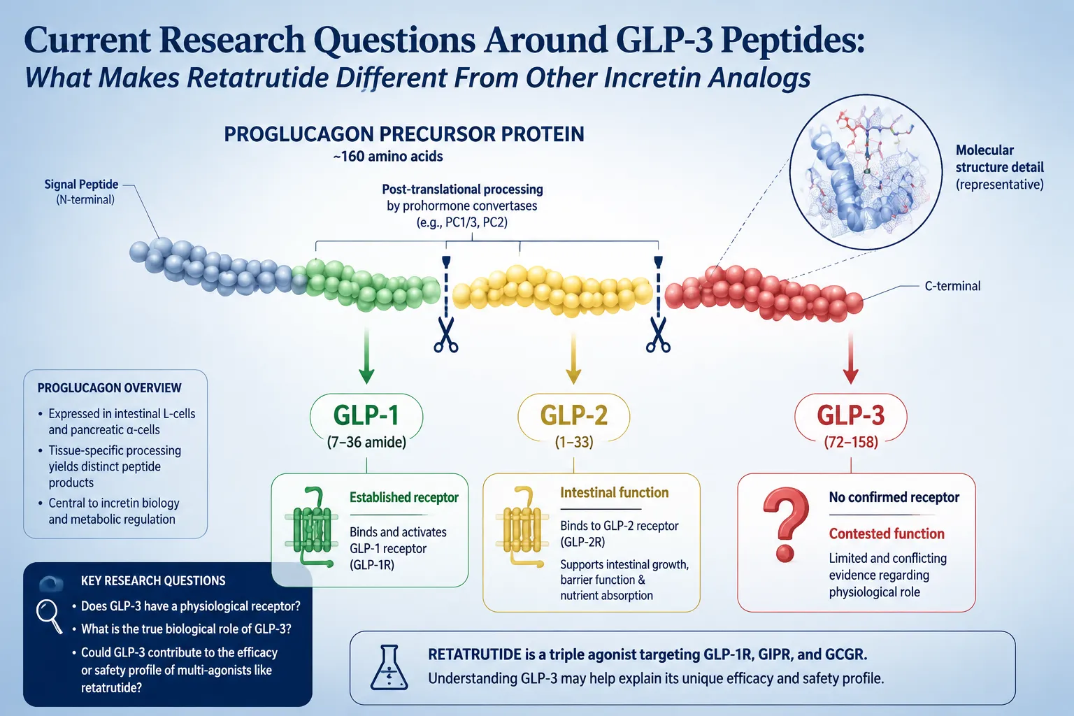
The term "GLP-3" does not refer to a real hormone. No such molecule exists in human physiology. The label emerged informally to describe retatrutide's position as the third generation of GLP-based obesity therapies:

  • Generation 1: GLP-1 single agonists (e.g., semaglutide / Wegovy)
  • Generation 2: GLP-1 + GIP dual agonists (e.g., tirzepatide / Zepbound)
  • Generation 3: GLP-1 + GIP + Glucagon triple agonists (retatrutide)

The correct scientific description is triple hormone receptor agonist. Researchers browsing retatrutide research and catalog resources or the GLP-1 Reta product tag will encounter both terms, but the informal "GLP-3" label should always be understood as generational shorthand rather than pharmacological classification.

Why the "GLP-3" Label Is Informal — and What It Actually Means

How Retatrutide Differs From GLP-1 Drugs: Receptor Targets and Clinical Outcomes

This is the core question for anyone asking what is GLP-3 retatrutide and why it is different from GLP-1. The differences operate on two levels: mechanistic and clinical.

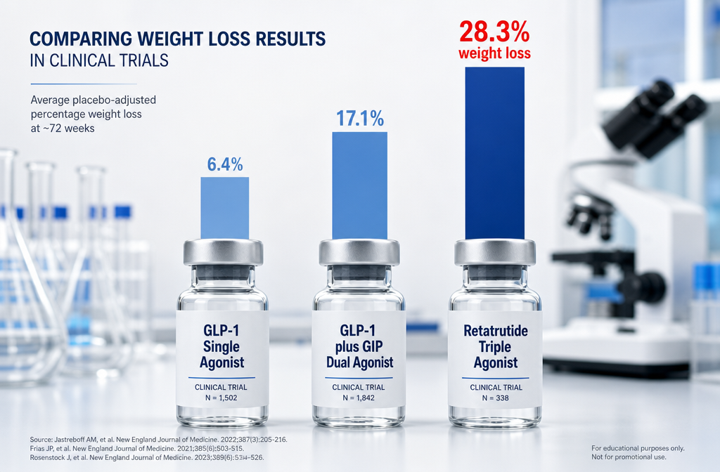
Mechanistically, semaglutide targets only GLP-1R. Tirzepatide adds GIPR. Retatrutide adds the glucagon receptor on top of both. That third receptor drives a meaningful increase in resting energy expenditure — the body burns more calories even at rest — which neither of the earlier drugs can replicate.

Clinically, the TRIUMPH-1 Phase 3 trial reported an average weight loss of 28.3% (approximately 70.3 pounds) over 80 weeks at the 12 mg weekly dose. By comparison, semaglutide typically produces roughly 15% weight loss, and tirzepatide reaches approximately 20-22%. Retatrutide also demonstrated an A1C reduction of up to 2.0% over 40 weeks in participants with type 2 diabetes, suggesting strong glycemic benefit beyond weight loss alone.

"Retatrutide's glucagon receptor component is the differentiating factor — it converts what would otherwise be a pure appetite-suppression strategy into a genuine energy-expenditure intervention."

Side effects remain consistent with the incretin drug class: nausea, diarrhea, constipation, and vomiting, all dose-dependent and generally manageable. Those interested in how metabolic peptides interact with energy systems may also find value in reviewing mitochondrial longevity research and AOD9604 metabolic research for broader context.

For researchers sourcing compounds for study, reviewing lab-tested peptide standards and certificate of analysis documentation ensures quality benchmarks are met before any research protocol begins.

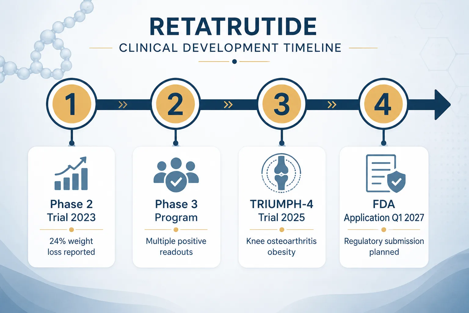
As of 2026, retatrutide is not FDA-approved. Eli Lilly anticipates filing for approval in 2026-2027, with potential market availability by 2027 or 2028. Those planning research timelines can consult the GLP-3 research planning and catalog navigation guide for sourcing and protocol considerations.

How Retatrutide Differs From GLP-1 Drugs: Receptor Targets and Clinical Outcomes

Conclusion

Retatrutide represents a genuine structural advance over existing GLP-1 therapies. Its triple-agonist biology — engaging GLP-1R, GIPR, and the glucagon receptor simultaneously — produces weight loss outcomes that approach bariatric surgery benchmarks and glycemic improvements that matter for type 2 diabetes management. The informal "GLP-3" label is a useful shorthand, but researchers should understand it as a generational marker, not a hormone designation.

Actionable next steps for researchers in 2026:

  • Review the TRIUMPH-1 Phase 3 trial data in detail to understand dose-response relationships.
  • Compare retatrutide's receptor profile against tirzepatide using the GLP-1 peptide generational research overview.
  • Verify compound purity standards before initiating any research protocol by consulting available COA documentation.
  • Monitor FDA filing timelines, currently projected for 2026-2027, to align research planning accordingly.
https://www.puretestedpeptides.com/wp-content/uploads/2026/06/What-Is-GLP-3-Retatrutide-Triple-Agonist-Biology-Receptor-Targets-and-Why-It-Is-Different-From-GLP-1.png 672 1024 Pure Tested https://www.puretestedpeptides.com/wp-content/uploads/2026/01/buy-peptides-online.jpg Pure Tested2026-06-09 13:05:142026-07-20 15:03:37What Is GLP-3 Retatrutide? Triple-Agonist Biology, Receptor Targets, and Why It Is Different From GLP-1
Peptides and Polypeptides in Cell Biology: How Experimental Peptides Interact With DNA, Mitochondria, and Hormone Receptors

Peptides and Polypeptides in Cell Biology: How Experimental Peptides Interact With DNA, Mitochondria, and Hormone Receptors

June 4, 2026/0 Comments/by Pure Tested

{"cover":"Professional landscape format (1536×1024) hero image with bold text overlay: 'Peptides & Polypeptides in Cell Biology' as main headline and subtitle 'How Experimental Peptides Interact With DNA, Mitochondria, and Hormone Receptors' in extra large 68pt bold white sans-serif font with deep navy semi-transparent overlay box, centered upper-third composition. Background shows a stunning macro scientific illustration of a glowing double-helix DNA strand intertwined with a luminous mitochondrion and molecular peptide chain structures, rendered in electric blue, teal, and white on deep navy background. Magazine cover aesthetic, editorial quality, high contrast, cinematic lighting, 2026 research theme.","content":["Detailed landscape format (1536×1024) scientific illustration showing intracellular peptide-DNA interaction: a close-up cross-section of a cell nucleus with glowing peptide chains (labeled EL28, PepH, Pep5) binding to a double-helix DNA strand. Molecular bonds shown as luminous gold nodes. Background features a translucent cell membrane with proteasome structures. Color palette: deep purple, gold, white. Clean lab-diagram aesthetic with annotated arrows, editorial quality, high contrast lighting from within the nucleus.","Detailed landscape format (1536×1024) visualization of mitochondrial targeting by peptides: a dramatic bird's-eye cross-section of a mitochondrion with its cristae structure clearly visible in electric teal and orange. Amphipathic peptide chains (MOTS-c labeled) are shown penetrating the outer mitochondrial membrane and localizing to the inner membrane. Surrounding cytoplasm shows cell-penetrating peptide vectors in motion. Deep charcoal background, bioluminescent glow effect, annotated molecular pathway arrows, editorial scientific illustration quality.","Detailed landscape format (1536×1024) diagram of hormone receptor signal transduction: a stylized cell surface showing multiple G protein-coupled receptors (GPCRs) with peptide hormone ligands docking onto receptor binding sites. Intracellular cascade shown with cAMP second messenger molecules, protein kinase activation chain, and nuclear receptor binding to DNA strand in the background. Color scheme: crimson red, white, slate blue. Clean infographic-style layout with labeled pathway steps, modern sans-serif annotations, editorial quality, high contrast."]

Professional landscape hero image () with : "Peptides and Polypeptides in Cell Biology: How Experimental Peptides Interact

Roughly 30% of all FDA-approved drugs work by targeting G protein-coupled receptors — proteins that respond directly to peptide signals. That single statistic reveals how deeply peptides and polypeptides in cell biology are woven into the machinery of life, and why research into experimental peptides has accelerated so sharply in 2026.

This article walks through the core mechanisms: how short amino acid chains reach the cell nucleus, penetrate mitochondrial membranes, and dock onto hormone receptors to trigger downstream signaling cascades.


Key Takeaways

  • Intracellular peptides such as EL28, PepH, and Pep5 interact directly with DNA-associated proteins and are studied as drug prototypes.
  • Peptide hormones are hydrophilic and cannot cross the lipid bilayer, so they bind cell surface receptors and activate second messengers like cyclic AMP.
  • Experimental peptides including MOTS-c can localize to mitochondria and influence energy regulation pathways.
  • GPCRs are the primary receptor family for peptide hormones and represent a major pharmacological target class.
  • Research-grade peptides such as CJC-1295 and GLP-1 analogs operate through receptor-mediated signaling with measurable downstream effects on gene expression.

Peptides and Polypeptides in Cell Biology: The Structural Foundation

Peptides and Polypeptides in Cell Biology: The Structural Foundation

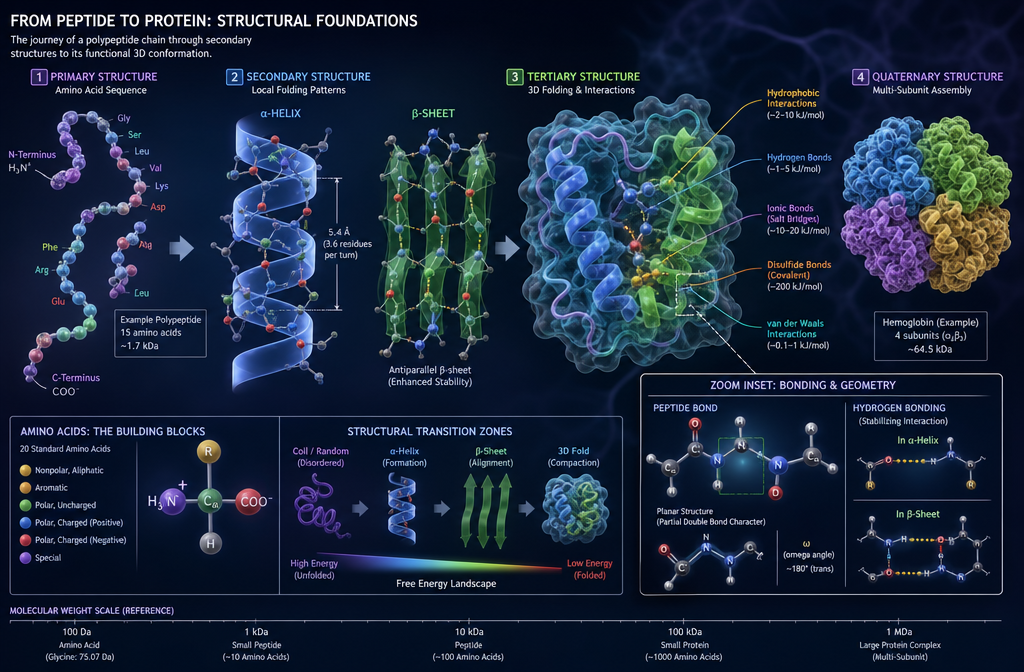
A peptide is a chain of two or more amino acids linked by peptide bonds. A polypeptide is simply a longer chain — typically more than 50 residues. When folded into functional shapes, polypeptides become proteins. The distinction matters in research because short peptides often behave differently from full proteins: they can slip through membranes, evade immune detection, and reach targets that larger molecules cannot.

Intracellular Peptides and DNA Interaction

Inside the cell, certain peptides operate in the nucleus itself. Intracellular peptides derived from proteasomal degradation — including EL28 (from proteasome regulatory subunit 4), PepH (from Histone H2B), and Pep5 (from cyclin D2) — have been identified as functional modulators of protein-protein interactions linked to gene regulation. These are not merely degradation byproducts; they act as prototype drug candidates because they already exist in the cellular environment and interact with DNA-associated machinery.

This opens a compelling research angle: if naturally occurring intracellular peptides can modulate transcription-linked proteins, then synthetic analogs designed to mimic or block those interactions could influence gene expression with high precision.


Mitochondrial Targeting: How Experimental Peptides Reach the Powerhouse

Mitochondrial Targeting: How Experimental Peptides Reach the Powerhouse

Mitochondria are not passive energy factories. They participate in intracrine signaling — internal signaling loops that influence cell survival, metabolism, and apoptosis. Peptides including angiotensin II and transforming growth factor-beta have been detected inside mitochondria, suggesting that peptide signaling extends well beyond the cell surface.

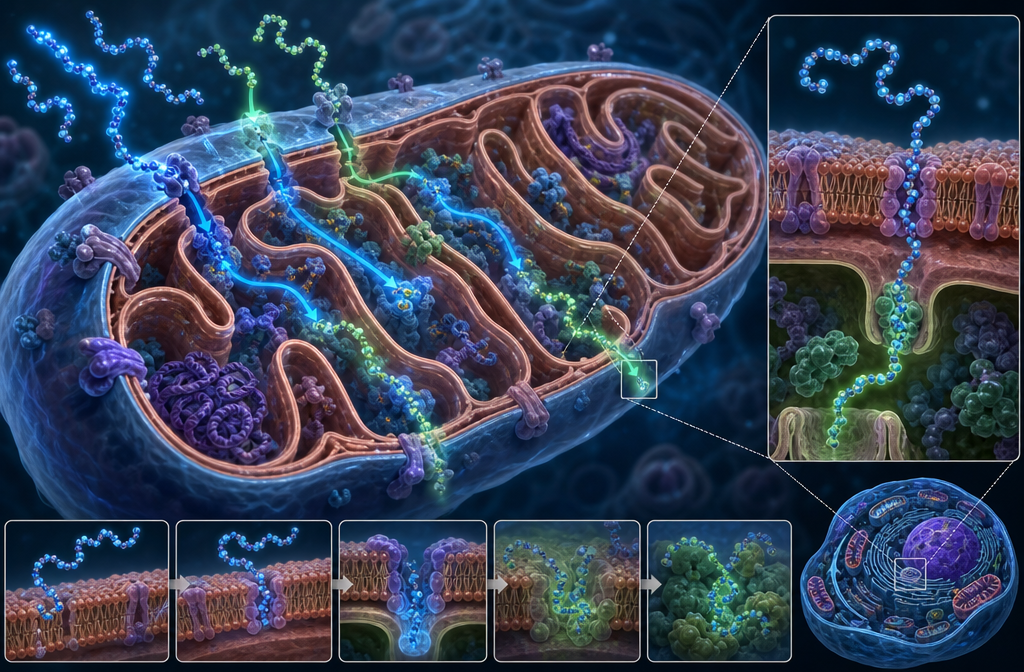
More recently, amphipathic proline-rich cell-penetrating peptides have been engineered to cross the plasma membrane and localize specifically to mitochondria. These vectors carry therapeutic payloads or act directly on mitochondrial membranes to stabilize cristae architecture and reduce oxidative stress.

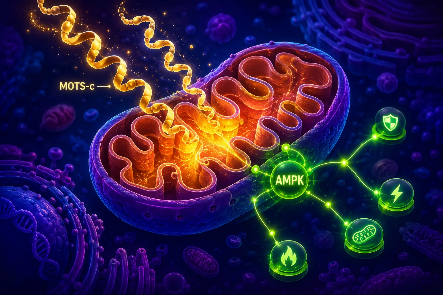
MOTS-c, a mitochondria-derived peptide encoded in mitochondrial DNA, is one of the most studied examples. Research into MOTS-c mitochondrial research themes shows that it translocates to the nucleus under metabolic stress and regulates gene expression — a striking example of cross-compartment peptide signaling. The compound MOTS-c and SLU-PP-332 pairing has also attracted attention for its potential effects on mitochondrial biogenesis pathways.

The SS-31 peptide (elamipretide) represents another mitochondria-targeted research compound. Its mechanism centers on cardiolipin stabilization within the inner mitochondrial membrane. Detailed research considerations are covered in this SS-31 10mg research peptide overview, and its broader mitochondrial dynamics are explored in SS-31 mitochondrial dynamics research.


Hormone Receptors and Signal Transduction: Where Peptides Meet Cell Biology

Hormone Receptors and Signal Transduction: Where Peptides Meet Cell Biology

Because peptide hormones are hydrophilic, they cannot diffuse through the fatty lipid bilayer of the cell membrane. Instead, they bind to receptors on the cell surface, which then relay the signal inward.

Three Major Receptor Classes for Peptide Hormones

Receptor Type Mechanism Example Peptide
G protein-coupled receptors (GPCRs) Activate G proteins, trigger cAMP GLP-1, GIP
Enzyme-linked receptors Direct kinase activation Insulin, IGF-1
Ion channel receptors Gate ion flow Neuropeptides

GPCRs dominate peptide hormone pharmacology. When a peptide ligand binds, the receptor activates a G protein, which in turn stimulates adenylyl cyclase to produce cyclic AMP (cAMP). This second messenger activates protein kinases that phosphorylate downstream targets — ultimately altering metabolism, proliferation, or secretion.

Research into GLP-1 dual receptor agonism and GIP receptor importance illustrates how next-generation peptide drugs exploit this pathway. Similarly, CJC-1295 research demonstrates GPCR-mediated growth hormone secretion through GHRH receptor activation.

Steroid hormones follow a different route — they diffuse through the membrane and bind nuclear receptors that act directly as transcription factors, binding DNA to switch genes on or off. Experimental peptides that mimic steroid hormone behavior are therefore studied for their potential to regulate gene expression without the systemic side effects of steroids.


Conclusion

Understanding peptides and polypeptides in cell biology — how experimental peptides interact with DNA, mitochondria, and hormone receptors — is no longer purely academic. In 2026, this knowledge directly informs the design of research-grade compounds targeting metabolic disease, mitochondrial dysfunction, and endocrine signaling.

Actionable next steps for researchers:

  • Review mitochondria-targeted compounds such as SS-31 and MOTS-c for models of intracellular peptide delivery.
  • Study GPCR-mediated pathways when evaluating GLP-1, GIP, and secretagogue peptides like CJC-1295 and ipamorelin.
  • Examine intracellular peptide prototypes (EL28, PepH) as templates for nucleus-targeted drug design.
  • Explore the full peptides research catalog to identify compounds relevant to specific signaling pathways.

The cell is not a black box. Peptides are the keys — and mapping how they fit each lock is the central challenge of modern molecular biology.


https://www.puretestedpeptides.com/wp-content/uploads/2026/06/Peptides-and-Polypeptides-in-Cell-Biology-How-Experimental-Peptides-Interact-With-DNA-Mitochondria-and-Hormone-Receptors.png 672 1024 Pure Tested https://www.puretestedpeptides.com/wp-content/uploads/2026/01/buy-peptides-online.jpg Pure Tested2026-06-04 13:04:432026-07-20 15:04:08Peptides and Polypeptides in Cell Biology: How Experimental Peptides Interact With DNA, Mitochondria, and Hormone Receptors
×

Helpful Links

  • My account
  • Cart
  • Checkout
  • Refund and Returns Policy
  • Privacy Policy
  • SMS Privacy Policy
  • Login
  • My Account
  • Logout

USA Made Lab Tested Peptides

All products are sold for research, laboratory, or analytical purposes only, and are not for human consumption


Pure Tested Peptides is a chemical supplier. Pure Tested Peptides is not a compounding / chemical compounding facility as defined under 503A of the Federal Food, Drug, and Cosmetic act. Pure Tested Peptides is not an outsourcing facility as defined under 503B of the Federal Food, Drug, and Cosmetic act.

The statements made within this website have not been evaluated by the US Food and Drug Administration. The products we offer are not intended to diagnose, treat, cure or prevent any disease.

Human/Animal Consumption Prohibited. Laboratory/In-Vitro Experimental Use Only

Scroll to top Scroll to top Scroll to top